Patients › Wrist
Fraktura ng Scaphoid
Scaphoid fractures — recognition, the high non-union risk, casting and percutaneous/open fixation.
Ang nararamdaman mo
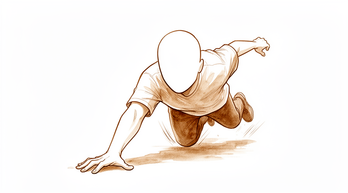
Maaaring maranasan mo ang sakit at pagkapagod sa base ng iyong hinlalaki, kaagad sa ilalim ng pulso. Ang lugar na ito ay kilala bilang anatomical snuffbox. Karaniwang nagsisimula ang sakit pagkatapos ng pagkabagsak sa isang nakabukas na kamay. Maaaring mapansin mo ang pamamaga o pagkabla-bla sa paligid ng pulso. Maaaring maging mahirap ang mga simpleng gawain. Maaaring masakit ang pag-abot sa likod upang isara ang bra. Maaaring magdulot ng matulis na katiwalian ang pagtatakip ng damit o ang pag-ikot ng hawakan ng pinto. Maaaring maramdaman na hindi matatag o masakit ang pag-angat ng mga bagay, kahit mga magaan.
Karaniwang lumalala ang sakit sa galaw. Ang paggamit ng iyong hinlalaki at pulso nang sabay-sabay ay nagdudulot ng stress sa nasirang buto. Maaaring mahirapan kang hawakan nang mahigpit ang mga bagay. Maaaring maging hamon ang paghawak ng tasa ng kape o telepono. Karaniwang tumutulong ang pahinga ng iyong kamay upang bawasan ang sakit. Gayunpaman, maaaring bumalik ang sakit sa huling bahagi ng araw pagkatapos ng aktibidad. Ilang pasyente ang nag-uulat na napapansin ang katiwalian sa gabi, lalo na kung sila ay natutulog sa gilid na iyon. Karaniwang gisingin ng matigas o masakit na pulso sa umaga.
Mahalagang malaman na hindi laging malinaw na ipinapakita ng karaniwang X-ray ang sirang buto. Sa katunayan, ang mga tunay na sirang buto ay natutukoy lamang sa humigit-kumulang 40% ng mga pasyente batay sa unang X-ray at klinikal na pagsusuri lamang. Ibig sabihin, maaaring maging malala ang iyong mga sintomas kahit normal ang unang scan. Kung ang iyong doktor ay nag-aalala ng sirang buto ngunit hindi malinaw ang X-ray, maaaring mag-order sila ng MRI. Ang scan na ito ay makakahanap ng mga nakatagong sugat na napapansin ng X-ray. Ang maagang pagtukoy ay tumutulong upang maiwasan ang mga komplikasyon.
Kung hindi gumaling nang maayos ang buto, maaari itong magdulot ng nonunion. Ito ay nangyayari kapag ang buto ay nabigo na mag-ugnay muli. Nananatiling mataas ang mga rate ng nonunion para sa mga sirang buto ng scaphoid. Sa ilang kaso, ang delayed o nonunion ay nangyayari sa higit sa 6% ng mga pasyente, kahit may tamang paunang paggamot. Karamihan sa mga pasyenteng ito ay nangangailangan ng operasyon upang ayusin ang buto. Ang maagang internal fixation ay lalong pinipili upang bawasan ang risk na ito. Ang iyong doktor ay talakayin ang pinakamainam na landas para sa iyo batay sa tiyak na detalye ng iyong sugat.
Ano ang nangyayari
Ang scaphoid ay isang maliit na buto na hugis ng walnuts sa iyong pulso. Ito ay gumaganap bilang isang kritikal na tulay sa pagitan ng iyong mga buto ng braso at kamay. Ang butong ito ay may masalimuot na suplay ng dugo, ibig sabihin ay hindi laging nakakakuha ng sapat na sustansya upang magpagaling nang sarili. Kapag nabasag ito, ang proseso ng paggaling ay maaaring huminto. Ito ay tinatawag na nonunion. Kahit na mayroong modernong diagnosis at operasyon, nananatiling mataas ang mga rate ng nonunion.
Kung hindi bumabalik ang buto sa pagkakaisa, nagbabago ang normal na galaw ng iyong pulso. Ang mga buto sa iyong pulso ay dapat gumalaw nang magkasamang parang sayaw. Ang isang basag na scaphoid ay nagdudulot ng pagkagambala sa ritmong ito. Bahagyang binabalewala nito ang itaas at ibabang hanay ng mga buto sa pulso. Ito ay nagdudulot ng hindi normal na galaw at pagkasira sa mga ibabaw ng kasu-kasuan. Sa paglipas ng panahon, ang pagkasira na ito ay maaaring magdulot ng arthritis. Maaari kang maranasan ang sakit at katigasan habang ang mga ibabaw ng kasu-kasuan ay nagkikiskisan nang walang karaniwang makinis na proteksyon.
Maaaring tulungan ng iyong doktor ang pagbabalik ng balanse na ito. Para sa ilang mga basag, isang simpleng turnilyo ang humahawak sa mga piraso habang nagpapagaling. Para sa mas kumplikadong mga kaso, maaaring gumamit ang iyong doktor ng bone graft. Ito ay nangangahulugang pagkuha ng isang maliit na piraso ng malusog na buto upang punan ang mga puwang at hikayatin ang paglago. Sa ilang sitwasyon, maaari ring baguhin ng iyong doktor ang hugis ng iyong buto ng braso. Ito ay nagpapalayo ng bigat mula sa nasirang scaphoid. Layunin ng mga hakbang na ito ang pagbabalik ng normal na galaw ng pulso at lakas ng hawak.
Ang layunin ay pigilan ang hindi normal na pagkasira bago ito magdulot ng pangmatagalang pinsala. Kung maagang tratuhin, kahit na mga displaced fractures ay maaaring magpagaling nang maayos. Kung ang buto ay magkakaisa, malamang na magkakaroon ka ng magandang resulta, anuman ang maliit na pagbabago sa hugis. Ang pokus ay magbigay sa iyo ng isang pulso na walang sakit at may kakayahang harapin ang mga pang-araw-araw na gawain nang walang pagkiskis o kawalan ng katatagan na dulot ng isang basag na tulay.
Ano ang maaari naming gawin dito
Ang iyong paggamot ay nagsisimula sa maingat na pagsubaybay at pahinga. Dahil ang karaniwang X-ray at dalawang pagsusuri ng klinikal ay tumutukoy lamang sa tunay na fracture sa humigit-kumulang 40% ng mga pasyente, maaaring gumamit ang iyong surgeon ng maagang MRI upang makakuha ng malinaw na sagot. Ang scan na ito ay tumpak na nakakahanap ng nakatagong pinsala at tumutulong upang mapatunayan ang kawalan ng fracture kapag ang mga unang resulta ay hindi malinaw. Habang naghihintay ka para sa diagnosis o habang nasa conservative treatment ka, dapat mong iwasan ang mga gawain na nagpapabigat sa iyong pulso. Layunin ng physiotherapy na ibalik ang iyong range of motion at lakas kapag nakagaling na ang buto. Para sa maraming pasyente, lalo na ang mga may nondisplaced fractures, epektibo ang nonoperative treatment. Ang mga rate ng union para sa mga kaso na ito ay umaabot o kahit lumampas sa mga rate ng surgery. Inaasahan mong magsuot ka ng cast o splint sa isang panahon na itatakda ng iyong surgeon. Walang iisang pinakamainam na protocol kung gaano katagal ang immobilization ng iyong pulso pagkatapos ng anumang proseso, kaya ang iyong team ang gabay sa iyo batay sa iyong paggaling.
Ang pamamahala ng sakit ay isang mahalagang bahagi ng iyong paggaling. Maaaring rekomendahan ng iyong surgeon ang mga over-the-counter pain relievers upang manatili kang komportable. Gayunpaman, kailangan mong maging maingat sa paggamit ng nonsteroidal anti-inflammatory drugs (NSAIDs). Kung kukunin mo ang mga gamot na ito sa loob ng unang buwan ng iyong pinsala, may mas mataas na risk na hindi magagaling ang buto (nonunion). Ang pagkabigo na ito ay maaaring magdulot ng mas kumplikadong salvage procedures sa hinaharap. Para sa karamihan ng acute fractures, hindi namin ginagamit ang cortisone, hyaluronic acid, o PRP injections bilang standard care. Sa halip, nakatuon kami sa pagprotekta sa buto habang ito ay nag-aayos ng sarili. Kung mayroon kang nonunion na nondisplaced at nonangulated, maaaring isaalang-alang ang minimally invasive bone grafting at compression screw fixation. Ang pamamaraang ito ay ligtas at epektibo sa pag-stabilize ng buto nang walang malaking surgery.
Ang surgery ay karaniwang inialok lamang para sa displaced fractures o mga kaso kung saan nabigo ang conservative care. Kung ang iyong fracture ay displaced, inirerekomenda ang operative intervention upang maayos na i-align ang buto. Para sa mga recent nonunions na hindi gumaling sa unang treatment, maaaring gawin ng iyong surgeon ang distal scaphoid resection o gumamit ng double antirotation screw fixation kasama ang arthroscopy. Layunin ng mga prosedurong ito ang pag-stabilize ng buto at pagpapalakas ng paggaling. Habang ang maagang internal fixation ay lalong pinapaboran para sa ilang acute fractures, hindi ito laging kinakailangan para sa mga nondisplaced na pinsala. Sa katunayan, walang tunay na long-term benefit sa surgery kumpara sa nonoperative treatment para sa acute nondisplaced o minimally displaced fractures. Ang iyong surgeon ang tumutulong sa iyo na pumili ng landas batay sa iyong mga personal na halaga at tolerance sa risk. Tandaan, nananatiling mataas ang mga rate ng nonunion kahit may mga improved techniques, kaya mahalaga ang malapit na follow-up anuman ang napiling treatment.
Ano ang inaasahan
Karamihan sa mga fracture ng scaphoid ay gumagaling nang maayos, lalo na sa mga bata. Para sa mga matatanda, ang prognosis ay nakadepende sa kung gaano kabilis ka makakuha ng paggamot at kung ang mga piraso ng buto ay lumipat o hindi. Kung ang fracture ay hindi displaced o bahagya lamang displaced, maaaring irekomenda ng iyong surgeon ang paggamit ng cast o operasyon. Parehong paraan ay nagdudulot ng katulad na long-term function. Gayunpaman, ang operasyon ay tumutulong sa iyo na bumalik sa trabaho nang humigit-kumulang 7 linggo mas mabilis kaysa sa paggamit ng cast lamang.
Kung ang buto ay hindi gumaling, tinatawag itong nonunion. Ito ay nangyayari sa higit sa 10% ng mga kaso pagkatapos ng operasyon para sa closed fractures. Mas karaniwan ito kung magpapakonsulta ka ng higit sa 21 araw pagkatapos ng pinsala. Ang pagkaantala sa paggamot ay nagpapataas ng risk ng pagkabigo ng cast. Ang nonunion ay maaaring magdulot ng progressive wear-and-tear arthritis sa pulso. Bagama't tila seryoso ito, maraming pasyente ay nag-uulat pa rin ng magandang galaw at lakas ng pulso sa loob ng mga taon, kahit na bahagya lamang ang pagbabago sa hugis ng buto.
Ang iyong surgeon ay magmamanman nang mahigpit sa iyong paggaling. Kung mayroon kang nonunion, maaaring kailanganin ang karagdagang operasyon. Ang mga ulit na prosedimiento ay mas hindi matagumpay kaysa sa unang pagtatangka. Sa ilang kaso kung saan umusbong na ang arthritis, ang pag-alis ng bahagi ng butong scaphoid ay maaaring magpahinga ng sakit. Humigit-kumulang 94% ng mga pasyente ay nasisiyahan sa prosedurang ito, at ito ay humihinto sa pagbagsak ng pulso.
Sa kabuuan, halos lahat ng scaphoid fractures na matagumpay na nag-union ay nagdudulot ng magandang resulta. Ang susi ay ang tiyakin na gumaling ang buto. Mahalaga ang maagang diagnosis dahil madalas na nakakaligtaan ng mga standard na X-ray ang mga pinsalang ito. Kung ikaw ay nasa grupo na may mas mataas na risk para sa delayed healing, tulad ng mga may ilang kondisyon sa mental health o mula sa mga mahihirap na komunidad, mahalaga ang karagdagang atensyon sa iyong follow-up. Sa tamang pag-aalaga, inaasahan mong mababawi ang normal na function at lakas ng kamay sa loob ng mga darating na buwan.
Kailan kumonsulta sa doktor
Kumonsulta sa iyong doktor kung mayroon kang patuloy na sakit, kahinaan, o kawalan ng katatagan sa iyong pulso. Ang mga sintomas na nakakaapekto sa pagtulog o trabaho ay nangangailangan ng pansin. Humingi ng pagsusuri ng espesyalista kung ang iyong pulso ay nakakabit o biglang bumabagsak. Ang biglang paglala ng sakit ay isang babala rin. Magkaroon ng kamalayan na ang pagkaantala sa pagdarasal ng 21 araw o higit pa pagkatapos ng sugat ay nagpapahiwatig ng mas mataas na panganib ng pagkabigo ng pagkakabit. Ang maling diagnosis ng mga fracture ay maaaring magdulot ng malalaking komplikasyon. Ang maagang diagnosis ay tumutulong iwasan ang mga isyung ito. Kung ang mga unang X-ray ay hindi malinaw, ang maagang MRI ay maaaring tukuyin ang mga sugat nang tumpak. Huwag balewalain ang mga sintomas, dahil ang hindi naaayos na fracture ay maaaring hindi gumaling nang maayos.
Evidence & references
Overview
- Pediatric scaphoid fractures have excellent outcomes [1].
- Some well-established and widely used principles of scaphoid fracture management are supported by an insufficient amount of evidence, with many decisions based on small case series [2].
- The clinical outcomes of malunited scaphoids after reconstruction for scaphoid fractures nonunion did not differ significantly from well-united scaphoids at a minimum 5-year follow-up [4].
- Internal fixation of scaphoid fractures is indicated in certain acute situations and in chronic nonunion cases [15].
- Appropriately performed acute percutaneous internal fixation is now a standard treatment option for a selected group of patients with acute scaphoid fracture [17].
- The definition of instability of scaphoid fractures and the indications for conservative treatment must be considered carefully [14].
- Nondisplaced scaphoid fractures can be effectively treated nonoperatively with union rates approaching or exceeding those of operative intervention, while operative intervention is recommended for displaced fractures [24].
- This study did not demonstrate a true long-term benefit of internal fixation, compared with nonoperative treatment, for acute nondisplaced or minimally displaced scaphoid fractures [5].
- For all indications, the scaphoid staple has a high union rate and a low complication rate [6].
- Patients with recent scaphoid fractures that failed treatment may also be treated with distal scaphoid resection [25].
- Despite improvements in diagnosis and surgical techniques, nonunion rates remain high and early internal fixation is increasingly favored even for nondisplaced fractures [28].
Anatomy & Pathophysiology
- The scaphoid is critical to the coordination of normal carpal kinematics [34].
- Scaphoid fracture has significant biomechanical consequences to the wrist [34].
- Scaphoid nonunions have a dramatic impact on carpal kinematics, partially uncoupling the proximal and distal carpal rows [35].
- Problem fractures and non-unions of the scaphoid are associated with major alterations in wrist kinematics [46].
- Problem fractures and non-unions of the scaphoid are associated with a higher incidence of premature carpal collapse and degenerative arthritis than previously appreciated [46].
- A foreshortened healed scaphoid will disrupt carpal kinematics [37].
- A foreshortened healed scaphoid negatively impacts results, including decreased wrist range of motion and diminished grip strength [37].
- Malunion or nonunion of an acute scaphoid fracture can lead to abnormal carpal kinematics and wrist arthrosis [61].
- Radiocarpal-based lunate morphology was not associated with scaphoid fracture [74].
- Anomalous carpal kinematics caused by lunotriquetral coalition may have predisposed both scaphoid bones to fracture, although causality cannot be proven [78].
Classification
- Pediatric scaphoid fractures have excellent outcomes [1].
- Even some well-established and widely used principles of scaphoid fracture management are supported by an insufficient amount of evidence, with many decisions based on small case series [2].
- The combination of conventional radiographs and two clinical examinations does not provide adequate diagnostic certainty for scaphoid fractures, as a true fracture was identified in only about 40% of patients [3].
- If there is a strong clinical suspicion of a scaphoid fracture which cannot be confirmed by conventional radiology, bone scintigraphy is a valuable diagnostic tool [8].
- The combination of conventional radiographs and clinical reassessment does not increase the accuracy of these diagnostic tests compared with the accuracy of conventional radiographs alone and is therefore also limited in diagnosing scaphoid fractures [9].
- There is no consensus regarding the imaging modality and measurements to use to define a scaphoid fracture as 'nondisplaced' [10].
- Due to low agreement between observers for the recognition of scaphoid fractures and poor diagnostic performance, 6-week radiographs are not adequate for evaluating suspected scaphoid fractures [12].
- Scaphoid fractures account for 2% of all fractures and are the most commonly injured carpal bone [13].
- The definition of instability of scaphoid fractures and the indications for conservative treatment must be considered carefully [14].
- Internal fixation of scaphoid fractures is indicated in certain acute situations and in chronic nonunion cases [15].
- Despite improvements in diagnosis and surgical techniques, nonunion rates remain high and early internal fixation is increasingly favored even for nondisplaced fractures [28].
- There is a need for a validated prognostic classification system for scaphoid nonunions that can allow comparisons between outcome studies [41].
- The authors hypothesise higher union rates in scaphoid fractures using more stable fixation systems [79].
- Scaphoid fracture and nonunion management continues to be an area of expanding evidence with opportunities to improve knowledge and familiarization with current evidence-based data [80].
Clinical Presentation
- Scaphoid fractures account for 2% of all fractures and are the most commonly injured carpal bone [13].
- Most scaphoid fractures are missed due to failure to consider the possibility of the injury and search for clinical signs [19].
- The combination of conventional radiographs and two clinical examinations does not provide adequate diagnostic certainty for scaphoid fractures, as a true fracture was identified in only about 40% of patients [3].
- The combination of conventional radiographs and clinical reassessment does not increase the accuracy of these diagnostic tests compared with the accuracy of conventional radiographs alone and is therefore also limited in diagnosing scaphoid fractures [9].
- 6-week radiographs are not adequate for evaluating suspected scaphoid fractures due to low agreement between observers for the recognition of scaphoid fractures and poor diagnostic performance [12].
- Ultrasonic assessment is not recommended for the early diagnosis of acute scaphoid fractures, with a sensitivity of only 50% and five missed scaphoid fractures in a small series [39].
- If there is a strong clinical suspicion of a scaphoid fracture which cannot be confirmed by conventional radiology, bone scintigraphy is a valuable diagnostic tool [8].
- Clinical examination along with early MRI scan should form the basis of diagnosing a suspected scaphoid fracture [20].
- The use of early MRI in patients with clinically suspected scaphoid fracture results in the accurate and reliable identification of a significant number of radiological occult injuries and early identification of patients without acute injuries [23].
- MRI-detected scaphoid fractures are not universally benign, with delayed or nonunion seen in over 6% despite appropriate initial immobilization, with most of these patients with nonunion requiring surgery to achieve union [22].
- The diagnosis of scaphoid and other fractures is reliable when using HRpQCT in patients with a clinically-suspected fracture [40].
- There is no consensus regarding the imaging modality and measurements to use to define a scaphoid fracture as 'nondisplaced' [10].
- Oblique scaphoid fractures are potentially unstable and may result in detrimental sequelae if overlooked in the acute stage [16].
Investigations
- Conventional radiographs combined with two clinical examinations provide inadequate diagnostic certainty for scaphoid fractures, identifying a true fracture in only about 40% of patients [3].
- The combination of conventional radiographs and clinical reassessment does not increase diagnostic accuracy compared to conventional radiographs alone [9].
- There is no consensus on the imaging modality or measurements used to define a scaphoid fracture as nondisplaced [10].
- Six-week radiographs are not adequate for evaluating suspected scaphoid fractures due to low inter-observer agreement and poor diagnostic performance [12].
- Most missed scaphoid fractures result from a failure to consider the injury possibility and search for clinical signs [19].
- Clinical examination combined with early MRI scan should form the basis for diagnosing suspected scaphoid fractures [20].
- MRI-detected scaphoid fractures are not universally benign, with delayed or nonunion occurring in over 6% of cases despite appropriate initial immobilization [22].
- Most patients with nonunion of MRI-detected scaphoid fractures require surgery to achieve union [22].
- Early MRI in patients with clinically suspected scaphoid fractures accurately and reliably identifies a significant number of radiological occult injuries [23].
- Early MRI in patients with clinically suspected scaphoid fractures allows for the early identification of patients without acute injuries [23].
- Early MRI provides an immediate diagnosis for suspected scaphoid fractures when initial radiographs are inconclusive [56].
- Early MRI for suspected scaphoid fractures when initial radiographs are inconclusive is cost-effective and minimizes complications [56].
- CT is a good way to screen for occult fractures but may not be superior to MRI or bone scanning in detecting scaphoid fractures without causing overtreatment [58].
- Multidetector computed tomography (MDCT) has a sensitivity of 86% and specificity of 100% for detecting occult scaphoid fractures in patients with negative radiographic examinations [59].
- MRI is the optimal second test for assessing a possible scaphoid fracture after a negative radiograph [60].
- CT is preferred over MRI when the fracture is visible for further assessment and surgical planning [60].
- There is variation in definitions of scaphoid fractures on MRI scans, highlighting a need for consensus to assess reliability and diagnostic performance [63].
- Bone scintigraphy is inappropriate for evaluating specificity and sensitivity against clinical examination [64].
- MRI is the recommended examination of choice for diagnosing occult scaphoid fractures over bone scintigraphy [64].
- MRI is considered the best diagnostic radiological test for triage of suspected scaphoid fractures according to existing literature [67].
- Bone scanning, CT, and ultrasound may be useful for suspected scaphoid fractures when MRI is not readily available [67].
- Routine MRI of suspected scaphoid fractures carries a notable risk of overdiagnosis and potential overtreatment [72].
- Nearly 70% of MRI findings in suspected scaphoid fractures are categorized as distracting and potentially misleading [72].
- Stopping the pursuit of occult fractures may prevent unnecessary treatment due to the risk of overdiagnosis with routine MRI [72].
- Better standardization of MRI definitions for scaphoid fractures is required to address diagnostic uncertainty [76].
- A definitive definition may not exist to solve the potentially unsolvable issue of diagnostic uncertainty in scaphoid fractures [76].
- Patients should participate in decisions regarding diagnostic and treatment strategies for scaphoid fractures due to diagnostic uncertainty [76].
- MRI is not 100% specific for diagnosing occult scaphoid fractures, with a specificity of 96% in healthy volunteers [77].
Treatment
Nonoperative Management
- Pediatric scaphoid fractures have excellent outcomes [1].
- Internal fixation of scaphoid fractures is indicated in certain acute situations and in chronic nonunion cases [15].
- Early treatment of acute scaphoid fractures is important, with union rates significantly greater when treatment is instituted prior to 4 weeks from injury [26].
- Nondisplaced scaphoid fractures can be effectively treated nonoperatively with union rates approaching or exceeding those of operative intervention, while operative intervention is recommended for displaced fractures [24].
- Surgical treatment for non-displaced and minimally displaced acute scaphoid fractures may be slightly favourable compared to conservative treatment for standardised functional outcome on the short term (within 2 years), with a significantly faster return to work (SMD of 7 weeks) [21].
- This study did not demonstrate a true long-term benefit of internal fixation, compared with nonoperative treatment, for acute nondisplaced or minimally displaced scaphoid fractures [5].
- We found no difference in functional outcome at 12 months for fractures of the waist of the scaphoid with ≤ 2 mm displacement treated operatively or nonoperatively [18].
- Non- and minimally displaced scaphoid waist fractures are best treated conservatively [36].
- Non-operative treatment of non-displaced scaphoid fractures may be preferred [53].
- A restricted period of cast immobilisation is usually adequate for the treatment of non-displaced scaphoid fractures [53].
- Nondisplaced fractures of the scaphoid heal with cast immobilization in most cases, but operative treatment is being offered with greater frequency to active patients to reduce the period of cast immobilization [57].
- The definition of instability of scaphoid fractures and the indications for conservative treatment must be considered carefully [14].
- Currently, there is insufficient evidence to support the most effective treatment for acute scaphoid fractures [51].
- Even some well-established and widely used principles of scaphoid fracture management are supported by an insufficient amount of evidence, with many decisions based on small case series [2].
- Among patients with nonoperatively managed scaphoid fractures, those prescribed NSAIDs within 1 month of diagnosis demonstrated an increased risk of nonunion and subsequent salvage procedures [54].
Operative Management
- Appropriately performed acute percutaneous internal fixation is now a standard treatment option for a selected group of patients with acute scaphoid fracture [17].
- The frequency of non-union after surgical management for closed scaphoid fractures exceeds 10% and remained consistent during the study period [11].
- For all indications, the scaphoid staple has a high union rate and a low complication rate [6].
- The use of 2 headless compression screws for the treatment of scaphoid nonunions is safe and effective [44].
- Patients with recent scaphoid fractures that failed treatment may also be treated with distal scaphoid resection [25].
- The optimal protocol for postoperative immobilization following operative treatment of scaphoid fractures remains controversial [62].
Special Populations and Considerations
- The authors prefer to treat nondisplaced acute scaphoid fractures in the athlete on an individualized basis [55].
- The clinical outcomes of malunited scaphoids after reconstruction for scaphoid fractures nonunion did not differ significantly from well-united scaphoids at a minimum 5-year follow-up [4].
- This case is interesting as the child is one of the youngest patients described in the literature with a scaphoid fracture, and the fracture went on to non-union despite immediate medical attention and rigorous treatment [49].
- The authors argue that comfort with uncertainty is key in suspected scaphoid fracture scenarios, as there is no best strategy; instead, clinicians should help patients choose a diagnostic and therapeutic course based on their individual values and risk tolerance [66].
Complications
- Pediatric scaphoid fractures have excellent outcomes [1].
- Many decisions regarding scaphoid fracture management are based on small case series due to insufficient evidence for well-established principles [2].
- The combination of conventional radiographs and two clinical examinations does not provide adequate diagnostic certainty for scaphoid fractures, as a true fracture was identified in only about 40% of patients [3].
- Clinical outcomes of malunited scaphoids after reconstruction for scaphoid fracture nonunion did not differ significantly from well-united scaphoids at a minimum 5-year follow-up [4].
- There is no true long-term benefit of internal fixation compared with nonoperative treatment for acute nondisplaced or minimally displaced scaphoid fractures [5].
- The scaphoid staple has a high union rate and a low complication rate for all indications [6].
- Subacute scaphoid fractures presenting within 6 months from injury can be expected to successfully heal with casting alone, even if the initial diagnosis is delayed [7].
- The frequency of nonunion after surgical management for closed scaphoid fractures exceeds 10% and remained consistent during the study period [11].
- Oblique scaphoid fractures are potentially unstable and may result in detrimental sequelae if overlooked in the acute stage [16].
- Appropriately performed acute percutaneous internal fixation is a standard treatment option for a selected group of patients with acute scaphoid fracture [17].
- There is no difference in functional outcome at 12 months for fractures of the waist of the scaphoid with ≤ 2 mm displacement treated operatively or nonoperatively [18].
- Persistent nonunion is common after surgery for scaphoid nonunion, and surgeries for persistent nonunion are even less successful [27].
- Delayed presentation of scaphoid fractures 21 days or more after injury predicts a greater risk of casting failure, although the union rate remains high with comparable time in cast [29].
- Nearly half of all patients with malunited acute scaphoid fractures demonstrated radiographic findings of early arthritis on CT imaging but had overall good clinical results on midterm follow-up [30].
- Increased likelihood for nonunion was found when the fracture was treated greater than 31 days from injury and when fracture volume was less than 38% of the entire scaphoid [32].
Recovery
- Pediatric scaphoid fractures have excellent outcomes [1].
- Clinical outcomes of malunited scaphoids after reconstruction for nonunion did not differ significantly from well-united scaphoids at a minimum 5-year follow-up [4].
- There is no true long-term benefit of internal fixation compared with nonoperative treatment for acute nondisplaced or minimally displaced scaphoid fractures [5].
- The scaphoid staple has a high union rate and a low complication rate for all indications [6].
- Subacute scaphoid fractures presenting within 6 months from injury can be expected to successfully heal with casting alone, even if the initial diagnosis is delayed [7].
- The frequency of nonunion after surgical management for closed scaphoid fractures exceeds 10% [11].
- Oblique scaphoid fractures are potentially unstable and may result in detrimental sequelae if overlooked in the acute stage [16].
- There is no difference in functional outcome at 12 months for fractures of the waist of the scaphoid with ≤ 2 mm displacement treated operatively or nonoperatively [18].
- Surgical treatment for non-displaced and minimally displaced acute scaphoid fractures may be slightly favourable compared to conservative treatment for standardised functional outcome on the short term (within 2 years) [21].
- Surgical treatment for non-displaced and minimally displaced acute scaphoid fractures results in a significantly faster return to work (SMD of 7 weeks) [21].
- Union rates are significantly greater when treatment is instituted prior to 4 weeks from injury [26].
- Persistent nonunion is common after surgery for scaphoid non-union, and surgeries for persistent nonunion are even less successful [27].
- Delayed presentation of scaphoid fractures 21 days or more after injury predicts a greater risk of casting failure [29].
- The union rate remains high with comparable time in cast despite delayed presentation of scaphoid fractures 21 days or more after injury [29].
- Nearly half of all patients with malunited acute scaphoid fractures demonstrated radiographic findings of early arthritis on CT imaging [30].
- Patients with malunited acute scaphoid fractures demonstrated overall good clinical results on midterm follow-up despite radiographic findings of early arthritis [30].
- From an 8- to 11-year perspective, patients with distal scaphoid fractures report normal self-assessed hand function [31].
- From an 8- to 11-year perspective, patients with distal scaphoid fractures report good wrist motion and strength [31].
- Increased likelihood for nonunion was found when the fracture was treated greater than 31 days from injury [32].
- Increased likelihood for nonunion was found when fracture volume was less than 38% of the entire scaphoid [32].
- Patients treated nonoperatively or with salvage procedures had similar long-term outcomes as those treated with a corrective scaphoid osteotomy [65].
- Patients with comorbid psychiatric conditions experienced increased rates of delayed scaphoid union [83].
- Dynamic imaging with time-intensity curve analysis does not provide additional predictive value over standard delayed enhanced imaging for acute scaphoid fracture viability assessment using contrast-enhanced MRI [84].
- Scaphoid nonunions demonstrate findings indicative of progression to union on CT at a mean of 6 weeks [86].
- Scaphoid nonunions demonstrate findings indicative of progression to union on CT as early as 3 weeks postoperatively [86].
Key Evidence
- [L1] Pediatric scaphoid fractures have excellent outcomes. [1] (10.1177/1558944717735948)
- [L5] Even some well-established and widely used principles of scaphoid fracture management are supported by an insufficient amount of evidence, with many decisions based on small case series. [2] (10.1177/1753193420977241)
- [L5] The combination of conventional radiographs and two clinical examinations does not provide adequate diagnostic certainty for scaphoid fractures, as a true fracture was identified in only about 40% of patients. [3] (10.1097/corr.0000000000002413)
- [L4] The clinical outcomes of malunited scaphoids after reconstruction for scaphoid fractures nonunion did not differ significantly from well-united scaphoids at a minimum 5-year follow-up. [4] (10.1016/j.otsr.2014.09.026)
- [L1] This study did not demonstrate a true long-term benefit of internal fixation, compared with nonoperative treatment, for acute nondisplaced or minimally displaced scaphoid fractures. [5] (10.2106/jbjs.g.00673)
- [L4] For all indications, the scaphoid staple has a high union rate and a low complication rate. [6] (10.1177/1558944716658747)
- [Paper] If there is a strong clinical suspicion of a scaphoid fracture which cannot be confirmed by conventional radiology, bone scintigraphy is a valuable diagnostic tool. [8] (10.1016/j.injury.2005.02.009)
- [L2] The combination of conventional radiographs and clinical reassessment does not increase the accuracy of these diagnostic tests compared with the accuracy of conventional radiographs alone and is therefore also limited in diagnosing scaphoid fractures. [9] (10.1097/corr.0000000000002310)
- [L5] There is no consensus regarding the imaging modality and measurements to use to define a scaphoid fracture as 'nondisplaced.' [10] (10.1016/j.jhsa.2012.10.025)
- [L3] The frequency of non-union after surgical management for closed scaphoid fractures exceeds 10% and remained consistent during the study period. [11] (10.1016/j.jhsa.2015.06.019)
- [L2] Due to low agreement between observers for the recognition of scaphoid fractures and poor diagnostic performance, 6-week radiographs are not adequate for evaluating suspected scaphoid fractures. [12] (10.1007/s00402-016-2438-4)
- [L5] Scaphoid fractures account for 2% of all fractures and are the most commonly injured carpal bone. [13] (10.1016/j.hcl.2017.04.003)
- [L5] The definition of instability of scaphoid fractures and the indications for conservative treatment must be considered carefully. [14] (10.1142/s0218810415400018)
- [L5] Internal fixation of scaphoid fractures is indicated in certain acute situations and in chronic nonunion cases. [15] (10.1016/s0749-0712(21)00118-9)
- [L4] Oblique scaphoid fractures are potentially unstable and may result in detrimental sequelae if overlooked in the acute stage. [16] (10.1016/j.injury.2009.07.078)
- [L4] Appropriately performed acute percutaneous internal fixation is now a standard treatment option for a selected group of patients with acute scaphoid fracture. [17] (10.5435/00124635-200708000-00004)
- [L1] We found no difference in functional outcome at 12 months for fractures of the waist of the scaphoid with ≤ 2 mm displacement treated operatively or nonoperatively. [18] (10.1302/0301-620x.104b8.bjj-2022-0085.r2)
- [L4] Most scaphoid fractures were missed due to failure to consider the possibility of the injury and search for clinical signs. [19] (10.1016/j.injury.2019.05.009)
- [L3] Clinical examination along with early MRI scan should form the basis of diagnosing a suspected scaphoid fracture. [20] (10.1177/1753193420979465)
- [L1] Surgical treatment for non-displaced and minimally displaced acute scaphoid fractures may be slightly favourable compared to conservative treatment for standardised functional outcome on the short term (within 2 years), with a significantly faster return to work (SMD of 7 weeks). [21] (10.1136/jisakos-2015-000024)
- [L3] MRI-detected scaphoid fractures are not universally benign, with delayed or nonunion seen in over 6% despite appropriate initial immobilization, with most of these patients with nonunion requiring surgery to achieve union. [22] (10.1302/0301-620x.106b4.bjj-2023-1171.r1)
- [L2] The use of early MRI in patients with clinically suspected scaphoid fracture results in the accurate and reliable identification of a significant number of radiological occult injuries and early identification of patients without acute injuries. [23] (10.1177/1753193412471008)
- [L1] Nondisplaced scaphoid fractures can be effectively treated nonoperatively with union rates approaching or exceeding those of operative intervention, while operative intervention is recommended for displaced fractures. [24] (10.2106/jbjs.rvw.15.00073)
- [L4] Patients with recent scaphoid fractures that failed treatment may also be treated with distal scaphoid resection. [25] (10.1016/j.jhsg.2024.03.013)
- [L5] Early treatment of acute scaphoid fractures is important, with union rates significantly greater when treatment is instituted prior to 4 weeks from injury. [26] (10.1016/s0749-0712(21)00580-1)
- [L4] Persistent nonunion is common after surgery for scaphoid non-union, and surgeries for persistent nonunion are even less successful. [27] (10.1016/j.jhsa.2015.06.022)
- [L5] This article reviews current concepts regarding the treatment of scaphoid fractures and nonunions, highlighting that despite improvements in diagnosis and surgical techniques, nonunion rates remain high and early internal fixation is increasingly favored even for nondisplaced fractures. [28] (10.1016/j.jhsa.2008.04.026)
- [L4] Delayed presentation of scaphoid fractures 21 days or more after injury predicts a greater risk of casting failure; however, the union rate remains high with comparable time in cast. [29] (10.1016/j.jhsa.2023.10.020)
- [L4] Nearly half of all patients with malunited acute scaphoid fractures demonstrated radiographic findings of early arthritis on CT imaging but overall good clinical results on midterm follow-up. [30] (10.1016/j.jhsa.2020.04.002)
- [L2] From an 8- to 11-year perspective, patients with distal scaphoid fractures report normal self-assessed hand function as well as good wrist motion and strength. [31] (10.1016/j.jhsa.2017.06.016)
- [L5] The scaphoid is critical to the coordination of normal carpal kinematics, and its fracture has significant biomechanical consequences to the wrist. [34] (10.1016/s0749-0712(21)01439-6)
- [L4] Scaphoid nonunions have a dramatic impact on carpal kinematics, partially uncoupling the proximal and distal carpal rows. [35] (10.1016/j.jhsa.2008.03.008)
- [L2] Non- and minimally displaced scaphoid waist fractures are best treated conservatively. [36] (10.1016/j.jhsa.2015.03.007)
- [L5] All scaphoid fractures that heal do not yield acceptable results, as a foreshortened healed scaphoid will disrupt carpal kinematics and negatively impact results, including decreased wrist range of motion and diminished grip strength. [37] (10.1016/s0749-0712(21)01437-2)
- [L4] With a sensitivity of only 50% and five missed scaphoid fractures in this small series, we can not recommend ultrasonic assessment for the early diagnosis of acute scaphoid fractures. [39] (10.1054/jhsb.2000.0432)
- [L4] The diagnosis of scaphoid and other fractures is reliable when using HRpQCT in patients with a clinically-suspected fracture. [40] (10.1302/0301-620x.102b4.bjj-2019-0632.r3)
- [L4] There is a need for a validated prognostic classification system for scaphoid nonunions that can allow comparisons between outcome studies. [41] (10.1177/1753193417739510)
- [L4] The use of 2 headless compression screws for the treatment of scaphoid nonunions is safe and effective. [44] (10.1016/j.jhsa.2014.02.030)
- [L5] Problem fractures and non-unions of the scaphoid are associated with major alterations in wrist kinematics and a higher incidence of premature carpal collapse and degenerative arthritis than previously appreciated. [46] (10.2106/00004623-199274030-00014)
- [L4] This case is interesting as the child is one of the youngest patients described in the literature with a scaphoid fracture, and the fracture went on to non-union despite immediate medical attention and rigorous treatment. [49] (10.2106/00004623-198365080-00026)
- [L1] Currently, there is insufficient evidence to support the most effective treatment for acute scaphoid fractures. [51] (10.1007/s11552-010-9276-6)
- [L4] A restricted period of cast immobilisation is usually adequate for the treatment of non-displaced scaphoid fractures. [53] (10.1016/j.injury.2008.10.028)
- [L2] Among patients with nonoperatively managed scaphoid fractures, those prescribed NSAIDs within 1 month of diagnosis demonstrated an increased risk of nonunion and subsequent salvage procedures. [54] (10.1016/j.jhsg.2026.100958)
- [L4] The authors prefer to treat nondisplaced acute scaphoid fractures in the athlete on an individualized basis. [55] (10.1016/s0749-0712(21)00181-5)
- [L5] Early magnetic resonance imaging (MRI) provides an immediate diagnosis for suspected scaphoid fractures when initial radiographs are inconclusive, which is cost-effective and minimizes complications. [56] (10.1016/j.jhsa.2013.03.055)
- [L5] Nondisplaced fractures of the scaphoid heal with cast immobilization in most cases, but operative treatment is being offered with greater frequency to active patients to reduce the period of cast immobilization. [57] (10.5435/00124635-200007000-00003)
- [Commentary] CT is a good way to screen occult fractures but may not be any better than MRI or bone scanning in detecting scaphoid fractures without some over treatment. [58] (10.1177/1753193412446273)
- [L2] Although MRI remains the best diagnostic tool after radiography for detecting occult scaphoid fractures, MDCT sensitivity was 86% and specificity was 100% in this study. [59] (10.1007/s11604-010-0520-3)
- [Paper] MRI is the optimal second test for assessing a possible scaphoid fracture after a negative radiograph, while CT is preferred when the fracture is visible for further assessment and surgical planning. [60] (10.1016/j.hcl.2019.03.001)
- [L5] Early diagnosis and vigilant care of an acute scaphoid fracture are warranted to prevent malunion or nonunion, which can lead to abnormal carpal kinematics and wrist arthrosis. [61] (10.2106/00004623-200612000-00026)
- [L4] The optimal protocol for postoperative immobilization following operative treatment of scaphoid fractures remains controversial. [62] (10.1177/15589447221093675)
- [L3] This review highlights the need for a consensus definition of scaphoid fractures on MRI scans to assess the reliability and diagnostic performance of MRI scans for diagnosing true scaphoid fractures, as well as their potential harms and benefits. [63] (10.1177/17531934251367541)
- [L5] The authors argue that bone scintigraphy is inappropriate for evaluating specificity and sensitivity against clinical examination, and that MRI is the recommended examination of choice for diagnosing occult scaphoid fractures. [64] (10.1016/j.injury.2007.12.013)
- [L4] Patients treated nonoperatively or with salvage procedures had similar long-term outcomes as those treated with a corrective scaphoid osteotomy. [65] (10.1177/1558944716643295)
- [L5] The authors argue that comfort with uncertainty is key in suspected scaphoid fracture scenarios, as there is no best strategy; instead, clinicians should help patients choose a diagnostic and therapeutic course based on their individual values and risk tolerance. [66] (10.1097/corr.0000000000003141)
- [L5] According to the existing literature, MRI is the best diagnostic radiological test for triage of suspected scaphoid fractures, but bone scanning, CT, and ultrasound may also be useful, particularly when MRI is not readily available. [67] (10.1016/j.jhsa.2008.04.016)
- [L5] Routine MRI of suspected scaphoid fractures carries a notable risk of overdiagnosis and potential overtreatment, with nearly 70% of MRI findings categorized as distracting and potentially misleading, suggesting that stopping the pursuit of occult fractures may prevent unnecessary treatment. [72] (10.1097/corr.0000000000002914)
- [L3] By contrast, radiocarpal-based lunate morphology was not associated with scaphoid fracture. [74] (10.1016/j.jhsa.2025.10.018)
- [L5] The authors argue that better standardization of MRI definitions for scaphoid fractures is required, but acknowledge that a definition may not exist to solve the potentially unsolvable issue of diagnostic uncertainty, suggesting patients should participate in decisions regarding diagnostic and treatment strategies. [76] (10.1177/17531934251394819)
- [Paper] MRI is not 100% specific for diagnosing an occult scaphoid fracture, with a specificity of 96% in healthy volunteers. [77] (10.1016/s0363-5023(10)60085-8)
- [L4] The patient may represent two isolated coexisting conditions, or the anomalous carpal kinematics caused by the lunotriquetral coalition may have predisposed both scaphoid bones to fracture, although causality cannot be proven. [78] (10.1016/j.jhsa.2015.07.003)
- [Paper] The authors hypothesise higher union rates in scaphoid fractures using more stable fixation systems. [79] (10.1007/s00402-016-2556-z)
- [L5] Scaphoid fracture and nonunion management continues to be an area of expanding evidence with opportunities to improve knowledge and familiarization with current evidence-based data. [80] (10.1016/j.jhsg.2024.06.013)
- [L3] Patients with comorbid psychiatric conditions experienced increased rates of delayed scaphoid union. [83] (10.1177/15589447221142894)
- [L4] Our data are consistent with previously reported data supporting contrast-enhanced MRI for assessment of viability, and showing that dynamic imaging with time-intensity curve analysis does not provide additional predictive value over standard delayed enhanced imaging for acute scaphoid fracture. [84] (10.1007/s00256-014-1981-8)
- [L4] Scaphoid nonunions demonstrate findings indicative of progression to union on CT at a mean of 6 weeks and as early as 3 weeks postoperatively. [86] (10.1016/j.jhsa.2016.07.051)
References
[1] Management Modalities and Outcomes Following Acute Scaphoid Fractures in Children: A Quantitative Review and Meta-Analysis. HAND. 2017. DOI: 10.1177/1558944717735948 [2] Questions regarding the evidence guiding treatment of displaced scaphoid fractures. Journal of Hand Surgery (European Volume). 2020. DOI: 10.1177/1753193420977241 [3] CORR Insights®: What Is the Diagnostic Performance of Conventional Radiographs and Clinical Reassessment Compared With HR-pQCT Scaphoid Fracture Diagnosis?. Clinical Orthopaedics & Related Research. 2022. DOI: 10.1097/corr.0000000000002413 [4] Clinical outcome of scaphoid malunion as a result of scaphoid fracture nonunion surgical treatment: A 5-year minimum follow-up study. Orthopaedics & Traumatology: Surgery & Research. 2015. DOI: 10.1016/j.otsr.2014.09.026 [5] Nonoperative Compared with Operative Treatment of Acute Scaphoid Fractures. The Journal of Bone & Joint Surgery. 2008. DOI: 10.2106/jbjs.g.00673 [6] The Scaphoid Staple: A Systematic Review. HAND. 2016. DOI: 10.1177/1558944716658747 [7] 10.1055-s-0035-1564983. n.d.. [8] Outcome of routine bone scintigraphy in suspected scaphoid fractures. Injury. 2005. DOI: 10.1016/j.injury.2005.02.009 [9] What Is the Diagnostic Performance of Conventional Radiographs and Clinical Reassessment Compared With HR-pQCT Scaphoid Fracture Diagnosis?. Clinical Orthopaedics & Related Research. 2022. DOI: 10.1097/corr.0000000000002310 [10] Diagnosis of Scaphoid Fracture Displacement. The Journal of Hand Surgery. 2013. DOI: 10.1016/j.jhsa.2012.10.025 [11] An Epidemiologic Perspective on Scaphoid Fracture Treatment and Frequency of Nonunion. The Journal of Hand Surgery. 2015. DOI: 10.1016/j.jhsa.2015.06.019 [12] 6-week radiographs unsuitable for diagnosis of suspected scaphoid fractures. Archives of Orthopaedic and Trauma Surgery. 2016. DOI: 10.1007/s00402-016-2438-4 [13] Treatment of Scaphoid Fractures. Hand Clinics. 2017. DOI: 10.1016/j.hcl.2017.04.003 [14] Scaphoid Fracture - Overview and Conservative Treatment. Hand Surgery. 2015. DOI: 10.1142/s0218810415400018 [15] INTERNAL FIXATION OF SCAPHOID FRACTURES. Hand Clinics. 1997. DOI: 10.1016/s0749-0712(21)00118-9 [16] Management of late-diagnosed scaphoid fractures. Injury. 2010. DOI: 10.1016/j.injury.2009.07.078 [17] Percutaneous Fixation of Scaphoid Fractures. Journal of the American Academy of Orthopaedic Surgeons. 2007. DOI: 10.5435/00124635-200708000-00004 [18] One-year outcome of surgery compared with immobilization in a cast for adults with an undisplaced or minimally displaced scaphoid fracture. The Bone & Joint Journal. 2022. DOI: 10.1302/0301-620x.104b8.bjj-2022-0085.r2 [19] Why scaphoid fractures are missed. A review of 52 medical negligence cases. Injury. 2019. DOI: 10.1016/j.injury.2019.05.009 [20] Reliability of clinical tests for prediction of occult scaphoid fractures and cost benefit analysis of a dedicated scaphoid pathway. Journal of Hand Surgery (European Volume). 2020. DOI: 10.1177/1753193420979465 [21] Surgical treatment of non- and minimally-displaced acute scaphoid fractures favours over-conservative treatment but only in the short term: an updated meta-analysis. Journal of ISAKOS. 2016. DOI: 10.1136/jisakos-2015-000024 [22] The rate of nonunion in the MRI-detected occult scaphoid fracture. The Bone & Joint Journal. 2024. DOI: 10.1302/0301-620x.106b4.bjj-2023-1171.r1 [23] Early magnetic resonance imaging in patients with a clinically suspected scaphoid fracture may identify occult wrist injuries. Journal of Hand Surgery (European Volume). 2012. DOI: 10.1177/1753193412471008 [24] Acute Scaphoid Fractures. JBJS Reviews. 2016. DOI: 10.2106/jbjs.rvw.15.00073 [25] Distal Scaphoid Excision for Chronic and Nonchronic Scaphoid Fracture Nonunion. Journal of Hand Surgery Global Online. 2024. DOI: 10.1016/j.jhsg.2024.03.013 [26] MANAGEMENT OF ACUTE SCAPHOID FRACTURES. Hand Clinics. 2000. DOI: 10.1016/s0749-0712(21)00580-1 [27] Importance of Computed Tomography in Determining Displacement of Scaphoid Fractures. The Journal of Hand Surgery. 2015. DOI: 10.1016/j.jhsa.2015.06.022 [28] Treatment of Scaphoid Fractures and Nonunions. The Journal of Hand Surgery. 2008. DOI: 10.1016/j.jhsa.2008.04.026 [29] Clinically Significant Treatment Delay in Pediatric Scaphoid Fractures. The Journal of Hand Surgery. 2024. DOI: 10.1016/j.jhsa.2023.10.020 [30] Scaphoid Malunion Clinical and Radiographic Outcomes at a Minimum of 4 Years Follow-Up. The Journal of Hand Surgery. 2020. DOI: 10.1016/j.jhsa.2020.04.002 [31] Long-Term Outcomes After Distal Scaphoid Fractures: A 10-Year Follow-Up. The Journal of Hand Surgery. 2017. DOI: 10.1016/j.jhsa.2017.06.016 [32] 10.1055-s-0039-3402769. n.d.. [34] EFFECTS OF SCAPHOID FRACTURES ON THE BIOMECHANICS OF THE WRIST. Hand Clinics. 2001. DOI: 10.1016/s0749-0712(21)01439-6 [35] Interfragmentary Motion in Patients With Scaphoid Nonunion. The Journal of Hand Surgery. 2008. DOI: 10.1016/j.jhsa.2008.03.008 [36] Conservative Treatment Versus Arthroscopic-Assisted Screw Fixation of Scaphoid Waist Fractures—A Randomized Trial With Minimum 4-Year Follow-Up. The Journal of Hand Surgery. 2015. DOI: 10.1016/j.jhsa.2015.03.007 [37] INCIDENCE, MECHANISM, AND NATURAL HISTORY OF SCAPHOID FRACTURES. Hand Clinics. 2001. DOI: 10.1016/s0749-0712(21)01437-2 [39] Ultrasound for Diagnosis of Scaphoid Fractures. Journal of Hand Surgery. 2000. DOI: 10.1054/jhsb.2000.0432 [40] The interobserver reliability of the diagnosis and classification of scaphoid fractures using high-resolution peripheral quantitative CT. The Bone & Joint Journal. 2020. DOI: 10.1302/0301-620x.102b4.bjj-2019-0632.r3 [41] Scaphoid nonunion: what is the role of the Zaidemberg 1,2 intercompartmental supraretinacular arterial flap?. Journal of Hand Surgery (European Volume). 2017. DOI: 10.1177/1753193417739510 [44] Scaphoid Nonunions Treated With 2 Headless Compression Screws and Bone Grafting. The Journal of Hand Surgery. 2014. DOI: 10.1016/j.jhsa.2014.02.030 [46] Compression-staple fixation for fractures, non-unions, and delayed unions of the carpal scaphoid.. The Journal of Bone & Joint Surgery. 1992. DOI: 10.2106/00004623-199274030-00014 [49] Carpal scaphoid fracture and non-union in an eight-year-old child. Report of a case.. The Journal of Bone & Joint Surgery. 1983. DOI: 10.2106/00004623-198365080-00026 [51] Treatment of Acute Scaphoid Fractures: A Systematic Review and Meta-Analysis. HAND. 2010. DOI: 10.1007/s11552-010-9276-6 [53] Non-operative treatment of non-displaced scaphoid fractures may be preferred. Injury. 2009. DOI: 10.1016/j.injury.2008.10.028 [54] Early Nonsteroidal Anti-Inflammatory Drug Prescriptions and Nonunion After Scaphoid Fractures: A TriNetX Matched Cohort Study. Journal of Hand Surgery Global Online. 2026. DOI: 10.1016/j.jhsg.2026.100958 [55] PERCUTANEOUS AND ARTHROSCOPIC SCREW FIXATION OF SCAPHOID FRACTURES IN THE ATHLETE. Hand Clinics. 1999. DOI: 10.1016/s0749-0712(21)00181-5 [56] The Role of Magnetic Resonance Imaging in Scaphoid Fractures. The Journal of Hand Surgery. 2013. DOI: 10.1016/j.jhsa.2013.03.055 [57] Acute Fractures of the Scaphoid. Journal of the American Academy of Orthopaedic Surgeons. 2000. DOI: 10.5435/00124635-200007000-00003 [58] Commentary on ‘Early CT for suspected occult scaphoid fractures’ by Stevenson et al. J Hand Surg Eur. 2012, 37: 447-51. Journal of Hand Surgery (European Volume). 2012. DOI: 10.1177/1753193412446273 [59] Diagnostic accuracy of multidetector computed tomography for patients with suspected scaphoid fractures and negative radiographic examinations. Japanese Journal of Radiology. 2011. DOI: 10.1007/s11604-010-0520-3 [60] Imaging for Acute and Chronic Scaphoid Fractures. Hand Clinics. 2019. DOI: 10.1016/j.hcl.2019.03.001 [61] Acute Fractures of the Scaphoid. The Journal of Bone & Joint Surgery. 2006. DOI: 10.2106/00004623-200612000-00026 [62] Postoperative Immobilization of Scaphoid Fractures: A Comprehensive Review of the Literature. HAND. 2022. DOI: 10.1177/15589447221093675 [63] Variation in definitions of scaphoid fracture on MRI scans for suspected fracture: a systematic review. Journal of Hand Surgery (European Volume). 2025. DOI: 10.1177/17531934251367541 [64] A prospective comparison for suspected scaphoid fractures: Bone scintigraphy versus clinical outcome. Injury. 2008. DOI: 10.1016/j.injury.2007.12.013 [65] Long-Term Outcomes of Scaphoid Malunion. HAND. 2016. DOI: 10.1177/1558944716643295 [66] Reply to the Letter to the Editor: Routine MRI Among Patients With a Suspected Scaphoid Fracture Risks Overdiagnosis. Clinical Orthopaedics & Related Research. 2024. DOI: 10.1097/corr.0000000000003141 [67] Imaging for Suspected Scaphoid Fracture. The Journal of Hand Surgery. 2008. DOI: 10.1016/j.jhsa.2008.04.016 [72] Editor’s Spotlight/Take 5: Routine MRI Among Patients With a Suspected Scaphoid Fracture Risks Overdiagnosis. Clinical Orthopaedics & Related Research. 2023. DOI: 10.1097/corr.0000000000002914 [74] The Association Between Lunate Morphology and Scaphoid Fractures: A Comparative Radiographic Assessment. The Journal of Hand Surgery. 2026. DOI: 10.1016/j.jhsa.2025.10.018 [76] Re: van Boxel et al. Variation in definitions of scaphoid fracture on MRI scans for suspected fracture: a systematic review. Journal of Hand Surgery (European Volume). 2025. DOI: 10.1177/17531934251394819 [77] False Positive MRI's for Scaphoid Fracture in Healthy Volunteers. The Journal of Hand Surgery. 2010. DOI: 10.1016/s0363-5023(10)60085-8 [78] A Rare Case of Bilateral Lunotriquetral Coalition and Bilateral Scaphoid Nonunion. The Journal of Hand Surgery. 2015. DOI: 10.1016/j.jhsa.2015.07.003 [79] Rotational stability in screw-fixed scaphoid fractures compared to plate-fixed scaphoid fractures. Archives of Orthopaedic and Trauma Surgery. 2016. DOI: 10.1007/s00402-016-2556-z [80] Scaphoid Fractures and Nonunion: A Survey-based Review of Hand Surgeon’s Practice and the Evidence. Journal of Hand Surgery Global Online. 2024. DOI: 10.1016/j.jhsg.2024.06.013 [83] Delayed Scaphoid Fracture Union in Patients With Comorbid Psychiatric Diagnoses: A Retrospective Analysis of 20 340 Patients. HAND. 2022. DOI: 10.1177/15589447221142894 [84] Usefulness of dynamic contrast-enhanced MRI in the evaluation of the viability of acute scaphoid fracture. Skeletal Radiology. 2014. DOI: 10.1007/s00256-014-1981-8 [86] Early Detection of Healing of Scaphoid Fracture Nonunions Using Computed Tomography. The Journal of Hand Surgery. 2016. DOI: 10.1016/j.jhsa.2016.07.051