Patients › Wrist
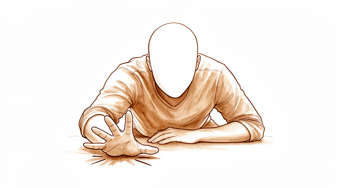
Fraktura ng Distal Radius
Distal radius fractures — assessment, casting, and indications for surgical fixation.
Ang nararamdaman mo
Maaaring masaktan ang iyong pulso at forearms kung saan nangyari ang pagkabasag. Karaniwang matalim ang sakit sa simula, pagkatapos ay magiging malalim na pananakit. Maaaring mapansin mo ang pamamaga at paitim na kumakalat pababa sa iyong kamay. Ang pangunahing layunin ng iyong doktor sa pagsasagawa ng operasyon ay maiwasan ang mga komplikasyon mula sa pinsalang ito. Mahalaga ang maagang pagdi-diagnose at paggamot upang maiwasan ang mga pangmatagalang epekto.
Ang mga simpleng galaw ay maaaring maging mahirap. Maaaring mahirapan kang umabot sa likod upang isara ang bra. Ang pagtatakip ng damit o pag-ikot ng hawakan ng pinto ay maaaring maging matigas o masakit. Ang pag-angat kahit ng magagaan na bagay ay maaaring magdulot ng paglala ng sakit. Gagawin ng iyong doktor ang kailangan upang ibalik ang normal na hugis ng iyong mga buto. Tinitiyak nito na mababawi mo ang iyong kakayahan at babawasan ang sakit sa paglipas ng panahon.
Maaaring lumala ang sakit sa gabi o pagkatapos ng aktibidad sa loob ng araw. May mga araw na mas maganda kaysa sa iba habang nagpapagaling. Bagama’t ang pagpapagaling ng buto ang pangunahing pokus, ang mga isyu sa malambot na tisyu ay maaari ring magdulot ng discomfort. Kasama rito ang iritasyon ng tendon o sensitibidad ng nerbiyo. Sa bihirang kaso, nangyayari ang pinagsamang isyu sa nerbiyo, ngunit sobrang bihira ito. Babantayan ng iyong doktor ang mga lugar na ito nang maigi.
Maaaring mag-alala ka tungkol sa pangmatagalang katigasan o arthritis. Ang pagkaantala sa pagdi-diagnose ng ilang sugat sa ligamento ay maaaring magdulot ng arthritis sa loob ng 10 taon kung hindi gagamutin. Gayunpaman, sa tamang pag-aalaga, gumagaling nang maayos ang karamihan sa mga tao. Napakababa ang risk na hindi magpagaling ang buto. Maaaring gumamit ang iyong doktor ng mga partikular na teknik upang istabilize ang basag. Layunin ng mga pamamaraang ito na magbigay ng istruktural na katatagan at tulungan kang bumalik sa mga pang-araw-araw na gawain nang mas maaga.
Iwasan ang pagsisikap na lumampas sa matinding sakit. Kung mararamdaman mo ang biglaang kawalan ng pakiramdam, pamamanhid, o pagtaas ng pamamaga, kumontak sa iyong care team. Maaaring ito ay mga senyales ng mga komplikasyon sa malambot na tisyu. Gabayan ka ng iyong doktor sa mga ligtas na galaw. Ang pagpapahinga ng braso at pagtaas nito ay makakatulong upang pamahalaan ang pamamaga. Sundin ang payo ng iyong doktor kung kailan magsimula ng banayad na galaw. Ang balanse na ito ay nagpoprotekta sa nagpapagaling na buto habang pinapanatili ang mobility ng iyong mga kasukasuan.
Ano ang nangyayari talaga
Ang distal radius fracture ay isang pagbasag sa malaking buto ng iyong forearms malapit sa pulso. Ang bahaging ito ay nagsisilbing kritikal na shock absorber para sa iyong kamay. Kapag nahulog ka sa isang nakalatag na kamay, ang puwersa ay umaakyat sa iyong braso at maaaring putulin ang butong ito. Karaniwang nagdudulot ang pagbasag na ang mga dulo ng buto ay lumilipat mula sa kanilang tamang posisyon, isang proseso na kilala bilang displacement. Ang maling pagkakahanay na ito ay nagdudulot ng pagkasira sa makinis na ibabaw ng kasukasuan, na nagiging sanhi ng sakit at katigasan sa paggalaw.
Ang pangunahing layunin ng iyong surgeon ay ibalik ang natural na hugis at pagkakahanay ng iyong pulso. Isipin ang iyong kasukasuan ng pulso bilang isang hinge sa pinto. Kung ang frame ng pinto ay nakabaluktot, hindi ito saras o maayos na bubuksan. Gayundin, kung ang radius ay hindi tama ang pagkakahanay, hindi maayos ang pagganap ng iyong pulso. Ang mga optimal na resulta ay nakadepende sa pagpapanatili at pagpapanumbalik ng anatomic alignment. Tinitiyak nito na ang mga buto ay tamang magkakasama, na nagbibigay-daan sa pagbawi ng lakas at range of motion.
Sa maraming kaso, ang pagbasag ay kasama ang metaphysis, ang mas malawak na bahagi ng buto sa itaas ng kasukasuan. Kung ang buto ay magbubulok sa bahaging ito, tinatawag itong comminution. Maaaring gumamit ang iyong surgeon ng bone graft substitutes upang punan ang mga puwang at magbigay ng structural stability. Ang mga materyales na ito ay nagsisilbing scaffolding, hawak ang mga piraso ng buto habang ang iyong katawan ay nagpapagaling. Ang risk na ang buto ay hindi magpapagaling, o nonunion, ay minimal sa mga fracture na ito.
Minsan, ang pagbasag ay kasama ng fracture ng ulnar styloid, isang maliit na bony bump sa kabilang buto ng forearms. Hindi mo kailangang mag-alala tungkol sa hiwalay na pagbasag na ito. Ang associated ulnar styloid fracture ay hindi nakakaapekto sa mga resulta ng iyong distal radial fracture. Ang iyong surgeon ay magpokus sa pag-stabilize ng radius, na ang pangunahing tagapagmana ng iyong pagbawi.
Ang mga komplikasyon ay biro pero kailangang pigilan. Ang nerve damage na kasama ang parehong median at ulnar nerves ay isang napakabiro na komplikasyon. Mahalaga ang maagang diagnosis at paggamot ng anumang isyu upang maiwasan ang mga long-term consequences. Ang iyong surgeon ay gagamit ng iba't ibang paraan upang ayusin ang buto, tulad ng external fixation na may percutaneous pins. Ang approach na ito ay nagbibigay ng reliable na magagandang resulta na may mababang rate ng komplikasyon para sa displaced fractures. Ang susi ay ang maagang pagkilala at pamamahala upang matiyak na bumalik ka sa buong function.
Mga maitutulong namin dito
Ang pangunahing alalahanin mo ngayon ay ang pag-iwas sa mga komplikasyon. Mahalaga ang maagang pagdi-diagnose at paggamot upang maiwasan ang mga pangmatagalang epekto. Dapat mong magpokus sa sariling pamamahala at pisikal na terapiya muna. Gabayin ka ng iyong doktor sa mga ligtas na galaw. Ang layunin ay muling ibalik ang kakayahan nang hindi nagdudulot ng karagdagang pinsala. Maaaring mas malala ang mga komplikasyon sa malambot na tisyu kaysa sa pinsala sa buto mismo. Kasama rito ang pinsala sa tendon, disfunkshon ng nerbiyos, mga problema sa balat, at complex regional pain syndrome. Kailangan mong agad na iulat ang anumang bagong pamamanhid o matinding pamamaga. Ang sabay-sabay na palsy ng median at ulnar nerve ay napakabihira, ngunit nangangailangan ito ng pamantayang estratehiya sa pamamahala kung ito ay mangyari.
Ang medikal na pamamahala ay nakatuon sa pagkontrol ng sakit at pamamaga. Maaaring magreseta ang iyong doktor ng gamot pang-alis ng sakit o anti-inflammatories upang makapagpakalma ka. Sa ilang kaso, maaaring isaalang-alang ang mga injeksyon tulad ng cortisone, hyaluronic acid, o PRP upang bawasan ang pamamaga at sakit. Layunin ng mga treatment na ito na magbigay ng ginhawa habang nagsisimula nang gumaling ang iyong katawan. Karaniwang limitado ang tagal ng epekto ng mga injeksyon na ito, na nagbibigay-daan sa iyo na makilahok sa terapiya. Mahalagang tandaan na ang mga bone graft substitutes ay pangunahing ginagamit upang magbigay ng istruktural na katatagan kaysa sa simpleng pag-alis ng sakit. Minimal ang panganib ng nonunion sa mga distal radius fracture, kaya’t nananatiling pokus ang pag-aalaga sa malambot na tisyu at pagkakahanay.
Isinasagawa ang operasyon kapag naabot na ng konservatibong paggamot ang hangganan nito. Maaaring rekomendahan ng iyong doktor ang operatibong paggamot kung may malaking deformity o kawalan ng katatagan. Ang mga radiographic na salik, tulad ng metaphyseal collapse ratio, ay tumutulong na ma-predict ang kawalan ng katatagan. Ang kasamang ulnar styloid fracture ay hindi nakakaapekto sa mga resulta ng distal radial fracture, kaya’t dapat mag-ingat ang mga kliniko sa pagpili ng operatibong paggamot para dito lamang. Ang external fixation na may dagdag na percutaneous pins ay isang mahusay na opsyon para sa mga displaced fracture. Ang paraang ito ay nagbibigay ng maaasahang magagandang resulta na may mababang rate ng reoperation at komplikasyon. Maaaring magbigay ang volar plating ng mas mahusay na radiological na resulta kumpara sa k-wiring, ngunit hindi ito laging nakakaugnay sa mas magandang functional na resulta sa 32 na buwan ng pagsubaybay. Ang mga optimal na resulta ay nakadepende sa maagang pagkilala at pamamahala ng anumang kasamang pinsala, tulad ng mga pinsala sa intrinsic carpal ligament. Ang pagkaantala sa pagdi-diagnose ng mga pinsalang ito sa ligament ay nagdudulot ng arthritis sa loob ng 10 taon kung hindi ito gagamutin.
Ano ang inaasahan
Ang pangunahing layunin ng iyong surgeon ay pigilan ang mga komplikasyon at siguraduhing gumaling ka nang tama. Mahalaga ang maagang pagdi-diyagnosis at paggamot upang maiwasan ang mga pangmatagalang isyu. Karamihan sa mga tao ay gumagaling nang maayos kapag ang fracture ay tamang pinamamahalaan. Napakababa ang risk na hindi gumaling ang buto (nonunion).
Maaaring makita mo ang kaunting pagbaba ng taas ng iyong wrist bone kukuha ka ng plate. Normal ito at karaniwang hindi nakakaapekto sa iyong final result. Kung mayroon kang maliit na fracture sa gilid ng iyong wrist (ulnar styloid), karaniwang hindi nito binabago ang iyong outcome. Ang iyong surgeon ang magdesisyon kung kailangan ng surgery batay sa pangunahing break, hindi lamang sa piraso sa gilid na ito.
Minsan, mas malala ang mga sugat sa malambot na bahagi (soft tissue injuries) kaysa sa sariling pagkabasag ng buto. Kasama rito ang iritasyon ng tendon, mga isyu sa nerve, o mga problema sa balat. Napaka-rare ang combined nerve damage. Kung mayroon kang complex na break na may dislocation, ang maagang pagkilala at pagpapanatili ng mga buto sa tamang posisyon ang susi sa magandang outcome. Ang pagka-delay na pagdi-diyagnosis ng mga sugat sa ligament sa wrist ay maaaring magdulot ng arthritis sa loob ng 10 taon kung hindi gagamutin.
Sa pamamagitan ng mga modernong teknik, maaaring ibalik ng iyong surgeon ang hugis at katatagan ng buto. Tumutulong ito upang muling makabalik ka sa iyong function nang mas maaga. Ang external fixation gamit ang mga pins ay isang mahusay na opsyon para sa mga displaced fractures, na nag-aalok ng maaasahang mga resulta na may mababang rate ng komplikasyon at reoperation. Ang volar plating ay nagbibigay ng mas mahusay na pagkakahanay ng buto sa X-rays kumpara sa mga wires, bagaman ang mga functional outcomes sa 32 buwan ay katulad.
Kung iiwan ito o gagamutin nang hindi tama, maaaring magpatuloy ang mga komplikasyon. Babantayan ka ng iyong surgeon nang mahigpit upang maunawaan ang anumang mga isyu nang maaga. Karamihan sa mga pasyente ay makakabalik sa magandang paggamit ng kanilang wrist. Inaasahan mo ang patuloy na pagpapabuti sa loob ng mga linggo at buwan. Sundin ang payo ng iyong surgeon upang protektahan ang iyong mga gumagaling na tisyu at maiwasan ang mga setback.
Kailan pumunta sa doktor
Kumonsulta sa iyong doktor kung mayroon kang patuloy na sakit na hindi gumagaling kahit pahinga, o kung napapansin mo ang kahinaan o kawalan ng katatagan sa iyong pulso. Humingi ng pagsusuri ng espesyalista kung ang iyong mga sintomas ay nakakaapekto sa pagtulog o trabaho, o kung may biglaang paglala. Mahalaga ang maagang pagdi-diagnos at paggamot upang maiwasan ang mga pangmatagalang epekto. Ang mga komplikasyon sa malambot na tisyu ay maaaring mas malala kaysa sa sugat sa buto mismo. Halimbawa, ang pagkaantala sa pagdi-diagnos ng mga pinsala sa intrinsic carpal ligament ay nagdudulot ng arthritis sa loob ng 10 taon kung hindi gagamutin. Ang pangunahing alalahanin ng iyong surgeon ay ang pag-iwas sa mga isyung ito sa pamamagitan ng maagang pagkilala at pamamahala.
Evidence & references
Overview
- Prevention of complications associated with distal radius fractures should be the treating surgeon's primary concern [1].
- Early diagnosis and treatment are important to avoid long-term consequences of distal radius fracture complications [1].
- Combined median and ulnar nerve palsy related to distal fractures of the radius is exceedingly rare [2].
- Combined median and ulnar nerve palsy complicating distal radius fractures require a standardised management strategy [2].
- Optimal outcomes in the treatment of forearm fracture–dislocations depend on early recognition and management [4].
- Restoration and maintenance of anatomic alignment are key principles for optimal outcomes in forearm fracture–dislocations [4].
- Novel locking plate designs have resulted in a rethinking of the contemporary approach to distal radius fracture fixation [6].
- A certain degree of radial height loss is noted in patients undergoing fracture fixation with volar locking plate for extra-articular distal radius fractures [7].
- An associated ulnar styloid fracture does not affect the outcomes of a distal radial fracture [8].
- Clinicians should be cautious in electing operative treatment for patients with an ulnar styloid fracture [8].
- Bone graft substitutes are primarily used to provide structural stability in distal radius fractures [9].
- Bone graft substitutes may facilitate early return to function in distal radius fractures [9].
- The risk of nonunion is minimal in distal radius fractures [9].
- External fixation supplemented with percutaneous pins is an excellent option for treating displaced fractures of the distal radius [10].
- External fixation supplemented with percutaneous pins yields reliably good results for displaced distal radius fractures [10].
- External fixation supplemented with percutaneous pins has a low reoperation rate for displaced distal radius fractures [10].
- External fixation supplemented with percutaneous pins has a low complication rate for displaced distal radius fractures [10].
- Die punch fragment size is not an indicator of the need for or use of a dorsal approach in distal radius fracture fixation [17].
Anatomy & Pathophysiology
- Prevention of complications associated with distal radius fractures should be the treating surgeon's primary concern, with early diagnosis and treatment being important to avoid long-term consequences [1].
- Combined median and ulnar nerve palsy related to distal fractures of the radius are exceedingly rare but require a standardized management strategy [2].
- Optimal outcomes in the treatment of forearm fracture–dislocations depend on early recognition and management, with restoration and maintenance of anatomic alignment being the key principles [4].
- A certain degree of radial height loss is noted in patients undergoing fracture fixation with volar locking plate for extra-articular distal radius fractures [7].
- An associated ulnar styloid fracture does not affect the outcomes of a distal radial fracture and clinicians should be cautious in electing operative treatment for patients with an ulnar styloid fracture [8].
- Bone graft substitutes are primarily used to provide structural stability and perhaps early return to function in distal radius fractures, where the risk of nonunion is minimal [9].
- Metaphyseal collapse ratio, a novel radiographic parameter, was found to provide a reliable measure of metaphyseal comminution, and to be significantly correlated with other radiographic parameters that predict distal radius fracture instability [11].
- There may be radiographic factors other than measures of deformity that some surgeons use to determine recommendations for surgery [12].
- Early accurate diagnosis of intrinsic carpal ligament injuries provides for best outcomes, while delayed diagnosis leads to arthritis within 10 years if not treated [16].
- DP fragment size is not an indicator of the need for or use of a dorsal approach in distal radius fracture fixation [17].
- Pronation effectively increases the proximal 'safe zone' of the posterior interosseous nerve, suggesting the forearm should be placed in pronation to minimize the risk of iatrogenic injury [18].
- CT scan should be requested only by experienced hand surgeons in order to help guide treatment, as it does not significantly improve inter- and intra-observer agreement for all classification systems [20].
Classification
- CT scans do not significantly improve inter- and intra-observer agreement for the AO, Fernandez, and Universal classification systems for distal radius fractures [20].
- The metaphyseal collapse ratio (MCR) is a novel radiographic parameter that provides a reliable measure of metaphyseal comminution [11].
- The metaphyseal collapse ratio (MCR) is significantly correlated with other radiographic parameters that predict distal radius fracture instability [11].
- There may be radiographic factors other than measures of deformity that some surgeons use to determine recommendations for surgery [12].
Clinical Presentation
- Prevention of complications associated with distal radius fractures should be the treating surgeon's primary concern [1].
- Early diagnosis and treatment are important to avoid long-term consequences of distal radius fracture complications [1].
- Combined median and ulnar nerve palsy related to distal fractures of the radius is exceedingly rare [2].
- Optimal outcomes in the treatment of forearm fracture–dislocations depend on early recognition and management [4].
- Restoration and maintenance of anatomic alignment are key principles for optimal outcomes in forearm fracture–dislocations [4].
- A certain degree of radial height loss is noted in patients undergoing fracture fixation with volar locking plate for extra-articular distal radius fractures [7].
- An associated ulnar styloid fracture does not affect the outcomes of a distal radial fracture [8].
- Clinicians should be cautious in electing operative treatment for patients with an ulnar styloid fracture [8].
- Bone graft substitutes are primarily used to provide structural stability and perhaps early return to function in distal radius fractures [9].
- The risk of nonunion is minimal in distal radius fractures [9].
- External fixation supplemented with percutaneous pins is an excellent option for treating displaced fractures of the distal radius [10].
- External fixation supplemented with percutaneous pins yields reliably good results for displaced distal radius fractures [10].
- External fixation supplemented with percutaneous pins has a low reoperation rate for displaced distal radius fractures [10].
- External fixation supplemented with percutaneous pins has a low complication rate for displaced distal radius fractures [10].
- Metaphyseal collapse ratio is a novel radiographic parameter that provides a reliable measure of metaphyseal comminution [11].
- Metaphyseal collapse ratio is significantly correlated with other radiographic parameters that predict distal radius fracture instability [11].
- There may be radiographic factors other than measures of deformity that some surgeons use to determine recommendations for surgery in distal radius fractures [12].
- Soft tissue complications encountered during the management of distal radius fractures include tendon injury, nerve dysfunction, vascular compromise, skin problems, compartment syndrome, and complex regional pain syndrome [15].
- Complications associated with soft tissues may be more problematic than the bone injury itself in distal radius fractures [15].
- Early accurate diagnosis of intrinsic carpal ligament injuries provides for best outcomes [16].
- Delayed diagnosis of intrinsic carpal ligament injuries leads to arthritis within 10 years if not treated [16].
- Pronation effectively increases the proximal 'safe zone' of the posterior interosseous nerve [18].
- The forearm should be placed in pronation to minimize the risk of iatrogenic injury to the posterior interosseous nerve [18].
- Monteggia fractures can be easily overlooked if radiographs of the elbow are not taken [19].
- Pre-existing congenital radial head dislocations can lead to inappropriate surgical intervention if not distinguished from Monteggia fractures [19].
- Early recognition and treatment of Essex-Lopresti injury is associated with improved outcomes [21].
Investigations
- Early diagnosis and treatment of complications associated with distal radius fractures are important to avoid long-term consequences [1].
- Combined median and ulnar nerve palsy related to distal radius fractures is exceedingly rare [2].
- Optimal outcomes in the treatment of forearm fracture–dislocations depend on early recognition and management [4].
- Restoration and maintenance of anatomic alignment are key principles in the treatment of forearm fracture–dislocations [4].
- An associated ulnar styloid fracture does not affect the outcomes of a distal radial fracture [8].
- Clinicians should be cautious in electing operative treatment for patients with an ulnar styloid fracture [8].
- Metaphyseal collapse ratio is a novel radiographic parameter that provides a reliable measure of metaphyseal comminution [11].
- Metaphyseal collapse ratio is significantly correlated with other radiographic parameters that predict distal radius fracture instability [11].
- There may be radiographic factors other than measures of deformity that some surgeons use to determine recommendations for surgery [12].
- Soft tissue complications encountered during the management of distal radius fractures include tendon injury, nerve dysfunction, vascular compromise, skin problems, compartment syndrome, and complex regional pain syndrome [15].
- Complications associated with soft tissues may be more problematic than the bone injury itself in distal radius fractures [15].
- Early accurate diagnosis of intrinsic carpal ligament injuries provides for best outcomes [16].
- Delayed diagnosis of intrinsic carpal ligament injuries leads to arthritis within 10 years if not treated [16].
- Monteggia fractures can be easily overlooked if radiographs of the elbow are not taken [19].
- Pre-existing congenital radial head dislocations can lead to inappropriate surgical intervention if misdiagnosed as Monteggia fractures [19].
Treatment
- Prevention of complications associated with distal radius fractures should be the treating surgeon's primary concern [1].
- Early diagnosis and treatment are important to avoid long-term consequences of distal radius fracture complications [1].
- Combined median and ulnar nerve palsy related to distal fractures of the radius is exceedingly rare [2].
- Combined median and ulnar nerve palsy complicating distal radius fractures require a standardised management strategy [2].
- Optimal outcomes in the treatment of forearm fracture–dislocations depend on early recognition and management [4].
- Restoration and maintenance of anatomic alignment are key principles in the treatment of forearm fracture–dislocations [4].
- A certain degree of radial height loss is noted in patients undergoing fracture fixation with volar locking plate for extra-articular distal radius fractures [7].
- An associated ulnar styloid fracture does not affect the outcomes of a distal radial fracture [8].
- Clinicians should be cautious in electing operative treatment for patients with an ulnar styloid fracture [8].
- Bone graft substitutes are primarily used to provide structural stability in distal radius fractures [9].
- Bone graft substitutes are used to perhaps provide early return to function in distal radius fractures [9].
- The risk of nonunion is minimal in distal radius fractures [9].
- External fixation supplemented with percutaneous pins is an excellent option for treating displaced fractures of the distal radius [10].
- External fixation supplemented with percutaneous pins for displaced distal radius fractures yields reliably good results [10].
- External fixation supplemented with percutaneous pins for displaced distal radius fractures has a low reoperation rate [10].
- External fixation supplemented with percutaneous pins for displaced distal radius fractures has a low complication rate [10].
- There may be radiographic factors other than measures of deformity that some surgeons use to determine recommendations for surgery in distal radius fractures [12].
- Superior radiological results were attained with volar plating compared to k-wiring for distal radius fractures [13].
- Superior radiological results with volar plating did not correlate with a better functional outcome compared to k-wiring at 32 months follow up [13].
Complications
- Prevention of complications associated with distal radius fractures should be the treating surgeon's primary concern [1].
- Early diagnosis and treatment of complications are important to avoid long-term consequences [1].
- Combined median and ulnar nerve palsy related to distal fractures of the radius is exceedingly rare [2].
- Combined median and ulnar nerve palsy complicating distal radius fractures require a standardised management strategy [2].
- Optimal outcomes in the treatment of forearm fracture–dislocations depend on early recognition and management [4].
- Restoration and maintenance of anatomic alignment are key principles for optimal outcomes in forearm fracture–dislocations [4].
- A certain degree of radial height loss is noted in patients undergoing fracture fixation with volar locking plate for extra-articular distal radius fractures [7].
- An associated ulnar styloid fracture does not affect the outcomes of a distal radial fracture [8].
- Clinicians should be cautious in electing operative treatment for patients with an ulnar styloid fracture [8].
- Bone graft substitutes are primarily used to provide structural stability in distal radius fractures [9].
- Bone graft substitutes may provide early return to function in distal radius fractures [9].
- The risk of nonunion in distal radius fractures is minimal [9].
- External fixation supplemented with percutaneous pins is an excellent option for treating displaced fractures of the distal radius [10].
- External fixation supplemented with percutaneous pins for displaced distal radius fractures yields reliably good results [10].
- External fixation supplemented with percutaneous pins for displaced distal radius fractures has a low reoperation rate [10].
- External fixation supplemented with percutaneous pins for displaced distal radius fractures has a low complication rate [10].
- Soft tissue complications encountered during the management of distal radius fractures include tendon injury, nerve dysfunction, vascular compromise, skin problems, compartment syndrome, and complex regional pain syndrome [15].
- Complications associated with soft tissues may be more problematic than the bone injury itself in distal radius fractures [15].
Recovery
- Prevention of complications associated with distal radius fractures should be the treating surgeon's primary concern [1].
- Early diagnosis and treatment of complications are important to avoid long-term consequences [1].
- Combined median and ulnar nerve palsy related to distal radius fractures is exceedingly rare [2].
- Combined median and ulnar nerve palsy complicating distal radius fractures requires a standardised management strategy [2].
- Optimal outcomes in the treatment of forearm fracture–dislocations depend on early recognition and management [4].
- Restoration and maintenance of anatomic alignment are key principles for optimal outcomes in forearm fracture–dislocations [4].
- A certain degree of radial height loss is noted in patients undergoing fracture fixation with volar locking plate for extra-articular distal radius fractures [7].
- An associated ulnar styloid fracture does not affect the outcomes of a distal radial fracture [8].
- Clinicians should be cautious in electing operative treatment for patients with an ulnar styloid fracture [8].
- Bone graft substitutes are primarily used to provide structural stability in distal radius fractures [9].
- Bone graft substitutes may facilitate early return to function in distal radius fractures [9].
- The risk of nonunion in distal radius fractures is minimal [9].
- External fixation supplemented with percutaneous pins is an excellent option for treating displaced fractures of the distal radius [10].
- External fixation supplemented with percutaneous pins yields reliably good results for displaced distal radius fractures [10].
- External fixation supplemented with percutaneous pins has a low reoperation rate for displaced distal radius fractures [10].
- External fixation supplemented with percutaneous pins has a low complication rate for displaced distal radius fractures [10].
- Metaphyseal collapse ratio is a novel radiographic parameter that provides a reliable measure of metaphyseal comminution [11].
- Metaphyseal collapse ratio is significantly correlated with other radiographic parameters that predict distal radius fracture instability [11].
- Volar plating attains superior radiological results compared to k-wiring for distal radius fractures [13].
- Superior radiological results with volar plating do not correlate with better functional outcomes compared to k-wiring at 32 months follow up [13].
- Early accurate diagnosis of intrinsic carpal ligament injuries provides for best outcomes [16].
- Delayed diagnosis of intrinsic carpal ligament injuries leads to arthritis within 10 years if not treated [16].
Key Evidence
- [Paper] Prevention of complications associated with distal radius fractures should be the treating surgeon's primary concern, with early diagnosis and treatment being important to avoid long-term consequences. [1] (10.1016/j.hcl.2014.12.002)
- [Paper] Combined median and ulnar nerve palsy related to distal fractures of the radius are exceedingly rare but require a standardised management strategy. [2] (10.1016/j.otsr.2018.04.026)
- [L5] Optimal outcomes in the treatment of forearm fracture–dislocations depend on early recognition and management, with restoration and maintenance of anatomic alignment being the key principles. [4] (10.1016/j.hcl.2015.01.010)
- [Paper] The management of distal radius fractures is in the midst of a renaissance with novel locking plate designs resulting in a rethinking of the contemporary approach to fracture fixation. [6] (10.1016/j.hcl.2005.04.001)
- [Paper] A certain degree of radial height loss is noted in patients undergoing fracture fixation with volar locking plate for extra-articular distal radius fractures. [7] (10.1016/j.otsr.2021.102842)
- [L1] Based on this meta-analysis, an associated ulnar styloid fracture does not affect the outcomes of a distal radial fracture and clinicians should be cautious in electing operative treatment for patients with an ulnar styloid fracture. [8] (10.1016/j.injury.2017.08.061)
- [L4] Bone graft substitutes are primarily used to provide structural stability and perhaps early return to function in distal radius fractures, where the risk of nonunion is minimal. [9] (10.1016/j.hcl.2012.02.004)
- [L1] External fixation supplemented with percutaneous pins is an excellent option for treating displaced fractures of the distal radius, with reliably good results, a low reoperation rate, and a low complication rate. [10] (10.1016/j.hcl.2009.08.008)
- [Paper] Metaphyseal collapse ratio, a novel radiographic parameter, was found to provide a reliable measure of metaphyseal comminution, and to be significantly correlated with other radiographic parameters that predict distal radius fracture instability. [11] (10.1016/j.otsr.2013.05.002)
- [Paper] There may be radiographic factors other than measures of deformity that some surgeons use to determine recommendations for surgery. [12] (10.1007/s12593-014-0164-0)
- [L3] Although superior radiological results were attained with volar plating, these results did not correlate with a better functional outcome compared to k-wiring at 32 months follow up. [13] (10.1016/j.injury.2015.08.040)
- [L5] This review focuses on soft tissue complications encountered during the management of distal radius fractures, including tendon injury, nerve dysfunction, vascular compromise, skin problems, compartment syndrome, and complex regional pain syndrome, noting that complications associated with soft tissues may be more problematic than the bone injury itself. [15] (10.1016/j.hcl.2009.11.002)
- [L5] Early accurate diagnosis of intrinsic carpal ligament injuries provides for best outcomes, while delayed diagnosis leads to arthritis within 10 years if not treated. [16] (10.1016/j.hcl.2015.01.003)
- [Paper] DP fragment size is not an indicator of the need for or use of a dorsal approach in DRF fixation. [17] (10.1055/s-0040-1712328)
- [Paper] Pronation effectively increases the proximal 'safe zone' of the nerve, suggesting the forearm should be placed in pronation to minimize the risk of iatrogenic injury. [18] (10.1016/j.injury.2015.01.028)
- [L4] Monteggia fractures can be easily overlooked if radiographs of the elbow are not taken, and pre-existing congenital radial head dislocations can lead to inappropriate surgical intervention. [19] (10.1016/j.injury.2005.08.028)
- [Paper] CT scan should be requested only by experienced hand surgeons in order to help guide treatment, as it does not significantly improve inter- and intra-observer agreement for all classification systems. [20] (10.1016/j.injury.2014.06.017)
- [L5] Early recognition and treatment is associated with improved outcomes. [21] (10.1016/j.hcl.2020.07.012)
References
[1] Management of Complications of Distal Radius Fractures. Hand Clinics. 2015. DOI: 10.1016/j.hcl.2014.12.002 [2] Combined median and ulnar nerve palsy complicating distal radius fractures. Orthopaedics & Traumatology: Surgery & Research. 2018. DOI: 10.1016/j.otsr.2018.04.026 [4] Management of Complications of Forearm Fractures. Hand Clinics. 2015. DOI: 10.1016/j.hcl.2015.01.010 [6] Distal Radius Fractures. Hand Clinics. 2005. DOI: 10.1016/j.hcl.2005.04.001 [7] Loss of radial height in extra-articular distal radial fracture following volar locking plate fixation. Orthopaedics & Traumatology: Surgery & Research. 2021. DOI: 10.1016/j.otsr.2021.102842 [8] Does concomitant ulnar styloid fracture and distal radius fracture portend poorer outcomes? A meta-analysis of comparative studies. Injury. 2017. DOI: 10.1016/j.injury.2017.08.061 [9] The Use of Bone Grafts and Substitutes in the Treatment of Distal Radius Fractures. Hand Clinics. 2012. DOI: 10.1016/j.hcl.2012.02.004 [10] External Fixation of Distal Radius Fractures. Hand Clinics. 2010. DOI: 10.1016/j.hcl.2009.08.008 [11] Distal radius fracture metaphyseal comminution: A new radiographic parameter for quantifying, the metaphyseal collapse ratio (MCR). Orthopaedics & Traumatology: Surgery & Research. 2013. DOI: 10.1016/j.otsr.2013.05.002 [12] Radiographs Versus Radiographic Measurements in Distal Radius Fractures. Journal of Hand and Microsurgery. 2015. DOI: 10.1007/s12593-014-0164-0 [13] Volar plate versus k-wire fixation of distal radius fractures. Injury. 2016. DOI: 10.1016/j.injury.2015.08.040 [15] Soft Tissue Complications of Distal Radius Fractures. Hand Clinics. 2010. DOI: 10.1016/j.hcl.2009.11.002 [16] Management of Complications of Ligament Injuries of the Wrist. Hand Clinics. 2015. DOI: 10.1016/j.hcl.2015.01.003 [17] The Die Punch Fragment: Analysis of Fragment Geometry and Need for Fixation. Journal of Hand and Microsurgery. 2022. DOI: 10.1055/s-0040-1712328 [18] The course of the posterior interosseous nerve in relation to the proximal radius: Is there a reliable landmark?. Injury. 2015. DOI: 10.1016/j.injury.2015.01.028 [19] When is a Monteggia fracture not a Monteggia fracture?. Injury Extra. 2007. DOI: 10.1016/j.injury.2005.08.028 [20] Does the CT improve inter- and intra-observer agreement for the AO, Fernandez and Universal classification systems for distal radius fractures?. Injury. 2014. DOI: 10.1016/j.injury.2014.06.017 [21] The Essex-Lopresti Injury:. Hand Clinics. 2020. DOI: 10.1016/j.hcl.2020.07.012