Patients › Elbow
Bali ng Olecranon
Olecranon fractures — patterns, non-operative care, and tension-band or plate fixation.
Ang iyong nararamdaman
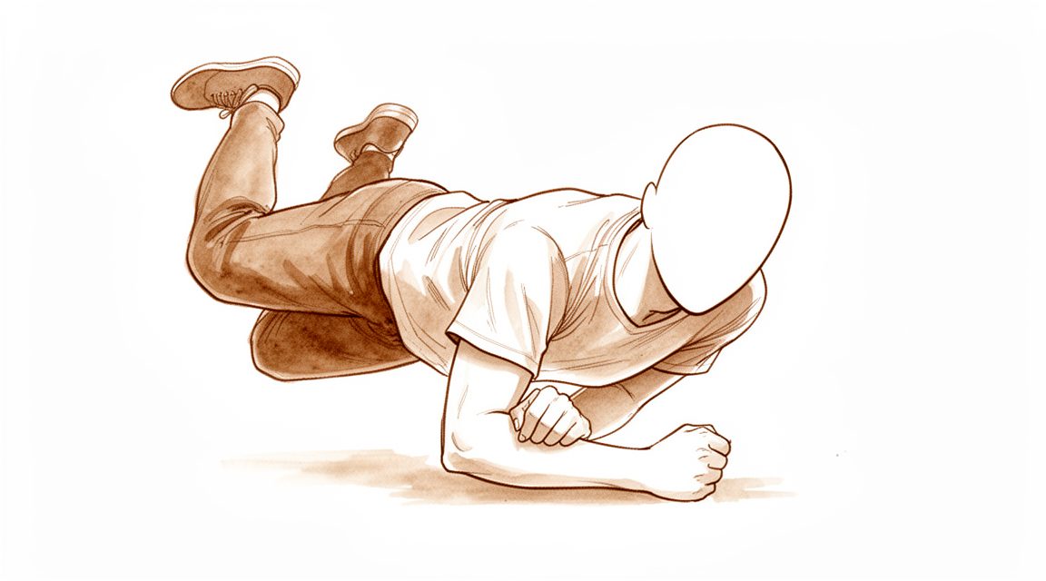
Ang olecranon fracture ay isang bali sa matulis na bahaging buto sa likod ng iyong siko. Karaniwan itong nangyayari sa isa sa dalawang paraan. Sa mga mas batang tao, kadalasan itong sumusunod sa isang malakas na pagkahulog o direktang pagkakauntog sa siko. Sa mga nakatatanda, madalas itong nagmumula sa simpleng pagkahulog mula sa pagkakatayo. Ang biglaan at malakas na pagtuwid ng braso ay maaari ring humila at tumanggal ng isang piraso ng buto mula sa dulo ng siko.
Karaniwan mong mararamdaman ang sakit mismo sa likod ng siko, at kadalasan itong masakit sa isang partikular na lugar sa halip na sa buong siko. Karaniwan ang pamamaga sa likod ng siko, at maaari itong maging matindi. Dahil ang matulis na bahaging buto na iyon ay nasa ilalim lang ng balat, kung minsan ay mararamdaman mo ang gilid ng bali sa kabila ng balat. Kung napunit ang balat sa pagkahulog, ang sugat ay karaniwang nasa likod ng siko, kung saan ito tumama sa lupa. Maaaring mahirap gamitin ang braso, at ang pagtuwid nito laban sa isang bagay, tulad ng pagtulak pataas mula sa upuan, ay maaaring pakiramdam na mahina o imposible.
Sa mga unang araw, kadalasang mas matindi ang sakit kapag sinusubukan mong igalaw ang siko, at maaari nitong guluhin ang iyong tulog. Ang mga pang-araw-araw na gawain na nagbabaluktot o nagpapabigat sa siko, tulad ng pagbuhat ng kettle, pagbibihis, o paghimlay sa iyong forearm, ay karaniwang masakit. Humuhupa ang pamamaga at pasa sa unang humigit-kumulang dalawang linggo, at unti-unting gumagaan ang sakit habang nagsisimulang maghilom ang buto.
May mga bagay na nangangailangan ng agarang pangangalaga. Pumunta sa emergency department sa parehong araw kung napunit ang balat sa ibabaw ng siko, kung nakalabas ang buto, o kung halatang wala sa hugis ang braso. Ganoon din kung ang iyong kamay o mga daliri ay namutla, nanlamig, pumuti o nangasul, o kung bigla kang nawalan ng pakiramdam o paggalaw sa braso pagkatapos ng pinsala. Kung ang mga sintomas ay hindi humuhupa, lumalala sa loob ng mga linggo, o ginigising ka sa gabi, magpatingin sa iyong GP o humingi ng specialist review. Kung hindi mo makontak ang klinika, pumunta sa pinakamalapit na emergency department.
Ano ang aktwal na nangyayari
Ang matulis na bahaging buto sa likod ng iyong siko ay tinatawag na olecranon. Ito ang itaas na dulo ng isa sa mga buto ng iyong forearm, at kumikilos ito na parang takip ng bisagra. Kapag itinutuwid mo ang iyong braso, dumudulas ang takip na iyon sa isang uka sa dulo ng buto ng iyong itaas na braso. Mahalaga ang bali dahil sa nakakabit dito: ang tendon mula sa iyong triceps, ang kalamnan sa likod ng iyong itaas na braso, ay nakaangkla mismo sa puntong iyon. Ang tendon na iyon ang nagpapahintulot sa iyo na ituwid ang iyong siko laban sa isang pasan.
Isipin ito na parang isang pinto na may malakas na spring na humihila dito pasara. Ang olecranon ang bracket na humahawak sa spring na iyon. Kapag nabali ang buto, patuloy na humihila ang spring, at kinakaladkad nito ang nabaling piraso pataas at palayo, na nag-iiwan ng puwang sa surface ng joint. Iyan ang dahilan kung bakit hindi normal na gumagana ang iyong braso ngayon: malakas pa rin ang kalamnan, ngunit humihila ito sa isang maluwag na piraso ng buto sa halip na igalaw ang iyong forearm. Nagiging mahina o imposible ang pagtuwid laban sa resistensya.
Naghihilom ang buto sa pamamagitan ng muling pagdudugtong, at ang isang nabaling piraso na may nakakabit na tendon ay maaaring muling kumabit habang nagdudugtong ang buto. Ngunit kailangang nasa tamang posisyon ang mga piraso para gumana ito nang maayos. Kapag hindi na-shift ang bali, maaaring panatilihin lamang na hindi gumagalaw ang mga piraso habang nagdudugtong ang mga ito. Kapag naghiwalay ang mga piraso, o kapag ang surface ng joint ay nabali sa ilang piraso, maaaring maging hindi matatag ang siko, at ang mga baling iyon ay karaniwang nangangailangan ng operasyon upang mapanatili ang mga ito sa pwesto habang naghihilom.
Inuuri ng mga surgeon ang mga baling ito ayon sa kung gaano kalayo ang paggalaw ng mga piraso at kung nananatiling matatag ang elbow joint. Sa unang grado, hindi na-shift ang bali. Sa ikalawang grado, na-shift ang bali ngunit gumagana pa rin ang joint. Ang ikatlong grado ay nangangahulugang ang mismong elbow joint ay hindi matatag. Ang mga gradong na-shift at hindi matatag ang kadalasang nangangailangan ng operasyon.
Karamihan sa mga baling ito ay gumagaling nang maayos, at karaniwan ang maliit na pagkawala ng ganap na pagtuwid.
Ano ang maaari naming gawin tungkol dito
Si Dr Kieran Hirpara, isang upper-limb surgeon sa Mater Private Hospital Rockhampton, ay itinutugma ang gamutan sa iyong partikular na pinsala. Ang ilan sa mga baling ito ay gumagaling nang walang operasyon, at ang iba ay nangangailangan ng surgery sa lalong madaling panahon pagkatapos ng pinsala, kaya mahalaga ang maagap na pagsusuri. Ang mga pasyente ay karaniwang nirerefer sa aming klinika ng kanilang GP; kung iminungkahi ng isang physiotherapist na magpatingin sa amin, kakailanganin mo pa rin ng referral mula sa iyong GP upang maging eligible para sa Medicare rebate. Sa klinika, kumukuha kami ng history, sinusuri ang iyong braso, at nag-aayos ng imaging kung kinakailangan.
Kung hindi na-shift ang bali, o kaunti lamang ang pag-shift nito, at matatag ang iyong siko, maaari naming irekomenda na panatilihin itong hindi gumagalaw habang naghihilom. Ibig sabihin nito ay isang splint o cast sa loob ng ilang panahon, na may mga pagsusuri gamit ang paulit-ulit na X-ray upang matiyak na nananatili sa pwesto ang mga piraso. Karaniwan ang landas na ito para sa mga nakatatandang pasyente, at para sa mga taong ginagamit ang braso sa mga pang-araw-araw na gawain sa halip na mabigat na trabaho. Naiiwasan nito ang operasyon at ang mga panganib na kasama nito. Ang kapalit ay maaaring hindi ganap na maituwid ang siko sa huli, at nahihirapan ang ilang tao na tanggapin iyon. Ibinabalik ang paggalaw nang paunti-unti kasama ang iyong hand therapist, na gagawa rin ng anumang splint na kailangan mo.
Kung naghiwalay ang mga piraso, o hindi matatag ang elbow joint, karaniwan naming irerekomenda ang operasyon mula sa simula. Ang layunin ay panatilihin ang mga piraso sa tamang posisyon upang makapagdugtong ang buto at maigalaw mo ang siko nang maaga. Ang pagpili kung paano hahawakan ang buto ay nakadepende sa pattern ng bali, at walang iisang paraan na angkop sa bawat fracture. Kung minsan, tunay na bukas ang parehong landas, halimbawa sa isang nakatatandang pasyente na may na-shift na bali. Pag-uusapan natin kung ano ang mahalaga sa iyo: kung gaano karaming sakit ang handa mong tiisin, at kung maaabala ka ba sa pagkawala ng huling ilang degrees ng pagtuwid.
Anumang landas ang piliin mo, magkatulad ang mga unang linggo. Pinapanatili kang komportable ng pain relief habang humuhupa ang pamamaga. Kailangang protektahan ang pinsala habang nagdudugtong ang buto, at sasabihin namin sa iyo kung ano ang maaari at hindi mo maaaring gawin gamit ang braso. Gagabayan ng iyong hand therapist ang iyong paggalaw sa tamang yugto, marahan sa simula, at unti-unting dinaragdagan habang naghihilom ang buto. Karamihan sa mga baling ito ay gumagaling nang maayos, at karaniwan ang maliit na pagkawala ng ganap na pagtuwid. Ang metalwork na nasa ilalim lang ng balat sa likod ng siko ay maaaring hindi komportable, at kailangan ng ilang tao ng maliit na operasyon sa kalaunan upang tanggalin ito.
Ano ang dapat asahan
Karamihan sa mga baling ito ay gumagaling nang maayos. Karaniwan ang maliit na pagkawala ng ganap na pagtuwid, at karamihan sa mga tao ay nananatiling maayos ang paggamit ng braso. Maganda ang pangmatagalang resulta pagkatapos ng operasyon para sa mga komplikadong bali, kahit na may lumitaw na kaunting wear-and-tear arthritis sa joint sa mga susunod na X-ray.
Kung ang iyong bali ay pinapanatiling hindi gumagalaw sa isang splint o cast, nagdudugtong ang buto sa loob ng mga linggo sa halip na mga araw. Magkakaroon ka ng paulit-ulit na X-ray upang matiyak na nananatili ang mga piraso kung saan sila dapat. Angkop ang landas na ito sa mga nakatatandang pasyente at sa mga taong ginagamit ang braso sa mga pang-araw-araw na gawain sa halip na mabigat na trabaho. Kasiya-siya ang mga resulta sa grupong iyon, sa maikli at sa pangmatagalang panahon. Ang kapalit ay maaaring hindi ganap na maituwid ang siko sa huli.
Kung sasailalim ka sa operasyon, hinahawakan sa pwesto ang buto gamit ang metal habang nagdudugtong ito. Nagsisimula nang maaga ang paggalaw at unti-unting dinaragdagan kasama ang iyong hand therapist. Inaasahan ang kaunting pagkawala ng ganap na pagtuwid pagkatapos ng pinsalang ito, anumang landas ang piliin mo. Sa isang pangmatagalang pag-aaral ng mga taong ginamot sa operasyon para sa na-shift na bali, 96% ang nagkaroon ng mabuti o napakahusay na resulta hanggang 25 taon pagkatapos ng pinsala.
Ang paggaling ay unti-unting pagbabalik sa iyong mga karaniwang ginagawa. Unang bumabalik ang mga magagaan na pang-araw-araw na gawain, at pagkatapos ay ang pagbubuhat at mas mabigat na paggamit habang lumalakas ang buto. Kung gaano kaaga ka makakapagmaneho, makakapagtrabaho o makakapaglaro ng sport ay nakadepende sa iyong trabaho, sa iyong braso at sa kung paano naghihilom ang buto, at gagabayan ka namin sa bawat yugto.
Mabuting malaman ang mga bagay na maaaring magkamali. Ang metalwork ay nasa ilalim lang ng balat sa likod ng siko, at maaari itong maging sapat na hindi komportable kaya pinipili ng ilang tao na ipatanggal ito sa kalaunan. Ito ay paulit-ulit na isyu pagkatapos ng operasyong ito. Ang iba pang mga problema, tulad ng paghilom ng buto sa hindi magandang posisyon o pananatiling matigas ng siko, ay hindi gaanong karaniwan. Kung ang iyong mga sintomas ay hindi humuhupa, lumalala sa loob ng mga linggo, o ginigising ka sa gabi, magpatingin sa iyong GP o humingi ng specialist review. Kung hindi mo makontak ang klinika, pumunta sa pinakamalapit na emergency department.
Kailan dapat magpatingin
Humingi ng agarang pangangalaga kung halatang wala sa hugis ang iyong siko, kung napunit ang balat sa ibabaw ng pinsala, o kung ang iyong kamay o mga daliri ay namutla, nanlamig, pumuti o nangasul. Ang biglaang pagkawala ng pakiramdam o paggalaw sa braso pagkatapos ng pinsala ay nangangailangan din ng pagpunta sa emergency department sa parehong araw. Ang mga palatandaang ito ay nangangahulugang kailangang masuri agad ang pinsala.
Para sa lahat ng iba pa, magsimula sa iyong GP. Masusuri nila ang iyong braso at makapag-aayos ng mga X-ray, na karaniwang sapat na upang maipakita ang ganitong uri ng bali. Kung sa tingin ng iyong GP ay dapat nang humawak ang isang espesyalista, ire-refer ka nila para sa pagsusuri.
Bumalik, o humingi ng specialist review, kung hindi humuhupa ang sakit, o kung ang pamamaga, paggalaw o paggamit ng iyong braso ay hindi bumubuti linggo-linggo habang naghihilom ang buto. Ang mga sintomas na lumalala sa loob ng mga linggo, gumigising sa iyo sa gabi, o pumipigil sa iyong pagtatrabaho o paggamit ng braso ay nararapat ding tingnang muli. Kung hindi mo makontak ang klinika, pumunta sa pinakamalapit na emergency department.
Higit pang detalye
Advanced reading: the deeper science (optional)
Ang seksyong ito ay lumalampas sa kailangan mo para sa iyong sariling mga desisyon sa paggamot. Ang olecranon fracture ay karapat-dapat sa karagdagang pagbabasa dahil sa isang natuklasan na bumabaligtad sa karaniwang lohika ng operasyon sa fracture: sa mga mas nakatatandang pasyente, ang isang displaced fracture ng olecranon ay maaaring iwanang hindi naka-fix, at ang siko ay gumagana pa rin.
Ang displaced fracture na hindi kailangang ayusin
Ang isang fracture sa olecranon ay naghihiwalay sa triceps mula sa forearm, kaya ang standard na pangangatwiran ay dapat itong ikabit muli kung hindi ay hindi maitutuwid ang siko laban sa resistance. Sa mga pasyenteng higit sa 70 taong gulang, ang pangangatwirang iyon ay hindi naaangkop. Isang systematic review ang nakatuklas na ang mga displaced olecranon fracture sa grupong ito ay maaaring epektibong mapamahalaan nang non-operatively, na nagreresulta sa mataas na satisfaction at isang functional range of motion [1].
Ang kapalit nito ay nasusukat: isang extension lag, kawalan ng kakayahang ituwid nang lubos ang siko, at nabawasang extension strength. Ang naiiiwasan ay isang sugat sa ibabaw ng subcutaneous bone sa manipis, madalas ay fragile na balat, at metalwork sa ilalim ng balat na iyon. Dahil ang prominent hardware ang pinakakaraniwang dahilan para sa pangalawang operasyon pagkatapos ng olecranon fixation, ang pag-iwas dito ay ganap na nag-aalis sa pinakakaraniwang komplikasyon.
Para sa isang matandang tao na ang braso ay ginagamit para sa mga pang-araw-araw na gawain sa halip na malakas na pagtulak, ang isang siko na kulang ng huling ilang degrees ng extension ay isang mas maliit na pasanin kaysa sa komplikasyon ng sugat. Para sa isang mas bata o higher-demand na pasyente ay hindi ito totoo, kung kaya't ang natuklasan ay partikular sa edad sa halip na pangkalahatan.
Kung saan ginagamit ang fixation, nagkaroon ng paghahati sa pagpili ng implant
Para sa karaniwang displaced-but-simple pattern, dalawang implant ang naglalaban: isang tension band construct ng mga wire at isang plate. Sa 827 na pasyenteng may Mayo type II fractures, nagpakita ang plate fixation ng mas mabuting efficacy at safety kaysa sa tension band wiring, kung saan binanggit ng mga may-akda na kakaunti lamang ang mga pag-aaral na naisama at kailangan pa rin ng mga higher-quality randomised trials [2]. Isang 2025 review ng 472 na pasyente na nagkukumpara ng mga modernong fixation technique laban sa tradisyonal na tension band wiring ang nagtuturo sa parehong direksyon [3].
Isa ito sa iilang bahagi ng site na ito kung saan ang paghahambing ng technique ay nagbibigay ng isang direksyon sa halip na pag-aalinlangan. Nararapat itong ituring nang may pag-iingat dahil sa evidence base na mismong binanggit ng mga may-akda.
Nagkakaroon ng arthritis, at kadalasan ay hindi ito mahalaga
Ang mga long-term imaging findings ay maaaring nakakabahala kapag iniulat nang walang konteksto. Pagkatapos ng mga isolated olecranon fractures, ang median incidence ng post-traumatic osteoarthritis ay 19% sa median follow-up na 41 buwan, at ang final patient-reported outcomes ay naglaro mula good hanggang excellent anuman ang uri ng fracture o ang pagkakaroon ng arthritis [4].
Ang dalawang bahagi ng pangungusap na iyon ay parehong mahalaga. Ang arthritis sa isang follow-up X-ray pagkatapos ng pinsalang ito ay karaniwan. Ito rin, sa timescale na ito, ay malaki ang pagkakaiba sa kung paano iniuulat ng mga tao ang paggana ng kanilang siko. Ang isang radiological finding ay hindi awtomatikong isang clinical problem, at hindi ito dapat maging trigger para sa karagdagang interbensyon nang mag-isa.
Mga Sanggunian
[1] Alvara CA, Biedron G, Dunn JC. Nonoperative management of olecranon fractures in elderly patients: a systematic review. Hand (N Y). 2020;17(4):734-9. https://doi.org/10.1177/1558944720944261
[2] Jia Y, Liu A, Guo T, Chen J, Yu W, Zhai J. Efficacy and safety of tension band wire versus plate for Mayo II olecranon fractures: a systematic review and meta-analysis. J Orthop Surg Res. 2022;17(1). https://doi.org/10.1186/s13018-022-03262-7
[3] Wang C, Li C. Modern fixation techniques versus traditional tension band wiring for olecranon fractures: a systematic review and meta-analysis. J Orthop Surg Res. 2025;20(1). https://doi.org/10.1186/s13018-025-06061-y
[4] Wiersma JP, de Klerk HH, Priester-Vink S, Doornberg JN, Bhasyam AR, van den Bekerom MP. Incidence of post-traumatic osteoarthritis in olecranon fractures and the role of fracture type: a systematic review. J Shoulder Elbow Surg. 2026. https://doi.org/10.1016/j.jse.2026.02.024
Evidence & references
This is the clinical evidence summary written for health professionals. It is technical, and it lists the research this page was built from. You do not need to read it to understand your treatment or to make a decision about it.
Overview
General Outcomes and Complications
- A majority of olecranon fractures heal uneventfully with good or excellent results, with a small loss of motion expected [11].
- 96% of individuals with a closed displaced fracture of the olecranon treated by open reduction and internal fixation had excellent or good outcomes up to 25 years after the fracture [4].
- Surgical treatment of olecranon fractures is associated with a high rate of complications [18].
- Patients undergoing revisions beyond implant removal after olecranon fracture fixation had poorer functional outcomes [18].
- No significant differences in functional outcomes or secondary operations were found with respect to fracture type, gender, or surgical method for olecranon fractures [18].
- The incidence of post-traumatic osteoarthritis following isolated olecranon fractures is 19% at a median follow-up of 41 months [22].
Operative Management
- No single technique is suitable for the management of all olecranon fractures [21].
- Low-profile double-plate osteosynthesis is a safe and effective alternative treatment for olecranon fractures with excellent subjective and objective clinical outcome measures [1].
- Plating of the olecranon leads to predictable union, though the most common complication was lack of full extension in 39% of patients [16].
- Both Kirschner wire tension band and anatomical locking plate procedures effectively treat Mayo type II olecranon fractures [9].
- Current randomized evidence suggests that tension band wiring and plate fixation may yield similar functional outcomes for displaced olecranon fractures, based on a limited number of randomized controlled trials [24].
- Tension band wiring remains an effective treatment for appropriately selected olecranon fractures and outperformed plate osteosynthesis in one cohort [40].
- The timing of fixation of displaced olecranon fractures does not significantly increase the rate of early complications or reoperation [15].
- Excision of the olecranon fragment with repair of the triceps mechanism is supported as a preferred method of treatment due to significantly less morbidity compared with internal fixation [65].
Non-Operative Management
- Close radiographic follow-up is recommended for nonsurgically treated olecranon fractures in children [2].
- Some isolated displaced (>2 mm) olecranon fractures in elderly patients can be successfully treated nonoperatively [6].
- Satisfactory short-term and long-term outcomes were found following the nonoperative management of isolated displaced olecranon fractures in older, lower-demand patients [13].
- Displaced olecranon fractures in patients older than 70 years may be effectively managed with nonoperative measures to produce high satisfaction and functional range of motion [23].
- Nonoperative management of olecranon fractures in elderly patients offers comparable functional outcomes to surgical intervention while significantly reducing exposure to potential complications and healthcare costs [41].
- Nonoperative treatment is a reasonable option for displaced stable olecranon fractures in elderly patients [29].
Elderly Population Specifics
- Olecranon fractures in the elderly have higher than expected 1-year mortality rates [5].
- Patients with olecranon fractures have essentially similar demographic characteristics compared to patients with distal radius fractures [10].
- The literature on the treatment of olecranon fractures in elderly patients is limited [7].
Anatomy & Pathophysiology
Bony Anatomy
- The olecranon and the coronoid process form the greater sigmoid notch, which articulates with the trochlea of the distal humerus [37].
- The intrinsic anatomy of the ulnohumeral articulation allows flexion/extension movement of the elbow joint and provides stability for the elbow [37].
- The olecranon serves as the insertion for the triceps tendon, which blends with the periosteum of the proximal ulna [37].
- The exposed, subcutaneous position of the olecranon renders it vulnerable to direct trauma and violent muscular contractions from the triceps [37].
- In children, the olecranon is predominantly cartilage, particularly in younger children, which reduces the chance of fracture occurring with a direct blow [48].
- In children, the thick periosteum and relatively thin metaphyseal cortex of the olecranon predispose it to minimally displaced greenstick fractures [48].
- Radiographic measurements of normal elbows guide the surgeon to obtain a more reliable length of the olecranon and maintain articular geometry during fixation [69].
Mechanisms of Injury
- Olecranon fractures can result from a direct blow, a fall on an outstretched hand with the elbow in flexion, or high-energy trauma associated with radial head fractures or elbow dislocation [37].
- Sudden and violent triceps muscle contraction can produce an avulsion fracture of varying size of the olecranon tip [37].
- A bimodal distribution of olecranon fractures is seen in young patients with high-energy trauma and elderly patients with low-energy trauma such as a fall from standing [37].
- Olecranon fractures typically occur as low-energy falls from standing in older patients or high-energy injury mechanisms in younger patients [38].
- The bone typically fails from a direct blow to the elbow or as a tensile force from the pull of the triceps tendon [38].
- Regardless of the mechanism of injury, the triceps acts as the main deforming mechanism pulling the fractured fragment proximally, creating a gap at the articular surface, and an incompetent extensor mechanism [38].
- In children, olecranon fractures are usually the result of a hyperextension injury [48].
- In children, flexion injuries are usually caused by a fall on an outstretched hand with the elbow flexed, resulting from a strong eccentric contraction of the triceps pulling the olecranon over the fulcrum of the distal humerus [48].
- In children, shear injuries result from a force to the proximal ulna just anterior to the humeral condyles, causing the olecranon to fracture through metaphyseal bone with anterior displacement of the distal fragment [48].
- Olecranon fractures commonly occur with a direct blow, generally resulting in comminuted fractures, or as an avulsion injury with triceps contracture [54].
- Contraction of the triceps often results in transverse or short oblique types of olecranon fractures [54].
- In children, hyperextension injuries are frequently associated with other elbow injuries, where the direction of the associated coronal plane force determines the corresponding injuries [48].
- In children, a valgus hyperextension force may produce an associated radial neck or medial epicondyle fracture [48].
- In children, a varus hyperextension injury may be associated with lateral dislocation of the radial head or a Bado type III Monteggia lesion [48].
- In children, flexion injuries are generally transverse (perpendicular to the axis of the ulna), displaced posteriorly rather than anteriorly, and rarely associated with other injuries [48].
Associated Injuries
- Olecranon fractures typically occur in isolation; however, given the subcutaneous location of the injury, there is a risk to soft tissues as well as risk of the fracture being open [38].
- In an epidemiologic study of olecranon fractures, 22% of patients had injuries to the ipsilateral limb and 6.4% of fractures were open [38].
- In children, olecranon fractures are associated with other elbow injuries (usually the medial epicondyle) in 20% to 50% of cases [48].
Classification
- The Mayo classification is the most common classification used for olecranon fractures, based on the amount of fracture displacement, the presence of comminution, and stability of the ulnohumeral joint [38].
- In the Mayo classification, Type I fractures are nondisplaced, Type II fractures are displaced, and Type III fractures have an unstable ulnohumeral joint [38].
- In the Mayo classification, modifier A indicates simple (noncomminuted) fractures and modifier B indicates comminuted fractures [38].
- The Mayo classification is simple to use, can help guide fracture management, and has good interobserver reliability [38].
- Displaced olecranon fracture lines entered along the medial side of the trochlear notch and exited at the base of the coronoid, while minimally displaced fractures entered and exited the trochlear notch at the base of the coronoid [38].
- The Schatzker and Colton classification attempted to classify olecranon fractures based on different fracture pattern morphology; however, its application was limited [38].
- A proposed fragment-specific classification system for complex olecranon fractures is anatomically based and considers the deforming forces from ligaments and tendons [64].
- In a study of 2462 fractures, the distribution included a larger share of comminuted central fractures (29%) compared to a single-center report from Scotland [8].
- In a study of 2462 fractures, an almost threefold larger share of distal olecranon fractures was observed compared to the 6.2% reported in a Scottish study of 64 olecranon fractures [8].
- In a study of 2462 fractures, the distribution differed from a single-center report from Scotland where 74% of the fractures were simple central fractures and 81% were central fractures [8].
Classification
Classification Systems and Reproducibility
- The Mayo classification is the most commonly used classification system in clinical practice for olecranon fractures [43].
- The Mayo classification consists of 3 types with a modifier to indicate comminution [43].
- Type I fractures in the Mayo classification are nondisplaced [43].
- Type II fractures in the Mayo classification are displaced [43].
- Type III fractures in the Mayo classification have accompanying injuries [43].
- The Mayo classification was designed to simplify categorization of olecranon fractures but does not achieve this goal due to poor reproducibility [27].
- Multiple classification systems for olecranon fractures exist, including AO, Mayo, Schatzker and Colton, but none are widely accepted or provide direct and reliable advice on operative strategies [88].
- The low reproducibility rates of existing olecranon fracture classification systems raise questions about their use in clinical and research contexts [88].
- The Colton classification had the best intra- and inter-observer agreement among the Mayo, AO/OTA, and Colton & Schatzker classifications [111].
- The AO classification subsumes olecranon fractures to proximal forearm injuries [88].
- Schatzker and Colton divide olecranon fractures into groups by quantity of fragments and fracture lines [88].
Fracture Morphology and Distribution
- Quantitative 3-dimensional computed tomography analysis further clarified the fracture morphology of Mayo type I, II, and III fractures [62].
- In a study of 2462 fractures from the Swedish Fracture Register, 29% of olecranon fractures were comminuted central fractures [8].
- The share of distal olecranon fractures in the Swedish Fracture Register was almost threefold larger than the 6.2% reported in a Scottish study of 64 olecranon fractures [8].
- In a study of 64 olecranon fractures, the most frequent injury according to the AO fracture classification was the 21-B1.1 type [94].
- In a study of 64 olecranon fractures, a simple isolated displaced olecranon fracture (Mayo type 2A) was the most common according to the Mayo classification [94].
- In a study of 44 patients treated with tension band wiring, Type IIA fractures were the most common pattern, accounting for 91% of cases [107].
- In a study of 57 patients treated with a shape-memory alloy concentrator, the Mayo classification system was used to distinguish fracture displacement, presence of comminution, and ulnohumeral stability [60].
- Mayo type III fractures are categorized into anterior and posterior olecranon fracture-dislocations based on the radio-ulnar relationship [106].
- Articular impaction is a common feature of geriatric olecranon fractures [32].
Pediatric and Adolescent Considerations
- Fracture of the olecranon in a child is often only part of a complex injury to the bone and soft tissues around the elbow [30].
- The olecranon apophyseal ossification system, when combined with age and sex, successfully predicts peak height velocity within a year in 90% of cases [93].
Clinical Presentation
Mechanisms and Epidemiology
- The triceps acts as the main deforming mechanism pulling the fractured fragment proximally, creating a gap at the articular surface, and an incompetent extensor mechanism [38].
- The incidence of olecranon fractures increased by 29% over a 20-year study period in Denmark [12].
- In a Swedish study of 2462 fractures, 29% were comminuted central fractures and the share of distal olecranon fractures was almost threefold larger than in a Scottish study [8].
- In a Swedish study, high-energy injury mechanisms accounted for 5.7% of female and 11.6% of male patients overall, while low-energy mechanisms accounted for 87.4% of female and 77.8% of male patients overall [8].
- Fractures of the ipsilateral olecranon associated with the radial neck are not as rare as previously reported [42].
Physical Examination
- Pain is usually localized to the posterior part of the elbow [37].
- Given the subcutaneous location of the olecranon, the fracture itself may be palpable [37].
- Extensive posterior swelling is typical [37].
- A careful examination of the integrity of the extensor mechanism with gravity eliminated can aid surgical decision making [37].
- If present, open wounds are typically posterior and result from the direct impact of the posterior surface of the elbow against an unyielding structure [37].
- The affected extremity from shoulder to hand should be thoroughly examined for any soft tissue compromise [38].
- There is a low suspicion for open fractures given the subcutaneous nature of the olecranon [38].
- The elbow will often present with a large fluid collection subcutaneously over the olecranon [38].
- Examination of the extensor mechanism both with and without gravity eliminated should be performed to assess competency [38].
Imaging
- Plain radiographs are usually sufficient for isolated fractures of the olecranon [37].
- A true lateral radiograph is necessary to accurately identify the plane of the fracture and the number of fracture fragments [37].
- The examiner should assess for fracture comminution and impaction on radiographs [37].
- In more complex cases, CT may help delineate the comminution or impaction better, but this is not routinely required [37].
- Anteroposterior and lateral radiographs of the elbow are required for olecranon fractures [38].
- If there is concern for a radial head fracture, a radiocapitellar view of the elbow can be obtained [38].
- In the setting of an elbow dislocation, post-reduction radiographs should be obtained [38].
- Computed tomography (CT) can be used for preoperative planning for comminuted fractures of the olecranon if there is an associated radial head or coronoid fracture, but this is not routinely utilized [38].
Classification
- The Mayo classification is based on the amount of fracture displacement, the presence of comminution, and stability of the ulnohumeral joint [38].
- The modifier A (simple) and B (comminuted) in the Mayo classification indicate the presence of comminution [38].
- The Schatzker and Colton classification attempted to classify olecranon fractures based on different fracture pattern morphology, but its application was limited [38].
Prognosis and Outcomes
- A majority of olecranon fractures heal uneventfully with good or excellent results with a small loss of motion to be expected [11].
- Primary elbow instability and fracture morphology are prognostic factors for elbow function and development of arthrosis after operative treatment of olecranon fractures [45].
- The median incidence of post-traumatic osteoarthritis following isolated olecranon fractures is 19% at a median follow-up of 41 months [22].
- Patients aged 50 years or more with olecranon fracture should be evaluated for osteoporosis and fracture risks for secondary prevention of subsequent fractures [36].
Investigations
Clinical Examination
- The history may help distinguish a triceps avulsion from an actual direct blow to the elbow [37].
- Extensive posterior swelling is typical of olecranon fractures [37].
- Open wounds, if present, are typically posterior and result from the direct impact of the posterior surface of the elbow against an unyielding structure [37].
- The affected extremity from shoulder to hand should be thoroughly examined to identify associated ipsilateral injuries [38].
- The arm should be examined for soft tissue compromise, with a low suspicion for open fractures maintained given the subcutaneous nature of the olecranon [38].
Imaging
- In more complex cases, CT may help delineate comminution or impaction better, though this is not routinely required [37].
- Quantitative analysis of olecranon fractures via CT further clarified fracture morphology of Mayo type I, II, and III fractures [62].
Classification
- The distribution of olecranon fractures in a Swedish register study showed a larger share of comminuted central fractures (29%) compared to a single-center report from Scotland [8].
- The Swedish register study observed an almost threefold larger share of distal olecranon fractures than the 6.2% reported in a Scottish study of 64 olecranon fractures [8].
Pediatric Specifics
- Olecranon fractures are associated with other elbow injuries (usually the medial epicondyle) in 20% to 50% of pediatric cases [48].
- In younger children, the olecranon is predominantly cartilage, which reduces the chance of a fracture occurring with a direct blow [48].
- The thick periosteum and relatively thin metaphyseal cortex of the pediatric olecranon predispose it to minimally displaced greenstick fractures [48].
Treatment
General Principles and Indications
- Conventional indications for surgical treatment of olecranon fractures include open fractures, displaced fractures, and fractures causing disruption to the extensor mechanism affecting the insertion of the triceps muscle into the olecranon [28].
- The aims of treatment for all olecranon fractures are restoration of function and stability to the elbow joint to allow early mobilization, while minimizing associated complications [113].
- Fixation must be secure enough to permit early motion to avoid significant stiffness of the elbow joint [20].
- Good results overall are to be expected after surgical management of olecranon fractures [43].
Non-Operative Management
- Mayo type I stable undisplaced fractures can be managed effectively with nonoperative management [113].
- Some authors suggest splinting the elbow in 45° to 90° of flexion for three to four weeks followed by supervised mobilization for stable undisplaced fractures [113].
- Early active mobilization is recommended where pain allows, and time in a cast should be minimized to reduce stiffness [113].
- The acceptable degree of fracture displacement for nonoperative management is commonly quoted as < 2 mm of articular displacement on the lateral radiograph [113].
- Aggregate data support the non-operative treatment of isolated undisplaced olecranon fractures in children with good results [33].
- Nonoperative management of isolated displaced olecranon fractures in older, lower-demand patients yields satisfactory short-term and long-term outcomes [13].
Operative Management: Techniques and Outcomes
- There are four common methods in the surgical management of olecranon fractures: tension band wiring, plate fixation, intramedullary fixation, and excision of the proximal fractured fragment with re-attachment of triceps tendon [28].
- Both operative procedures (Kirschner wire tension band and anatomical locking plate) effectively treat Mayo type II olecranon fractures [9].
- Current randomized evidence suggests that tension band wiring (TBW) and plate fixation (PF) may yield similar functional outcomes for displaced olecranon fractures, based on a limited number of RCTs [24].
- Among active patients with a simple isolated, displaced fracture of the olecranon, no difference was found between TBW and plate fixation in patient-reported outcome at 1 year following surgery [35].
- Plate fixation has better efficacy and safety for Mayo II olecranon fractures compared to tension band wiring [51].
- TBW remains an effective treatment for appropriately selected olecranon fractures and outperformed plate osteosynthesis in one cohort [40].
- Internal fixation by cable pin system (CPS) is associated with a shorter healing time, fewer complications, and better function than TBW [34].
- Low-profile double-plate osteosynthesis is a safe and effective alternative treatment of olecranon fractures with excellent subjective and objective clinical outcome measures [1].
- Low-profile double-plate osteosynthesis for treating olecranon fractures resulted in good clinical outcomes [85].
- Suture tension band fixation reduces hardware complications in olecranon fractures [3].
- In all patients treated with percutaneously placed K-wires and absorbable tension-band sutures for displaced olecranon fractures, fracture united within 6 weeks and full painless range of movement was regained within 6 months [47].
- Fractures through an ununited olecranon apophysis are successfully treated with plate and screw fixation with bone grafting [71].
- The Nickel-Titanium olecranon memory connector (OMC) can be an effective alternative to treat olecranon fractures [77].
- The study supports the rationale for excision of the fragment of the olecranon with repair of the triceps mechanism as the preferred method of treatment due to significantly less morbidity compared with internal fixation [65].
- Functional results after at least 2 years were equally good with tension band and screwing techniques, but there was a considerably higher rate of separation at the fracture after screwing than after inserting a tension band [46].
Complications and Risk Factors
- Patients undergoing revisions beyond implant removal had poorer functional outcomes after olecranon fracture fixation [18].
- No significant differences in functional outcomes or secondary operations were found with respect to fracture type, gender, or surgical method for olecranon fracture fixation [18].
- Patients who have operative fixation of a fracture of the olecranon can be counseled that most patients keep their implants, that only 3% experience implant migration, and that technical factors such as the type or configuration of an implant seem less important than personal factors in determining who requests a second surgery for implant removal [25].
- Risk factors associated with a poorer outcome following operative treatment of displaced olecranon fractures are patient factors, fracture morphology, and associated elbow instability or fractures [113].
Complications
General Outcomes and Mortality
- ORIF for olecranon fractures demonstrates low short-term complication rates [101].
- Specific comorbidities such as smoking and ascites were associated with increased risk of short-term complications following ORIF for olecranon fractures [101].
Hardware and Fixation Complications
- Symptomatic hardware requiring removal is a recurring and frequent problem in olecranon fracture management [43].
- The most common complication following plating of the olecranon was lack of full extension in 39% of patients [16].
- Only 3% of patients who undergo operative fixation of an olecranon fracture experience implant migration [25].
- Technical factors such as the type or configuration of an implant seem less important than personal factors in determining who requests a second surgery for implant removal [25].
- Internal fixation by cable pin system (CPS) is associated with fewer complications than tension band wiring (TBW) for olecranon fractures [34].
- Suture tension band fixation reduces hardware complications in olecranon fractures compared to traditional techniques [3].
Long-Term and Specific Complications
- Articular impaction in olecranon fractures is associated with worse postoperative outcomes [67].
- Inadequate reduction of articular impaction contributes to increased step-off and poorer function in olecranon fractures [67].
Recovery
General Outcomes
- Articular impaction in olecranon fractures is associated with worse postoperative outcomes, with inadequate impaction reduction contributing to increased step-off and poorer function [67].
Operative Recovery
- Surgical treatment of olecranon fractures is associated with a high rate of complications, and patients undergoing revisions beyond implant removal had poorer functional outcomes [18].
- No significant differences in functional outcomes or secondary operations were found with respect to fracture type, gender, or surgical method following open reduction and fixation of olecranon fractures [18].
- Both tension band wiring and plate fixation yield excellent or good clinical outcomes with minimal loss of physical capacity, little pain, and disability in the majority of patients with simple and comminuted displaced olecranon fractures [68].
- Among active patients with a simple isolated, displaced fracture of the olecranon, no difference was found between tension band wiring and plate fixation in the patient-reported outcome at 1 year following surgery [35].
- Internal fixation by cable pin system is an effective method for olecranon fracture and is associated with a shorter healing time, fewer complications, and better function than tension band wiring [34].
- Tension band wiring remains an effective treatment for appropriately selected olecranon fractures and in one cohort outperformed plate osteosynthesis [40].
- Both Kirschner wire tension band combined with anatomical locking plate and standard operative procedures effectively treat Mayo type II olecranon fractures [9].
Non-Operative Recovery
- Close radiographic follow-up for nonsurgically treated olecranon fractures is recommended due to the risk of further displacement after initial nonsurgical treatment in children [2].
- All 10 cases of olecranon fractures in children treated with a sling were free of symptoms and had a full range of elbow movement at 3 weeks [17].
Elderly-Specific Outcomes
Key Evidence
- [L3] Low-profile double-plate osteosynthesis is a safe and effective alternative treatment of olecranon fractures with excellent subjective and objective clinical outcome measures. [1] (10.1016/j.otsr.2019.08.019)
- [L4] Close radiographic follow-up for nonsurgically treated olecranon fractures is recommended. [2] (10.5435/jaaos-d-25-00821)
- [L3] Future studies should focus on the long-term outcomes of this technique, as compared to traditional and more established techniques to treat olecranon fractures. [3] (10.1016/j.jseint.2026.101734)
- [L3] 96% of individuals with a closed displaced fracture of the olecranon treated by open reduction and internal fixation had excellent or good outcomes up to 25 years after the fracture. [4] (10.1067/mse.2002.124548)
- [L3] Olecranon fractures in the elderly have higher than expected 1 year mortality rates. [5] (10.1177/1758573221994860)
- [L4] Some isolated displaced (>2 mm) olecranon fractures in elderly patients can be successfully treated nonoperatively. [6] (10.1016/j.ocl.2016.08.011)
- [L4] The literature on the treatment of olecranon fractures in elderly patients is limited. [7] (10.1007/s11678-018-0488-7)
- [L4] [8] (10.1007/s00068-021-01765-2)
- [L3] Both operative procedures effectively treat Mayo type II olecranon fractures. [9] (10.1186/s12891-025-08843-1)
- [L3] Patients with olecranon fractures have essentially similar demographic characteristics compared to patients with distal radius fractures. [10] (10.1177/17585732221124301)
- [L4] A majority of olecranon fractures heal uneventfully with good/excellent results with a small loss of motion to be expected. [11] (10.1016/j.hcl.2015.07.003)
- [L3] The incidence of olecranon fractures increased by 29% over the 20-year study period. [12] (10.1186/s13018-025-05970-2)
- [L4] We found satisfactory short-term and long-term outcomes following the nonoperative management of isolated displaced olecranon fractures in older, lower-demand patients. [13] (10.2106/jbjs.l.01137)
- [L3] The timing of fixation of displaced olecranon fractures does not significantly increase the rate of early complications or reoperation. [15] (10.1016/j.jhsg.2023.09.002)
- [L3] Plating of the olecranon leads to predictable union, though the most common complication was lack of full extension in 39% of patients. [16] (10.1016/j.injury.2016.04.015)
- [L4] All 10 cases treated with a sling were free of symptoms and had a full range of elbow movement at 3 weeks. [17] (10.1016/0020-1383(75)90056-x)
- [L4] Surgical treatment of olecranon fractures is associated with a high rate of complications, and patients undergoing revisions beyond implant removal had poorer functional outcomes; however, no significant differences in functional outcomes or secondary operations were found with respect to fracture type, gender, or surgical method. [18] (10.1016/j.xrrt.2025.08.004)
- [L5] Fixation must be secure enough to permit early motion to avoid significant stiffness of the elbow joint. [20] (10.5435/00124635-200007000-00007)
- [Paper] No one technique is suitable for the management of all olecranon fractures. [21] (10.1016/j.injury.2008.12.013)
- [L4] This review identified a median OA incidence of 19% at a median follow-up of 41 months following isolated olecranon fractures. [22] (10.1016/j.jse.2026.02.024)
- [L4] Displaced olecranon fractures in patients older than 70 years may be effectively managed with nonoperative measures to produce high satisfaction and functional range of motion. [23] (10.1177/1558944720944261)
- [L1] Current randomized evidence suggests that TBW and PF may yield similar functional outcomes for displaced olecranon fractures; however, these findings are based on a limited number of RCTs, with several secondary outcomes derived from only 2 studies. [24] (10.1016/j.xrrt.2026.100817)
- [L3] Patients who have operative fixation of a fracture of the olecranon can be counseled that most patients keep their implants, that only 3% experience implant migration, and that technical factors such as the type or configuration of an implant seem less important than personal factors in determining who requests a second surgery for implant removal. [25] (10.1007/s11999-015-4488-2)
- [L5] The Mayo classification was designed to simplify categorization of olecranon fractures but does not achieve this goal due to poor reproducibility. [27] (10.1097/corr.0000000000000614)
- [L1] [28] (10.1002/14651858.cd010144.pub2)
- [L1] This supports nonoperative treatment as a reasonable option for displaced stable olecranon fractures in elderly patients. [29] (10.2106/jbjs.24.00655)
- [L4] Fracture of the olecranon in a child is often only part of a complex injury to the bone and soft tissues around the elbow. [30] (10.1016/0020-1383(80)90009-1)
- [L4] Articular impaction is a common feature of geriatric olecranon fractures. [32] (10.5435/jaaos-d-20-01293)
- [L4] Aggregate data support the non-operative treatment of isolated undisplaced olecranon fractures with good results, and support the operative treatment of fractures displaced ≥4 mm. [33] (10.1302/2058-5241.5.190082)
- [L1] Internal fixation by CPS is an effective method for olecranon fracture and is associated with a shorter healing time, fewer complications and better function than TBW. [34] (10.1177/147323001204000324)
- [L1] Among active patients with a simple isolated, displaced fracture of the olecranon, no difference was found between TBW and plate fixation in the patient-reported outcome at 1 year following surgery. [35] (10.2106/jbjs.16.00773)
- [L4] Our results suggest that patients aged 50 years or more with olecranon fracture should be evaluated for osteoporosis and fracture risks for secondary prevention of subsequent fractures. [36] (10.11005/jbm.2017.24.3.175)
- [L4] TBW remains an effective treatment for appropriately selected olecranon fractures and in this cohort outperformed plate osteosynthesis. [40] (10.1007/s00590-015-1724-0)
- [L5] Nonoperative management of olecranon fractures in elderly patients offers comparable functional outcomes to surgical intervention while significantly reducing exposure to potential complications and healthcare costs. [41] (10.1016/j.injury.2025.112579)
- [L3] Fractures of the ipsilateral olecranon associated with the radial neck are not so rare as previously reported. [42] (10.1186/s13018-021-02373-x)
- [L4] [43] (10.1016/j.jhsa.2012.12.036)
- [L3] Primary elbow instability and fracture morphology are prognostic factors for elbow function and development of arthrosis after operative treatment of olecranon fractures. [45] (10.1016/j.injury.2003.12.002)
- [L4] [46] (10.1016/0020-1383(87)90386-x)
- [L4] In all patients, fracture was united within 6 weeks and all patients regained full painless range of movement of elbow within 6 months. [47] (10.1097/bte.0b013e318254642f)
- [L1] Plate has better efficacy and safety for Mayo II olecranon fractures. [51] (10.1186/s13018-022-03262-7)
- [L4] [60] (10.1186/s13018-020-01982-2)
- [L4] Quantitative analysis of olecranon fractures further clarified fracture morphology of Mayo type I, II, and III fractures. [62] (10.1016/j.jse.2015.10.002)
- [L4] This proposed classification system is anatomically based and considers the deforming forces from ligaments and tendons. [64] (10.1016/j.jse.2023.12.021)
- [L3] The study supports the rationale for excision of the fragment of the olecranon with repair of the triceps mechanism as the preferred method of treatment of fractures of the olecranon due to significantly less morbidity compared with internal fixation. [65] (10.2106/00004623-198163050-00005)
- [L3] Articular impaction in olecranon fractures is associated with worse postoperative outcomes, with inadequate impaction reduction contributing to increased step-off and poorer function. [67] (10.1302/0301-620x.108b1.bjj-2025-0444.r1)
- [L3] Both TBW and plate fixation yield excellent/good clinical outcomes with minimal loss of physical capacity, little pain and disability in the majority of patients with simple and comminuted displaced olecranon fractures. [68] (10.1007/s00402-014-2021-9)
- [L4] The authors conclude that these measurements guide the surgeon to obtain a more reliable length of the olecranon and maintain articular geometry during fixation. [69] (10.1002/ca.20431)
- [L5] Fractures through an ununited olecranon apophysis are successfully treated with plate and screw fixation with bone grafting. [71] (10.1142/s2424835520720017)
- [L2] The study showed that OMC could be an effective alternative to treat olecranon fractures. [77] (10.1007/s00264-013-1878-5)
- [L3] Low-profile double-plate osteosynthesis for treating olecranon fractures resulted in good clinical outcomes. [85] (10.1016/j.jse.2020.01.091)
- [L4] [88] (10.1016/j.jor.2019.09.017)
- [L3] When combined with age and sex, the olecranon system successfully predicts PHV within a year in 90% of cases, establishing a single lateral view of the olecranon as a simple alternative to more complex grading systems. [93] (10.2106/jbjs.20.01856)
- [L3] [94] (10.1016/j.injury.2011.10.017)
- [L4] ORIF for olecranon fractures demonstrates low short-term complication rates; however, specific comorbidities such as smoking and ascites were associated with increased risk. [101] (10.1016/j.jseint.2025.06.017)
- [Paper] [106] (10.1007/s00402-016-2593-7)
- [L4] [107] (10.1016/j.jhsa.2013.05.012)
- [L4] [111] (10.1016/j.injury.2024.111496)
- [L4] [113] (10.1302/0301-620x.105b2.bjj-2022-0703.r1)
References
[1] Clinical evaluation of double-plate osteosynthesis for olecranon fractures: A retrospective case-control study. Orthopaedics & Traumatology: Surgery & Research. 2019. DOI: 10.1016/j.otsr.2019.08.019
[2] Further Displacement After Initial Nonsurgical Treatment of Minimally Displaced Olecranon Fractures in Children. Journal of the American Academy of Orthopaedic Surgeons. 2026. DOI: 10.5435/jaaos-d-25-00821
[3] Suture tension band fixation reduces hardware complications in olecranon fractures: a comparative study. JSES International. 2026. DOI: 10.1016/j.jseint.2026.101734
[4] Comparison of tension-band and figure-of-eight wiring techniques for treatment of olecranon fractures. Journal of Shoulder and Elbow Surgery. 2002. DOI: 10.1067/mse.2002.124548
[5] Complications and mortality associated with olecranon fractures in the elderly: a retrospective cohort comparison from a large level one trauma centre. Shoulder & Elbow. 2021. DOI: 10.1177/1758573221994860
[6] Controversies in Fractures of the Proximal Ulna. Orthopedic Clinics of North America. 2017. DOI: 10.1016/j.ocl.2016.08.011
[7] Nonoperative treatment of olecranon fractures in the elderly—a systematic review. Obere Extremität. 2018. DOI: 10.1007/s11678-018-0488-7
[8] Epidemiology, classification and treatment of olecranon fractures in adults: an observational study on 2462 fractures from the Swedish Fracture Register. European Journal of Trauma and Emergency Surgery. 2021. DOI: 10.1007/s00068-021-01765-2
[9] Efficacy evaluation of Kirschner wire tension band combined with anatomical locking plate in the treatment of Mayo type II olecranon fractures. BMC Musculoskeletal Disorders. 2025. DOI: 10.1186/s12891-025-08843-1
[10] Mortality and subsequent fractures of patients with olecranon fractures compared to other upper extremity osteoporotic fractures. Shoulder & Elbow. 2022. DOI: 10.1177/17585732221124301
[11] Olecranon Fractures. Hand Clinics. 2015. DOI: 10.1016/j.hcl.2015.07.003
[12] Epidemiology and Treatment of Olecranon Fractures: a nationwide register-based analysis of 27,880 cases in Denmark from 1999 to 2018. Journal of Orthopaedic Surgery and Research. 2025. DOI: 10.1186/s13018-025-05970-2
[13] Nonoperative Management of Displaced Olecranon Fractures in Low-Demand Elderly Patients. Journal of Bone and Joint Surgery. 2014. DOI: 10.2106/jbjs.l.01137
[15] Timing of Olecranon Fracture Fixation Does Not Affect Early Complication or Reoperation Rates. Journal of Hand Surgery Global Online. 2024. DOI: 10.1016/j.jhsg.2023.09.002
[16] Outcomes after plating of olecranon fractures: A multicenter evaluation. Injury. 2016. DOI: 10.1016/j.injury.2016.04.015
[17] Olecranon fractures in children. Injury. 1975. DOI: 10.1016/0020-1383(75)90056-x
[18] Risk factors for complications and poor function after open reduction and fixation of olecranon fractures. JSES Reviews, Reports, and Techniques. 2025. DOI: 10.1016/j.xrrt.2025.08.004
[20] Olecranon Fractures: Treatment Options. Journal of the American Academy of Orthopaedic Surgeons. 2000. DOI: 10.5435/00124635-200007000-00007
[21] Olecranon fractures. Injury. 2009. DOI: 10.1016/j.injury.2008.12.013
[22] Incidence of Post-traumatic Osteoarthritis in Olecranon Fractures and the Role of Instability and Comminution in its Development: A Systematic Review. Journal of Shoulder and Elbow Surgery. 2026. DOI: 10.1016/j.jse.2026.02.024
[23] Nonoperative Management of Olecranon Fractures in Elderly Patients: A Systematic Review. HAND. 2020. DOI: 10.1177/1558944720944261
[24] Comparative efficacy and safety of tension band wiring versus plate fixation for olecranon fractures: a meta-analysis and trial sequential analysis of randomized clinical trials. JSES Reviews, Reports, and Techniques. 2026. DOI: 10.1016/j.xrrt.2026.100817
[25] Factors Associated With Reoperation After Fixation of Displaced Olecranon Fractures. Clinical Orthopaedics & Related Research. 2016. DOI: 10.1007/s11999-015-4488-2
[27] Classifications in Brief: Mayo Classification of Olecranon Fractures. Clinical Orthopaedics & Related Research. 2018. DOI: 10.1097/corr.0000000000000614
[28] Surgical interventions for treating fractures of the olecranon in adults. Cochrane Database of Systematic Reviews. 2014. DOI: 10.1002/14651858.cd010144.pub2
[29] Surgery for Olecranon Fractures in the Elderly (SOFIE). Journal of Bone and Joint Surgery. 2025. DOI: 10.2106/jbjs.24.00655
[30] Fractures of the olecranon in children. Injury. 1980. DOI: 10.1016/0020-1383(80)90009-1
[32] Incidence and Management of Articular Impaction in Geriatric Olecranon Fractures. Journal of the American Academy of Orthopaedic Surgeons. 2021. DOI: 10.5435/jaaos-d-20-01293
[33] Paediatric olecranon fractures: a systematic review. EFORT Open Reviews. 2020. DOI: 10.1302/2058-5241.5.190082
[34] Randomized Prospective Study of Olecranon Fracture Fixation: Cable Pin System versus Tension Band Wiring. Journal of International Medical Research. 2040. DOI: 10.1177/147323001204000324
[35] Plate Versus Tension-Band Wire Fixation for Olecranon Fractures. Journal of Bone and Joint Surgery. 2017. DOI: 10.2106/jbjs.16.00773
[36] Olecranon Fractures Have Features of Osteoporotic Fracture. Journal of Bone Metabolism. 2017. DOI: 10.11005/jbm.2017.24.3.175
[37] Aaos Comprehensive Orthopaedic Review 3. Fractures of the Elbow > II. Olecranon Fractures.
[38] Rockwood And Green S Fractures In Adults. 40: Fractures of the Proximal Forearm: Olecranon, Proximal Radius, and Radial Head > Assessment of Olecranon Fractures.
[40] Outcome after olecranon fracture repair: Does construct type matter?. European Journal of Orthopaedic Surgery & Traumatology. 2015. DOI: 10.1007/s00590-015-1724-0
[41] How should displaced olecranon fractures in the elderly be treated - nonoperatively or operatively?. Injury. 2025. DOI: 10.1016/j.injury.2025.112579
[42] Management strategies of pediatric ipsilateral olecranon with associated radial neck fractures based on multicenter experience. Journal of Orthopaedic Surgery and Research. 2021. DOI: 10.1186/s13018-021-02373-x
[43] Olecranon Fractures. The Journal of Hand Surgery. 2013. DOI: 10.1016/j.jhsa.2012.12.036
[45] Olecranon fractures in adults: factors influencing outcome. Injury. 2004. DOI: 10.1016/j.injury.2003.12.002
[46] The complications of surgical treatment of displaced fractures of the olecranon. Injury. 1987. DOI: 10.1016/0020-1383(87)90386-x
[47] Outcome of Open Reduction and Internal Fixation With Percutaneously Placed K-wires and Absorbable Tension-band Sutures for Displaced Olecranon Fractures in Children. Techniques in Shoulder & Elbow Surgery. 2012. DOI: 10.1097/bte.0b013e318254642f
[48] Tachdjian S Pediatric Orthopaedics From The Texas Scottish Rite Hospital For Children E Book. Pigmented Villonodular Synovitis and Giant Cell Tumor of the Tendon Sheath > Olecranon Fractures.
[51] Efficacy and safety of tension band wire versus plate for Mayo II olecranon fractures: a systematic review and meta-analysis. Journal of Orthopaedic Surgery and Research. 2022. DOI: 10.1186/s13018-022-03262-7
[54] A Lange Medical Book Current Diagnosis Treatment In Orthopedics Fifth Edition. 2Musculoskeletal Trauma Surgery > OLECRANON FRACTURES (ICD-9:813.01).
[60] Application of a novel shape-memory alloy concentrator in displaced olecranon fractures: a report of the technique and mid-term clinical results. Journal of Orthopaedic Surgery and Research. 2020. DOI: 10.1186/s13018-020-01982-2
[62] Quantitative 3-dimensional computed tomography analysis of olecranon fractures. Journal of Shoulder and Elbow Surgery. 2016. DOI: 10.1016/j.jse.2015.10.002
[64] A novel fragment specific classification of complex olecranon fractures: 3-dimensional model design, radiological validation, and proposed surgical algorithm. Journal of Shoulder and Elbow Surgery. 2024. DOI: 10.1016/j.jse.2023.12.021
[65] Operative treatment of olecranon fractures. Excision or open reduction with internal fixation.. The Journal of Bone & Joint Surgery. 1981. DOI: 10.2106/00004623-198163050-00005
[67] Articular impaction of olecranon fracture is associated with poor postoperative clinical outcome. The Bone & Joint Journal. 2026. DOI: 10.1302/0301-620x.108b1.bjj-2025-0444.r1
[68] Simple and comminuted displaced olecranon fractures: a clinical comparison between tension band wiring and plate fixation techniques. Archives of Orthopaedic and Trauma Surgery. 2014. DOI: 10.1007/s00402-014-2021-9
[69] Radiographic measurements of normal elbows: Clinical relevance to olecranon fractures. Clinical Anatomy. 2006. DOI: 10.1002/ca.20431
[71] Olecranon Fracture through Persistent Olecranon Apophysis in a 21-Year-Old Male: A Case Report and Systematic Review of the Literature. The Journal of Hand Surgery (Asian-Pacific Volume). 2020. DOI: 10.1142/s2424835520720017
[77] Design and application of Nickel-Titanium olecranon memory connector in treatment of olecranon fractures: a prospective randomized controlled trial. International Orthopaedics. 2013. DOI: 10.1007/s00264-013-1878-5
[85] Can low-profile double-plate osteosynthesis for olecranon fractures reduce implant removal? A retrospective multicenter study. Journal of Shoulder and Elbow Surgery. 2020. DOI: 10.1016/j.jse.2020.01.091
[88] Plate fixation and tension band wiring after isolated olecranon fracture comparison of outcome and complications. Journal of Orthopaedics. 2020. DOI: 10.1016/j.jor.2019.09.017
[93] The Relationship of Olecranon Apophyseal Ossification and Sanders Hand Scores with the Timing of Peak Height Velocity in Adolescents. Journal of Bone and Joint Surgery. 2021. DOI: 10.2106/jbjs.20.01856
[94] The epidemiology of fractures of the proximal ulna. Injury. 2012. DOI: 10.1016/j.injury.2011.10.017
[101] Short-term complications of open reduction and internal fixation of olecranon fractures: a national database study. JSES International. 2025. DOI: 10.1016/j.jseint.2025.06.017
[106] Fracture line distribution of olecranon fractures. Archives of Orthopaedic and Trauma Surgery. 2016. DOI: 10.1007/s00402-016-2593-7
[107] Use of Kirschner Wires With Eyelets for Tension Band Wiring of Olecranon Fractures. The Journal of Hand Surgery. 2013. DOI: 10.1016/j.jhsa.2013.05.012
[111] Olecranon fractures: An old fixation device for a new surgical technique. Injury. 2024. DOI: 10.1016/j.injury.2024.111496
[113] Olecranon fractures. The Bone & Joint Journal. 2023. DOI: 10.1302/0301-620x.105b2.bjj-2022-0703.r1