Patients › Hand
Deformidad ng Boutonnière
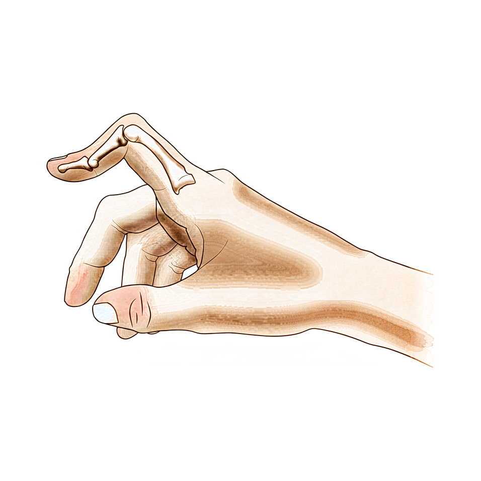
Extensor central-slip injury at the middle finger joint; early splinting prevents the zigzag deformity, established cases need surgery.
Ano ang nararamdaman mo
Maaaring mapansin mong ang kasu-kasuan ng gitnang daliri ay yumuyuko palabas habang ang kasu-kasuan ng dulo ay lumalabas. Ang tiyak na anyong ito ay tinatawag na boutonniere deformity. Ito ay nangyayari kapag ang mga tendon sa itaas ng iyong daliri ay lumipat sa kanilang posisyon. Maaari mong makita ang pagbabagong ito na unti-unting bumubuo sa loob ng panahon, o maaari itong lumabas bigla-bagla pagkatapos ng isaksak.
Ang sakit ay kadalasang nakasentro sa paligid ng gitnang kasu-kasuan ng iyong daliri. Ang hindi komportableng pakiramdam ay maaaring maramdaman bilang isang malalim na sakit o matalim na tusok kapag gumagalaw ang daliri. Maaaring makita mong ang pag-yuyuko ng daliri nang buo ay nagdudulot ng mas maraming sakit kaysa sa pagpapanatili nito ng tuwid. Ang mga gawain na nangangailangan ng paghawak o pagpipitak ay maaaring maging mahirap. Ang mga simpleng gawain tulad ng pag-button ng damit, pag-ikot ng hawakan ng pinto, o pag-type ay maaaring maramdaman na hindi komportable o masakit.
Ang iyong daliri ay maaaring maramdaman na matigas, lalo na sa umaga. Maaari mong mapansin ang pamamaga sa paligid ng gitnang kasu-kasuan. Ang katigasan na ito ay maaaring magpahirap sa paggawa ng kamay na nakapit. Kung mayroon kang rheumatoid arthritis, ang deformity ay maaaring umunlad nang iba kaysa kung ito ay dulot ng aksidente. Kung walang arthritis o pinsala, humigit-kumulang 13% ng mga tao ang nakakaranas ng kondisyong ito.
Ang dulo ng iyong daliri ay maaaring maramdaman na hindi matatag. Sa ilang kaso, ang kasu-kasuan ng dulo ay yumuyuko pababa higit pa sa karaniwan. Ito ay maaaring magpahirap sa pagpahinga ng iyong daliri nang patag sa isang mesa. Maaari kang mahirapan sa pagbukas ng mabibigat na pinto o pag-angat ng magaan na mga bagay gamit ang kamay na iyon. Ang pagtulog sa gilid ay maaaring magpindot sa deformity na daliri, na nagdudulot ng hindi komportableng pakiramdam.
Mahalagang kilalanin ang tunay na deformity na ito mula sa isang katulad na pinsala na tinatawag na pseudoboutonniere. Ang dalawang kondisyon ay nangangailangan ng iba't ibang pag-aalaga. Ang iyong doktor-surgeon ay magmamasid nang maigi sa kung paano gumagalaw ang iyong daliri upang paghiwalayin ang dalawa. Ang pag-unawa sa eksaktong nararamdaman mo ay tumutulong upang gabayan ang pinakamainam na plano ng paggamot para sa iyo.
Ano ang nangyayari talaga
Ang kasukasuan ng iyong daliri ay isang kumplikadong bisagra na binubuo ng mga buto, tendon, at isang protektibong balot na tinatawag na joint capsule. Sa isang malusog na daliri, may isang sentral na tendon na tumatakbo nang tuwid sa gitna ng iyong daliri upang tulungan itong tuwidin. Ang tendon na ito ay gumagana tulad ng pangunahing lubid na humihila upang buksan ang iyong daliri.
Sa Boutonnière deformity, nasira o mahina ang sentral na tendon na ito. Maaari itong mabasag dahil sa trauma o mahabagin ng mga kondisyon tulad ng rheumatoid arthritis. Kapag nabigo ang sentral na suportang ito, nagbabago ang balanse ng mga puwersa sa iyong daliri. Ang mga tendon sa gilid, na karaniwang tumutulong sa pagliko ng daliri, ay nagsisimulang humila nang sobra.
Isipin mo ito tulad ng zipper na lumipat sa track. Ang mga bahagi ay nandoon pa rin, ngunit hindi na ito dumudulas nang maayos magkasama. Dahil hindi hawak ng sentral na tendon ang mga bagay sa tamang posisyon, ang mga tendon sa gilid ay dumudulas patungo sa mga gilid. Ito ang nagdudulot ng pagliko paitaas ng gitnang kasukasuan ng iyong daliri, habang ang kasukasuan sa dulo ay maaaring lumabas o tumayo.
Ang paglipat sa tensyon na ito ang dahilan kung bakit tila kulot ang iyong daliri at mahigpit ito. Ang joint capsule, na karaniwang nagpapanatili ng katatagan ng kasukasuan, ay nagiging mahigpit at hindi balanse. Sa paglipas ng panahon, ang mga tissue ay umaangkop sa bagong, maling posisyong ito. Ito ang dahilan kung bakit maaaring manatili ang deformity kahit subukan mo ang mga konservatibong paggamot tulad ng paggamit ng splint o terapiya.
Ang pinakamahalagang salik sa problemang ito ay ang pagbabago sa iyong mga tendon at kaugnay na istraktura. Nangyayari ang mga pagbabagong ito nang maaga, kaya kritikal ang tumpak na diagnosis. Kailangang kilalanin ng iyong doktor ang pagkakaiba ng tunay na Boutonnière deformity at isang katulad na isyu na tinatawag na pseudoboutonniere injury. Ang landas ng paggamot ay lubos na nakadepende sa tamang pagkakaalam ng pagkakaibang ito.
Kung maagang nahuli ang pinsala, maaaring magtuon ang iyong doktor sa pagpapanumbalik ng posisyon ng sentral na tendon. Sa ilang kaso, maaari silang gumamit ng maliit na piraso ng ibang tendon upang muling itayo ang sentral na slip. Ang bagong graft na ito ay nagmimimito sa orihinal na tungkulin ng tendon habang pinapanatili ang ibang galaw ng daliri. Ang layunin ay panatilihin ang kasukasuan sa gitna at matatag upang muling makagalaw ka nito.
Gayunpaman, kung matagal nang naroroon ang deformity, maaaring magkaroon ng permanenteng pagbabago ang mga tissue. Sa mga kronikong kaso na ito, maaaring hindi maging epektibo ang simpleng pagkukumpuni. Ang natural na pag-unlad ng kondisyong ito ay madalas na nagdudulot ng patuloy na mga isyu, lalo na kung kasama ang rheumatoid arthritis. Maaaring hindi maaasahan ang pangmatagalang resulta ng rekonstruksyon ng malambot na tissue sa mga kaso na ito. Minsan, kailangan ng mas definitibong salvage procedure upang ayusin ang hugis at function ng iyong daliri.
Mga maaari naming gawin para dito
Simulan namin ang hindi operasyonal na paggamot upang muling mabawi ang galaw. Subukan ninyo ang serial casting para sa sapat na extension. Sunod nito ay tatlong buwan ng paggamit ng relative motion flexion orthotic. Maaari ring irekomenda ng inyong surgeon ang pisyoterapiya. Isang hanggang dalawang antas ng pagpapabuti sa range of motion ay maaaring makamit sa pamamagitan ng nonoperative treatment. Gayunpaman, maaaring manatili ang deformity kahit matapos ang dedikadong conservative management. Gumagamit kami ng relative motion flexion orthoses upang mapataas ang aktibong flexion ng distal interphalangeal joint. Tumutulong ito upang mapabuti ang extension ng gitnang joint ng daliri. Kailangan ninyong bigyan ng oras ang paraang ito bago isaalang-alang ang operasyon.
Ang medical management ay nakatuon sa kaginhawaan at pamamaga. Kung ang inyong deformity ay may kaugnayan sa rheumatoid arthritis, aayusin namin ang aktibidad ng pangunahing sakit. Tumutulong ang mga gamot sa sakit at anti-inflammatories upang pamahalaan ang discomfort. Maaaring mag-alok ng mga injection upang bawasan ang pamamaga sa joint. Layunin ng mga paggamot na ito na panatilihin ang joint na mobile at walang sakit habang dumadaan kayo sa therapy. Ang layunin ay istabilisuhin ang kondisyon at mapabuti ang inyong pang-araw-araw na pagganap nang walang operasyon.
Isasaalang-alang ang operasyon kapag naabot na ng conservative care ang hangganan. Ihihiwalay namin ang tunay na boutonniere deformity mula sa pseudoboutonniere injury bago magdesisyon. Kritikal ang pagkakaibang ito sa pagtatakda ng clinical management. Ang tagumpay ng operative result ay nakadepende sa kumpletong preoperative examination, tamang staging ng deformity, at tamang pagkakataon ng treatment. Kung kinakailangan ang soft tissue reconstruction, nauunawaan namin na maaaring hindi maaasahan ang long-term results. Ang recurrent o persistent na deformity ay pinakamainam na gamutin sa pamamagitan ng salvage procedure. Sa ilang kaso, nagbibigay ang Y-shaped tendon graft ng magagandang o mahusay na resulta. Itatakda ng inyong surgeon ang tunay na etiyoloji bago ang surgical intervention. Tinitiyak nito na ang piniling procedure ay tugma sa inyong partikular na anatomia at pangangailangan.
Ano ang inaasahan
Ang iyong prognosis ay malaki ang nakadepende sa kung ito ba ay tunay na deformity o isang katulad na sugat na tinatawag na pseudoboutonniere. Kailangan muna ng iyong surgeon na kumpirmahin ang diagnosis dahil ang landas ng paggamot ay ganap na nagbabago base sa pagkakaiba-iba na ito. Kung mayroon kang rheumatoid arthritis, madalas na hindi maaasahan ang pangmatagalang resulta mula sa pag-aayos ng malambot na tisyu. Sa mga kaso na ito, ang patuloy o muling paglitaw ng deformity ay maaaring magdulot ng pangangailangan para sa isang salvage procedure sa huli.
Para sa karamihan ng mga tao na walang rheumatoid arthritis, hindi laging nalulutas ang kondisyon nang sarili. Maaaring mapabuti ng nonoperative treatment ang iyong range of motion ng isang hanggang dalawang grado. Gayunpaman, maaaring manatili ang kitang-kitang deformity kahit matapos mo na ang dedikadong conservative management. Kung hindi sapat ang conservative care, ang surgery ay nag-aalok ng malakas na opsyon. Nagbibigay ang Y-shaped tendon graft ng magandang o mahusay na resulta sa 16 sa 18 na pasyente sa mga na-ulat na serye. Ang tagumpay ay nakadepa din sa iyong surgeon na gumawa ng kumpletong pagsusuri, tama ang pag-stage ng deformity, at tamang pagpili ng oras para sa intervention.
Ang paggaling ay isang unti-unting proseso. Kung magsisimula ka sa nonoperative care, maaaring gumamit ka ng serial casting upang tuwirin ang daliri, na sinundan ng tatlong buwan ng paggamit ng relative motion flexion orthotic. Ang pamamaraang ito ay nagdudulot ng katulad na resulta sa ibang mga paraan para sa mga chronic case at karaniwang sinusubukan bago isaalang-alang ang surgery. Inaasahan mong magdala ng orthosis sa loob ng ilang buwan upang mapanatili ang extension at mapabuti ang flexion.
Kung kailangan ang surgery, ang layunin ay ibalik ang function at alignment. Kailangan mong sundin nang mahigpit ang tiyak na mga tagubilin ng iyong surgeon. Maaaring mag-iba ang natural na kasaysayan ng deformity na ito, ngunit ang maagang at tumpak na pamamahala ay nagdudulot ng pinakamagandang mga resulta. Magpakatitiyaga sa proseso ng paggaling. Kailangan ng oras upang ang mga tendon at kasukasuan ay umangkop sa kanilang mga bagong posisyon. Gabay ng iyong surgeon ang iyong bawat yugto upang masiguro ang pinakamagandang pagbabalik sa iyong mga pang-araw-araw na gawain.
Kailan kumonsulta sa doktor
Kumonsulta sa iyong GP (General Practitioner) kung napansin mo ang pagliko sa gitnang kasu-kasuan ng iyong daliri na hindi natatuwid. Humingi ng pagsusuri ng espesyalista kung mayroon kang patuloy na sakit na hindi gumagaling kahit magpahinga. Humingi ng medikal na atensyon kung nararamdaman mong kahinaan o hindi katatagan sa daliri. Pumunta sa doktor kung ang daliri ay nakakabit o biglang bumabagsak habang ginagamit. Kontakin ang iyong surgeon kung ang mga sintomas ay nakakaapekto sa iyong pagtulog o trabaho. Ang biglang paglala ng deformity ay nangangailangan din ng mabilisang atensyon. Mahalaga ang tumpak na diagnosis para sa tamang paggamot. Ang pagkakaiba ng tunay na deformity mula sa katulad na sugat ay tumutulong upang matukoy ang angkop na pag-aalaga. Ang maagang pagsusuri ay nagtitiyak ng pinakamahusay na resulta para sa iyong pag-andar ng kamay.
Evidence & references
Overview
- Differentiating a true boutonniere deformity from a pseudoboutonniere injury is critical in determining clinical management [2].
- An understanding of the anatomy, clinical presentation, treatment options, and expected outcomes is crucial for optimal treatment of posttraumatic boutonniere and swan neck deformities [4].
- The natural history of the boutonnière deformity in rheumatoid arthritis is outlined, and a simple method of repair is described [3].
- The prevalence of boutonnière deformity without rheumatoid arthritis or trauma is approximately 13% [5].
- One to two grades of ROM improvement can be achieved with nonoperative treatment, although deformity can persist even after dedicated conservative management [8].
- Similar results occurred for chronic boutonniere deformity using serial casting for adequate extension followed by 3 months of RMF orthotic use, which should be attempted prior to surgical intervention [1].
- Long-term results following soft tissue reconstruction for boutonniere deformity in rheumatoid arthritis are unreliable, and recurrent or persistent deformity is best treated with a salvage procedure [9].
- A successful operative result for swan-neck and boutonniere deformities in the rheumatoid hand depends on complete preoperative examination, correct staging of the deformity, and proper timing of treatment [10].
- The Y-shaped tendon graft can be a useful procedure for the correction of chronic boutonniere deformity, providing good or excellent results in 16 of 18 patients in one series [6].
- Detachment of up to two-thirds of the phalangeal length was effective in reducing extensor lag of the DIP joint and did not cause any boutonniere deformity in a cadaveric model of fractional Fowler tenotomy for chronic mallet finger [7].
Anatomy & Pathophysiology
- Boutonnière deformity can persist even after dedicated conservative management [8].
- One to two grades of range of motion improvement can be achieved with nonoperative treatment of Boutonnière deformity [8].
- Accurate diagnosis and treatment of finger metacarpophalangeal joint injuries begins with an understanding of all potential diagnoses [15].
- Hand surgery and hand therapy practice interventions, including use of relative motion flexion orthoses for management of non-surgical and surgical extensor mechanism injuries, may benefit from an in-depth look at extensor mechanism zone III and IV anatomy and biomechanics [19].
- The most important factor in the development of finger deformities is the changes occurring in the tendons and related structures, especially in early stages [21].
- Reconstruction of the extensor central slip using a distally based flexor digitorum superficialis slip provides a robust repair that anatomically mimics the extensor central slip while maintaining the function of the donor FDS tendon [24].
- The main goals of any treatment of a proximal interphalangeal joint complication are maintaining concentric reduction of the joint, restoring joint stability, and facilitating early range-of-motion exercises [33].
Classification
- Differentiating a true boutonniere deformity from a pseudoboutonniere injury is critical in determining clinical management [2].
- The natural history of the boutonnière deformity in rheumatoid arthritis is outlined [3].
- The prevalence of boutonnière deformity without rheumatoid arthritis or trauma is approximately 13% [5].
- A modified Terrono classification for Type 1 thumb deformity in rheumatoid arthritis could detect advanced deformity earlier and was more strongly correlated with hand function [17].
Clinical Presentation
- Differentiating a true boutonniere deformity from a pseudoboutonniere injury is critical in determining clinical management [2].
- An understanding of the clinical presentation is crucial for optimal treatment of posttraumatic boutonnière and swan neck deformities [4].
- Accurate diagnosis of finger metacarpophalangeal joint injuries begins with an understanding of all potential diagnoses [15].
- The natural history of the boutonnière deformity in rheumatoid arthritis is outlined in historical literature [3].
- The prevalence of boutonnière deformity without rheumatoid arthritis or trauma is approximately 13% [5].
- The swan neck deformity can progress significantly with time due to increasing distal interphalangeal joint flexion contracture [14].
Investigations
- Differentiating a true boutonniere deformity from a pseudoboutonniere injury is critical in determining clinical management [2].
- An understanding of the anatomy, clinical presentation, treatment options, and expected outcomes is crucial for optimal treatment of posttraumatic boutonnière and swan neck deformities [4].
- Accurate diagnosis and treatment of finger metacarpophalangeal joint injuries begins with an understanding of all potential diagnoses [15].
- It is necessary to determine the true etiology before surgical intervention [12].
- A successful operative result depends on complete preoperative examination, correct staging of the deformity, and proper timing of treatment [10].
- Cortical breaks were commonly visualized in MCP and PIP joints with HR-pQCT and microCT [37].
Treatment
- Serial casting for adequate extension followed by 3 months of relative motion flexion (RMF) orthotic use should be attempted prior to surgical intervention for chronic boutonniere deformity [1].
- Differentiating a true boutonniere deformity from a pseudoboutonniere injury is critical in determining clinical management [2].
- A simple method of repair is described for the boutonnière deformity in rheumatoid arthritis [3].
- Understanding the anatomy, clinical presentation, treatment options, and expected outcomes is crucial for optimal treatment of posttraumatic boutonnière and swan neck deformities [4].
- The prevalence of boutonnière deformity without rheumatoid arthritis or trauma is approximately 13% [5].
- The Y-shaped tendon graft is a useful procedure for the correction of chronic boutonniere deformity, providing good or excellent results in 16 of 18 patients in a reported series [6].
- Detachment of up to two-thirds of the phalangeal length is effective in reducing extensor lag of the DIP joint and does not cause any boutonniere deformity in a cadaveric model [7].
- One to two grades of ROM improvement can be achieved with nonoperative treatment, although deformity can persist even after dedicated conservative management [8].
- Long-term results following soft tissue reconstruction for boutonniere deformity in rheumatoid arthritis are unreliable, and recurrent or persistent deformity is best treated with a salvage procedure [9].
- A successful operative result for swan-neck and boutonniere deformities in the rheumatoid hand depends on complete preoperative examination, correct staging of the deformity, and proper timing of treatment [10].
- Metacarpophalangeal joint arthroplasty improves function and deformity and achieves nearly uniform patient satisfaction in rheumatoid arthritis [11].
- One technique does not treat all finger deformities uniformly, highlighting the need to determine the true etiology before surgical intervention [12].
- The use of relative motion flexion orthoses (RMFO) is effective in increasing active distal interphalangeal joint flexion and improving PIP extension in patients with Burton stage 1 chronic boutonniere deformity [13].
Complications
- Differentiating a true boutonniere deformity from a pseudoboutonniere injury is critical in determining clinical management [2].
- The prevalence of boutonniere deformity without rheumatoid arthritis or trauma is approximately 13% [5].
- Detachment of up to two-thirds of the phalangeal length was effective in reducing extensor lag of the DIP joint and did not cause any boutonniere deformity in a cadaveric model [7].
- Long-term results following soft tissue reconstruction for boutonniere finger deformity in rheumatoid arthritis are unreliable [9].
- Recurrent or persistent deformity is best treated with a salvage procedure [9].
- A successful operative result depends on complete preoperative examination, correct staging of the deformity, and proper timing of treatment [10].
- One technique does not treat all deformities uniformly, highlighting the need to determine the true etiology before surgical intervention [12].
- Swan neck deformity can progress significantly with time due to increasing DIPJ flexion contracture [14].
Recovery
- Serial casting for adequate extension followed by 3 months of relative motion flexion (RMF) orthotic use yields similar results for chronic boutonniere deformity and should be attempted prior to surgical intervention [1].
- One to two grades of range of motion (ROM) improvement can be achieved with nonoperative treatment, although deformity can persist even after dedicated conservative management [8].
- The Y-shaped tendon graft is a useful procedure for the correction of chronic boutonniere deformity, providing good or excellent results in 16 of 18 patients in a reported series [6].
- The use of relative motion flexion orthoses (RMFO) is effective in increasing active distal interphalangeal joint flexion and improving proximal interphalangeal (PIP) extension in patients with Burton stage 1 chronic boutonniere deformity [13].
- Long-term results following soft tissue reconstruction for boutonniere deformity in rheumatoid arthritis are unreliable, and recurrent or persistent deformity is best treated with a salvage procedure [9].
- A successful operative result for boutonniere deformity depends on complete preoperative examination, correct staging of the deformity, and proper timing of treatment [10].
Key Evidence
- [L4] Similar results occurred for chronic boutonniere deformity using serial casting for adequate extension followed by 3 months of RMF orthotic use, which should be attempted prior to surgical intervention. [1] (10.1016/j.jht.2023.02.005)
- [L5] Differentiating a true boutonniere deformity from a pseudoboutonniere injury is critical in determining clinical management. [2] (10.1016/j.jhsa.2022.10.019)
- [L4] The natural history of the boutonnière deformity in rheumatoid arthritis is outlined, and a simple method of repair is described. [3] (10.2106/00004623-196951070-00009)
- [L5] An understanding of the anatomy, clinical presentation, treatment options, and expected outcomes is crucial for optimal treatment of posttraumatic boutonnière and swan neck deformities. [4] (10.5435/jaaos-d-14-00272)
- [L3] The prevalence of boutonnière deformity without rheumatoid arthritis or trauma is approximately 13%. [5] (10.1177/1753193417704610)
- [L4] The Y-shaped tendon graft can be a useful procedure for the correction of chronic boutonniere deformity; in our patient series, this provided good or excellent results in 16 of 18 patients. [6] (10.1016/j.jhsa.2021.01.003)
- [L5] Detachment of up to two-thirds of the phalangeal length was effective in reducing extensor lag of the DIP joint and did not cause any boutonniere deformity in this cadaveric model. [7] (10.1016/j.jhsa.2012.07.039)
- [L3] One to two grades of ROM improvement can be achieved, although deformity can persist even after dedicated conservative management. [8] (10.1016/j.jht.2025.02.013)
- [L5] Long-term results following soft tissue reconstruction are unreliable, and recurrent or persistent deformity is best treated with a salvage procedure. [9] (10.1016/j.jhsa.2011.05.029)
- [L5] A successful operative result depends on complete preoperative examination, correct staging of the deformity, and proper timing of treatment. [10] (10.5435/00124635-199903000-00002)
- [L5] Follow-up studies show that this surgery improves function and deformity and achieves nearly uniform patient satisfaction. [11] (10.5435/00124635-200305000-00005)
- [L5] It emphasizes that one technique does not treat all deformities uniformly and highlights the need to determine the true etiology before surgical intervention. [12] (10.1016/j.jhsa.2022.07.008)
- [L4] The use of RMFO is effective in increasing active distal interphalangeal joint flexion and improving PIP extension in patients with Burton stage 1 chronic boutonniere deformity. [13] (10.1016/j.jhsa.2022.08.007)
- [L5] The swan neck deformity in this individual progressed significantly with time because of increasing DIPJ flexion contracture. [14] (10.1016/j.jht.2009.11.005)
- [L5] Accurate diagnosis and treatment of finger metacarpophalangeal joint injuries in athletes begins with an understanding of all potential diagnoses, allowing for safe and early return to play. [15] (10.5435/jaaos-d-21-01031)
- [L3] The modified classification could detect advanced deformity earlier and was more strongly correlated with hand function. [17] (10.1177/1753193419886719)
- [L5] Hand surgery and hand therapy practice interventions, including use of RMF orthoses for management of non-surgical and surgical EM injuries may benefit from an in-depth look at the EM zone III and IV anatomy and biomechanics. [19] (10.1016/j.jht.2023.01.002)
- [L4] The most important factor in the development of finger deformities is the changes occurring in the tendons and related structures, especially in early stages. [21] (10.2106/00004623-195739030-00006)
- [L4] The modified technique provides a robust repair that anatomically mimics the extensor central slip yet maintains the function of the donor FDS tendon. [24] (10.1016/j.jhsa.2009.01.025)
- [L5] The main goals of any treatment of a PIP joint complication are maintaining concentric reduction of the joint, restoring joint stability, and facilitating early range-of-motion exercises. [33] (10.1016/j.hcl.2017.12.014)
- [L4] Cortical breaks were commonly visualized in MCP and PIP joints with HR-pQCT and microCT. [37] (10.1186/s12891-016-1148-y)
References
[1] The relative motion concept in acute and chronic boutonniere deformity: Invited commentary. Journal of Hand Therapy. 2023. DOI: 10.1016/j.jht.2023.02.005 [2] Boutonniere Versus Pseudoboutonniere Deformities: Pathoanatomy, Diagnosis, and Treatment. The Journal of Hand Surgery. 2023. DOI: 10.1016/j.jhsa.2022.10.019 [3] Correction of the Rheumatoid Boutonnière Deformity. The Journal of Bone & Joint Surgery. 1969. DOI: 10.2106/00004623-196951070-00009 [4] Posttraumatic Boutonnière and Swan Neck Deformities. Journal of the American Academy of Orthopaedic Surgeons. 2015. DOI: 10.5435/jaaos-d-14-00272 [5] Thumb boutonnière deformity without rheumatoid arthritis or trauma. Journal of Hand Surgery (European Volume). 2017. DOI: 10.1177/1753193417704610 [6] Y-Shaped Tendon Graft—A Technique in the Reconstruction of Posttraumatic Chronic Boutonniere Deformity. The Journal of Hand Surgery. 2021. DOI: 10.1016/j.jhsa.2021.01.003 [7] Fractional Fowler Tenotomy for Chronic Mallet Finger: A Cadaveric Biomechanical Study. The Journal of Hand Surgery. 2012. DOI: 10.1016/j.jhsa.2012.07.039 [8] Nonoperative treatment of the Boutonniere deformity: Is there a difference in outcomes?. Journal of Hand Therapy. 2025. DOI: 10.1016/j.jht.2025.02.013 [9] Treatment of Boutonniere Finger Deformity in Rheumatoid Arthritis. The Journal of Hand Surgery. 2011. DOI: 10.1016/j.jhsa.2011.05.029 [10] Operative Correction of Swan-Neck and Boutonniere Deformities in the Rheumatoid Hand. Journal of the American Academy of Orthopaedic Surgeons. 1999. DOI: 10.5435/00124635-199903000-00002 [11] Metacarpophalangeal Joint Arthroplasty in Rheumatoid Arthritis. Journal of the American Academy of Orthopaedic Surgeons. 2003. DOI: 10.5435/00124635-200305000-00005 [12] Clarification of Extensor Tenotomy for Finger Deformities. The Journal of Hand Surgery. 2022. DOI: 10.1016/j.jhsa.2022.07.008 [13] The Use of Relative Motion Flexion Orthoses for Chronic Boutonniere Deformity. The Journal of Hand Surgery. 2024. DOI: 10.1016/j.jhsa.2022.08.007 [14] Swan Neck Deformity after Distal Interphalangeal Joint Flexion Contractures: A Biomechanical Analysis. Journal of Hand Therapy. 2010. DOI: 10.1016/j.jht.2009.11.005 [15] Finger Metacarpophalangeal Joint Injuries in Athletes: Evaluation, Diagnosis, Treatment, and Return to Play. Journal of the American Academy of Orthopaedic Surgeons. 2023. DOI: 10.5435/jaaos-d-21-01031 [17] A modified Terrono classification for Type 1 thumb deformity in rheumatoid arthritis: a cross-sectional analysis. Journal of Hand Surgery (European Volume). 2019. DOI: 10.1177/1753193419886719 [19] An in-depth look at zone III and IV anatomy of the finger extensor mechanism and some clinical implications for use of the relative motion flexion orthosis. Journal of Hand Therapy. 2023. DOI: 10.1016/j.jht.2023.01.002 [21] Finger Deformities Caused by Rheumatoid Arthritis. The Journal of Bone & Joint Surgery. 1957. DOI: 10.2106/00004623-195739030-00006 [24] Reconstruction of the Extensor Central Slip Using a Distally Based Flexor Digitorum Superficialis Slip. The Journal of Hand Surgery. 2009. DOI: 10.1016/j.jhsa.2009.01.025 [33] Complications of Proximal Interphalangeal Joint Injuries. Hand Clinics. 2018. DOI: 10.1016/j.hcl.2017.12.014 [37] Visual detection of cortical breaks in hand joints: reliability and validity of high-resolution peripheral quantitative CT compared to microCT. BMC Musculoskeletal Disorders. 2016. DOI: 10.1186/s12891-016-1148-y