Patients › Hand
Pag-release ng trigger finger
Trigger finger release — understanding the condition, conservative treatments, and surgical options for a stuck or clicking finger.
Bakit ito ang inirekomendang operasyon
Ang trigger finger release ay isang maikling proseduro na naglilaya sa masikip na tendon sheath sa palad upang pigilan ang pagkakadikit o pagkakasara ng iyong daliri. Posibleng inirekomenda ng iyong doktor ang operasyong ito dahil sa patuloy na sakit, pagkabuo ng buntong sa palad, o pagkakadikit ng daliri, lalo na kung hindi nagbigay ng pangmatagalang ginhawa ang mga steroid injection. Bagama’t karamihan sa mga tao ay sumusubok muna ng mga injection, ang operasyon ang pinakamainam na pagpipilian kapag nabigo ang mga treatment na iyon o kung ikaw ay may diabetes at nangangailangan ng maaasahang solusyon.
Karaniwang mababa ang panganib at mataas ang epektibidad ng operasyong ito. Humigit-kumulang 97% ng mga pasyente ay nakakaranas ng ganap na paglaya ng kanilang mga sintomas pagkatapos ng proseduro. Ang pangunahing layunin nito ay ibalik ang maayos na galaw at alisin ang sakit na dulot ng pagkakadikit ng iyong daliri sa nakaduduong na posisyon.
Bago ang operasyon
Kailangan mong mag-fasting ng ilang oras bago ang iyong operasyon at mag-arrange ng taong magdadala sa iyo pauwi. Mangyaring dalhin ang listahan ng lahat ng kasalukuyang gamot na iyong iniiinom at magsuot ng komportableng damit. Maaaring mag-order ang iyong surgeon ng mga simpleng pagsusuri tulad ng X-ray, blood work, o pagsusuri ng anestesiya upang matiyak na ligtas ka para sa proseso. Ang mga pagsusuring ito ay tumutulong sa amin na maunawaan ang iyong pangkalahatang kalusugan at magplano ng pinakamainam na pag-aalaga para sa iyo. Bibigyan ka ng tiyak na mga tagubilin ng iyong surgeon tungkol sa mga gamot na dapat itigil bago ang araw ng iyong operasyon. Ang bukas na operasyong ito ay gumagamit ng isang maliit na hiwa sa ibabaw ng daliri upang paluwagin ang mahigpit na tendon sheath.
Sa araw ng operasyon
Dadating ka sa klinika at makikilala ang iyong anestesiyolohista upang talakayin ang kontrol sa sakit. Ang operasyong ito ay maaaring gawin sa ilalim ng lokal na anestesya (isang injeksyon na nagpapababa ng pakiramdam lamang sa lugar ng operasyon, habang ikaw ay gising) o sa ilalim ng pangkalahatang anestesya (ganap na natutulog). Karamihan sa mga tao ay pumipili ng lokal: mas mabilis ang paggaling at maaari kang umuwi agad pagkatapos. Kung mas gusto mong natutulog, ito rin ay isang makatwirang pagpili; talakayin ito sa iyong doktor at anestesiyolohista.
Pagkatapos, pupunta ka sa silid-operasyon kung saan gagawa ang iyong doktor ng isang maliit na hiwa sa ibabaw ng daliri upang paluwagin ang mahigpit na banda. Maikli at ligtas ang proseso. Pagkatapos, gumigising ka sa recovery area kung saan hahawakan ka ng mga staff hanggang sa handa ka nang umuwi. Maaaring magkaroon ng mga maliit na isyu tulad ng pagkadikit sa peklat o bahagyang pagkasira, ngunit karamihan sa mga pasyente ay mararamdaman ang agad na pagpapagaan mula sa pagkakakulong.
Ano ang kinabibilangan ng operasyon
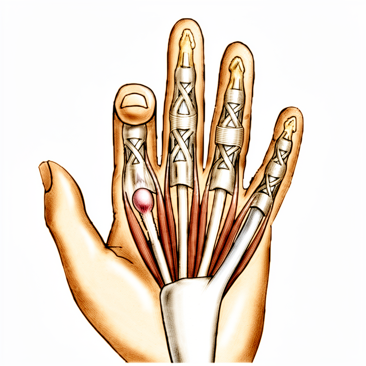
Gagawa ang iyong doktor ng isang hiwa na humigit-kumulang 2 cm ang haba sa palad ng iyong daliri o hinlalaki. Ang eksaktong lokasyon ay nakadepende sa kung aling daliri ang apektado, kadalasan ay direkta sa ilalim ng pangunahing guhit ng iyong kamay. Ang hiwang ito ay nagbibigay-daan sa malapit na banda ng tisyu, na tinatawag na A1 pulley, na humahawak sa iyong tendon.
Sa loob, guguho ng iyong doktor ang malapit na banda upang malaya ang tendon upang muling makadulas nang maayos. Kung mayroon kang rheumatoid arthritis, maaaring tanggalin ng iyong doktor ang isang maliit na bahagi ng tendon sa halip upang protektahan ang pagkakahanay ng iyong daliri. Kapag inilaya na ang banda, galawin mo ang iyong daliri papunta at palayo upang kumpirmahin na ito ay gumagalaw nang malaya nang walang pagkakadikit.
Isasara ang hiwa gamit ang mga tahi na tatanggalin sa loob ng 10 hanggang 14 araw. Maglalagay ng compression dressing na tatanggalin pagkatapos ng 48 oras. Inirerekomenda na gamitin ang iyong daliri nang normal kapag naalis na ang dressing.
Pagkatapos ng operasyon
Gising ka sa isang recovery area kung saan sinusuri ng iyong surgeon ang iyong kamay. Mababa ang risk ng open surgery na ito, bagaman ang humigit-kumulang 1 sa 20 mga daliri ay maaaring magkaroon ng mild, temporaryong isyu. Day case ito, kaya papalabas ka sa parehong araw. Balot ng dressing ang iyong kamay; karaniwang hindi kailangan ng sling o brace para sa hinlalaki. Ang sakit ay pinamamahalaan gamit ang standard na gamot. Dapat may kasama kang tao na mananatili sa iyo sa loob ng unang 24 oras upang tulungan ka. Karamihan sa mga pasyente ay mabilis na nakakaramdam ng pagpapabuti, at humigit-kumulang 97% ay may kumpletong resolusyon pagkatapos ng prosedura.
Pagbawi
Maramdaman mong matigas at masakit ang iyong kamay sa unang ilang araw. Maaaring makita mong may pamamaga o bahagyang pagkulubot sa paligid ng iyong daliri. Karaniwan ito. Maaaring imungkahi ng iyong doktor ang paggamit ng ice pack upang mapagaan ang hindi komportableng pakiramdam. May ilang pasyente ang nakakakita na nakakatulong ang paggalaw ng kanilang daliri nang dahan-dahan upang bawasan ang katigasan nang hindi nakakasama.
Maaari kang makagalaw ng iyong daliri sa araw ng operasyon. Maaaring payuhan ka ng iyong doktor na panatilihing itaas ang kamay upang bawasan ang pamamaga. Karaniwang maaari mong hugasan nang dahan-dahan ang iyong kamay pagkatanggal ng panapos. Iwasan ang mabigat na pagkapit o pag-angat hangga’t hindi pa sinasabi ng iyong doktor na ligtas na ito. Ang mga simpleng gawain tulad ng pagkain o pag-type ay madalas ay okay na agad pagkatapos ng operasyon.
Kung mayroon kang diabetes, maaaring konting tumanda ang antas ng asukal sa dugo pagkatapos ng prosedura. Babantayan ito ng iyong doktor nang mahigpit. Ang bawat isa ay gumagaling sa magkakaibang bilis. Gabayan ka ng iyong doktor at terapistang kung kailan ka makakabalik sa trabaho o pagmamaneho batay sa iyong partikular na pag-unlad. Maniwala sa iyong katawan at sundin ang kanilang payo para sa pinakamainam na resulta.
Maaaring mangyari
Karamihan sa mga pasyente ay magaling, ngunit minsan ay may mga problema na maaaring mangyari. Ang iyong surgeon at ang team ay magbabantay nang maigi upang maagang makita ang anumang isyu.
Maaaring mapansin mo ang pamumula, pamamaga, o sakit na lumalala imbes na lumilipas pagkatapos ng iyong operasyon. Maaaring ito ay senyales ng malalim na impeksyon, lalo na kung nakuha mo ang steroid injection noong nakaraang buwan. Kung nararamdaman mo ang malalim, pulso-pulso na sakit na hindi nababawasan ng simpleng gamot sa sakit, o kung nakikita mong kumakalat ang pamumula mula sa sugat, tawagan agad ang klinika. Maaaring kailanganin mong operahin upang linisin ang lugar.
Minsan ay bahagyang hihiwalay ang mga gilid ng sugat, o maaaring maging napakabaliw ng kamao. Maaari ring maramdaman mong bumababa ang kakayahan mong yumuko o tuwidin ang iyong daliri. Ito ay mga maliit na isyu, ngunit kung nakakaabala ito, ibahagi ito sa susunod na review.
Sa bihirang kaso, maaaring masira ang nerbiyos sa gilid ng iyong hinlalaki habang ginagawa ang release. Maaaring maramdaman mo ang kawalan ng pakiramdam, pangangati, o kakaibang pakiramdam sa lugar na iyon. Kung mapansin mo ang biglaang pagbabago sa pakiramdam o lakas, ipaalam agad sa iyong surgeon.
Kung mayroon kang rheumatoid arthritis, maaaring magpalala ng pag-ikot ng iyong joint sa daliri ang operasyon. Maaaring mapansin mong lumiliko ang iyong daliri papunta sa iba pang daliri kaysa dati. Talakayin ang riskong ito sa iyong surgeon bago ang operasyon.
Minsan, maaaring magsimulang mag-click o mag-lock muli ang daliri pagkatapos ng operasyon. Maaaring ito ay mangyari kung ang tendon ay nakakabit sa scar tissue o gumaling nang may maliit na buntot. Karaniwan, lumalagpas ang pakiramdam na ito sa paglipas ng panahon, ngunit kung patuloy ito, ibahagi ito sa iyong team.
Ang talahanayan ng mga komplikasyon sa pahinang ito ay naglalaman ng karaniwang mga rate kung nais mo ang mga detalye.
Kailan tawagan ang aming klinika
Tawagan kami kung ikaw ay magkaroon ng lagnat, lumalalang pamumula, o paglabas ng dugo o tubig mula sa sugat. Humingi ng agad na paggamot kung biglaang mabigat ang sakit, may bagong pamamanhid, o kung hindi mo na makagalaw ang iyong daliri. Pumunta agad sa emergency room kung napansin mo ang pamamaga sa binti o hirap sa paghinga. Maaaring magpahiwatig ang mga senyales na ito ng impeksyon, pinsala sa nerbiyos, o dugo na nakadugo na nangangailangan ng agad na pag-aalaga.
Evidence & references
Overview
- Local anesthetic infiltration in the palm proximal to the incision site is preferred for trigger finger release [5].
- A pneumatic arm tourniquet may be helpful, although a high forearm Esmarch wrap is usually sufficient [5].
- A transverse incision about 2 cm long is made several millimeters distal to the distal palmar crease for middle, ring, and small trigger finger releases [5].
- A transverse incision about 2 cm long is made several millimeters distal to the proximal palmar crease for index trigger finger releases [5].
- Trigger thumb releases can be performed through incisions either distal or proximal to the metacarpophalangeal joint flexion crease [5].
- Alternative incisions for fingers can be made obliquely or longitudinally between the metacarpophalangeal and distal palmar creases [5].
- Alternative incisions for the thumb can be made obliquely across the thumb metacarpophalangeal flexion crease [5].
- Digital nerves on the thumb are more palmar and closer to the flexor sheath than might be anticipated [5].
- The thumb radial digital nerve is especially vulnerable during trigger finger release [5].
- Subcutaneous tissues are spread away from the underlying annular pulley system to ensure digital nerves are safely protected [5].
- Trigger thumbs require release of only the A1 pulley [5].
- Trigger digits require division of the A1 and A0, or proximal palmar pulley [5].
- Pulley division is usually accomplished with an initial opening of the pulley with a No. 15 knife blade and a pair of tenotomy scissors [5].
- For trigger thumb release, cutting too far distally should be avoided to prevent disruption of the oblique pulley [5].
- The sheath is incised from proximal to distal approximately 1 cm during trigger finger release [5].
- The patient should be asked to actively flex and extend the digit after sheath incision to reassess for triggering [5].
- Persistent triggering implies that either the A1 and palmar pulleys are incompletely released or an alternate site of triggering is present [5].
- When the distal A1 pulley edge is released, the divided pulley leaves are parallel rather than ending in a V-shaped pattern [5].
- The patient should be encouraged to actively flex and extend the digit after the tendon sheath has been released to ensure the release is complete [5].
- Other fingers can be found to trigger at the same surgical setting and can be managed at the same time [5].
- The skin is closed and a small, dry compression dressing is applied after trigger finger release [5].
- The compression dressing is removed after 48 hours postoperatively [5].
- Sutures are removed at 10 to 14 days postoperatively [5].
- Normal use of the finger or thumb is encouraged postoperatively [5].
Anatomy & Pathophysiology
- Stenosing tenosynovitis (trigger finger) involves mechanical impingement of the flexor tendons at the A1 pulley [9].
- The pathologic examination of affected pulleys demonstrates a proliferation of chondrocytes and increased type III collagen [9].
- The flexor digitorum profundus tendon often demonstrates a pathologic nodule, while the flexor digitorum superficialis is often unaffected [9].
- Trigger finger occurs in 2% to 3% of the general population [9].
- Women are more commonly affected than men [9].
- The digits are affected in the following order of decreasing prevalence: thumb, ring, long, little, and index [9].
- Middle and ring finger involvement is most common in adults [1, 2].
- Trigger finger is more common in patients with diabetes mellitus than in nondiabetic patients [4].
- When multiple digits are involved, the possibility of diabetes should be considered [4].
- Trigger finger is associated with systemic diseases including diabetes mellitus (10% to 20% lifetime incidence), hypothyroidism, sarcoidosis, rheumatoid arthritis, and septic tenosynovitis [9].
- Trigger finger is associated with gout, where monosodium urate precipitation elicits a fulminant inflammatory reaction in the tenosynovium [9].
- Trigger finger is associated with calcific tendinitis, where calcium salt deposition in the tenosynovium can resemble an infection [9].
- Trigger finger is associated with pseudogout, where calcium pyrophosphate dihydrate crystal deposition is often localized to the triangular fibrocartilage or within the carpal tunnel [9].
- Trigger finger is associated with amyloidosis, characterized by the deposition of beta-2-microglobulin in thick, plaque-like accumulations along the flexor tendons [9].
- Amyloidosis-associated trigger finger is most commonly seen in patients with renal failure undergoing peritoneal dialysis or hemodialysis [9].
- Trigger finger is associated with inflammatory arthropathy [1, 2].
- Trigger finger may be associated with repetitive grasping activities [1, 2].
- Fibrocartilaginous metaplasia occurs in the pulley and/or flexor digitorum superficialis (FDS) tendon [1, 2].
- A palpable lump or knot in the palm may represent a thickened area in the first annular pulley or a nodule/fusiform swelling of the flexor tendon just distal to it [7].
- The tendon nodule is usually just proximal to the anulus at the metacarpophalangeal joint level [7].
- In rheumatoid patients, a nodule distal to the metacarpophalangeal joint level may cause triggering [7].
- Triggering is often more pronounced in the morning than later in the day [4].
- Triggering may result from catching of the tendon on the palmar aponeurosis transverse fibers [7].
- A partially lacerated flexor tendon at the level of the A1 pulley may heal with a nodule sufficiently large to cause triggering [7].
- In adults, trigger thumb is a distinctly separate entity from congenital trigger thumb [7].
- Stenosing tenosynovitis in adults is usually seen in individuals older than 45 years of age [7].
- When associated with a collagen disease, several fingers may be involved, most often the long and ring fingers [7].
- Trigger finger is more common in patients with diabetes mellitus [9].
- Trigger finger is associated with Dupuytren's disease, with a high percentage of concurrent cases in the middle and ring finger [11].
- The percentage of patients suffering from both trigger finger and Dupuytren's disease increases with age [11].
- In Stage I Dupuytren's disease, thickening of the pulley wall leads to narrowing of the A1 pulley and synovial congestion [12].
- In more progressed stages of Dupuytren's contracture (Stages II or III), concomitant trigger finger is rarely seen [12].
- The absence of trigger finger in advanced Dupuytren's contracture may be explained by reduced range of motion reducing mechanical irritation at the A1 pulley [12].
- The absence of trigger finger in advanced Dupuytren's contracture may be explained by the tendon becoming slightly thinner distal to the chiasm of the deep and superficial flexor tendon [12].
- A variable annular pulley (fourth pulley) is found in 75% of patients and may contribute to stenosis [1, 2].
- Trigger finger is associated with carpal tunnel syndrome in 40% to 60% of patients [1, 2].
- Congenital trigger digit involves narrowing and thickening of the sheath, with occasional formation of a ganglion cyst [10].
- An intratendinous nodule may be present proximal to the first annular pulley, often referred to as Notta's nodule [10].
- Chronic inflammation is frequent in congenital trigger digits [10].
- Congenital trigger digit occurs far more commonly in the thumb [10].
- Congenital trigger digit is bilateral in about 25% of patients [10].
- Congenital trigger digit has been associated with trisomy 13 [10].
- Congenital trigger digit has been associated with mucopolysaccharidosis [10].
- Spontaneous resolution occurs in about 30% of children in whom congenital trigger digit appears within the first year of life [10].
- Spontaneous resolution occurs in about 12% of children in whom congenital trigger digit appears between 6 months and 2 years of age [10].
- Baek et al. noted spontaneous resolution in 63% of congenital trigger digits over a median of 48 months [10].
- Trigger finger is not often associated with a fixed flexion deformity in children, unlike congenital trigger thumb [10].
- Trigger finger in children may not respond to a simple A-pulley release [10].
- Surgical intervention for pediatric trigger finger may require excision of one or both slips of the flexor digitorum superficialis tendon and release of the A3 pulley [10].
- Triggering in adults is often caused by catching of the tendon on the palmar aponeurosis transverse fibers [7].
- Intraarticular disorders such as loose bodies, degenerative joint disease, and fractures can cause symptoms similar to trigger finger [7].
- Common extensor tendon subluxation can cause symptoms similar to trigger finger [7].
- A volar retinacular ganglion cyst may be present between the A1 and A2 pulleys [9].
- Fixed flexion deformity of the proximal interphalangeal (PIP) joint may be present in trigger finger [9].
- Triggering is often more pronounced in the morning than later in the day [4].
- Patients frequently state that the problem is in the proximal interphalangeal joint with trigger finger or in the proximal interphalangeal joint with trigger thumb [7].
- Pressure accentuates the apparent snapping or triggering of the more distal joints [7].
- Local tenderness may be present but is not a prominent complaint in trigger finger [7].
- Triggering is often more pronounced in the morning than later in the day [4].
- Triggering is often more pronounced in the morning than later in the day [4].
Classification
- Trigger finger demographics include women older than 50 years of age [1].
- The middle and ring fingers are the most commonly involved digits in adults with trigger finger [1].
- Trigger finger is associated with diabetes as a comorbidity [1].
- Trigger finger is associated with inflammatory arthropathy as a comorbidity [1].
- Repetitive grasping activities are possibly associated with the etiology of trigger finger [1].
- Histology of trigger finger shows fibrocartilaginous metaplasia in the pulley and/or flexor digitorum superficialis (FDS) tendon [1].
- Presentation of trigger finger includes pain and tenderness in the distal palm [1].
- Trigger finger presentation progresses from pain to mechanical catching and locking [1].
- Trigger finger may become fixed in advanced presentation [1].
- Referred pain at the dorsal metacarpophalangeal (MCP) or proximal interphalangeal (PIP) area is a common complaint in trigger finger [1].
- Concomitant trigger finger and carpal tunnel syndrome (CTS) occurs in 40% to 60% of patients [1].
- A fourth pulley, described as a variable annular pulley, is found in 75% of patients with thumb trigger finger [1].
- The presence of a fourth pulley in the thumb may contribute to stenosis [1].
- Grade I trigger finger is defined as pain and tenderness at the A1 pulley [1].
- Grade II trigger finger is defined as catching of the finger [1].
- Grade III trigger finger is defined as locking of the finger that is passively correctable [1].
- Grade IV trigger finger is defined as a fixed, locked finger [1].
Clinical Presentation
- Trigger finger is most common in women older than 50 years of age [1, 2].
- The middle and ring fingers are the most commonly involved digits in adults [1, 2].
- Trigger finger is associated with diabetes and inflammatory arthropathy [1, 2].
- Repetitive grasping activities are possibly associated with the etiology of trigger finger [1, 2].
- Histology of trigger finger demonstrates fibrocartilaginous metaplasia of the pulley and/or flexor digitorum superficialis (FDS) tendon [1, 2].
- Trigger finger occurs in 2% to 3% of the general population [9].
- The digits are affected in decreasing order of prevalence: thumb, ring, long, little, and index [9].
- Trigger finger is more common in patients with systemic diseases including diabetes mellitus (10% to 20% lifetime incidence), hypothyroidism, sarcoidosis, rheumatoid arthritis, and septic tenosynovitis [9].
- Gout can mimic infectious tenosynovitis with initial presentation of marked pain, erythema, swelling, and warmth [9].
- Calcific tendinitis can resemble infection and result in triggering, with males affected five times more frequently than females [9].
- Amyloidosis is characterized by deposition of beta-2-microglobulin along flexor tendons, most commonly seen in patients with renal failure undergoing dialysis [9].
- Clinical presentation includes pain and tenderness in the distal palm at the proximal edge of the digital A1 pulley [4].
- Patients frequently note catching or triggering of the affected finger or thumb after forceful flexion [4].
- In severe cases, the opposite hand must be used to force the finger or thumb passively into extension [4].
- In the most severe cases, the finger becomes locked in a flexed position [4].
- Triggering is often more pronounced in the morning than later in the day [4].
- Stenosing tenosynovitis is more common in diabetic patients than in nondiabetic patients [4].
- When multiple digits are involved, the possibility of diabetes should be considered [4].
- A common complaint is referred pain at the dorsal MCP or PIP area [1, 2].
- Concomitant trigger finger and carpal tunnel syndrome (CTS) occurs in 40% to 60% of patients [1, 2].
- A palpable lump or knot may be present in the palm, representing a thickened area in the first annular pulley or a nodule/fusiform swelling of the flexor tendon [7].
- The tendon nodule is usually just proximal to the anulus at the metacarpophalangeal joint level [7].
- In rheumatoid patients, a nodule distal to the metacarpophalangeal joint level may cause triggering [7].
- Local tenderness may be present but is not a prominent complaint [7].
- Pressure accentuates the apparent snapping or triggering of the more distal joints [7].
- Patients frequently state that the problem is in the proximal interphalangeal joint with trigger finger or in the proximal interphalangeal joint with trigger thumb [7].
- Physical examination may reveal a palpable triggering or pain with flexion and extension of the finger [9].
- Physical examination may reveal nodularity of the flexor tendon just proximal to the A1 pulley [9].
- A volar retinacular ganglion cyst may be present between the A1 and A2 pulleys [9].
- A fixed flexion deformity of the proximal interphalangeal (PIP) joint may be present [9].
- Green classification Grade I is defined as pain and tenderness at the A1 pulley [1, 2, 9].
- Green classification Grade II is defined as catching of the finger [1, 2, 9].
- Green classification Grade III is defined as locking of the finger that is passively correctable [1, 2, 9].
- Green classification Grade IV is defined as a fixed, locked finger [1, 2, 9].
- Triggering in adults is often seen in individuals older than 45 years of age [7].
- When associated with a collagen disease, several fingers may be involved, most often the long and ring fingers [7].
- Triggering may occur after operative release due to catching of the tendon on the palmar aponeurosis transverse fibers [7].
- Occasionally, a partially lacerated flexor tendon at this level heals with a nodule sufficiently large to cause triggering [7].
- Other conditions such as intraarticular disorders (loose bodies, degenerative joint disease, fractures) and common extensor tendon subluxation can cause similar symptoms [7].
- Pathologic examination demonstrates a proliferation of chondrocytes and increased type III collagen in the affected pulleys [9].
- The flexor digitorum profundus tendon often demonstrates a pathologic nodule, while the flexor digitorum superficialis is often unaffected [9].
- Newer evidence has found a fourth pulley (variable annular pulley) in 75% of patients, which may contribute to stenosis [1, 2].
Investigations
- Trigger finger demographics include women older than 50 years of age [1, 2].
- The middle and ring fingers are the most commonly involved digits in adults [1, 2].
- Trigger finger is associated with comorbidities including diabetes and inflammatory arthropathy [1, 2].
- Etiology is possibly associated with repetitive grasping activities [1, 2].
- Histology demonstrates fibrocartilaginous metaplasia of the pulley and/or flexor digitorum superficialis (FDS) tendon [1, 2].
- Pathologic examination of affected pulleys demonstrates a proliferation of chondrocytes and increased type III collagen [9].
- The flexor digitorum profundus tendon often demonstrates a pathologic nodule, while the flexor digitorum superficialis is often unaffected [9].
- Presentation includes pain and tenderness in the distal palm at the proximal edge of the digital A1 pulley [4].
- Patients frequently note catching or triggering of the affected finger or thumb after forceful flexion [4].
- In more severe cases, the opposite hand must be used to force the finger or thumb passively into extension [4].
- In the most severe cases, the finger becomes locked in a flexed position [4].
- Triggering is often more pronounced in the morning than later in the day [4].
- Stenosing tenosynovitis is more common in diabetic patients than in nondiabetic patients [4].
- When multiple digits are involved, the possibility of diabetes should be considered [4].
- A common complaint is referred pain at the dorsal MCP or PIP area [1, 2].
- Concomitant trigger finger and carpal tunnel syndrome (CTS) occurs in 40% to 60% of patients [1, 2].
- Trigger finger occurs in 2% to 3% of the general population [9].
- Women are more commonly affected than men [9].
- The digits are affected in the following order of decreasing prevalence: thumb, ring, long, little, and index [9].
- Trigger finger is more common in patients with systemic diseases such as diabetes mellitus (10% to 20% lifetime incidence), hypothyroidism, sarcoidosis, rheumatoid arthritis, and septic tenosynovitis [9].
- Physical examination may reveal tenderness to palpation of the flexor tendon at the level of the A1 pulley [9].
- Physical examination may reveal palpable triggering or pain with flexion and extension of the finger [9].
- Physical examination may reveal nodularity of the flexor tendon just proximal to the A1 pulley [9].
- A volar retinacular ganglion cyst may be present between the A1 and A2 pulleys [9].
- A fixed flexion deformity of the proximal interphalangeal (PIP) joint may be present [9].
- Green classification Grade I is defined as pain and tenderness at the A1 pulley [1, 2, 9].
- Green classification Grade II is defined as catching of the finger [1, 2, 9].
- Green classification Grade III is defined as locking of the finger that is passively correctable [1, 2, 9].
- Green classification Grade IV is defined as a fixed, locked finger [1, 2, 9].
- Newer evidence has found a fourth pulley (variable annular pulley) in 75% of patients with trigger thumb, which may contribute to stenosis [1, 2].
- Gout can mimic infectious tenosynovitis with initial presentation of marked pain, erythema, swelling, and warmth [9].
- Definitive diagnosis of gout is made by tenosynovial aspiration or biopsy showing negatively birefringent urate crystals under polarized light microscopy [9].
- Calcific tendinitis can resemble an infection and result in triggering [9].
- Radiographs of calcific tendinitis reveal fluffy ectopic calcification in the soft tissues [9].
- Pseudogout is characterized by calcium pyrophosphate dihydrate crystal deposition, often localized to the triangular fibrocartilage or within the carpal tunnel [9].
- Pathology of pseudogout reveals rhomboid-shaped crystals with positive birefringence [9].
- Amyloidosis is characterized by deposition of beta-2-microglobulin in thick, plaque-like accumulations along the flexor tendons [9].
- Amyloidosis is most commonly seen in patients with renal failure undergoing peritoneal dialysis or hemodialysis [9].
- Trigger finger is more common in patients with diabetes mellitus [9].
- Corticosteroid injection into the flexor tendon sheath is a nonoperative treatment option [1, 2].
- Injection is "curative" in about 60% of patients initially [1, 2].
- Diabetic patients are generally less responsive to corticosteroid injection [1, 2].
- There is no difference between soluble and insoluble corticosteroid preparations [1, 2].
- 65% to 90% of patients who do not have diabetes obtain relief of symptoms with one or two injections [9].
- Relief of symptoms after injection in patients with diabetes may depend on chronic glucose levels (hemoglobin A1c levels) [9].
- Surgical release of the A1 pulley is curative in digits refractory to steroid injection [4].
- Surgical release of the A1 pulley provides satisfactory results in greater than 90% of patients [9].
- Surgical treatment is required more often in patients with systemic diseases [9].
- In patients with rheumatoid arthritis, preference is to excise a slip of the FDS tendon rather than to release the A1 pulley [1, 2].
- In patients with rheumatoid arthritis, the entire annular pulley system should be preserved to prevent further ulnar drift of the fingers [4].
- Release of the A1 pulley in rheumatoid arthritis patients carries a chance that ulnar drift at the MCP joint can be exacerbated [1, 2].
- Percutaneous release of the A1 pulley may be accomplished with a needle on the middle and ring fingers, especially if they actively lock [4].
- Percutaneous release of the A1 pulley may be accomplished with a 25-gauge hypodermic needle with corticosteroid infiltration [3].
- The radial digital nerve is at risk of iatrogenic injury during thumb trigger finger release due to its superficial location [1, 2].
- Minor complications of surgical release include wound dehiscence, scar tenderness, and decreased range of motion [1, 2].
- Preoperative hypoglycemia increases infection risk after trigger finger injection and release [3].
Treatment
- Nonoperative treatment for trigger finger includes corticosteroid injection into the flexor tendon sheath [1].
- Corticosteroid injection is "curative" in about 60% of patients initially [1].
- Diabetic patients are generally less responsive to corticosteroid injection than non-diabetic patients [1].
- There is no difference in efficacy between soluble and insoluble corticosteroid preparations for trigger finger [1].
- Repeat corticosteroid injections provided symptomatic relief for a year or more in 50% of patients in a study of 292 injections [7].
- Corticosteroid injections may elevate serum glucose levels for 5 days or more in patients with diabetes mellitus [7].
- Patients with unstable diabetes may be better treated without corticosteroid injection [7].
- Immediate surgical release in the clinic was identified as the most cost-effective treatment strategy for trigger finger in diabetic patients [7].
- Surgical release of the A1 pulley is curative for digits refractory to steroid injection [4].
- Surgical release reliably relieves the problem for most patients, with approximately 97% having complete resolution after operative treatment [7].
- Persistence of triggering is more common than recurrence after trigger finger release [7].
- Surgical treatment options include open or percutaneous release of the A1 pulley [1].
- In patients with rheumatoid arthritis, the preference is to excise a slip of the FDS tendon rather than to release the A1 pulley [1].
- Excision of an FDS slip in rheumatoid arthritis patients is preferred because release of the A1 pulley carries a risk of exacerbating ulnar drift at the MCP joint [1].
- In patients with rheumatoid arthritis, the entire annular pulley system should be preserved to prevent further ulnar drift of the fingers [4].
- Triggering in rheumatoid arthritis patients is treated by tenosynovectomy and excision of one slip of the flexor digitorum superficialis [4].
- The radial digital nerve is at risk of iatrogenic injury during thumb trigger finger release due to its superficial location [1].
- Digital nerves on the thumb are more palmar and closer to the flexor sheath than might be anticipated, making them especially vulnerable [5].
- Minor complications of surgical release include wound dehiscence, scar tenderness, and decreased range of motion [1].
- Percutaneous release of the A1 pulley may be accomplished with a needle on the middle and ring fingers, especially if they actively lock [4].
- Percutaneous release using a needle or push knife has literature support for safety and effectiveness [7].
- Incomplete pulley release and damage to flexor tendons and digital nerves remain concerns with limited exposure techniques, especially in the index finger and thumb [7].
- For trigger thumb, the A1 pulley is released while avoiding cutting too far distally to disrupt the oblique pulley [5].
- Trigger digits require division of the A1 and A0 (proximal palmar) pulleys, whereas trigger thumbs require release of only the A1 pulley [5].
- If persistent triggering occurs after initial release, it implies either incomplete release of the A1 and palmar pulleys or an alternate site of triggering [5].
- When the distal A1 pulley edge is released, the divided pulley leaves are parallel rather than ending in a V-shaped pattern [5].
- Percutaneous release can be performed using an 18- or 19-gauge needle [8].
- During percutaneous release, the needle bevel should be oriented longitudinally parallel to the flexor tendons [8].
- A scraping or grating sensation felt during needle movement indicates the sheath is being incised during percutaneous release [8].
- Loss of the grating sensation as the pulley is cut indicates completion of percutaneous release [8].
- Injection of corticosteroid is optional during percutaneous trigger finger release [8].
- For combined trigger finger and Dupuytren's disease, an isolated opening of the pulley without touching Dupuytren tissue followed by corticosteroid application in the area of the opened pulley is a recommended strategy [13].
- Resecting Dupuytren's tissue concomitantly with trigger finger release resulted in a 58% recurrence rate with induration within 1 year [13].
- Normal use of the finger or thumb is encouraged after surgical release [5].
- Active hand and finger use with stretching exercises is encouraged after percutaneous release [8].
- Sutures are removed at 10 to 14 days following open surgical release [5].
- The compression dressing is removed after 48 hours following open surgical release [5].
- The needle entry site is covered with an adhesive bandage or light nonrestrictive dressing after percutaneous release [8].
- Triggering is often more pronounced in the morning than later in the day [4].
- In severe cases, the opposite hand must be used to force the finger or thumb passively into extension [4].
- In the most severe cases, the finger becomes locked in a flexed position [4].
- Stenosing tenosynovitis is more common in diabetic patients than in nondiabetic patients [4].
- When multiple digits are involved, the possibility of diabetes should be considered [4].
- Triggering in patients may occur after operative release due to catching of the tendon on the palmar aponeurosis transverse fibers, which usually resolves with time [7].
- Occasionally, a partially lacerated flexor tendon at the level of the pulley heals with a nodule sufficiently large to cause triggering [7].
- Local tenderness may be present but is not a prominent complaint in trigger finger [7].
- Pressure accentuates the apparent snapping or triggering of the more distal joints in trigger finger [7].
- Patients frequently state that the problem is in the proximal interphalangeal joint with trigger finger or in the proximal interphalangeal joint with trigger thumb [7].
- Some adjacent finger triggering may become obvious only after a given finger is released, and both can be released at the same surgical setting [7].
- Percutaneous release with steroid injection was more effective than steroid injection alone for trigger thumb [6].
- Preoperative hypoglycemia increases infection risk after both trigger finger injection and release [3].
- Database review found that preoperative hypoglycemia increased infection risk after both procedures [7].
Complications
- Concomitant trigger finger and carpal tunnel syndrome occurs in 40% to 60% of patients [1, 2].
- Diabetic patients are generally less responsive to corticosteroid injections than non-diabetic patients [1, 2].
- Preoperative hypoglycemia increases the risk of infection after both trigger finger injection and release procedures [3, 7].
- Corticosteroid injections may elevate serum glucose levels for 5 days or more in patients with diabetes mellitus [7].
- Patients with unstable diabetes may be better treated without corticosteroid injection [7].
- In patients with rheumatoid arthritis, release of the A1 pulley carries a risk of exacerbating ulnar drift at the MCP joint [1, 2, 4].
- The radial digital nerve is at risk of iatrogenic injury during thumb trigger finger release due to its superficial location [1, 2, 5].
- Incomplete pulley release and damage to flexor tendons and digital nerves remain concerns with limited exposure techniques [7].
- Minor complications following surgical release include wound dehiscence, scar tenderness, and decreased range of motion [1, 2].
- Persistence of triggering is more common than recurrence after trigger finger release [7].
- Triggering may occur after operative release due to catching of the tendon on the palmar aponeurosis transverse fibers [7].
- Occasionally, a partially lacerated flexor tendon at the level of the pulley may heal with a nodule sufficiently large to cause triggering [7].
References
[1] Miller S Review Of Orthopaedics. SECTION 16 PATELLAR TRACKING IN TOTAL KNEE ARTHROPLASTY > 2. Flexor tendon injury > 3. Stenosing tenosynovitis (trigger finger). [2] Miller S Review Of Orthopaedics. 2. Flexor tendon injury > 3. Stenosing tenosynovitis (trigger finger). [3] Campbell S Operative Orthopaedics 4 Volume Set. EXCISION OF NECROTIC MUSCLES COMBINED WITH NEUROLYSIS OF MEDIAN AND ULNAR NERVES FOR SEVERE CONTRACTURE > REFERENCES > TRIGGER THUMB AND TRIGGER FINGER. [4] A Lange Medical Book Current Diagnosis Treatment In Orthopedics Fifth Edition. 9Hand Surgery > 2. Flexor Tenosynovitis (Trigger Finger and Trigger Thumb). [5] Campbell S Operative Orthopaedics 4 Volume Set. EXCISION OF NECROTIC MUSCLES COMBINED WITH NEUROLYSIS OF MEDIAN AND ULNAR NERVES FOR SEVERE CONTRACTURE > TRIGGER FINGER AND THUMB > SURGICAL RELEASE OF TRIGGER FINGER. [6] Campbell S Operative Orthopaedics 4 Volume Set. EXCISION OF NECROTIC MUSCLES COMBINED WITH NEUROLYSIS OF MEDIAN AND ULNAR NERVES FOR SEVERE CONTRACTURE > TRIGGER THUMB AND TRIGGER FINGER. [7] Campbell S Operative Orthopaedics 4 Volume Set. EXCISION OF NECROTIC MUSCLES COMBINED WITH NEUROLYSIS OF MEDIAN AND ULNAR NERVES FOR SEVERE CONTRACTURE > TRIGGER FINGER AND THUMB. [8] Campbell S Operative Orthopaedics 4 Volume Set. EXCISION OF NECROTIC MUSCLES COMBINED WITH NEUROLYSIS OF MEDIAN AND ULNAR NERVES FOR SEVERE CONTRACTURE > PERCUTANEOUS RELEASE OF TRIGGER FINGER. [9] Aaos Comprehensive Orthopaedic Review 3. Tendinopathy of the Hand and Wrist* > II. Trigger Finger. [10] Campbell S Operative Orthopaedics 4 Volume Set. MULTIPLE Z-PLASTY RELEASE OF A CONGENITAL RING > TRIGGER FINGERS. [11] Dupuytren S Disease And Related Hyperproliferative Disorders. 31. The Influence of Dupuytren’s Disease on Trigger Fingers and Vice Versa > 31.5 Discussion. [12] Dupuytren S Disease And Related Hyperproliferative Disorders. 31. The Influence of Dupuytren’s Disease on Trigger Fingers and Vice Versa > 31.5 Discussion > 31.5.1 Why Is DD Concurring with TF?. [13] Dupuytren S Disease And Related Hyperproliferative Disorders. 31. The Influence of Dupuytren’s Disease on Trigger Fingers and Vice Versa > 31.5 Discussion > 31.5.3 Surgical Strategies and Recommendations.