Patients › Hand
Mallet Finger
Mallet finger causes fingertip drooping after extensor tendon injury; splinting is key, surgery occasionally needed.
Ano ang nararamdaman mo
Maaaring mapansin mo ang sakit sa dulo ng iyong daliri. Karaniwang nakatuon ang hindi komportableng pakiramdam sa huling kasukasuan, kung saan ang tendon ay nakakabit sa buto. Sa ilang kaso, maaaring mararamdaman mo ang sakit na ito sa parehong kamay nang sabay-sabay. Maaaring magkasama ng pagputol ng tendon o maliit na fracture ang pinsala kung saan ito nakakabit.
Karaniwang lumalala ang sakit kapag sinusubukan mong tuwidin ang iyong daliri. Maaaring mahirap para sa iyo na itulak laban sa resistensya o itaas ang mabibigat na mga bagay. Ang mga simpleng gawain tulad ng pagtatakip ng damit o pag-abot sa likod para ikabit ang bra ay maaaring maging hamon. Maaaring maging matigas ang iyong daliri, lalo na kapag gising ka pa lang sa umaga.
Maraming tao ang nakakakita na nakakatulong ang pahinga ng daliri upang bawasan ang sakit. Gayunpaman, mahalaga ang pagpapanatili ng tuwid na daliri para sa paggaling. Kung baluktot mo ang dulo, maaaring mararamdaman mo ang matulis na hilaw o pagtaas ng sakit. Ito ay dahil ang tendon ay sinusubukang hilahin ang kasukasuan laban sa tamang pagkakahanay.
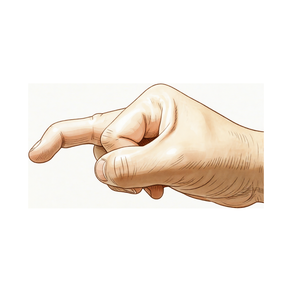
Sa mga malubhang kaso, maaaring mapansin mo ang pagbaba ng dulo ng iyong daliri pababa. Ang pagbaba na ito ay maaaring magpahirap sa paghawak nang mahigpit sa mga bagay. Maaari ka ring maranasan ang pamamaga sa paligid ng kasukasuan. Habang ang karamihan sa mga pinsala ay gumagaling nang maayos gamit ang splint, ang ilang kaso ay nangangailangan ng mas kumplikadong pag-aalaga. Itatatakda ng iyong doktor kung kailangan mo ng operasyon batay sa laki ng anumang fragment ng buto o sa posisyon ng kasukasuan.
Kung mayroon kang malaking fragment ng buto o kung lumilipat ang kasukasuan laban sa tamang posisyon, maaaring irekomenda ng iyong doktor ang isang operasyon. Karaniwang ginagawa ito upang ibalik ang tamang pagkakahanay at pag-andar. Parehong ang mga surgical at non-surgical na paggamot ay karaniwang nagdudulot ng mahusay na resulta. Inaasahan mong mababalik ang buong paggamit ng iyong daliri na may tamang pag-aalaga.
Ano ang nangyayari
May maliit na tendon sa dulo ng iyong daliri na tinatawag na terminal tendon. Ito ay gumagana tulad ng lubid na nag-uugnay sa iyong kalamnan sa buto sa pinakadulo ng iyong daliri. Ang lubid na ito ang nagbibigay-daan sa iyo na tuwidin ang iyong daliri. Kapag nasugatan ang lugar na ito, napuputol ang lubid o hinahatak nito ang maliit na piraso ng buto palayo sa kasukasuan. Humihinto dito ang signal para tuwidin ang dulo ng iyong daliri.
Ang pinakakaraniwang sanhi ay biglaang puwersa na yumuyuko pabalik sa iyong tuwid na daliri. Isipin ang pagkakadikit ng iyong daliri sa isang bola o frame ng pinto habang ito ay nakatuwid. Biglang yumuko ang kasukasuan nang masyadong mabilis. Ang biglaang pag-unat ay pumutol sa tendon mula sa buto. Sa ilang kaso, may kasamang maliit na piraso ng buto na nabasag kasama ng tendon. Pareho ang klinikal na itsura kung ang tendon mismo ang naputol o kung ang piraso ng buto ang nahatak. Sa parehong kaso, sira ang koneksyon.
Dahil hindi na nakakabit ang lubid para sa pagtutwid, nananatiling yumuko ang iyong daliri. Hindi mo kayang aktibong itaas ang dulo ng iyong daliri upang ituwid ito. Ito ay tinatawag na mallet finger deformity. Gayunpaman, kayang pa-aktibo mong ituwid ito gamit ang iyong ibang kamay. Mananatiling intact ang joint capsule at mga ligamento, kaya hindi lumalabas ang kasukasuan mismo. Ang posisyon na yumuko ay simpleng resulta ng walang laban na hilang kalamnan mula sa kabilang panig ng daliri.
Ang pinsalang ito ay kadalasang tumatama sa maliit, singsing, o gitnang daliri ng iyong dominanteng kamay. Mas karaniwan ito sa mga lalaki. Bagama't kadalasang dulot ito ng trauma, may ilang matatanda na may arthritis na dulot ng pagkasira na maaaring umusbong sa ganitong postura nang walang tiyak na pinsala. Sa mga bata, maaaring tumama ang pinsala sa growth plate imbes na sa tendon. Anuman ang sanhi, pareho ang resulta: hindi na nakakonekta ang mekanismo na nagtatayo ng dulo ng iyong daliri, na nag-iiwan nito na bumababa.
Ano ang maaari naming gawin dito
Karamihan sa mga sugat sa mallet finger ay gumagaling nang walang operasyon. Malamang na mag-iisip ka ng splint upang panatilihing tuwid ang dulo ng iyong daliri. Pinapayagan nito ang tendon na gumaling sa tamang posisyon. Maaaring gamutin ka ng hand therapist nang kasing epektibo ng isang surgeon para sa mga simpleng kaso. Gumagamit sila ng mga pamamaraan na bihirang magdulot ng mga problema sa balat. Maaari mo ring subukan ang paggamit ng splint sa gabi, ngunit hindi nito pinapabuti ang iyong panghuling resulta. May ilang pasyente ang gumagamit ng mga ehersisyo kasama ang splinting, bagaman hindi malinaw ang ebidensya para dito. Kailangan mong isuot ang iyong splint nang tuloy-tuloy sa loob ng panahong inirekomenda ng iyong surgeon. Huwag itong alisin upang hugasan o baluktotin ang daliri. Ang konsistensya ang susi sa tagumpay.
Ang pamamahala ng sakit ay nakatuon sa kaginhawahan habang gumagaling ang tendon. Maaari kang kumain ng mga over-the-counter na panlunas sa sakit o anti-inflammatories kung kinakailangan. Tumutulong ito upang pamahalaan mo ang iyong mga pang-araw-araw na gawain habang hindi gumagalaw ang iyong daliri. May ilang pasyente ang nagtatanong tungkol sa mga injeksyon. Gayunpaman, hindi sinusuportahan ng ebidensya ang mga injeksyon ng cortisone, hyaluronic acid, o PRP para sa partikular na sugat na ito. Hindi bahagi ng pamantayang plano ng pag-aalaga ang mga tratong ito para sa mallet finger. Dapat manatili ang iyong pokus sa pagpapanatili ng tuwid na kasukasuan gamit ang iyong splint. Kung mayroon kang malaking sakit, talakayin ang mga ligtas na pagpipilian sa iyong surgeon. Iwasan ang agresibong masahe o pag-stretch, na maaaring makagambala sa paggaling.
Ang operasyon ay isinasaalang-alang lamang kung nabigo ang konservatibong pag-aalaga o kung malala ang sugat. Maaaring irekomenda ng iyong surgeon ang isang operasyon kung mayroon kang malaking fracture na kabilang ang higit sa isang-tatlo ng ibabaw ng kasukasuan. Indikado rin ang operasyon kung lumipat ang fragment ng buto mula sa kanyang orihinal na posisyon. May ilang pasyente ang pumipili ng operasyon dahil hindi sila makakapagtrabaho habang may splint. Kung mayroon kang kronikong sugat na hindi gumaling pagkatapos ng ilang buwan ng splinting, maaaring maging pagpipilian ang operasyon. Inaayos ng pamamaraang ito ang nasirang tendon upang ibalik ang kakayahan mong tuwidin ang daliri. Ito ay isang maliit na operasyon na may mataas na rate ng tagumpay. Talakayin ng iyong surgeon ang partikular na mga panganib at benepisyo sa iyo kung magiging kinakailangan ang landas na ito.
Ano ang inaasahan
Karamihan sa mga sugat sa mallet finger ay gumagaling nang maayos nang walang operasyon. Posibleng irekomenda ng iyong doktor ang paggamit ng splint upang panatilihing tuwid ang dulo ng iyong daliri. Pinapayagan nito ang tendon o buto na muling kumonekta. Parehong ang mga operasyon at hindi-operasyon na paggamot ay nagdudulot ng mahusay na klinikal na resulta para sa karamihan ng mga pasyente. Inaasahan mo ang mataas na antas ng kasiyahan sa iyong resulta ng paggamot.
Ang landas patungo sa paggaling ay nakadepende sa kabiguan ng iyong sugat. Para sa mga simpleng kaso, sapat na karaniwang ang isang simpleng splint o isang dorsal glued splint. Kung ang iyong sugat ay may kasamang malaking fragment ng buto o kung ang kasukasuan ay lumipat mula sa tamang posisyon, posibleng irekomenda ang operasyon. Ang operasyon ay isang pagpipilian din para sa mga kronikong kaso o kung nabigo ang mga nakaraang paggamot. Sa mga bata, hindi gaanong malinaw ang pangangailangan para sa operasyon, ngunit nananatiling epektibo ang hindi-operasyon na pamamahala para sa karamihan.
Dapat mong malaman na hindi laging garantisado ang buong pagwawasto ng posisyon ng daliri. Kung mayroon kang isang matinding kronikong mallet finger na may malaking liko, mas hindi konsistente ang buong pagwawasto. Gayunpaman, kahit sa mga komplikadong kasing ito, madalas na maganda hanggang mahusay ang mga resulta sa pangmatagalan. Mababa ang mga rate ng komplikasyon para sa konservatibong pamamahala. Bihirang ang mga seryosong isyu tulad ng impeksyon o depekto sa kuko, lalo na sa mga makabagong teknika sa operasyon.
Ang paggaling ay isang unti-unting proseso. Maaaring mapansin mo ang ilang stiffness o isang maliit na natitirang liko sa dulo ng iyong daliri. Karaniwan ito at madalas ay hindi malaki ang epekto nito sa iyong pang-araw-araw na gawain. Hindi pinapabuti ng supplemental na paggamit ng splint sa gabi ang iyong resulta sa terms ng kapansanan o kasiyahan, kaya maaaring hindi mo na kailangang isuot ito pagkatapos ng iyong unang yugto ng paggaling.
Kung mayroon kang isang tiyak na uri ng fracture na may kasamang higit sa isang-tatlong bahagi ng ibabaw ng kasukasuan, mayroong humigit-kumulang 50% na tsansang hindi ito magpatuloy sa joint subluxation (pagdulas mula sa tamang posisyon). Ang laki ng fracture at kung gaano kabilis ka nagsimulang isuot ang iyong splint ay mga pangunahing salik sa iyong paggaling. Ang pagkaantala sa paggamot ay maaaring dagdagan ang risk ng mga komplikasyon, kaya mahalagang sundin ang payo ng iyong doktor sa timing.
Sa kabuuan, maaari kang maging confident na ang mallet finger ay isang highly treatable na kondisyon. Kung pipiliin mo ang paggamit ng splint o operasyon, ang layunin ay ibalik ang function at bawasan ang sakit. Karamihan sa mga pasyente ay bumabalik sa kanilang normal na mga gawain na may functional na daliri. Gabay ng iyong doktor ang iyong landas patungo sa pinakamainam na paraan para sa iyong partikular na sugat upang masiguro ang pinakamainam na posibleng pananaw.
Kailan makipag-ugnayan sa doktor
Kumonsulta sa iyong doktor kung mayroon kang patuloy na sakit na hindi gumagaling kahit magpahinga. Humingi ng pagsusuri ng espesyalista kung napapansin mo ang kahinaan o kawalan ng katatagan sa daliri. Humingi ng tulong kung ang kasukasuan ay nakakabit o biglang sumasayaw. Kontakin ang iyong doktor kung ang mga sintomas ay nakakaapekto sa iyong tulog o trabaho. Humingi ng tulong para sa anumang biglaang paglala ng iyong kondisyon. Minsan, ang mga sugat sa Mallet finger ay nangyayari sa parehong kamay sabay-sabay. Bihirang, ang mga sugat na ito ay naglalaman ng kombinasyon ng pinsala sa tendon at fracture ng buto sa mga mas batang pasyente. Maaari ring magkaroon ng papel ang mga biochemical na pagbabago sa paraan kung paano nabubuo ang mga sugat na ito. Ang maagang pagsusuri ay tumutulong upang masiguro ang pinakamahusay na resulta para sa iyong daliri.
Evidence & references
Overview
- Absolute indications for surgical intervention for mallet fingers in pediatric populations remain unclear [1].
- Large-fragment mallet finger cases can be effectively managed conservatively with low complication rates [2].
- Most mallet finger injuries can be managed non-surgically with splinting [3].
- Surgery is occasionally recommended for acute or chronic mallet finger cases or for salvage of failed prior treatment [3].
- Both surgical and nonsurgical treatments of mallet finger injuries lead to excellent clinical outcomes [4].
- All cases of mallet finger are proposed to be treated with a dorsal glued splint except for stage IV mallet finger, which is treated with extra-articular pinning [5].
- A simple splint is recommended as an alternative means of treating mallet finger [8].
- Surgery is generally indicated in mallet fractures involving more than one-third of the articular surface [9].
- Surgery is generally indicated in all patients who develop volar subluxation of the distal phalanx [9].
- A significant advantage of surgical management over conservative management in complicated cases (large fragment or volar subluxation) has yet to be clearly proven [9].
- The majority of hand fractures can be treated without surgery, though surgery offers distinct advantages in properly selected cases [12].
- Pullout wire fixation together with distal interphalangeal joint Kirschner wire stabilization for acute combined tendon and bone (double level) mallet finger injury reports good to excellent long-term results with no reported complications such as infection, nonunion, or nail deformity [13].
- Central slip tenotomy with distal repair is a technique described for severe chronic mallet fingers with deformities exceeding 36 degrees [18].
- Full correction of extensor lag is less consistent with greater degrees of preoperative flexion deformity in severe chronic mallet fingers treated with central slip tenotomy and distal repair [18].
- The Ishiguro extension block technique produces satisfactory results and is effective and minimally invasive for mallet finger fractures when properly applied [54].
Anatomy & Pathophysiology
- Mallet finger deformity is characterized by a loss of active distal interphalangeal (DIP) joint extension with full passive range of motion evident [19].
- The deformity reflects the loss of normal extensor force transmission via the terminal tendon insertion onto the distal phalanx [19].
- The unopposed flexor digitorum profundus pulls the distal joint into flexion [19].
- The usual mechanism of injury involves sudden passive flexion of an actively extended DIP joint [19].
- Disruption of the terminal tendon may be entirely confined to the tendon or may involve an avulsed fracture fragment from the dorsal lip of the distal phalanx proximal articular surface [19].
- The clinical appearance of soft tissue and bony mallet fingers is similar because the avulsed fragment includes the terminal tendon insertion [19].
- The distal joint rests in flexion, a posture that cannot be actively changed [19].
- Full passive extension of the DIP joint is possible in mallet finger [19].
- Mallet finger most commonly involves a closed rupture of the terminal tendon with or without associated fracture of the distal phalanx [37].
- Snagging the extending finger on an object that suddenly flexes the DIP joint is a frequent cause of mallet finger [37].
- Less commonly, a forceful hyperextension injury of the DIP joint may result in a large fracture of the base of the distal phalanx involving one-third or more of the articular surface [37].
- Elderly patients with osteoarthritis of the DIP joint may have mallet deformities that are not related to trauma [37].
- Individuals with hyperlax joints may have multiple pseudomallet swan neck postures that are unrelated to trauma [37].
- Open mallet injuries are uncommon [37].
- The most frequently involved digits are the small, ring, and middle fingers of the dominant hand [37].
- There is a male predominance in mallet finger incidence [37].
- Tendinous mallet fingers have been reported to occur from age 11 onward [37].
- In skeletally immature individuals, a transepiphyseal plate fracture may be seen instead of tendon rupture [37].
- There may be a familial predisposition to mallet fingers [37].
- Displacement of the epiphysis of the distal phalanx can cause the digit to assume a mallet finger posture [14].
- Hyperextension of the phalanx usually affords satisfactory reduction of a displaced epiphysis [14].
- The extensor apparatus of the fingers includes the interosseous muscle, extensor digitorum communis tendon, lumbrical muscle, flexor tendon sheath, sagittal bands, transverse metacarpal ligament, interosseous hood, interosseous hood oblique fibers, extensor lateral band, extensor middle band, interosseous middle band, interosseous lateral band, oblique retinacular ligament, central extensor lateral, spiral fibers, transverse retinacular ligament, lateral extensor tendon, triangular lamina, and terminal extensor tendon [14].
- Injuries to the finger extensor apparatus are very common and may produce chronic deformity and loss of function [34].
Classification
- Absolute indications for surgical intervention for mallet fingers in pediatric populations remain unclear [1].
- Doyle type 4c mallet finger cases can be effectively managed conservatively with low complication rates [2].
- Most mallet finger injuries can be managed non-surgically with splinting, although surgery is occasionally recommended for acute or chronic cases or for salvage of failed prior treatment [3].
- Both surgical and nonsurgical treatments of mallet finger injuries lead to excellent clinical outcomes [4].
- All cases of mallet finger are proposed to be treated with a dorsal glued splint except for stage IV mallet finger, which is treated with extra-articular pinning [5].
- Approximately 50% of patients with a mallet fracture involving more than one-third of the articular surface of the distal phalanx do not progress to subluxation of the DIP joint [6].
- Fracture size and time to application of finger immobilizer are independent risk factors for the development of DIP joint subluxation in mallet fracture [6].
- A simple splint is recommended as an alternative means of treating mallet finger [8].
- Deepithelialised pedicled skin flap technique is a new reliable alternative in the treatment of chronic mallet finger [11].
- A hand therapist can treat mallet finger injuries of type 1 as effectively as a surgeon, with a method of immobilisation that offers practically no complications regarding skin condition [16].
- A modification to the Doyle classification is proposed to make it more encompassing and less prone to interobserver error [35].
- The interrater reliability of the Kellgren & Lawrence and OARSI classification systems for post-traumatic osteoarthritis in the distal interphalangeal joint after mallet finger fractures is considerably lower than initially assumed [41].
- Non-operative management of mallet fractures, regardless of fracture classification, joint congruence or pre-existing degenerate change in the DIP joint, is safe and yields predictably good outcomes in most patients [46].
- Most acute mallet fingers can be treated by continuous splinting, while fracture dislocations require open reduction and internal fixation [50].
Clinical Presentation
- Mallet finger injuries can present as bilateral cases [21].
- Biochemical abnormalities may play a role in the etiology of mallet fingers [21].
- Mallet finger injuries can involve a rare combination of tendon avulsion and fracture in juvenile patients [26].
Investigations
- Mallet finger deformity is characterized by a loss of active distal interphalangeal (DIP) joint extension with full passive range of motion evident [19].
- The deformity reflects the loss of normal extensor force transmission via the terminal tendon insertion onto the distal phalanx [19].
- The unopposed flexor digitorum profundus pulls the distal joint into flexion [19].
- The usual mechanism of injury involves sudden passive flexion of the actively extended distal interphalangeal joint [19].
- Disruption of the terminal tendon may be entirely confined to the tendon or may involve an avulsed fracture fragment from the dorsal lip of the distal phalanx proximal articular surface [19].
- Because the avulsed fragment includes the terminal tendon insertion, the clinical appearance of soft tissue and bony mallet fingers is similar [19].
- The distal joint rests in flexion, a posture that cannot be actively changed [19].
- Full passive extension of the distal interphalangeal joint is possible [19].
- A radiograph should be obtained to determine whether a fracture is present [19].
- If a fracture is present, radiographs should assess whether the dorsal fragment is large and whether the distal phalanx is subluxed palmarward [19].
Treatment
Non-Operative Management
- Most mallet finger injuries can be managed non-surgically with splinting [3].
- Both surgical and nonsurgical treatments of mallet finger injuries lead to excellent clinical outcomes [4].
- A dorsal glued splint is proposed for the treatment of all cases of mallet finger except stage IV injuries [5].
- A simple splint is recommended as an alternative means of treating mallet finger [8].
- Supplemental night splinting does not improve the outcome of mallet finger in terms of extensor lag, disability, or satisfaction with treatment [15].
- A hand therapist can treat type 1 mallet finger injuries as effectively as a surgeon [16].
- A hand therapist can treat type 1 mallet finger injuries as effectively as a surgeon, with a method of immobilisation that offers practically no complications regarding skin condition [39].
- Conservative therapeutic management of acute, closed mallet finger is diverse and varied, with exercises and interventions supplementary to splinting commonly utilised [23].
- An alternative simple and custom-made orthosis manages the mallet finger by allowing for PIP flexion while inhibiting full extension or hyperextension [25].
- The clinical efficacy of elastic taping for the treatment of mallet finger injuries remains to be tested vigorously [32].
Operative Management
- Surgery is occasionally recommended for acute or chronic mallet cases or for salvage of failed prior treatment [3].
- Surgical management may be considered for acute and chronic mallet lesions in patients who have failed nonsurgical treatment [10].
- Surgical management may be considered for acute and chronic mallet lesions in patients who are unable to work with the splint in position [10].
- Surgical management may be considered for acute and chronic mallet lesions in patients who have a fracture involving more than one third of the joint surface [10].
- Surgery is generally indicated in the case of mallet fractures involving more than one-third of the articular surface [9].
- Surgery is generally indicated in all patients who develop volar subluxation of the distal phalanx [9].
- A significant advantage of surgical management even in complicated cases (fracture >1/3 articular surface or volar subluxation) has yet to be clearly proven [9].
- Complication rates for conservative management of Doyle type 4c mallet finger are low, suggesting large-fragment cases can be effectively managed conservatively [2].
- Absolute indications for surgical intervention for mallet fingers in the pediatric population remain unclear [1].
Surgical Techniques
- The Thompson procedure is an effective technique for the salvage of a closed mallet injury with an associated swan neck deformity following failed treatment [27].
- A deepithelialised pedicled skin flap technique is a new reliable alternative in the treatment of chronic mallet finger [11].
- Scar overlapping suture for treating chronic tendinous mallet finger in children is safe and effective [36].
- For chronic mallet finger, if the distal phalanx droops severely but passive extension in the distal interphalangeal joint is still satisfactory, surgery may be indicated depending on the patient’s needs [14].
- Surgical treatment for chronic mallet finger involves making a small V-shaped or U-shaped incision on the dorsum of the finger, convex distally, with the tip no closer than 5 mm proximal to the nail base [14].
- The surgical technique for chronic mallet finger involves developing a flap between the tendon and subcutaneous fat to expose the extensor tendon with intervening scar [14].
- The surgical technique for chronic mallet finger involves identifying the junction of normal tendon with scar, severing the tendon transversely proximal to the joint, and resecting sufficient scar or tendon to allow closure with the finger in maximal extension [14].
- The surgical repair of chronic mallet finger is supported by a transarticular 0.045-inch Kirschner wire [14].
- The extensor tendon in chronic mallet finger surgery is repaired with 4-0 monofilament nylon or 4-0 monofilament wire as a pull-out roll stitch [14].
- Postoperative care for chronic mallet finger surgery involves removing sutures at 10 to 14 days and maintaining the distal joint in extension with Kirschner wire protection for 4 weeks [14].
- The Kirschner wire is removed after 4 to 6 weeks in chronic mallet finger surgery, with the repair protected by a splint for an additional 8 weeks [14].
Complications
- Absolute indications for surgical intervention for mallet fingers in pediatric populations remain unclear [1].
- Complication rates for conservative management of Doyle type 4c mallet fingers are low [2].
- Surgery is occasionally recommended for acute or chronic cases or for salvage of failed prior treatment [3].
- Both surgical and nonsurgical treatments of mallet finger injuries lead to excellent clinical outcomes [4].
- Stage IV mallet finger is treated with extra-articular pinning rather than dorsal glued splinting [5].
- Approximately 50% of patients with a mallet fracture involving more than one-third of the articular surface of the distal phalanx do not progress to subluxation of the DIP joint [6].
- Fracture size is an independent risk factor for the development of DIP joint subluxation in mallet fracture [6].
- Time to application of finger immobilizer is an independent risk factor for the development of DIP joint subluxation in mallet fracture [6].
- Pullout wire fixation together with distal interphalangeal joint Kirschner wire stabilization for acute combined tendon and bone mallet finger injuries reports no complications such as infection, nonunion, or nail deformity [13].
- Supplemental night splinting does not improve outcomes in terms of extensor lag, disability, or satisfaction with treatment [15].
- Full correction of extensor lag is less consistent with greater degrees of preoperative flexion deformity in severe chronic mallet fingers treated with central slip tenotomy and distal repair [18].
- The Thompson procedure is an effective technique for the salvage of a closed mallet injury with an associated swan neck deformity following failed treatment [27].
- The most common cause of procedural failure in closed reductions using an extension-block pin for bony mallet finger is inaccurate insertion of the K-wire to fix the distal interphalangeal joint [56].
Recovery
- Most mallet finger injuries can be managed non-surgically with splinting [3].
- Surgery is occasionally recommended for acute or chronic cases or for salvage of failed prior treatment [3].
- Both surgical and nonsurgical treatments of mallet finger injuries lead to excellent clinical outcomes [4].
- Complication rates for conservative management of large-fragment mallet finger cases are low [2].
- Supplemental night splinting does not improve the outcome of mallet finger in terms of extensor lag, disability, or satisfaction with treatment [15].
- LIPUS therapy may be recommended as an option to treat type I mallet finger fracture in children for whom initiation of treatment was delayed up to 8 weeks [29].
- Approximately 50% of patients with a mallet fracture involving more than one-third of the articular surface of the distal phalanx do not progress to subluxation of the DIP joint [6].
- Fracture size and time to application of finger immobilizer are independent risk factors for the development of DIP joint subluxation in mallet fracture [6].
- Full correction of extensor lag is less consistent with greater degrees of preoperative flexion deformity in severe chronic mallet fingers treated with central slip tenotomy with distal repair [18].
- The surgical technique of pullout wire fixation together with distal interphalangeal joint Kirschner wire stabilization for acute combined tendon and bone mallet finger injuries reports good to excellent long-term results [13].
- No reported complications such as infection, nonunion, or nail deformity were observed in the series of acute combined tendon and bone mallet finger injuries treated with pullout wire fixation and DIP joint Kirschner wire stabilization [13].
Key Evidence
- [L4] Absolute indications for surgical intervention for mallet fingers in this population remain unclear. [1] (10.1016/j.jhsa.2018.03.037)
- [L4] Complication rates were low, suggesting that large-fragment mallet finger cases can be effectively managed conservatively. [2] (10.1186/s12891-026-09787-w)
- [L5] Most mallet finger injuries can be managed non-surgically with splinting, although surgery is occasionally recommended for acute or chronic cases or for salvage of failed prior treatment. [3] (10.1007/s11552-014-9609-y)
- [L4] Both surgical and nonsurgical treatments of mallet finger injuries lead to excellent clinical outcomes. [4] (10.1016/j.jhsa.2017.10.004)
- [L5] The authors propose to treat all cases of mallet finger with a dorsal glued splint except for stage IV mallet finger, which they treat with extra-articular pinning. [5] (10.5999/aps.2016.43.2.134)
- [L2] Approximately 50% of patients with a mallet fracture involving more than one-third of the articular surface of the distal phalanx do not progress to subluxation of the DIP joint; fracture size and time to application of finger immobilizer are independent risk factors for the development of DIP joint subluxation in mallet fracture. [6] (10.1177/1753193414554556)
- [L2] The study recommends this splint as an alternative means of treating mallet finger. [8] (10.1136/emj.10.3.244)
- [L4] Although surgery is generally indicated in the case of mallet fractures involving more than one-third of the articular surface as well as in all patients who develop volar subluxation of the distal phalanx, a significant advantage of surgical management even in those complicated cases has yet to be clearly proven. [9] (10.1177/1558944716642763)
- [L5] Surgical management may be considered for acute and chronic mallet lesions in patients who have failed nonsurgical treatment, are unable to work with the splint in position, or have a fracture involving more than one third of the joint surface. [10] (10.5435/00124635-200509000-00007)
- [Paper] This method seems to be a new reliable alternative in the treatment of chronic mallet finger. [11] (10.1016/j.injury.2013.01.013)
- [L5] The majority of hand fractures can be treated without surgery, though surgery offers distinct advantages in properly selected cases. [12] (10.1016/j.jhsa.2013.02.017)
- [L4] The study describes a surgical technique for acute combined tendon and bone mallet fingers and reports good to excellent long-term results with no reported complications such as infection, nonunion, or nail deformity in the series. [13] (10.1016/j.jhsa.2014.11.011)
- [L1] Supplemental night splinting does not improve the outcome of mallet finger in terms of extensor lag, disability, or satisfaction with treatment. [15] (10.1007/s11552-013-9600-z)
- [L4] A hand therapist can treat mallet finger injuries of type 1 as effectively as a surgeon, with a method of immobilisation that offers practically no complications regarding skin condition. [16] (10.1177/175899830501000103)
- [L4] The article describes a technique combining central slip tenotomy with distal repair for severe chronic mallet fingers with deformities exceeding 36 degrees, noting that full correction of extensor lag is less consistent with greater degrees of preoperative flexion deformity. [18] (10.1016/j.jhsa.2014.01.040)
- [L4] This case raises a question regarding the possible role of biochemical abnormalities causing mallet fingers. [21] (10.1177/175899830400900103)
- [L4] Conservative therapeutic management of acute, closed mallet finger is diverse and varied, with exercises and interventions supplementary to splinting commonly utilised. [23] (10.1177/1758998316664822)
- [L5] An alternative simple and custom-made orthosis has been presented to manage the mallet finger, allowing for PIP flexion while inhibiting full extension or hyperextension. [25] (10.1016/j.jht.2016.01.001)
- [L4] The authors describe a rare combination of tendon avulsion and fracture in a juvenile mallet finger, noting that while few mallet fingers require open treatment, this combined injury may be more common than thought. [26] (10.1177/1753193415571777)
- [L4] It is an effective technique for the salvage of a closed mallet injury with an associated swan neck deformity following failed treatment. [27] (10.1016/j.jhsa.2013.04.011)
- [L3] LIPUS therapy may be recommended as an option to treat type I mallet finger fracture in children for whom initiation of treatment was delayed up to 8 weeks. [29] (10.1177/1558944717692095)
- [L4] The clinical efficacy of the proposed method of elastic taping for the treatment of mallet finger injuries remains to be tested vigorously. [32] (10.1016/j.jht.2014.02.005)
- [L5] Injuries to the finger extensor apparatus are very common and may produce chronic deformity and loss of function. [34] (10.1016/j.hcl.2013.03.003)
- [L4] This article provides a topical review of the contemporary literature concerning acute mallet finger injuries and proposes a modification to the Doyle classification to make it more encompassing and less prone to interobserver error. [35] (10.1016/j.jhsa.2022.10.013)
- [L4] Scar overlapping suture for treating chronic tendinous mallet finger in children is safe and effective. [36] (10.1186/s13018-019-1106-0)
- [L4] A hand therapist can treat type 1 mallet finger injuries as effectively as a surgeon. [39] (10.1197/j.jht.2008.04.002)
- [L4] The interrater reliability of the Kellgren & Lawrence and OARSI classification systems for post-traumatic osteoarthritis in the distal interphalangeal joint after mallet finger fractures is considerably lower than initially assumed. [41] (10.1016/j.jhsa.2024.03.012)
- [L3] Non-operative management of mallet fractures, regardless of fracture classification, joint congruence or pre-existing degenerate change in the DIP joint, is safe and yields predictably good outcomes in most patients. [46] (10.1177/1753193421992986)
- [L5] Most acute mallet fingers can be treated by continuous splinting, while fracture dislocations require open reduction and internal fixation. [50] (10.1097/01.blo.0000205903.51727.62)
- [L4] The extension block technique, when properly applied, produces satisfactory results and is effective and minimally invasive for mallet finger fractures. [54] (10.1054/jhsb.2001.0733)
- [Paper] The most common cause of procedural failure was inaccurate insertion of the K-wire to fix the distal interphalangeal joint. [56] (10.1055/s-0040-1701318)
References
[1] Outcomes of Splinting in Pediatric Mallet Finger. The Journal of Hand Surgery. 2018. DOI: 10.1016/j.jhsa.2018.03.037 [2] Surgical versus conservative management of Doyle type 4c mallet finger: a comparative study. BMC Musculoskeletal Disorders. 2026. DOI: 10.1186/s12891-026-09787-w [3] Current Concepts: Mallet Finger. HAND. 2014. DOI: 10.1007/s11552-014-9609-y [4] Surgical and Nonsurgical Management of Mallet Finger: A Systematic Review. The Journal of Hand Surgery. 2018. DOI: 10.1016/j.jhsa.2017.10.004 [5] Review of Acute Traumatic Closed Mallet Finger Injuries in Adults. Archives of Plastic Surgery. 2016. DOI: 10.5999/aps.2016.43.2.134 [6] The risk factors associated with subluxation of the distal interphalangeal joint in mallet fracture. Journal of Hand Surgery (European Volume). 2014. DOI: 10.1177/1753193414554556 [8] The conservative treatment of mallet finger with a simple splint: a case report.. Emergency Medicine Journal. 1993. DOI: 10.1136/emj.10.3.244 [9] The Diagnosis and Management of Mallet Finger Injuries. HAND. 2016. DOI: 10.1177/1558944716642763 [10] Mallet Finger. Journal of the American Academy of Orthopaedic Surgeons. 2005. DOI: 10.5435/00124635-200509000-00007 [11] A new surgical treatment for mallet finger deformity: Deepithelialised pedicled skin flap technique. Injury. 2013. DOI: 10.1016/j.injury.2013.01.013 [12] Hand Fractures: A Review of Current Treatment Strategies. The Journal of Hand Surgery. 2013. DOI: 10.1016/j.jhsa.2013.02.017 [13] Pullout Wire Fixation Together With Distal Interphalangeal Joint Kirschner Wire Stabilization for Acute Combined Tendon and Bone (Double Level) Mallet Finger Injury. The Journal of Hand Surgery. 2015. DOI: 10.1016/j.jhsa.2014.11.011 [14] Campbell S Operative Orthopaedics 4 Volume Set. RECONSTRUCTION OF FINGER FLEXORS: SINGLE-STAGE TENDON GRAFT > CHRONIC MALLET FINGER (SECONDARY REPAIR). [15] A Prospective Randomized Controlled Trial Comparing Night Splinting with No Splinting after Treatment of Mallet Finger. HAND. 2014. DOI: 10.1007/s11552-013-9600-z [16] Hand Therapist-led Management of Mallet Finger. The British Journal of Hand Therapy. 2005. DOI: 10.1177/175899830501000103 [18] Central Slip Tenotomy With Distal Repair in the Treatment of Severe Chronic Mallet Fingers. The Journal of Hand Surgery. 2014. DOI: 10.1016/j.jhsa.2014.01.040 [19] A Lange Medical Book Current Diagnosis Treatment In Orthopedics Fifth Edition. 9Hand Surgery > 3. Mallet Finger. [21] Bilateral Mallet Fingers: A Case Study. The British Journal of Hand Therapy. 2004. DOI: 10.1177/175899830400900103 [23] How do hand therapists conservatively manage acute, closed mallet finger? A survey of members of the British Association of Hand Therapists. Hand Therapy. 2016. DOI: 10.1177/1758998316664822 [25] The challenge of the mallet orthosis: A simple solution. Journal of Hand Therapy. 2016. DOI: 10.1016/j.jht.2016.01.001 [26] Combined tendon avulsion and fracture in a mallet finger injury in a juvenile. Journal of Hand Surgery (European Volume). 2015. DOI: 10.1177/1753193415571777 [27] The Thompson Procedure for Chronic Mallet Finger Deformity. The Journal of Hand Surgery. 2013. DOI: 10.1016/j.jhsa.2013.04.011 [29] Comparison of Treatment Results for Mallet Finger Fractures in Children Between Low-Intensity Pulsed Ultrasound Stimulation and Ishiguro’s Method. HAND. 2017. DOI: 10.1177/1558944717692095 [32] A novel way of treating mallet finger injuries. Journal of Hand Therapy. 2014. DOI: 10.1016/j.jht.2014.02.005 [34] Diagnosis and Treatment of Finger Deformities Following Injuries to the Extensor Tendon Mechanism. Hand Clinics. 2013. DOI: 10.1016/j.hcl.2013.03.003 [35] Acute Mallet Finger Injuries—A Review. The Journal of Hand Surgery. 2023. DOI: 10.1016/j.jhsa.2022.10.013 [36] Scar overlapping suture for treating chronic tendinous mallet finger in children. Journal of Orthopaedic Surgery and Research. 2019. DOI: 10.1186/s13018-019-1106-0 [37] Green S Operative Hand Surgery. CASE STUDY 5.2 Unusual Mallet Finger Presentation. [39] Hand Therapist-led Management of Mallet Finger. Journal of Hand Therapy. 2008. DOI: 10.1197/j.jht.2008.04.002 [41] Rater Agreement of Post-Traumatic Osteoarthritis of the Distal Interphalangeal Joint 12 Years After a Mallet Finger Fracture. The Journal of Hand Surgery. 2024. DOI: 10.1016/j.jhsa.2024.03.012 [46] The non-operative management of bony mallet injuries. Journal of Hand Surgery (European Volume). 2021. DOI: 10.1177/1753193421992986 [50] Tendon Avulsion Injuries of the Distal Phalanx. Clinical Orthopaedics and Related Research. 2006. DOI: 10.1097/01.blo.0000205903.51727.62 [54] The Ishiguro Extension Block Technique for the Treatment of Mallet Finger Fracture: Indications and Clinical Results. Journal of Hand Surgery. 2003. DOI: 10.1054/jhsb.2001.0733 [56] Causes of Procedural Failures of Closed Reductions using an Extension-Block Pin for Bony Mallet Finger. Journal of Hand and Microsurgery. 2021. DOI: 10.1055/s-0040-1701318