Patients › Shoulder
Distal Clavicle Excision (Mumford Procedure)
A Mumford procedure (distal clavicle excision) removes the small worn outer tip of the collarbone to ease shoulder pain at the AC joint. What the keyhole operation involves and what recovery looks like.
Why this operation has been suggested
Dr Kieran Hirpara, an upper-limb surgeon at Mater Private Hospital Rockhampton, starts with the least invasive options that suit your condition. Patients are generally referred to our clinic by their GP; if a physiotherapist has suggested you see us, you will still need a referral from your GP in order to be eligible for the Medicare rebate. Our assessment, which includes your history, an examination and scans where needed, works out what is causing your pain. For this kind of long-standing joint wear we usually try non-operative care first, such as changing your activities and physiotherapy, and we consider surgery when that has not given enough improvement.
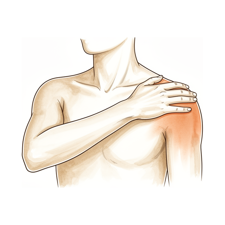
This operation removes a small piece of bone from the outer end of your collarbone, the joint where it meets the top of your shoulder. It is offered to people whose pain comes from wear at that joint, or from the bone breaking down after repeated heavy lifting, and only when the ligaments holding the joint steady are sound. Removing that small piece of bone stops the rough surfaces grinding on each other. The aim is less pain and freer movement of your shoulder, and both keyhole and open forms of this operation bring significant pain reduction at 1 year.
Before the operation
Your surgeon will plan the operation using scans you have already had, such as an X-ray, MRI or ultrasound of the shoulder. These show the joint clearly and help decide how much bone needs to be removed. On the day, stop eating and drinking seven hours before your operation. We ask for seven rather than six so you can be brought forward if the theatre list runs early. Your surgeon will tell you which of your usual medicines to skip and which to take. Bring a written list of everything you take. Arrange for someone to drive you home afterwards. Wear loose, comfortable clothing that is easy to get on and off. If you have other medical conditions, you may need blood tests or a review with the anaesthetist.
On the day
You arrive at the hospital's surgical admissions unit, where you are checked in and prepared for theatre. The anaesthetist will meet you before the operation and talk you through both parts. This operation is done under general anaesthetic combined with a regional nerve block. You are then taken into the operating theatre, where the operation is performed.
You wake up in the recovery area, where nurses monitor you while the anaesthetic wears off. Once you are stable, you either go to the ward or go home, depending on the procedure and your recovery.
What the operation involves
This is keyhole surgery. Your surgeon makes a few small cuts around your shoulder, including at least one at the back, and works with a small camera inside the joint. The camera shows the joint on a screen, so no large incision is needed to see it.
Through these small cuts, your surgeon removes about 5 mm of bone from the outer end of your collarbone. This is enough to stop the rough, worn surfaces at the joint grinding on each other. Your surgeon takes care to remove a controlled amount of bone, enough to relieve pain while protecting the ligaments and capsule that keep the joint steady. Protecting these structures matters, because removing too much bone can leave the collarbone unstable.
The small cuts are closed with stitches. A dressing goes over the top, and you keep that dressing on for about 10 days.
After the operation
Most patients stay one night in hospital after this operation, though some are able to go home the same day. Please arrange for someone to stay with you for the first 24 hours after you get home. You will have pain for the first day or two; we will give you medicine to keep it comfortable, and the shoulder may feel numb for a while because of the nerve block. Numbness and weakness in the first 24 hours after a nerve block are expected. Call the clinic if you cannot feel or move the arm, hand or fingers once the nerve block has worn off, at about 24 hours. Your arm rests in a simple sling for comfort, which comes off for exercises and washing. We leave the dressing on for about 10 days; please do not take it off before then unless we tell you to. We change or remove it when we see you.
Recovery
For the first day or two your shoulder will be sore and may feel swollen. The pain medicine we give you keeps this comfortable, and the numbness from the nerve block helps at first too. Resting with your arm supported and doing your exercises gently usually settles the discomfort. Most people find the soreness eases steadily over the first couple of weeks.
Your arm rests in a simple sling for comfort. You take it off for your exercises and for washing, and your physiotherapist will show you which movements to start with. These begin gently and build up as the pain settles. You can use your hand for light tasks at home, such as eating or writing, once you feel steady. Avoid lifting anything heavy or pushing through pain.
As movement returns, everyday activities get easier. Getting dressed, reaching overhead and sleeping on that side all improve gradually. Sleeping propped up with extra pillows can be more comfortable in the early days. Once your surgeon clears you to drive, typically at the six-week review, you can get back behind the wheel; see Driving after upper-limb surgery for more detail. Desk work and light daily tasks usually come back within 2 to 6 weeks, and heavier activity such as sport or gym work takes longer, from 3 to 6 months.
Everyone heals at their own pace, so your timeline may differ. Your surgeon and physiotherapist will guide you at each review.
What can go wrong
Most patients do well, but problems can occasionally happen. Your surgeon and the team monitor you closely to spot any issue early.
Sometimes pain continues after the operation. This is the most common problem with this surgery. It can happen if slightly too much or too little bone was removed, or if a small piece of bone grows back. If your shoulder still aches the way it did before, or the pain returns after a period of relief, bring it up at your next review. Sometimes a further operation is needed to fix it.
Removing too much bone can cause a different problem. The ligaments and capsule that hold the collarbone steady can be disturbed, leaving the joint loose or unstable. You might notice the collarbone feeling like it shifts or clicks, or a sense that the joint is not secure when you move your arm. If the joint was already loose before surgery, this risk is greater. An unstable joint may need further surgery to rebuild the ligaments. Tell your surgeon at your next review if the joint feels loose or moves in a new way.
Other problems are uncommon but worth knowing about. These include infection, stiffness, fracture, the joint fusing on its own, and a lasting pain condition called complex regional pain syndrome. Infection signs to watch for are fever, spreading redness around the wound, or fluid or pus leaking from it. Call the clinic the same day if you notice any of these. Pain that keeps getting worse despite your pain medicines also needs a same-day call to the clinic. The joint coming out of place after keyhole surgery has also been reported. If your shoulder suddenly changes shape, drops forward, or feels very painful and unstable after a knock or fall, contact the clinic straight away.
The complications table on this page lists typical rates if you want the specifics.
When to call us
Most problems are picked up early when you tell us about them. Call the clinic the same day if you have a fever, spreading redness around the wound, or fluid or pus leaking from it. Call the clinic the same day too if pain keeps getting worse despite your pain medicines. Go to the emergency department if you have swelling or pain in your calf, or shortness of breath or chest pain. These can be signs of a blood clot. Go to the emergency department as well if your fingers or hand turn pale, cold, white, blue or dark. Call the clinic if you cannot feel or move the arm, hand or fingers once the nerve block has worn off, at about 24 hours. If you cannot reach the clinic, go to your nearest emergency department.
Where to read more about the condition
This page is about the operation itself. The two conditions it treats have their own pages, each covering what the evidence shows about when surgery helps and when it does not: wear at the joint is covered on the AC Joint Osteoarthritis page, and the weightlifter's form, where the end of the collarbone breaks down, on the Distal Clavicle Osteolysis page.
Evidence & references
This is the clinical evidence summary written for health professionals. It is technical, and it lists the research this page was built from. You do not need to read it to understand your treatment or to make a decision about it.
Overview
- Simple excision of the outer end of the clavicle has yielded satisfactory results in patients with complete dislocation and subluxation of the acromioclavicular joint, with no residual upward displacement disturbing the patients [2].
- Excision of the outer end of the clavicle is preferred for old dislocations, while open reduction and internal fixation are not recommended due to complications and poor functional results [16].
- Open distal clavicle resection yields good to excellent results in properly selected patients and does not create significant subjective or objective weakness [3].
- In appropriately selected patients, open or arthroscopic distal clavicle resection is necessary to relieve symptoms of painful conditions of the acromioclavicular joint [7].
- Arthroscopic distal clavicle resection has provided more 'good or excellent' results than the open procedure, but is comprised of low-level evidence [6].
- Arthroscopic and open distal clavicle excisions both provide significant pain reduction at 1 year with no significant difference in outcome measures between groups, except for VAS pain score improvement [8].
- Among patients undergoing distal clavicle excision for acromioclavicular joint pathology, those having an arthroscopic procedure, specifically through the direct approach, can expect a faster return to activities while obtaining similar long-term outcomes compared with the open procedure [9].
- A well-performed distal clavicle excision will likely perform better than a poorly performed one, regardless of whether an open or arthroscopic approach is chosen [5].
- In carefully selected patients with isolated ACJ pathology, arthroscopic distal clavicle excision results in statistically and clinically significant improvements in range of motion and patient-reported outcome measures [18].
- Both the direct superior approach and the indirect subacromial approach to the arthroscopic distal clavicle resection result in successful clinical outcome with clinically insignificant difference at final follow-up [19].
- Incomplete excision and regrowth of the distal clavicle are the most common causes of revision [12].
- Regeneration of the excised portion of clavicle occurs frequently and may produce an unacceptable result, and may be more likely to occur in the younger patient population [13].
- Horizontal instability of the clavicle is evident with distal clavicle resection of greater than 10 mm [15].
- Although distal clavicle excision with 2.5 mm of bone was successful in many specimens, a 5 mm resection guaranteed no bone-to-bone abutment [17].
- Routine distal clavicle resection in the setting of rotator cuff repair does not result in improved outcomes for patients with no difference being observed at 24 months post surgery [11].
- A records review found that 10 of 894 (1.1%) rotator cuff repairs underwent subsequent distal clavicle resection [40].
- Regardless of the technique chosen for distal clavicle resection, portal placement remains paramount in both facilitating surgery and avoiding injury to adjacent extra-articular structures [1].
- The use of intraoperative ultrasound and cannulated dilators allows surgeons to perform distal clavicle excisions in a more efficient, reproducible and safer manner [4].
- A novel anterior portal technique for arthroscopic distal clavicle excision yields a safe corridor for placement of instruments in a reliable manner that does not disrupt the important stabilizers of the glenohumeral or AC joints [20].
- Clavicular reconstruction may be the treatment of choice in patients with excess resection of the clavicle [14].
Anatomy & Pathophysiology
Bony Anatomy
- The clavicle is the first bone to ossify, occurring at the fifth week of gestation [59].
- The clavicle is the only long bone to ossify by intramembranous ossification [59, 65].
- The medial (sternal) epiphysis of the clavicle is the last ossification center to fuse, occurring at age 20 to 25 years [59].
- The primary blood supply to the clavicle is periosteal, and no nutrient artery is present [59, 65].
- The distal clavicle is flat in the anteroposterior plane [65].
- The scapula has only one true diarthrodial articulation, the acromioclavicular joint [59].
- The acromion has three ossification centers: the metacromion (base), the mesoacromion (middle), and the preacromion (tip) [59].
- Failure of fusion of the acromial ossification centers results in os acromiale [59].
- The coracoid process serves as an attachment site for the coracoacromial ligament, coracoclavicular ligaments, conjoined tendon, and pectoralis minor muscle [70].
Ligaments and Joint Structures
- The acromioclavicular joint is a small diarthrodial joint with an interposed fibrocartilaginous disk [59].
- The acromioclavicular joint is a planar diarthroidal joint that couples scapular and sternoclavicular motion [32].
- A fibrocartilaginous load-bearing disk cushions the high contact stress of the acromioclavicular joint when load is placed across the small articular surface area [32].
- The superior and posterior acromioclavicular ligaments are the primary stabilizers to anterior and posterior (horizontal) translation of the clavicle [59].
- The coracoclavicular ligaments consist of the conoid (medial) and trapezoid (lateral) components [59, 65].
- The coracoclavicular ligaments are the primary stabilizers to superior (vertical) translation of the distal clavicle [59, 65].
- The trapezoid ligament is located approximately 25 mm from the acromioclavicular joint [70].
- The conoid ligament is located approximately 45 mm from the acromioclavicular joint and is the stronger of the two coracoclavicular ligaments [70].
- The superior shoulder suspensory complex is composed of the glenoid, coracoid process, coracoclavicular ligaments, distal clavicle, acromioclavicular joint, and acromion [59].
- The superior shoulder suspensory complex provides a stable connection between the scapula and the axial skeleton [59].
Pathophysiology and Biomechanics
- The acromioclavicular joint articulation helps provide a stable base for the glenohumeral joint to position the arm in space [32].
- Distal clavicle resection does not address instability and may accentuate it when horizontal or vertical instability exists [23].
- Distal clavicle resection must be reserved for patients in whom the coracoclavicular ligaments are intact and there is no concomitant instability [23].
- Kinematic changes resulting from acromioclavicular joint dislocation could be a potential source of pain and dysfunction in the shoulder [87].
- A combined injury of the acromioclavicular and coracoclavicular ligaments significantly alters glenohumeral kinematics during abduction [117].
- Large kinematic differences exist between the intact state and a Rockwood V lesion during both humerothoracic and scapulothoracic movements [118, 126].
- The distal clavicle has a wide range of motion during shoulder abduction, which does not support the concept of synchronous motion with the scapula [123].
- The trapezoid and conoid ligaments have unique functions in normal shoulder kinematics due to their anatomic attachments [86].
- Scapular and clavicular kinematics are affected in acromioclavicular separation models [89].
- Type I and II acromioclavicular joint disruptions impair long-term shoulder function in about half of patients 10 years after injury [101].
- The normal coracoclavicular distance measures approximately 1.1 to 1.3 cm [150].
- Injury to the acromioclavicular ligaments in a type II injury causes an increase of 3.6 mm in anterior and 6.4 mm in posterior translation [150].
Classification
- The Rockwood classification system groups similar diagnoses, provides prognostic information, and guides treatment options [147].
- Studies have shown fair to moderate reliability in the use of the Rockwood classification for classification and surgical decision making [147].
- The ISAKOS Upper Extremity Committee suggests adding grade IIIA and grade IIIB injuries to a modified Rockwood classification to distinguish between stable type III injuries and unstable grade III injuries with therapy-resistant scapular dysfunction and overriding clavicle [154].
- In the Rockwood classification, type I is established when 0% to <10% superior displacement of the distal clavicle is found [144].
- In the Rockwood classification, type II is present when the affected side differs by 10% to ≤25% superior displacement of the distal clavicle [144].
- In the Rockwood classification, type III dislocation is defined as a coracoclavicular distance difference of >25% to ≤100% compared with the contralateral side [144].
- In the Rockwood classification, a coracoclavicular distance difference of >100% compared with the contralateral side indicates a type V separation [144].
- A new classification of AC joint instability defines Group 1 as having a coracoclavicular distance difference ≤30%, which includes all Rockwood type I, type II, and borderline low-grade type III patients [144].
- A new classification of AC joint instability defines Group 2 as having a coracoclavicular distance difference >30%, which represents high-grade AC joint dislocations including all Rockwood type V patients and the majority of Rockwood type III patients [144].
- The Rockwood type IV AC injury includes disruption of AC ligaments, disruption of coracoclavicular ligaments, and posterior translation of the clavicle [33].
- Methods to diagnose both superior and posterior translation of the clavicle need further debate [34].
- Fractures of the clavicle may be divided into three groups: Group I (middle third), Group II (distal to the coracoclavicular ligament), and Group III (proximal end) [24].
- Neer classified fractures of the distal end of the clavicle into two types [24].
- Robinson classified clavicular injuries and defined the lateral one-fifth of the clavicle as Type 3 [52].
Clinical Presentation
Indications and Selection
- In appropriately selected patients, open or arthroscopic distal clavicle resection is necessary to relieve symptoms [7].
- Distal clavicle excision must be reserved for patients in whom the coracoclavicular ligaments are intact and there is no concomitant instability [23].
- When horizontal or vertical instability exists, results of distal clavicle excision are compromised because the technique does not address instability and may accentuate it [23].
- Operation for acromioclavicular dislocation should be considered only in thin patients with a prominent clavicle, those doing heavy work, or those whose work requires frequent shoulder abduction and flexion [49].
- For chronic symptomatic acromioclavicular injuries, partial claviculectomy is believed to be the best procedure, offering negligible morbidity and rapid return to function [35].
- Clavicular resection reliably produced significant improvement in patients with persistent pain or posttraumatic arthritis following acromioclavicular joint dislocation [22].
- Simple excision of the outer end of the clavicle has yielded satisfactory results in patients with complete dislocation and subluxation, with no residual upward displacement disturbing the patients [2].
- Routine distal clavicle resection in the setting of rotator cuff repair does not result in improved outcomes for patients, with no difference observed at 24 months post-surgery [11].
Diagnostic Considerations
- There may be misdiagnosis due to associated or underlying superior labral detachment causing symptoms similar to those of acromioclavicular joint pathology [127].
- Clinical examination and surgical treatment should address anatomic restoration of individual structures to optimize the mechanical capability of the claviscapular segment [25].
Investigations
Radiographic Evaluation
- The Zanca view is a special radiographic view indicated for the acromioclavicular joint, performed as an AP with 10° cephalic tilt centered over the AC joint [83].
- The Zanca view technique requires the patient to be supine and uses only one-half the voltage of a routine shoulder AP view to create a soft-tissue view [83].
- Weighted stress radiographs significantly increase the measured elevation of the clavicle and the coracoclavicular distance compared to non-weighted views [155].
- The normal coracoclavicular distance is 1.1 to 1.3 cm [83].
- Methods to diagnose both superior and posterior translation of the clavicle require further debate [34].
- Plain radiographs can observe arthritis, calcific tendinitis, and osteolysis of the distal clavicle [82].
- The axillary view provides good visualization of the coracoid process, acromion, and distal clavicle [83].
Advanced Imaging
- CT imaging is frequently used to evaluate fractures of the shoulder, assess for bony lesions in recurrent instability cases, or for preoperative templating for shoulder arthritis [82].
- MRI is the modality of choice for evaluating the rotator cuff, biceps, and subacromial/subdeltoid bursa [82].
- Ultrasonography is a low-cost alternative to MRI and arthrography for evaluating both skeletal and soft-tissue structures of the shoulder [82].
- Ultrasonography can provide immediate, real-time visualization of the rotator cuff, biceps tendon, and calcific deposits [82].
- Ultrasonography is highly operator dependent and is not as useful for evaluating labral tears or rotator cuff tears that are very small or larger than 3 cm [82].
- Intraoperative ultrasound allows surgeons to safely identify and access the acromioclavicular joint for distal clavicle excision [4].
- The cross-sectional A-frame morphology of the superior cortex of the distal clavicle provides a reproducible landmark that is eliminated approximately 1.0 cm medial to the distal, lateral end of the clavicle, which can be used intraoperatively to determine when adequate resection has been completed [28].
Treatment
Indications and Contraindications
- Distal clavicle resection is indicated for symptomatic acromioclavicular joint pathology in appropriately selected patients [7].
- The procedure is reserved for patients with intact coracoclavicular ligaments and no concomitant instability [23].
- Results are compromised when horizontal or vertical instability exists because the technique does not address instability and may accentuate it [23].
- Clavicular resection reliably produced significant improvement in patients with persistent pain or posttraumatic arthritis following dislocation [22].
- Routine distal clavicle excision is not absolutely necessary, even in patients with symptomatic acromioclavicular joint osteoarthritis [136].
Outcomes and Efficacy
- Simple excision of the outer end of the clavicle has yielded satisfactory results with no residual upward displacement disturbing the patients [2].
- Patients undergoing arthroscopic distal clavicle excision through the direct approach can expect a faster return to activities while obtaining similar long-term outcomes compared with the open procedure [9].
- Both the direct superior approach and the indirect subacromial approach to arthroscopic distal clavicle resection result in successful clinical outcome with clinically insignificant difference at final follow-up [19].
Surgical Technique and Landmarks
- Portal placement remains paramount in both facilitating surgery and avoiding injury to adjacent extra-articular structures regardless of the technique chosen for distal clavicle resection [1].
- Intraoperative use of ultrasound and cannulated dilators allows surgeons to perform distal clavicle excisions in a more efficient, reproducible and safer manner [4].
- Beveling the inferior 20% to 25% of the clavicle to make it co-planar with the decompressed acromion is safe and is not an etiologic factor in acromioclavicular joint pain or instability for appropriate clinical indications [139].
Complications and Revision
- A 5-mm distal clavicle resection guaranteed no abutment but decreased joint stiffness [29].
- The early results of clavicular reconstruction after failure of partial excision of the lateral end of the clavicle are encouraging and may be the treatment of choice in patients with excess resection of the clavicle [14].
Complications
Resection-Related Complications
- Incomplete excision and regrowth of the distal clavicle are the most common causes of revision surgery [12].
- Regeneration of the excised portion of the clavicle occurs frequently and may produce an unacceptable result, with a higher likelihood in younger patients [13].
- The most common complication of distal clavicle resection is persistent pain, which can result from over- or under-resection [140].
- Incomplete resection can occur due to poor visualization [140].
- Overexuberant resection of the clavicle or disruption of the acromioclavicular and coracoclavicular ligamentous system can result in iatrogenic instability of the acromioclavicular joint [140].
- Posterior translation of the acromioclavicular joint is increased by 32% after a distal clavicle resection with an acromioclavicular capsular incision [140].
- Posterior translation can be reduced to 13% if the distal clavicle resection is completed with a coracoacromial ligament augmentation procedure [140].
- Postoperative iatrogenic instability may require revision surgery or coracoclavicular ligament reconstruction [140].
- Other complications of distal clavicle resection include infection, stiffness, fracture, spontaneous fusion, and complex regional pain syndrome [140].
- A 5-mm distal clavicle resection guaranteed no bone-to-bone abutment but decreased joint stiffness [29].
- Distal clavicle excision with 2.5 mm of bone was successful in many specimens, whereas a 5 mm resection guaranteed no bone-to-bone abutment [17].
- The high failure rate in patients with even subtle acromioclavicular instability is 42% [167].
- When horizontal or vertical instability exists, results of distal clavicle resection are compromised because the technique does not address instability and may accentuate it [23].
Instability and Dislocation
- Acromioclavicular dislocation can occur after arthroscopic distal clavicle resection [1].
- Portal placement is paramount in avoiding injury to adjacent extra-articular structures during distal clavicle resection [1].
- Distal clavicle resection should be reserved for patients with intact coracoclavicular ligaments and no concomitant instability [23].
- Clavicular resection reliably produced significant improvement in patients with persistent pain or posttraumatic arthritis following late loss of reduction [22].
- Simple excision of the outer end of the clavicle yielded satisfactory results with no residual upward displacement disturbing patients [2].
Fractures and Bony Complications
- Fracture of the distal clavicle or coracoid process after coracoclavicular ligament repair or reconstruction is a rare but serious complication that can occur independent of bone tunnels created during the index procedure [100].
- Clavicle and coracoid fractures occurred in 1.9 out of 100 cases in a review of operative acromioclavicular joint separations in an active population [161].
- Retaining the clavicular hook-plate indefinitely carries a potential risk of developing osteolysis and fracture around the implant [51].
- Subacromial osteolysis is a complication associated with hook plate fixation for acromioclavicular dislocation [163].
- Hook plates used for lateral clavicle fractures can have complications including rotator cuff tear, impingement pain, and need for removal [52].
- Clavicular reconstruction after failure of partial excision of the lateral end of the clavicle has encouraging early results and may be the treatment of choice in patients with excess resection [14].
Infection and Other Complications
- Older patients and females were more likely to experience postoperative complications requiring reoperations, including revision acromioclavicular joint reconstruction, distal clavicle excision, and irrigation and debridement [37].
- The incidence of complications in operative acromioclavicular joint separations in an active population was 1.35 per 100 person-years [161].
Recovery
Outcomes and Functional Improvement
- Arthroscopic distal clavicle excision results in statistically and clinically significant improvements in range of motion and patient-reported outcome measures in carefully selected patients with isolated ACJ pathology [18].
- For chronic symptomatic injuries, partial claviculectomy is believed to be the best procedure, offering negligible morbidity and rapid return to function [35].
- Clavicular resection reliably produced significant improvement in patients with persistent pain or posttraumatic arthritis [22].
- Open and arthroscopic distal clavicle excision are both effective surgeries to treat recalcitrant acromioclavicular joint pain, providing similarly good to excellent results regarding patient satisfaction and shoulder function at intermediate-term follow-up [166].
- Arthroscopic distal clavicle resection has provided more 'good or excellent' results than has the open procedure, but is comprised of low-level evidence [6].
- Less residual pain was found using the arthroscopic technique compared to the open technique [166].
Return to Activity
- More than 90% of patients manage to return to driving within 4 weeks and to work within 6 weeks following arthroscopic subacromial decompression and acromioclavicular joint excision [97].
Complications and Revision
- Older patients and females were more likely to experience postoperative complications requiring reoperations, including revision ACJR, distal clavicle excision, and irrigation and debridement [37].
Key Evidence
- [Case_report] Regardless of the technique chosen for distal clavicle resection, portal placement remains paramount in both facilitating surgery and avoiding injury to adjacent extra-articular structures. [1] (10.1016/j.jse.2010.08.032)
- [L4] Open distal clavicle resection yields good to excellent results in properly selected patients and does not create significant subjective or objective weakness. [3] (10.1016/s1058-2746(10)80006-0)
- [L5] The technique will allow surgeons to perform distal clavicle excisions in a more efficient, reproducible and safer manner. [4] (10.1016/j.eats.2024.103331)
- [L5] A well-performed distal clavicle excision will likely perform better than a poorly performed one, regardless of whether an open or arthroscopic approach is chosen. [5] (10.1016/j.arthro.2018.03.004)
- [L3] Arthroscopic distal clavicle resection has provided more 'good or excellent' results than has the open procedure, but is comprised of low-level evidence. [6] (10.1097/blo.0b013e31802f5450)
- [L5] In appropriately selected patients, open or arthroscopic distal clavicle resection is necessary to relieve symptoms. [7] (10.5435/00124635-199905000-00004)
- [L1] Arthroscopic and open distal clavicle excisions both provide significant pain reduction at 1 year with no significant difference in outcome measures between groups, except for VAS pain score improvement. [8] (10.1016/j.jse.2006.10.006)
- [L3] Among patients undergoing distal clavicle excision for acromioclavicular joint pathology, those having an arthroscopic procedure, specifically through the direct approach, can expect a faster return to activities while obtaining similar long-term outcomes compared with the open procedure. [9] (10.1016/j.arthro.2009.12.007)
- [L2] Routine distal clavicle resection in the setting of rotator cuff repair does not result in improved outcomes for patients with no difference being observed at 24 months post surgery. [11] (10.1177/1758573217741124)
- [L4] Incomplete excision and regrowth of the distal clavicle are the most common causes of revision. [12] (10.1016/j.arthro.2009.06.010)
- [L4] Regeneration of the excised portion of clavicle occurs frequently and may produce an unacceptable result, and may be more likely to occur in the younger patient population. [13] (10.1016/s1058-2746(05)80049-7)
- [L4] The early results are encouraging and may be the treatment of choice in patients with excess resection of the clavicle. [14] (10.1016/s1058-2746(95)80128-6)
- [L4] Horizontal instability of the clavicle is evident with distal clavicle resection of greater than 10 mm. [15] (10.1016/j.xrrt.2021.05.003)
- [L4] Excision of the outer end of the clavicle is preferred for old dislocations, while open reduction and internal fixation are not recommended due to complications and poor functional results. [16] (10.2106/00004623-196345080-00024)
- [Abstract] Although distal clavicle excision with 2.5 mm of bone was successful in many specimens, a 5 mm resection guaranteed no bone-to-bone abutment. [17] (10.1016/j.jse.2007.02.105)
- [L4] In carefully selected patients with isolated ACJ pathology, arthroscopic distal clavicle excision results in statistically and clinically significant improvements in range of motion and patient-reported outcome measures. [18] (10.1016/j.jseint.2023.07.014)
- [L2] Both the direct superior approach and the indirect subacromial approach to the arthroscopic distal clavicle resection result in successful clinical outcome with clinically insignificant difference at final follow-up. [19] (10.1177/0363546506294855)
- [L4] The technique was validated using cadaveric dissection and yields a method of establishing the anterior portal for distal clavicle excision with a safe corridor for placement of instruments in a reliable manner that does not disrupt the important stabilizers of the glenohumeral or AC joints. [20] (10.1097/bte.0000000000000033)
- [L3] Late loss of reduction was common, and clavicular resection reliably produced significant improvement in patients with persistent pain or posttraumatic arthritis. [22] (10.2106/00004623-198769070-00013)
- [L5] [23] (10.5435/00124635-200904000-00002)
- [L4] [24] (10.2106/00004623-196749040-00024)
- [L5] Clinical examination and surgical treatment should address anatomic restoration of individual structures to optimize the mechanical capability of the claviscapular segment. [25] (10.5435/jaaos-d-24-00360)
- [L5] The cross-sectional A-frame morphology of the superior cortex of the distal clavicle provides a reproducible landmark that is eliminated approximately 1.0 cm medial to the distal, lateral end of the clavicle, which can be used intraoperatively to determine when adequate resection has been completed. [28] (10.1016/j.jse.2021.10.013)
- [L5] A 5-mm distal clavicle resection guaranteed no abutment but decreased joint stiffness. [29] (10.1016/j.arthro.2007.07.004)
- [Paper] [32] (10.1097/bte.0000000000000044)
- [L4] [33] (10.1016/j.arthro.2016.06.013)
- [L4] Methods to diagnose both superior and posterior translation of the clavicle need further debate. [34] (10.1016/j.jseint.2019.11.006)
- [L4] Older patients and females were more likely to experience postoperative complications requiring reoperations, including revision ACJR, distal clavicle excision, and irrigation and debridement. [37] (10.1007/s00167-016-4206-y)
- [L3] This records review found that 10 of 894 (1.1%) rotator cuff repairs underwent subsequent distal clavicle resection. [40] (10.1177/2325967119844295)
- [L1] Operation should be considered only in thin patients with a prominent clavicle, those doing heavy work, or those whose work requires frequent shoulder abduction and flexion. [49] (10.2106/00004623-198668040-00011)
- [L5] Retaining the clavicular hook-plate indefinitely carries a potential risk of developing osteolysis and fracture around the implant. [51] (10.1016/j.injury.2004.08.010)
- [L4] [52] (10.1177/1758573214536535)
- [L5] The trapezoid and conoid ligaments have unique functions in normal shoulder kinematics because of their anatomic attachments. [86] (10.1016/j.arthro.2009.12.031)
- [L5] The kinematic changes could be a potential source of pain and dysfunction in the shoulder with AC joint dislocation. [87] (10.1177/0363546512458571)
- [L5] Scapular and clavicular kinematics were affected in AC separation models. [89] (10.1016/j.jse.2013.01.004)
- [L3] The results obtained in the present study suggest that more than 90% of the patients manage to return to driving within 4 weeks and to work within 6 weeks following arthroscopic subacromial decompression and acromio-clavicular joint excision. [97] (10.1111/j.1758-5740.2010.00048.x)
- [L4] Fracture of the distal clavicle or coracoid process after CC ligament repair or reconstruction is a rare but serious complication that can occur independent of bone tunnels created during the index procedure. [100] (10.1177/03635465211036713)
- [L4] Type I and II acromioclavicular joint disruptions impair long-term shoulder function in about half of patients 10 years after injury. [101] (10.1177/0363546508319047)
- [L5] A combined injury of the AC and CC ligaments significantly alters GH kinematics during abduction. [117] (10.1186/s12891-016-1330-2)
- [L5] There are large kinematic differences between the intact state and a Rockwood V lesion not only during humerothoracic, but also during scapulothoracic movements. [118] (10.1016/j.jse.2022.01.096)
- [L5] The distal clavicle had a wide range of motion during shoulder abduction, which did not support the concept of synchronous motion with the scapula. [123] (10.1016/j.arthro.2012.04.001)
- [L5] Large kinematic differences were seen between the intact state and a Rockwood V lesion not only during humerothoracic movements but also during scapulothoracic movements in the cadaveric model. [126] (10.1177/03635465211053016)
- [L4] The successful treatment of 15 patients by SLAP lesion repair after initial distal clavicle resection indicates that there may be misdiagnosis due to associated or underlying superior labral detachment causing similar symptoms. [127] (10.1016/s1058-2746(96)80319-3)
- [L2] Routine distal clavicle excision is not absolutely necessary, even in patients with symptomatic ACJ osteoarthritis. [136] (10.1007/s00167-020-06098-y)
- [L4] We conclude that for appropriate clinical indications, beveling the inferior 20% to 25% of the clavicle to make it co-planar with the decompressed acromion is safe and is not an etiologic factor in acromioclavicular joint pain or instability. [139] (10.1067/mse.2000.109560)
- [L5] [140] (10.1177/0363546513485359)
- [L1] [144] (10.1016/j.jse.2020.10.026)
- [L5] The Rockwood classification system groups similar diagnoses, provides prognostic information, and guides treatment options, though studies have shown fair to moderate reliability in its use for classification and surgical decision making. [147] (10.1007/s11999-016-5079-6)
- [L5] [150] (10.1016/j.jse.2010.10.030)
- [L5] The ISAKOS Upper Extremity Committee suggests adding grade IIIA and grade IIIB injuries to a modified Rockwood classification to distinguish between stable type III injuries and unstable grade III injuries with therapy-resistant scapular dysfunction and overriding clavicle. [154] (10.1016/j.arthro.2013.11.005)
- [L4] Weighted stress radiographs significantly increased the measured elevation of the clavicle and the coracoclavicular distance compared to non-weighted views. [155] (10.1016/j.jseint.2023.06.011)
- [L3] This review demonstrated an incidence of 1.35 complications per 100 person-years, with clavicle and coracoid fractures occurring in 1.9 out of 100 cases. [161] (10.1177/2325967121s00330)
- [L1] The current analysis suggests coracoclavicular ligament reconstruction as an effective surgical approach for decreasing the incidence of subacromial osteolysis. [163] (10.1016/j.jse.2024.03.018)
- [L3] Open and arthroscopic distal clavicle excision are both effective surgeries to treat recalcitrant acromioclavicular joint pain, providing similarly good to excellent results regarding patient satisfaction and shoulder function at intermediate-term follow-up, though less residual pain was found using the arthroscopic technique. [166] (10.1177/0363546511419633)
- [L4] The high failure rate in patients with even subtle acromioclavicular instability (42%) suggests that in these cases formal stabilization with ligament reconstruction should be considered in addition to resection of the distal clavicle. [167] (10.1016/s1058-2746(10)80007-2)
References
[1] Acromioclavicular dislocation after arthroscopic distal clavicle resection: a case report. Journal of Shoulder and Elbow Surgery. 2011. DOI: 10.1016/j.jse.2010.08.032
[2] Complete Dislocation and Subluxation of the Acromioclavicular Joint: End Result in Seventy-three Cases.. The Journal of Bone and Joint Surgery. American Volume. 1961.
[3] Surgical resection of the distal clavicle. Journal of Shoulder and Elbow Surgery. 1995. DOI: 10.1016/s1058-2746(10)80006-0
[4] Intraoperative Use of Ultrasound and Cannulated Dilators to Safely Identify and Access the Acromioclavicular Joint for Distal Clavicle Excision: A Technique Guide. Arthroscopy Techniques. 2024. DOI: 10.1016/j.eats.2024.103331
[5] Editorial Commentary: The “Mumford” & Sons: For Distal Clavicle Excisions, What Are Our Young Surgeons Doing, and How Well Are They Doing It?. Arthroscopy. 2018. DOI: 10.1016/j.arthro.2018.03.004
[6] Surgical Treatment of Symptomatic Acromioclavicular Joint Problems. Clinical Orthopaedics and Related Research. 2007. DOI: 10.1097/blo.0b013e31802f5450
[7] Painful Conditions of the Acromioclavicular Joint. Journal of the American Academy of Orthopaedic Surgeons. 1999. DOI: 10.5435/00124635-199905000-00004
[8] Arthroscopic versus open distal clavicle excision: Comparative results at six months and one year from a randomized, prospective clinical trial. Journal of Shoulder and Elbow Surgery. 2007. DOI: 10.1016/j.jse.2006.10.006
[9] Open Versus Arthroscopic Distal Clavicle Resection. Arthroscopy. 2010. DOI: 10.1016/j.arthro.2009.12.007
[11] Is routine distal clavicle resection necessary in rotator cuff repair surgery? A systematic review and meta-analysis. Shoulder & Elbow. 2017. DOI: 10.1177/1758573217741124
[12] Open Versus Arthroscopic Acromioclavicular Joint Resection: A Retrospective Comparison Study. Arthroscopy. 2009. DOI: 10.1016/j.arthro.2009.06.010
[13] Proximal clavicle excision: An analysis of results. Journal of Shoulder and Elbow Surgery. 1995. DOI: 10.1016/s1058-2746(05)80049-7
[14] Clavicular reconstruction after failure of partial excision of the lateral end of the clavicle. Journal of Shoulder and Elbow Surgery. 1995. DOI: 10.1016/s1058-2746(95)80128-6
[15] The reverse coracoacromial ligament transfer for “horizontal” acromioclavicular joint instability. JSES Reviews, Reports, and Techniques. 2021. DOI: 10.1016/j.xrrt.2021.05.003
[16] COMPLETE DISLOCATION OF THE ACROMIOCLAVICULAR JOINT. The Journal of Bone & Joint Surgery. 1963. DOI: 10.2106/00004623-196345080-00024
[17] Arthroscopic Distal Clavicle Resection: A Biomechanical Analysis In A Cadaver Model. Journal of Shoulder and Elbow Surgery. 2007. DOI: 10.1016/j.jse.2007.02.105
[18] Patient outcomes following arthroscopic distal clavicle excision: a prospective case series. JSES International. 2023. DOI: 10.1016/j.jseint.2023.07.014
[19] Arthroscopic Distal Clavicle Resection in Athletes. The American Journal of Sports Medicine. 2007. DOI: 10.1177/0363546506294855
[20] A Novel and Reproducible Anterior Portal for Arthroscopic Distal Clavicle Excision. Techniques in Shoulder & Elbow Surgery. 2014. DOI: 10.1097/bte.0000000000000033
[22] Dislocation of the acromioclavicular joint. An end-result study.. The Journal of Bone & Joint Surgery. 1987. DOI: 10.2106/00004623-198769070-00013
[23] Acromioclavicular Joint Injuries: Diagnosis and Management. Journal of the American Academy of Orthopaedic Surgeons. 2009. DOI: 10.5435/00124635-200904000-00002
[24] Fractures and Ligamentous Injuries of the Clavicle and Its Articulation. The Journal of Bone & Joint Surgery. 1967. DOI: 10.2106/00004623-196749040-00024
[25] Effect of Acromioclavicular Joint Injuries on the Acromioclavicular Joint Complex and Scapulohumeral Rhythm: A Functional and Mechanical Perspective. Journal of the American Academy of Orthopaedic Surgeons. 2025. DOI: 10.5435/jaaos-d-24-00360
[28] Distal clavicle “A-frame” morphology: a reliable intraoperative guide for arthroscopic distal clavicle excision. Journal of Shoulder and Elbow Surgery. 2022. DOI: 10.1016/j.jse.2021.10.013
[29] Arthroscopic Distal Clavicle Resection: A Biomechanical Analysis of Resection Length and Joint Compliance in a Cadaveric Model. Arthroscopy. 2007. DOI: 10.1016/j.arthro.2007.07.004
[32] How to Perform a Distal Clavicle Resection During Rotator Cuff Repair Surgery. Techniques in Shoulder & Elbow Surgery. 2015. DOI: 10.1097/bte.0000000000000044
[33] Posterior Distal Clavicle Beveling for Chronic Nonincarcerated Type IV Acromioclavicular Separations: Surgical Technique and Early Clinical Outcomes. Arthroscopy. 2016. DOI: 10.1016/j.arthro.2016.06.013
[34] Methods used to assess the severity of acromioclavicular joint separations in Japan: a survey. JSES International. 2020. DOI: 10.1016/j.jseint.2019.11.006
[35] Acromioclavicular-Joint Injury: AN END-RESULT STUDY.. The Journal of Bone and Joint Surgery. American Volume. 1966.
[37] Early complications of acromioclavicular joint reconstruction requiring reoperation. Knee Surgery, Sports Traumatology, Arthroscopy. 2016. DOI: 10.1007/s00167-016-4206-y
[40] Preoperative Factors Associated With Subsequent Distal Clavicle Resection After Rotator Cuff Repair. Orthopaedic Journal of Sports Medicine. 2019. DOI: 10.1177/2325967119844295
[49] Conservative or surgical treatment of acromioclavicular dislocation. A prospective, controlled, randomized study.. The Journal of Bone & Joint Surgery. 1986. DOI: 10.2106/00004623-198668040-00011
[51] Clavicular hook–plate: complications of retaining the implant. Injury. 2005. DOI: 10.1016/j.injury.2004.08.010
[52] Open reduction and fixation of displaced lateral clavicle fractures using the Minimally Invasive Acromioclavicular Joint Reconstruction (MINAR®) technique: a case series review. Shoulder & Elbow. 2014. DOI: 10.1177/1758573214536535
[59] Aaos Comprehensive Orthopaedic Review 3. Anatomy of the Shoulder, Arm, and Elbow > I. Shoulder.
[65] Aaos Comprehensive Orthopaedic Review 3. Fractures of the Clavicle, Scapula, and Glenoid > I. Clavicular Fractures.
[70] Miller S Review Of Orthopaedics. Genetics of musculoskeletal conditions and abnormalities are summarized in Table 1.27 > UPPER EXTREMITY > SHOULDER.
[82] Orthopaedic Knowledge Update 13 Ebook Without Multimedia. Shoulder Anatomy and Biomechanics, Clinical Evaluation, Imaging > Clinical Evaluation > Imaging.
[83] Aaos Comprehensive Orthopaedic Review 3. Imaging of the Shoulder and Elbow > I. Shoulder.
[86] A Biomechanical Analysis of the Native Coracoclavicular Ligaments and Their Influence on a New Reconstruction Using a Coracoid Tunnel and Free Tendon Graft. Arthroscopy. 2010. DOI: 10.1016/j.arthro.2009.12.031
[87] The Function of the Acromioclavicular and Coracoclavicular Ligaments in Shoulder Motion. The American Journal of Sports Medicine. 2012. DOI: 10.1177/0363546512458571
[89] Acromioclavicular joint ligamentous system contributing to clavicular strut function: a cadaveric study. Journal of Shoulder and Elbow Surgery. 2013. DOI: 10.1016/j.jse.2013.01.004
[97] Return to Work and Driving following Arthroscopic Subacromial Decompression and Acromioclavicular Joint Excision. Shoulder & Elbow. 2010. DOI: 10.1111/j.1758-5740.2010.00048.x
[100] Coracoid or Clavicle Fractures Associated With Coracoclavicular Ligament Reconstruction. The American Journal of Sports Medicine. 2021. DOI: 10.1177/03635465211036713
[101] Long-Term Shoulder Function after Type I and II Acromioclavicular Joint Disruption. The American Journal of Sports Medicine. 2008. DOI: 10.1177/0363546508319047
[117] Influence of disruption of the acromioclavicular and coracoclavicular ligaments on glenohumeral motion: a kinematic evaluation. BMC Musculoskeletal Disorders. 2016. DOI: 10.1186/s12891-016-1330-2
[118] Kinematic Alterations In The Shoulder Complex In Rockwood V Acromioclavicular Dislocations During Humerothoracic And Scapulothoracic Movements. Journal of Shoulder and Elbow Surgery. 2022. DOI: 10.1016/j.jse.2022.01.096
[123] Dynamic Function of Coracoclavicular Ligament at Different Shoulder Abduction Angles: A Study Using a 3‐Dimensional Finite Element Model. Arthroscopy. 2012. DOI: 10.1016/j.arthro.2012.04.001
[126] Kinematic Alterations in the Shoulder Complex in Rockwood V Acromioclavicular Injuries During Humerothoracic and Scapulothoracic Movements: A Whole-Cadaver Study. The American Journal of Sports Medicine. 2021. DOI: 10.1177/03635465211053016
[127] The slap lesion as a cause of continued symptoms and failure of distal clavicle resection. Journal of Shoulder and Elbow Surgery. 1996. DOI: 10.1016/s1058-2746(96)80319-3
[136] Acromioclavicular joint arthritis is not an indication for routine distal clavicle excision in arthroscopic rotator cuff repair. Knee Surgery, Sports Traumatology, Arthroscopy. 2020. DOI: 10.1007/s00167-020-06098-y
[139] Midterm results of arthroscopic co-planing of the acromioclavicular joint. Journal of Shoulder and Elbow Surgery. 2000. DOI: 10.1067/mse.2000.109560
[140] Degenerative Joint Disease of the Acromioclavicular Joint. The American Journal of Sports Medicine. 2013. DOI: 10.1177/0363546513485359
[144] The ligamentous injury pattern in acute acromioclavicular dislocations and its impact on clinical and radiographic parameters. Journal of Shoulder and Elbow Surgery. 2021. DOI: 10.1016/j.jse.2020.10.026
[147] Classifications in Brief: Rockwood Classification of Acromioclavicular Joint Separations. Clinical Orthopaedics & Related Research. 2017. DOI: 10.1007/s11999-016-5079-6
[150] Acromioclavicular joint injuries: indications for treatment and treatment options. Journal of Shoulder and Elbow Surgery. 2011. DOI: 10.1016/j.jse.2010.10.030
[154] ISAKOS Upper Extremity Committee Consensus Statement on the Need for Diversification of the Rockwood Classification for Acromioclavicular Joint Injuries. Arthroscopy. 2014. DOI: 10.1016/j.arthro.2013.11.005
[155] Position of scapula and clavicle in acute acromioclavicular joint dislocations: depressed scapula or elevated distal clavicle?. JSES International. 2023. DOI: 10.1016/j.jseint.2023.06.011
[161] Characteristics and Complications of Operative Acromioclavicular Joint Separations in an Active Population (222). Orthopaedic Journal of Sports Medicine. 2021. DOI: 10.1177/2325967121s00330
[163] Subacromial osteolysis following hook plate fixation for acromioclavicular dislocation: a systematic review and meta-analysis. Journal of Shoulder and Elbow Surgery. 2024. DOI: 10.1016/j.jse.2024.03.018
[166] Arthroscopic Versus Open Distal Clavicle Excision. The American Journal of Sports Medicine. 2011. DOI: 10.1177/0363546511419633
[167] Arthroscopic resection of the distal clavicle with a superior approach. Journal of Shoulder and Elbow Surgery. 1995. DOI: 10.1016/s1058-2746(10)80007-2