Patients › Elbow
Hội chứng ống cẳng tay quay
Radial tunnel syndrome — causes forearm pain, weakness straightening fingers, and is distinct from tennis elbow.
Những cảm giác bạn đang trải qua
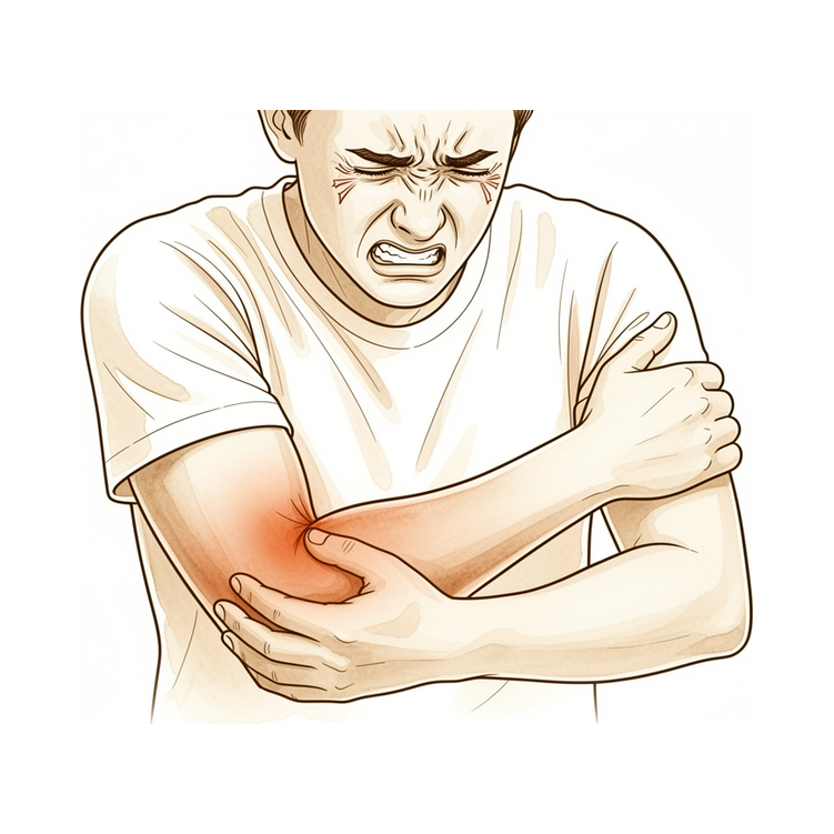
Triệu chứng chính là đau ở phía ngoài cẳng tay, gần khuỷu tay. Cơn đau thường nặng nhất tại một điểm đau khi ấn, cách chỗ lồi xương ở ngoài khuỷu tay vài centimet về phía dưới. Ấn vào điểm đó thường gây đau. Cơn đau có thể lan xuống mặt sau cẳng tay về phía cổ tay.
Một số động tác làm cơn đau tệ hơn. Xoay ngửa lòng bàn tay lên khi có lực cản, hoặc duỗi ngón giữa khi có lực cản, có thể gây ra cơn đau. Các hoạt động liên tục dồn lực lên cẳng tay cũng vậy. Nhiều người nhận thấy cơn đau bùng lên sau khi làm việc hoặc chơi thể thao hơn là lúc nghỉ ngơi.
Tình trạng này ảnh hưởng đến một dây thần kinh chi phối cơ chứ không chi phối da. Điều đó có nghĩa là bạn ít có khả năng bị tê hoặc ngứa ran, và bản thân dây thần kinh thường vẫn hoạt động bình thường. Vấn đề là đau, chứ không phải yếu hay mất cảm giác.
Trong sinh hoạt hằng ngày, cơn đau thường xuất hiện khi làm những việc cần nắm chặt và xoay vặn. Nhấc ấm đun nước, vặn tay nắm cửa, dùng tuốc-nơ-vít hoặc vắt khăn đều có thể gây đau. Vì điểm đau khi ấn nằm gần phía ngoài khuỷu tay, tình trạng này thường bị nhầm với bệnh khuỷu tay quần vợt, và hai tình trạng này có thể xảy ra cùng lúc.
Không có một xét nghiệm duy nhất nào xác nhận được tình trạng này. Bác sĩ phẫu thuật đưa ra chẩn đoán dựa trên những gì bạn kể và việc thăm khám, kiểm tra điểm đau khi ấn đặc trưng đó và kiểu đau của bạn.
Hãy để ý những dấu hiệu sau. Hãy đến gặp bác sĩ đa khoa hoặc đề nghị được khám chuyên khoa nếu các triệu chứng không dịu đi, nặng dần lên qua nhiều tuần, khiến bạn thức giấc vào ban đêm, hoặc khiến bạn không thể làm việc hay sử dụng cánh tay. Hãy đến phòng cấp cứu ngay trong ngày nếu cánh tay trở nên nóng, đỏ, sưng và đau, đặc biệt là khi kèm theo sốt, hoặc nếu bàn tay chuyển sang nhợt nhạt, lạnh, trắng hoặc xanh tím. Nếu bạn không thể liên lạc với phòng khám ngoài giờ làm việc hoặc vào cuối tuần, hãy đến phòng cấp cứu gần nhất.
Điều gì đang thực sự xảy ra
Cơn đau đến từ một dây thần kinh, chứ không phải từ bản thân khớp khuỷu. Dây thần kinh giống như một sợi dây cáp truyền tín hiệu giữa não và các cơ của bạn. Một nhánh của dây thần kinh quay chạy dọc phía ngoài cẳng tay, đi qua khuỷu tay, để chi phối các cơ giúp duỗi cổ tay và các ngón tay.
Dọc theo đoạn đó, có nhiều cấu trúc riêng biệt nằm sát dây thần kinh. Mép của một cơ, một dải mô sợi, hoặc một nhóm nhỏ các mạch máu bắt chéo đều có thể đè lên nó. Đây không phải là một lối đi hẹp duy nhất. Nó giống như một con đường mòn đi bộ với nhiều điểm có thể bị thắt hẹp dọc đường, và bất kỳ điểm nào cũng có thể chèn ép sợi dây cáp chạy bên cạnh.
Khi một trong những cấu trúc đó đè lên dây thần kinh, dây thần kinh bị kích ứng và đau. Đó là lý do cơn đau nằm ở một điểm đau khi ấn bên dưới chỗ lồi xương ở ngoài khuỷu tay, và vì sao xoay ngửa lòng bàn tay lên hoặc duỗi ngón giữa khi có lực cản làm cơn đau tệ hơn. Những động tác đó kéo lên các cơ do dây thần kinh này chi phối, và dây thần kinh bị kích ứng sẽ “phàn nàn”.
Dây thần kinh vẫn hoạt động, nên các cơ của bạn vẫn hoạt động và bạn không bị tê hay ngứa ran. Vấn đề là kích ứng và đau, chứ không phải mất tín hiệu. Vì điểm đau nằm rất gần phía ngoài khuỷu tay, cơn đau dễ bị nhầm với bệnh khuỷu tay quần vợt, một tình trạng ảnh hưởng đến một gân ở cùng khu vực. Hai tình trạng này thậm chí có thể xảy ra cùng lúc.
Những biện pháp chúng tôi có thể áp dụng
Bác sĩ Kieran Hirpara, bác sĩ phẫu thuật chi trên tại Bệnh viện tư nhân Mater Rockhampton, sẽ bắt đầu bằng các phương pháp ít xâm lấn nhất phù hợp với tình trạng của bạn. Thông thường, bệnh nhân được bác sĩ đa khoa giới thiệu đến phòng khám chúng tôi; nếu vật lý trị liệu viên khuyên bạn nên gặp chúng tôi, bạn vẫn cần có giấy giới thiệu từ bác sĩ đa khoa để được hưởng mức hoàn trả từ Medicare. Tại phòng khám, chúng tôi sẽ hỏi bệnh sử, khám cánh tay và chỉ định chụp chiếu nếu điều đó có ích. Đối với tình trạng này, chúng tôi thường thử điều trị không phẫu thuật trước, và cân nhắc phẫu thuật khi cách đó không mang lại đủ cải thiện.
Bước đầu tiên là thay đổi cách bạn sử dụng cánh tay. Giảm bớt những việc cần nắm chặt và xoay vặn làm cơn đau bùng lên sẽ cho dây thần kinh bị kích ứng cơ hội dịu lại. Trị liệu bàn tay nhằm làm dịu cơn đau và xây dựng lại khả năng chịu lực của cẳng tay, để công việc và các hoạt động hằng ngày trở nên dễ chịu trở lại. Chúng tôi đề nghị bạn cho các biện pháp này một thời gian thử hợp lý trước khi nghĩ đến bất kỳ điều gì xa hơn.
Thuốc giảm đau và thuốc chống viêm có thể giúp bạn vượt qua đợt bùng phát. Chúng làm dịu cơn đau trong khi các biện pháp khác phát huy tác dụng. Chúng không giải quyết được tình trạng chèn ép lên bản thân dây thần kinh.
Nếu điều trị không phẫu thuật không mang lại đủ cải thiện, phẫu thuật là một lựa chọn. Ca phẫu thuật này được gọi là giải phóng ống quay. Nó giải phóng dây thần kinh tại từng vị trí có cấu trúc lân cận đang đè lên nó. Trước khi đề xuất, chúng tôi kiểm tra kỹ vị trí dây thần kinh đang bị chèn ép, để ca phẫu thuật nhắm đúng chỗ và ít làm ảnh hưởng nhất đến các mô xung quanh. Vì chẩn đoán dựa trên những gì bạn kể và việc thăm khám chứ không phải một xét nghiệm rõ ràng, chúng tôi xem quyết định phẫu thuật là quyết định chung, được đưa ra cùng nhau khi bạn đã hiểu những gì phẫu thuật có thể và không thể giải quyết.
Những điều có thể xảy ra
Đối với hầu hết mọi người, tình trạng này không làm tổn thương dây thần kinh. Dây thần kinh vẫn hoạt động, nên các cơ vẫn hoạt động và bạn không bị mất cảm giác. Vấn đề là đau, và cơn đau do dây thần kinh bị kích ứng có thể mất nhiều thời gian mới dịu đi. Nó hiếm khi hết chỉ sau một đêm, và thường bùng lên khi làm những việc cần nắm chặt và xoay vặn trước khi giảm dần.
Thành thật mà nói, tình trạng này khó dự đoán. Một số người đỡ hơn nhờ nghỉ ngơi, thay đổi hoạt động và trị liệu bàn tay. Những người khác vẫn tiếp tục bị đau dù đã được điều trị không phẫu thuật tốt. Không có xét nghiệm đáng tin cậy nào để xác nhận chẩn đoán, điều này khiến khó nói trước bạn sẽ thuộc nhóm nào. Điều chúng tôi có thể nói là cơn đau có xu hướng lúc có lúc không tùy theo mức độ bạn dồn lực lên cẳng tay, chứ không tăng dần đều.
Nếu điều trị không phẫu thuật có hiệu quả, cơn đau giảm dần trong nhiều tuần đến nhiều tháng khi dây thần kinh bớt bị kích ứng. Bạn có thể sẽ thấy các việc hằng ngày như nhấc ấm đun nước hoặc vặn tay nắm cửa bớt đau trước, rồi sau đó công việc nặng hơn mới cảm thấy bình thường trở lại. Nếu phẫu thuật được thực hiện sau khi điều trị không phẫu thuật không giúp ích đủ, quá trình hồi phục cũng diễn ra từ từ chứ không tức thì, và một số người vẫn còn đau phần nào sau đó.
Nếu không điều trị, tình trạng này có thể tự dịu đi, nhưng cũng có thể kéo dài hoặc tiếp tục bùng lên mỗi khi bạn quay lại những việc làm nó nặng hơn. Không có cách nào biết trước điều gì sẽ xảy ra. Điều thường quan trọng là cho việc điều trị một thời gian thử hợp lý và không cố chịu đựng cơn đau không dịu đi.
Nếu các triệu chứng không dịu đi, nặng dần lên qua nhiều tuần, khiến bạn thức giấc vào ban đêm, hoặc khiến bạn không thể làm việc hay sử dụng cánh tay, hãy đến gặp bác sĩ đa khoa hoặc đề nghị được khám chuyên khoa. Hãy đến phòng cấp cứu ngay trong ngày nếu cánh tay trở nên nóng, đỏ, sưng và đau, đặc biệt là khi kèm theo sốt, hoặc nếu bàn tay chuyển sang nhợt nhạt, lạnh, trắng hoặc xanh tím. Nếu bạn không thể liên lạc với phòng khám ngoài giờ làm việc hoặc vào cuối tuần, hãy đến phòng cấp cứu gần nhất.
Khi nào nên gặp bác sĩ
Các dấu hiệu cảnh báo của tình trạng này liên quan đến cơn đau không dịu đi, chứ không phải về nguy hiểm. Hãy đến gặp bác sĩ đa khoa hoặc đề nghị được khám chuyên khoa nếu cơn đau cẳng tay không dịu đi sau một thời gian thử nghỉ ngơi và thay đổi hoạt động hợp lý, hoặc nếu nó cứ khiến bạn không thể làm việc hay sử dụng cánh tay. Hãy đi khám sớm thay vì chờ đợi nếu điểm đau khi ấn bên dưới phía ngoài khuỷu tay là một đặc điểm nổi bật, hoặc nếu việc xoay ngửa lòng bàn tay lên hoặc duỗi ngón giữa khi có lực cản luôn gây ra cơn đau. Những kiểu biểu hiện này gợi ý tình trạng này hơn là bệnh khuỷu tay quần vợt, và đáng được làm rõ đúng cách.
Tình trạng này thường không làm tổn thương dây thần kinh, nên yếu đột ngột ở cổ tay hoặc các ngón tay không phải là biểu hiện điển hình của nó. Nếu bạn nhận thấy mới bị yếu khi duỗi cổ tay, các ngón tay hoặc ngón cái, hãy đề nghị được khám chuyên khoa sớm thay vì chờ đợi, vì kiểu biểu hiện đó gợi ý một vấn đề thần kinh khác, được đánh giá theo mốc thời gian riêng.
Hãy đến phòng cấp cứu ngay trong ngày nếu cánh tay trở nên nóng, đỏ, sưng và đau, đặc biệt là khi kèm theo sốt, hoặc nếu bàn tay chuyển sang nhợt nhạt, lạnh, trắng hoặc xanh tím. Nếu bạn không thể liên lạc với phòng khám ngoài giờ làm việc hoặc vào cuối tuần, hãy đến phòng cấp cứu gần nhất.
Phân tích sâu hơn
Advanced reading: the deeper science (optional)
Phần này đi sâu hơn mức cần thiết để bạn đưa ra quyết định điều trị. Hội chứng ống cẳng tay quay là một chủ đề đáng để tìm hiểu thêm, nhưng vì một lý do không mấy dễ chịu: đây là tình trạng bệnh lý tại vùng này có cơ sở bằng chứng y khoa yếu nhất; hiện chưa có xét nghiệm nào có thể xác nhận chẩn đoán; việc hiểu rõ mức độ không chắc chắn này là điều cần thiết khi cân nhắc việc phẫu thuật.
Không có xét nghiệm xác nhận nào
Hầu hết các trường hợp chèn ép dây thần kinh đều có thể được chứng minh thông qua các xét nghiệm. Hội chứng ống cổ tay làm chậm tốc độ dẫn truyền thần kinh ở vùng cổ tay; hội chứng ống khuỷu tay cũng gây hiện tượng tương tự ở vùng khuỷu tay. Ngược lại, hội chứng ống quay thường không gây ra thay đổi như vậy; nhánh dây thần kinh bị ảnh hưởng chủ yếu chi phối cơ chứ không phải da, nên không có triệu chứng tê bì để xác định, và kết quả đo dẫn truyền thần kinh thường vẫn bình thường.
Vì vậy, chẩn đoán chủ yếu dựa vào mô hình đau, vùng sưng đau cách mỏm khuỷu tay vài centimet về phía ngoài chứ không phải ngay tại mỏm khuỷu, cùng phản ứng của bệnh nhân với việc tiêm thuốc tê tại chỗ. Mỗi yếu tố trên đều có tính gợi ý nhưng không đủ để khẳng định chẩn đoán. Các nghiên cứu hiện đại cho thấy vẫn còn nhiều tranh cãi liên quan đến cả chẩn đoán lẫn kết quả điều trị [1].
Hệ quả thực tế là tình trạng nhầm lẫn khá phổ biến giữa hội chứng ống quay và chứng viêm gân khuỷu tay – hai bệnh này nằm gần nhau và có thể xuất hiện đồng thời. Trường hợp viêm gân khuỷu tay kéo dài không đáp ứng với các phương pháp điều trị hiệu quả chính là hoàn cảnh phổ biến nhất khi người ta bắt đầu cân nhắc đến khả năng mắc hội chứng ống quay.
Bằng chứng về các phương pháp điều trị thực sự còn rất hạn chế
Một phân tích có hệ thống về các biện pháp can thiệp cho thấy có xu hướng cho thấy phẫu thuật giải ép có thể mang lại hiệu quả; điều đáng chú ý hơn nữa là hiệu quả của các phương pháp điều trị nội khoa vẫn chưa được xác định, vì hầu hết các phương pháp này đều chưa có nghiên cứu nào được thực hiện [2].
Đây không phải là trường hợp thông thường khi người ta nói “bằng chứng còn hạn chế”. Đối với phần lớn các phương pháp điều trị không phẫu thuật dành cho tình trạng này, các nghiên cứu vẫn chưa được tiến hành. Các phân tích hiện tại vẫn ưu tiên các biện pháp điều trị không phẫu thuật, đồng thời coi phẫu thuật giải ép là một lựa chọn khả thi đối với những trường hợp khó điều trị [1]; việc sắp xếp như vậy thể hiện sự thận trọng hợp lý chứ không phải do phương pháp phẫu thuật được chứng minh là vượt trội hơn.
Ý nghĩa đối với việc ra quyết định
Có hai khía cạnh cần xem xét, và chúng đi theo hai hướng trái ngược nhau.
Phản đối việc phẫu thuật sớm: Chẩn đoán chưa thể được xác nhận; do đó ca phẫu thuật có thể chỉ giúp giải áp cho một dây thần kinh không phải là nguyên nhân gây bệnh. Trong trường hợp phẫu thuật giải áp không thành công, thường rất khó xác định liệu do kỹ thuật phẫu thuật chưa đủ tốt hay do chẩn đoán ban đầu sai lệch.
Khi cân nhắc phẫu thuật cho bệnh nhân phù hợp: Hiện chưa có bằng chứng khoa học nào ủng hộ các phương pháp điều trị thay thế; vì vậy việc chờ đợi không phải là lựa chọn được khoa học chứng minh như nhiều người vẫn nghĩ. Đó chỉ đơn giản là phương án ít rủi ro hơn mà thôi.
Quan điểm hợp lý là: Việc chẩn đoán chính xác có ý nghĩa quan trọng hơn bất kỳ yếu tố nào khác được đề cập trên trang này. Cần có tiền sử bệnh điển hình, kết quả khám lâm sàng cho thấy tổn thương nằm ở vùng ống thần kinh quay chứ không phải mỏm khuỷu, hình ảnh chẩn đoán loại trừ khả năng có khối u gây chèn ép; và lý tưởng nhất là có phản ứng tích cực rõ rệt sau khi tiêm thuốc gây tê chẩn đoán, trước khi tiến hành bất kỳ can thiệp phẫu thuật không thể đảo ngược nào.
Một vấn đề liên quan nhưng khác biệt
Liệt dây thần kinh gian xương sau – biểu hiện bằng tình trạng yếu cơ gây khó khăn trong việc duỗi các ngón tay và ngón cái thay vì đau đớn – là một tình trạng riêng biệt có phương pháp điều trị rõ ràng. Trong trường hợp hình ảnh chẩn đoán không cho thấy tổn thương gây chèn ép, nên áp dụng phương pháp điều trị nội khoa trước; phẫu thuật chỉ được thực hiện khi có bằng chứng về tổn thương chèn ép hoặc khi phương pháp điều trị nội khoa thất bại [3]. Khi không có tổn thương gây chèn ép, nên thử áp dụng phương pháp điều trị không phẫu thuật; việc mổ thám sát mới được chỉ định nếu sau 6 tuần theo dõi vẫn không thấy dấu hiệu hồi phục cơ hoặc nếu tình trạng yếu cơ ngày càng nặng lên [4].
Đây là những ngưỡng cụ thể; sự tồn tại của chúng là minh chứng rõ ràng nhất cho sự khác biệt giữa các trường hợp: khi dây thần kinh ngừng hoạt động rõ ràng, các tài liệu y khoa mới có thể chỉ dẫn cách điều trị và thời điểm thực hiện. Còn khi chỉ có triệu chứng đau, thì không thể làm được như vậy.
Tài liệu tham khảo
[1] Wolf JM, Patel R, Ghosh K. Hội chứng ống quay: tổng quan và các bằng chứng tốt nhất. J Am Acad Orthop Surg. 2023;31(15):813-9. https://doi.org/10.5435/JAAOS-D-23-00314
[2] Huisstede B, Miedema HS, van Opstal T, de Ronde MT, Verhaar JA, Koes BW. Các phương pháp can thiệp điều trị hội chứng ống quay: tổng quan có hệ thống các nghiên cứu quan sát. J Hand Surg Am. 2008;33(1):72.e1-72.e10. https://doi.org/10.1016/j.jhsa.2007.10.001
[3] McGraw I. Liệt dây thần kinh gian xương sau tự phát đơn thuần: tổng quan về nguyên nhân và cách xử trí. J Hand Surg Eur Vol. 2018;44(3):310-6. https://doi.org/10.1177/1753193418813788
[4] Sigamoney KV, Rashid A, Ng CY. Cách xử trí tình trạng liệt dây thần kinh gian xương sau không do chấn thương. J Hand Surg Am. 2017;42(10):826-30. https://doi.org/10.1016/j.jhsa.2017.07.026
Evidence & references
This is the clinical evidence summary written for health professionals. It is technical, and it lists the research this page was built from. You do not need to read it to understand your treatment or to make a decision about it.
Overview
- Radial tunnel syndrome is relatively uncommon but is an important cause of lateral forearm pain [1].
- Clinical examination is a crucial part of the diagnosis of radial tunnel syndrome [1].
- The duration of symptoms averaged 2.3 years before a definitive diagnosis of radial tunnel syndrome was made [2].
- The diagnosis of radial tunnel syndrome remains clinical [5].
- Adherence to a defined protocol is useful in diagnosing radial tunnel syndrome [5].
- The issue surrounding radial tunnel syndrome traditionally has been properly identifying it clinically [7].
- Compression of the radial nerve at the elbow is of moderate frequency compared to the ulnar tunnel syndrome [9].
- A case of bilateral radial tunnel syndrome with signs discordant with traditionally used clinical diagnostic tests was successfully relieved with surgical decompression [6].
- The Rule-of-Nine test effectively specifies the site of tenderness for the diagnosis of radial tunnel syndrome [11].
Anatomy & Pathophysiology
Clinical Presentation & Diagnostic Challenges
- Radial tunnel syndrome is a relatively uncommon but important cause of lateral forearm pain [1].
- Clinical examination is a crucial component of the diagnosis for radial tunnel syndrome [1].
- The average duration of symptoms before a definitive diagnosis of radial tunnel syndrome was 2.3 years [2].
- Prominent focal tenderness in the area of the radial tunnel is a principal diagnostic criterion for radial tunnel syndrome [20].
- In radial tunnel syndrome, symptoms do not occur in the distribution of the purportedly affected nerve, which distinguishes focal tenderness from a positive Phalen's test in carpal tunnel syndrome [20].
- Skeptics note that radial tunnel syndrome presents with normal neurologic function and no confirmatory electrodiagnostic evidence of nerve dysfunction, contrasting with other well-described entrapment neuropathies [20].
Etiology & Pathophysiology
- There is dispute over the etiology of radial tunnel syndrome, with some skeptics questioning its status as a viable entrapment neuropathy [20].
- The posterior interosseous nerve carries unmyelinated (group IV) afferent fibers from the wrist capsule and small myelinated (group IIA) afferent fibers from muscles along its distribution [20].
- Unmyelinated group IV fibers are associated with nociception and pain [20].
- The current best evidence for the conservative management of radial tunnel syndrome consists primarily of expert opinion and inferences from studies on other nerve compressions [3].
Relevant Elbow Anatomy
- The elbow is a trocho-ginglymoid joint consisting of medial and lateral articulations that afford bony stability [63].
- The ulnohumeral joint is formed by the articulation of the trochlea with the ulna within the greater sigmoid notch [63].
- The radiocapitellar joint is formed by the articulation of the capitellum and radial head [63].
- The radius is held in close approximation to the ulna at the proximal radioulnar joint by the annular ligament [63].
- The lateral epicondyle is the origin of the lateral extensor musculature [63].
- The radial nerve enters the interval between the brachialis and brachioradialis muscles in the proximal angle of the lateral approach to the elbow [66].
- The deep branch of the radial nerve enters the supinator muscle [66].
- The posterior interosseous nerve is located just superficial to the anterior joint capsule at the level of the radiocapitellar joint [75].
- At the level of the radial neck, the posterior interosseous nerve may come in direct contact with the joint capsule [75].
- Supination increases the linear distance between the posterior interosseous nerve and the radial head [107].
Classification
- Radial tunnel syndrome (RTS) and posterior interosseous nerve (PIN) compression are distinct entities with different clinical presentations but share identical potential sites of nerve interference [29].
- The author of [29] proposes unifying RTS and PIN compression as mild and severe forms of one disease to simplify nomenclature [29].
- The authors of [52] recommend viewing radial tunnel syndrome and posterior interosseous nerve syndrome as a single condition presenting along a spectrum of nerve entrapment [52].
- Radial tunnel syndrome is a pain syndrome caused by compression of the posterior interosseous nerve at the proximal forearm [22].
- The diagnosis of radial tunnel syndrome remains clinical, and adherence to a defined protocol is useful in diagnosing this condition [5].
- A differential latency of ≥0.30 ms was considered indicative of radial tunnel syndrome [16].
- Radial tunnel syndrome is an illness construct based on a speculative pathophysiology with no verifiable pathophysiology or accepted reference standard for diagnosis [14].
- Skeptics note that the signs and symptoms of RTS contrast from other well-described entrapment neuropathies in that there is prominent focal tenderness, normal neurologic function, and no confirmatory electrodiagnostic evidence of nerve dysfunction [20].
- Prominent focal tenderness in the area of the radial tunnel remains one of the principal diagnostic criteria for RTS [20].
- Focal tenderness at the radial tunnel in RTS differs from a positive Phalen's test in carpal tunnel syndrome in that the symptoms do not occur in the distribution of the purportedly affected nerve [20].
- There has been no randomized controlled trial that compares surgical with nonsurgical treatment or with a placebo for RTS [20].
- Skeptics of RTS point to the great variability of surgical results reported in the literature as one of the characteristics of placebo surgery [20].
Clinical Presentation
- Radial tunnel syndrome is a relatively uncommon cause of lateral forearm pain [1].
- Radial tunnel syndrome and posterior interosseous nerve compression are distinct entities with different clinical presentations [29].
- The author proposes unifying radial tunnel syndrome and posterior interosseous nerve compression as mild and severe forms of one disease to simplify nomenclature [29].
- The authors recommend viewing radial tunnel syndrome and posterior interosseous nerve syndrome as a single condition presenting along a spectrum of nerve entrapment [52].
- Prominent focal tenderness in the area of the radial tunnel is one of the principal diagnostic criteria for radial tunnel syndrome [20].
- In radial tunnel syndrome, symptoms do not occur in the distribution of the purportedly affected nerve, which differs from a positive Phalen's test in carpal tunnel syndrome [20].
- A diagnosis of radial tunnel syndrome should be considered in patients with forearm and wrist pain that has not responded to more conventional treatment [13].
- The diagnosis of radial tunnel syndrome remains clinical, and adherence to a defined protocol is useful in diagnosing the condition [5].
- The issue surrounding radial tunnel syndrome has traditionally been properly identifying it clinically [7].
- A case of bilateral radial tunnel syndrome presented with signs discordant with traditionally used clinical diagnostic tests [6].
- Muscle denervation edema or atrophy along the distribution of the posterior interosseous nerve is the most common MR finding in radial tunnel syndrome [32].
- A symptomatic double-level entrapment of the posterior interosseous branch of the radial nerve has been described [51].
- Compression of the radial nerve at the elbow is of moderate frequency compared to ulnar tunnel syndrome [9].
- The annual incidence rate of posterior interosseous nerve entrapment is estimated to be 0.03% [88].
- The most frequent location of radial nerve entrapment is around the elbow, specifically involving the posterior interosseous nerve branch [88].
- The radial tunnel spans 5 cm, extending from the humero-radial joint and running distally between the deep and superficial heads of the supinator [88].
- The radial tunnel is bounded laterally by extensor carpi radialis longus and extensor carpi radialis brevis muscles and medially by biceps tendon and the brachialis [88].
- The roof of the radial tunnel is formed by brachioradialis [88].
- The floor of the radial tunnel is the elbow-joint capsule [88].
- Potential sites of entrapment in the radial tunnel include capsular tissue of the radiocapitellar joint, hypertrophic crossing branches of leash of Henry, the leading proximal tendinous and medial edge of ECRB, the arcade of Frohse, and the distal border of the supinator between its two heads [88].
- The arcade of Frohse is noted to be the most common site of entrapment in the radial tunnel [88].
- Clinical criteria for radial tunnel syndrome include activity-related pain, maximal tenderness 3–5 cm distal to the lateral epicondyle, pain exacerbation with forearm supination, radiation to the dorsoradial aspect of the forearm, and a positive Lister test [81].
- A clinical diagnosis of radial tunnel syndrome requires 4 of 5 diagnostic signs and symptoms, with one being maximal tenderness 3–5 cm distal to the lateral epicondyle [81].
- Physical examination for radial tunnel syndrome includes wrist flexion and forearm pronation, the Rule of Nines test, and assessment of weakness and pain with resisted long finger extension [86].
- The history for radial tunnel syndrome includes extensor musculature "forearm aching" [86].
Investigations
Clinical Diagnosis
- A diagnosis of radial tunnel syndrome should always be born in mind when dealing with patients with forearm and wrist pain that has not responded to more conventional treatment [13].
- It is important to understand the anatomic course and distribution of the radial nerve in order to make an accurate diagnosis [24].
Electrodiagnostic Studies
Imaging
- Neuroimaging should be considered as a complementary diagnostic method in posterior interosseous neuropathy [132].
- Dynamic ultrasonographic assessment of changes in the AP diameter and CSA is an effective diagnostic tool for identifying radial tunnel syndrome [121].
- Ultrasonography, computed tomography, and magnetic resonance imaging revealed the location of the ganglion in every patient with radial nerve palsy caused by a ganglion [124].
- The authors acknowledge that their study does not establish diagnostic accuracy or a reference standard for radial tunnel syndrome [49].
Treatment
Non-Operative Management
- Current best evidence for the conservative management of radial tunnel syndrome consists primarily of expert opinion and inferences taken from studies on other nerve compressions and related syndromes [3].
- The effectiveness of conservative treatments for radial tunnel syndrome is unknown because, for most treatments, no studies are available [48].
- Prospective evaluation of a single corticosteroid injection demonstrated improvement in standardized outcomes measures of pain and function at one year in 75% of patients presenting with symptoms of radial tunnel syndrome [8].
- Nonsurgical management is first-line for radial tunnel syndrome [4].
- The two most common nerve entrapment disorders about the elbow, including radial tunnel, should be initially managed conservatively before considering surgical intervention [80].
Operative Management
- Surgical decompression remains a viable option for refractory cases of radial tunnel syndrome despite ongoing controversy regarding diagnosis and outcomes [4].
- There is a tendency that surgical decompression of the radial tunnel might be effective in patients with radial tunnel syndrome [48].
- The authors advocate for surgery in high radial nerve entrapment neuropathy cases resistant to conservative treatment, emphasizing the importance of dissecting the entire length of the fibrous tunnel [17].
- This study indicates that pain in patients with radial tunnel syndrome may be treated successfully by surgical decompression of the superficial branch of the radial nerve [104].
- The results of radial tunnel release have been sufficiently good thus far as to warrant some attention, with an overall significant improvement in 99 of 108 cases (91.7%) when combined with other series [19].
- Nineteen patients (20 extremities) felt satisfied and helped and believed they gained improved function because of surgical decompression of the radial tunnel [27].
- The authors believe that a high rate of morbidity is associated with both the disease and its treatment, suggesting that great caution has to be taken before performing radial tunnel release [26].
- The authors suggest that great caution be taken before performing radial tunnel release and strict adherence to the indications noted during the preoperative examination [50].
Evidence Limitations and Controversy
- There has been no randomized controlled trial that compares surgical with nonsurgical treatment or with a placebo for radial tunnel syndrome [20].
- Skeptics of radial tunnel syndrome point to the great variability of surgical results reported in the literature as one of the characteristics of placebo surgery [20].
- Although radial tunnel syndrome is classically described as a nerve compression and entrapment syndrome, there is dispute over its etiology [20].
Complications
- A high rate of morbidity is associated with both radial tunnel syndrome and its treatment [26].
- In a case of congenital compression of the radial nerve, the patient was followed for an additional 3 months without clinical improvement in radial nerve function [12].
- Nerve injury is an uncommon though troublesome complication of fractures or dislocations of the elbow [39].
Recovery
- A patient with congenital compression of the radial nerve was followed for an additional 3 months without clinical improvement in radial nerve function [12].
- In a patient with posterior interosseous-nerve syndrome secondary to rheumatoid synovitis where the duration of entrapment was more than two years, a tendon transfer was used as treatment [139].
- Isolated posterior interosseous nerve neurectomy has shown excellent clinical outcomes, with few patients experiencing recurrent pain at long-term follow-up [123].
Key Evidence
- [L5] Radial tunnel syndrome is relatively uncommon but is an important cause of lateral forearm pain, with clinical examination being a crucial part of the diagnosis. [1] (10.1197/j.jht.2006.02.005)
- [L4] The duration of symptoms averaged 2.3 years before a definitive diagnosis of radial tunnel syndrome was made. [2] (10.1016/s0363-5023(83)80201-9)
- [L5] Current best evidence for the conservative management of radial tunnel syndrome (RTS) consists primarily of expert opinion and inferences taken from studies on other nerve compressions and related syndromes. [3] (10.1197/j.jht.2006.02.020)
- [L4] The article reviews the anatomy, diagnosis, and treatment of radial tunnel syndrome, noting that while nonsurgical management is first-line, surgical decompression remains a viable option for refractory cases despite ongoing controversy regarding diagnosis and outcomes. [4] (10.5435/jaaos-d-23-00314)
- [L4] The diagnosis of radial tunnel syndrome remains clinical, and adherence to a defined protocol is useful in diagnosing this condition. [5] (10.1016/s0266-7681(98)80015-6)
- [L5] A case of bilateral radial tunnel syndrome with signs discordant with traditionally used clinical diagnostic tests was successfully relieved with surgical decompression. [6] (10.1177/15589447211029045)
- [L5] The issue surrounding radial tunnel syndrome traditionally has been properly identifying it clinically. [7] (10.1097/01.bth.0000231580.32406.71)
- [L4] Prospective evaluation of corticosteroid injection demonstrated improvement in standardized outcomes measures of pain and function at one year in 75% of patients presenting with symptoms of radial tunnel syndrome. [8] (10.1016/j.jhsa.2017.06.095)
- [Paper] Compression of the radial nerve at the elbow is of moderate frequency compared to the ulnar tunnel syndrome. [9] (10.1016/j.main.2004.10.006)
- [L4] The Rule-of-Nine test effectively specifies the site of tenderness for the diagnosis of radial tunnel syndrome. [11] (10.1177/230949900401200115)
- [L5] The patient was followed for an additional 3 months, without clinical improvement in radial nerve function. [12] (10.1016/s0363-5023(89)80099-1)
- [L4] A diagnosis of radial tunnel syndrome should always be born in mind when dealing with patients with forearm and wrist pain that has not responded to more conventional treatment. [13] (10.1016/s0266-7681(05)80152-4)
- [L5] Radial tunnel syndrome is an illness construct based on a speculative pathophysiology with no verifiable pathophysiology or accepted reference standard for diagnosis. [14] (10.1016/j.jhsa.2010.03.020)
- [L4] A differential latency of ≥0.30 ms was considered indicative of radial tunnel syndrome. [16] (10.1016/s0363-5023(98)80163-9)
- [Case_report] The authors advocate for surgery in high radial nerve entrapment neuropathy cases resistant to conservative treatment, emphasizing the importance of dissecting the entire length of the fibrous tunnel. [17] (10.1016/j.jse.2025.02.060)
- [L4] The results of radial tunnel release have been sufficiently good thus far as to warrant some attention, with an overall significant improvement in 99 of 108 cases (91.7%) when combined with other series. [19] (10.1016/s0363-5023(79)80105-7)
- [L5] [20] (10.1016/j.jhsa.2009.10.016)
- [L5] Radial tunnel syndrome is a pain syndrome caused by compression of the posterior interosseous nerve at the proximal forearm. [22] (10.1016/j.ocl.2012.07.022)
- [L5] It is important to understand the anatomic course and distribution of the radial nerve in order to make an accurate diagnosis. [24] (10.1007/s11420-011-9238-8)
- [L5] The authors believe that a high rate of morbidity is associated with both the disease and its treatment, suggesting that great caution has to be taken before performing radial tunnel release. [26] (10.1097/00130911-200212000-00010)
- [L4] Nineteen patients (20 extremities), however, felt satisfied and helped and believed they gained improved function because of surgical decompression of the radial tunnel. [27] (10.1016/s0363-5023(97)80086-x)
- [L5] Radial tunnel syndrome (RTS) and posterior interosseous nerve (PIN) compression are distinct entities with different clinical presentations but share identical potential sites of nerve interference; the author proposes unifying them as mild (RTS) and severe (PIN compression) forms of one disease to simplify nomenclature. [29] (10.1177/1753193420953990)
- [L4] Muscle denervation edema or atrophy along the distribution of the posterior interosseous nerve is the most common MR finding in radial tunnel syndrome. [32] (10.1148/radiol.2401050028)
- [L4] Nerve injury is an uncommon though troublesome complication of fractures or dislocations of the elbow. [39] (10.1016/s0020-1383(79)80015-7)
- [L4] [48] (10.1016/j.jhsa.2007.10.001)
- [Paper] The authors acknowledge that their study does not establish diagnostic accuracy or a reference standard for radial tunnel syndrome. [49] (10.1177/17531934261463150)
- [L4] The authors suggest that great caution be taken before performing radial tunnel release and strict adherence to the indications noted during the preoperative examination. [50] (10.1053/jhsu.1999.0566)
- [L5] A symptomatic double-level entrapment of the posterior interosseous branch of the radial nerve is described. [51] (10.1016/s0363-5023(83)80202-0)
- [L5] The authors advocate for consistent use of the terminology distinguishing the deep branch of the radial nerve (DBRN) and the posterior interosseous nerve (PIN), and recommend viewing radial tunnel syndrome and posterior interosseous nerve syndrome as a single condition presenting along a spectrum of nerve entrapment. [52] (10.1177/17531934241254706)
- [L2] [81] (10.1016/j.jhsa.2024.09.023)
- [L5] [88] (10.1016/j.jisako.2024.03.001)
- [L4] This study indicates that pain in patients with radial tunnel syndrome may be treated successfully by surgical decompression of the superficial branch of the radial nerve. [104] (10.1177/1753193408099832)
- [L5] Supination increases the linear distance between the PIN and radial head and should be considered to increase the safe working volume whenever intra-articular procedures are performed on the anterolateral aspect of the elbow. [107] (10.1016/j.jse.2018.08.019)
- [L4] Dynamic ultrasonographic assessment of changes in the AP diameter and CSA is an effective diagnostic tool for identifying radial tunnel syndrome. [121] (10.1177/17531934261443138)
- [L4] Isolated PINN have shown excellent clinical outcomes, with few patients experiencing recurrent pain at long-term follow-up. [123] (10.1177/1558944717692093)
- [L4] Ultrasonography, computed tomography, and magnetic resonance imaging revealed the location of the ganglion in every patient. [124] (10.1016/s0363-5023(10)80102-9)
- [L4] Neuroimaging should be considered as a complementary diagnostic method in PINS. [132] (10.1212/wnl.0000000000003287)
- [L4] In the third patient, in whom the duration of entrapment was more than two years, a tendon transfer was used as treatment. [139] (10.2106/00004623-197355040-00009)
References
[1] Radial Tunnel Syndrome: A Surgeon's Perspective. Journal of Hand Therapy. 2006. DOI: 10.1197/j.jht.2006.02.005
[2] Radial tunnel syndrome: A spectrum of clinical presentations. The Journal of Hand Surgery. 1983. DOI: 10.1016/s0363-5023(83)80201-9
[3] Management of Radial Tunnel Syndrome: A Therapist's Clinical Perspective. Journal of Hand Therapy. 2006. DOI: 10.1197/j.jht.2006.02.020
[4] Radial Tunnel Syndrome: Review and Best Evidence. Journal of the American Academy of Orthopaedic Surgeons. 2023. DOI: 10.5435/jaaos-d-23-00314
[5] Radial Tunnel Syndrome: Diagnosis and Management. Journal of Hand Surgery. 1998. DOI: 10.1016/s0266-7681(98)80015-6
[6] Radial Tunnel Syndrome: Case Report and Comprehensive Critical Review of a Compression Neuropathy Surrounded by Controversy. HAND. 2021. DOI: 10.1177/15589447211029045
[7] A Unified Approach to Radial Tunnel Syndrome and Lateral Tendinosis. Techniques in Hand & Upper Extremity Surgery. 2006. DOI: 10.1097/01.bth.0000231580.32406.71
[8] Prospective Evaluation of Single Corticosteroid Injection in Radial Tunnel Syndrome. The Journal of Hand Surgery. 2017. DOI: 10.1016/j.jhsa.2017.06.095
[9] Compressions du nerf radial au coude. Chirurgie de la Main. 2004. DOI: 10.1016/j.main.2004.10.006
[11] A New Clinical Test for Radial Tunnel Syndrome—The Rule-of-Nine Test: A Cadaveric Study. Journal of Orthopaedic Surgery. 2001. DOI: 10.1177/230949900401200115
[12] Congenital compression of the radial nerve. The Journal of Hand Surgery. 1989. DOI: 10.1016/s0363-5023(89)80099-1
[13] Radial Tunnel Syndrome. Journal of Hand Surgery. 1995. DOI: 10.1016/s0266-7681(05)80152-4
[14] Radial Tunnel Syndrome. The Journal of Hand Surgery. 2010. DOI: 10.1016/j.jhsa.2010.03.020
[16] Differential latency testing: A more sensitive test for radial tunnel syndrome. The Journal of Hand Surgery. 1998. DOI: 10.1016/s0363-5023(98)80163-9
[17] High radial nerve entrapment neuropathy: an anatomical cadaver study and case report. Journal of Shoulder and Elbow Surgery. 2025. DOI: 10.1016/j.jse.2025.02.060
[19] The radial tunnel syndrome. The Journal of Hand Surgery. 1979. DOI: 10.1016/s0363-5023(79)80105-7
[20] Unusual Compression Neuropathies of the Forearm, Part I: Radial Nerve. The Journal of Hand Surgery. 2009. DOI: 10.1016/j.jhsa.2009.10.016
[22] Radial Tunnel Syndrome. Orthopedic Clinics of North America. 2012. DOI: 10.1016/j.ocl.2012.07.022
[24] Posterior Interosseous Neuropathy: Electrodiagnostic Evaluation. HSS Journal®: The Musculoskeletal Journal of Hospital for Special Surgery. 2012. DOI: 10.1007/s11420-011-9238-8
[26] Radial Tunnel Syndrome. Techniques in Hand and Upper Extremity Surgery. 2002. DOI: 10.1097/00130911-200212000-00010
[27] Radial tunnel syndrome: Long-term results of surgical decompression. The Journal of Hand Surgery. 1997. DOI: 10.1016/s0363-5023(97)80086-x
[29] Radial tunnel syndrome: definition, distinction and treatments. Journal of Hand Surgery (European Volume). 2020. DOI: 10.1177/1753193420953990
[32] MR Imaging Features of Radial Tunnel Syndrome: Initial Experience. Radiology. 2006. DOI: 10.1148/radiol.2401050028
[39] Acute nerve injury as a complication of closed fractures or dislocations of the elbow. Injury. 1979. DOI: 10.1016/s0020-1383(79)80015-7
[48] Interventions for Treating the Radial Tunnel Syndrome: A Systematic Review of Observational Studies. The Journal of Hand Surgery. 2008. DOI: 10.1016/j.jhsa.2007.10.001
[49] Re: Role of high-resolution dynamic ultrasonography in the evaluation of posterior interosseous nerve compression at radial tunnel: a prospective case-control study. Journal of Hand Surgery (European Volume). 1934. DOI: 10.1177/17531934261463150
[50] Results of surgical treatment for radial tunnel syndrome. The Journal of Hand Surgery. 1999. DOI: 10.1053/jhsu.1999.0566
[51] Double-entrapment radial tunnel syndrome. The Journal of Hand Surgery. 1983. DOI: 10.1016/s0363-5023(83)80202-0
[52] Nomenclature of the radial nerve: distinguishing between the deep branch of the radial nerve and the posterior interosseous nerve. Journal of Hand Surgery (European Volume). 2024. DOI: 10.1177/17531934241254706
[63] Orthopaedic Knowledge Update 13 Ebook Without Multimedia. Anatomy, Biomechanics, Physical Examination, and Imaging of the Elbow > Anatomy > Bony Anatomy.
[66] Campbell S Operative Orthopaedics 4 Volume Set. LATERAL APPROACHES.
[75] Green S Operative Hand Surgery. PERTINENT ANATOMY AND PORTALS.
[80] Orthopaedic Knowledge Update 13 Ebook Without Multimedia. Elbow Degenerative Conditions and Nerve Disorders > Summary.
[81] Investigating the Effect of Triamcinolone Local Injection on Clinical Outcomes of Patients With Radial Tunnel Syndrome: A Placebo-Controlled Clinical Trial. The Journal of Hand Surgery. 2025. DOI: 10.1016/j.jhsa.2024.09.023
[86] Orthopaedic Knowledge Update 13 Ebook Without Multimedia. Anatomy, Biomechanics, Physical Examination, and Imaging of the Elbow > Biomechanics > Clinical Examination.
[88] Current concepts of surgical approach for radial nerve entrapment around the elbow. Journal of ISAKOS. 2024. DOI: 10.1016/j.jisako.2024.03.001
[104] Radial Tunnel Syndrome: Emphasis on the Superficial Branch of the Radial Nerve. Journal of Hand Surgery (European Volume). 2009. DOI: 10.1177/1753193408099832
[107] The posterior interosseous nerve crosses the radial head midline and increases its distance from bony structures with supination of the forearm. Journal of Shoulder and Elbow Surgery. 2019. DOI: 10.1016/j.jse.2018.08.019
[121] Role of high-resolution dynamic ultrasonography in the evaluation of posterior interosseous nerve compression at radial tunnel: a prospective case-control study. Journal of Hand Surgery (European Volume). 2026. DOI: 10.1177/17531934261443138
[123] Outcomes Following Isolated Posterior Interosseous Nerve Neurectomy: A Systematic Review. HAND. 2017. DOI: 10.1177/1558944717692093
[124] Diagnosis of radial nerve palsy caused by ganglion with use of different imaging techniques. The Journal of Hand Surgery. 1991. DOI: 10.1016/s0363-5023(10)80102-9
[132] Posterior interosseous neuropathy. Neurology. 2016. DOI: 10.1212/wnl.0000000000003287
[139] Posterior Interosseous-Nerve Syndrome Secondary to Rheumatoid Synovitis. The Journal of Bone & Joint Surgery. 1973. DOI: 10.2106/00004623-197355040-00009