Patients › Wrist
Giải ép ống cổ tay
Carpal tunnel release surgery relieves median nerve compression—when it’s needed and what to expect.
Lý do phẫu thuật này được đề xuất
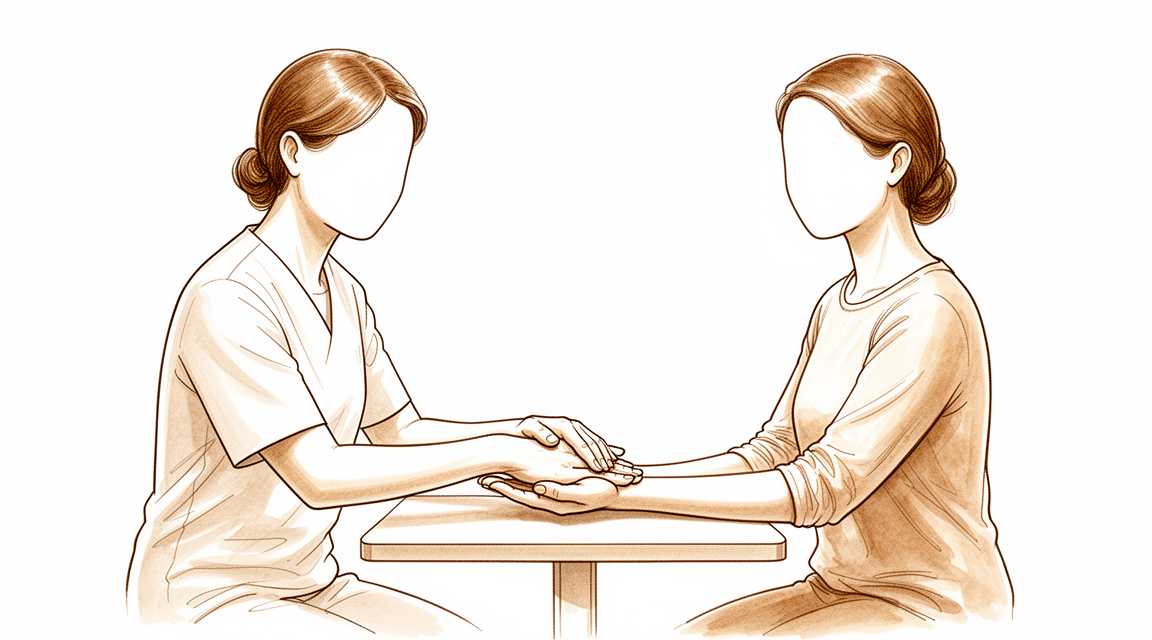
Bác sĩ Kieran Hirpara, bác sĩ phẫu thuật chi trên tại Bệnh viện tư nhân Mater Rockhampton, sẽ bắt đầu bằng các phương pháp điều trị ít xâm lấn nhất phù hợp với tình trạng của bạn. Thông thường, bệnh nhân được bác sĩ đa khoa giới thiệu đến phòng khám chúng tôi; nếu vật lý trị liệu viên khuyên bạn nên đến gặp chúng tôi, bạn vẫn cần có giấy giới thiệu từ bác sĩ đa khoa để được hưởng mức hoàn trả từ chương trình Medicare. Trong buổi khám, chúng tôi sẽ hỏi bệnh sử, khám tay bạn và chỉ định các xét nghiệm hình ảnh hoặc kiểm tra dây thần kinh nếu cần thiết. Hội chứng ống cổ tay xảy ra khi dây thần kinh ở cổ tay bị chèn ép, gây ra cảm giác tê, ngứa ran và đau ở tay. Thông thường chúng tôi bắt đầu bằng các biện pháp điều trị không phẫu thuật như đeo nẹp, vật lý trị liệu hoặc tiêm thuốc. Phẫu thuật được cân nhắc khi các phương pháp điều trị này chưa mang lại hiệu quả đáng kể. Ca phẫu thuật này được gọi là giải phóng ống cổ tay; nó giúp giảm áp lực lên dây thần kinh bằng cách cắt một dải mô căng chặt băng ngang cổ tay. Sau ca phẫu thuật, có thể thấy sự cải thiện rõ rệt về mức độ các triệu chứng và chức năng tay, bất kể tuổi tác, các bệnh lý đi kèm hay tình trạng bảo hiểm lao động của bệnh nhân. Chúng tôi sẽ cùng bạn thảo luận về các phương án điều trị và quyết định xem phẫu thuật có phù hợp với bạn hay không.
Trước khi phẫu thuật
Bác sĩ phẫu thuật sẽ đưa ra những hướng dẫn cụ thể cho bạn, nhưng dưới đây là những điều bạn có thể mong đợi. Bạn cần ngừng ăn và uống 7 giờ trước khi phẫu thuật. Chúng tôi yêu cầu thời gian 7 giờ thay vì 6 giờ để có thể sắp xếp phẫu thuật sớm hơn nếu danh sách mổ diễn ra sớm hơn dự kiến. Hãy mang theo danh sách các loại thuốc bạn đang dùng và thông báo cho bác sĩ về bất kỳ loại thuốc chống đông máu nào, vì một số loại có thể cần phải ngưng sử dụng trước phẫu thuật. Hãy sắp xếp người đưa đón bạn về nhà sau khi phẫu thuật. Hãy mặc quần áo rộng rãi, thoải mái và có tay áo dễ cởi ra. Có thể cần thực hiện một số phương pháp chẩn đoán hình ảnh như chụp X-quang, MRI hoặc siêu âm để lên kế hoạch phẫu thuật. Nếu bạn mắc các bệnh lý khác, có thể cần làm xét nghiệm máu hoặc được bác sĩ gây mê thăm khám; tuy nhiên hầu hết mọi người đều không cần.
Vào ngày phẫu thuật
Vào ngày phẫu thuật, bạn đến đơn vị tiếp nhận bệnh nhân phẫu thuật của bệnh viện. Nhân viên ở đó sẽ làm thủ tục nhập viện và chuẩn bị cho bạn trước khi vào phòng mổ. Sau đó, bạn sẽ gặp bác sĩ gây mê – người chịu trách nhiệm tiêm thuốc gây mê và theo dõi bạn trong suốt ca phẫu thuật. Ca phẫu thuật này có thể được thực hiện dưới gây tê tại chỗ (tiêm thuốc làm tê vùng cần phẫu thuật, trong khi bạn vẫn tỉnh táo) hoặc gây mê toàn thân (bạn sẽ ngủ hoàn toàn). Hầu hết mọi người chọn phương pháp gây tê tại chỗ vì thời gian hồi phục nhanh hơn và bạn có thể về nhà sớm sau đó. Nếu bạn muốn ngủ trong suốt ca phẫu thuật, đó cũng là lựa chọn hợp lý; hãy thảo luận với bác sĩ phẫu thuật và bác sĩ gây mê. Sau đó bạn sẽ được đưa vào phòng mổ để tiến hành ca phẫu thuật. Sau khi hoàn tất, bạn tỉnh dậy tại khu hồi sức, nơi các y tá theo dõi tình trạng sức khỏe của bạn cho đến khi thuốc mê hết tác dụng. Khi sức khỏe ổn định, tùy thuộc vào loại phẫu thuật và mức độ hồi phục, bạn sẽ được về nhà hoặc ở lại bệnh viện một thời gian ngắn.
Quy trình phẫu thuật
Phẫu thuật này được gọi là thủ thuật giải phóng ống cổ tay. Bác sĩ phẫu thuật sẽ thực hiện một vết mổ duy nhất tại vùng cổ tay và lòng bàn tay cần điều trị. Qua vết mổ này, bác sĩ tiếp cận được dải mô chặt chẽ tạo thành “trần” của ống cổ tay – một đường hầm hẹp nơi dây thần kinh đi qua. Việc cắt đứt dải mô này giúp mở rộng ống cổ tay và giảm áp lực lên dây thần kinh. Dải mô này không cần phải khâu lại; nó sẽ tự lành và tạo ra không gian rộng hơn cho dây thần kinh.
Vết mổ sau đó được khâu lại bằng chỉ khâu. Tiếp theo, bác sĩ sẽ băng bó tay và cổ tay bạn bằng lớp băng dày có đệm. Bạn cần giữ lớp băng này trong khoảng 10 ngày, như sẽ được mô tả ở phần tiếp theo.
Quy trình phẫu thuật diễn ra nhanh chóng; bạn có thể về nhà ngay trong ngày. Trước khi bạn ký vào giấy đồng ý phẫu thuật, bác sĩ sẽ giải thích lại các bước thực hiện, và bạn hoàn toàn có thể đặt câu hỏi bất cứ lúc nào.
Sau phẫu thuật
Bạn sẽ tỉnh dậy tại khu vực hồi sức, nơi các điều dưỡng sẽ theo dõi tình trạng của bạn cho đến khi thuốc gây mê hết tác dụng. Bàn tay của bạn sẽ được băng bó bằng lớp băng dày có lót mút; chúng tôi cũng sẽ cung cấp thuốc giảm đau để giúp bạn cảm thấy dễ chịu hơn. Đây là ca phẫu thuật ngoại trú, vì vậy bạn có thể về nhà ngay trong ngày. Vui lòng sắp xếp người thân ở bên cạnh bạn trong 24 giờ đầu tiên. Chúng tôi để băng gạc trên vết thương khoảng 10 ngày; xin đừng tháo nó ra trước thời hạn đó trừ khi chúng tôi yêu cầu. Chúng tôi sẽ thay hoặc gỡ băng gạc khi khám lại cho bạn. Khi nghỉ ngơi, hãy giữ bàn tay ở tư thế nâng cao, và sử dụng tay nhẹ nhàng cho các công việc đơn giản khi bạn cảm thấy đủ khỏe. Hầu hết mọi người có thể lái xe trở lại trong vòng một đến hai tuần, khi vết thương không còn khó chịu và họ có thể nắm và xoay vô lăng mà không cần bảo vệ bàn tay vừa phẫu thuật. Xem thêm mục Lái xe sau phẫu thuật chi trên.
Quá trình hồi phục
Trong vài ngày đầu, bàn tay của bạn sẽ bị đau và sưng, và lớp băng gạc đệm sẽ khiến tay cảm thấy cồng kềnh. Hãy giữ tay ở vị trí cao khi nghỉ ngơi; điều này giúp giảm sưng và giảm cảm giác khó chịu. Thông thường, các loại thuốc giảm đau thông thường là đủ. Rất nhiều người sau ca phẫu thuật này hoàn toàn không cần dùng thuốc giảm đau mạnh.
Khi cảm thấy đủ khỏe, bạn có thể dùng tay một cách nhẹ nhàng để thực hiện các công việc nhẹ nhàng. Băng gạc sẽ được giữ nguyên cho đến khi chúng tôi khám lại cho bạn; sau khi tháo băng, bạn có thể rửa tay và tắm bình thường. Các mũi khâu sẽ được cắt bỏ trong lần tái khám tiếp theo. Các bài tập trị liệu cho tay sau phẫu thuật sẽ do Ruby Doolan tại Extend Rehabilitation hướng dẫn. Ruby sẽ hướng dẫn bạn các bài tập và nếu cần thiết sẽ chế tạo nẹp để giúp bạn cảm thấy thoải mái hơn. Việc vận động nhẹ nhàng giúp dây thần kinh ổn định dần; vì vậy theo thời gian, bạn sẽ sử dụng tay nhiều hơn.
Khi tình trạng sưng giảm dần, bạn sẽ nhận thấy cảm giác tê và ngứa ran cũng giảm đi, đồng thời khả năng nắm tay cũng dần hồi phục. Khi bạn có thể nắm tay và xoay cổ tay mà không còn đau đớn, các hoạt động sinh hoạt hàng ngày sẽ trở lại bình thường. Việc giảm áp lực lên dây thần kinh kịp thời giúp cảm giác và chức năng của bàn tay hồi phục. Nếu bạn bị triệu chứng tương tự ở cả hai tay, chúng tôi có thể bàn bạc về việc phẫu thuật cả hai tay trong cùng một lần.
Quá trình hồi phục có thể khác nhau tùy từng người. Lộ trình phục hồi của bạn có thể khác đi; bác sĩ phẫu thuật và chuyên viên trị liệu sẽ hướng dẫn bạn trong suốt quá trình này.
Những biến chứng có thể xảy ra
Hầu hết bệnh nhân đều hồi phục tốt, nhưng đôi khi vẫn có thể gặp phải các vấn đề. Bác sĩ phẫu thuật và đội ngũ y tế sẽ theo dõi sát sao để phát hiện sớm bất kỳ dấu hiệu bất thường nào.
Nhiễm trùng. Dấu hiệu nhiễm trùng vết mổ gồm vùng da đỏ lan rộng ra từ vết cắt, cảm giác nóng ran hoặc có dịch tiết ra. Bạn có thể cảm thấy đau sâu, đau theo nhịp đập mà thuốc giảm đau thông thường không làm dịu; hoặc có thể bị sốt. Nếu nhận thấy bất kỳ triệu chứng nào, hãy gọi cho phòng khám ngay trong ngày. Nếu cảm thấy không khỏe hoặc vùng da đỏ lan rộng nhanh chóng, hãy đến phòng cấp cứu. Một số yếu tố có thể làm tăng nguy cơ nhiễm trùng, chẳng hạn như việc tiêm steroid vào cổ tay ngay trước phẫu thuật hoặc một số bệnh lý sẵn có. Chúng tôi sẽ trao đổi với bạn về những yếu tố liên quan trước khi tiến hành phẫu thuật.
Các vấn đề về vết thương. Đôi khi vết mổ mất nhiều thời gian hơn để lành, hở ra một chút, hoặc máu tích tụ dưới da gây sưng. Bạn có thể thấy chỉ khâu bị tách ra hoặc xuất hiện khối cứng, đau nhức gần vết cắt. Hãy nói với bác sĩ tại buổi tái khám, hoặc gọi cho phòng khám sớm nếu tình trạng có vẻ tồi tệ hơn.
Kích ứng dây thần kinh. Dây thần kinh cần được giải phóng nằm gần vùng phẫu thuật, nên có thể bị bầm hoặc kích ứng trong quá trình mổ. Triệu chứng thường là cảm giác ngứa ran, tê hoặc kiến bò mới xuất hiện ở bàn tay; hoặc một vùng da có cảm giác khác lạ so với trước đó. Hầu hết các trường hợp kích ứng dây thần kinh sẽ tự hết theo thời gian, nhưng một số thay đổi có thể kéo dài. Nếu thấy tình trạng tê mới xuất hiện mà không cải thiện, hãy báo cho bác sĩ phẫu thuật tại lần khám tiếp theo.
Các triệu chứng tái phát hoặc không hết hẳn. Thỉnh thoảng áp lực lên dây thần kinh không được giảm hoàn toàn, hoặc ống cổ tay lại bị hẹp trở lại sau vài tháng, vài năm. Bạn có thể nhận thấy các triệu chứng như ngứa ran, tê hoặc đau về đêm xuất hiện trở lại. Nếu điều này xảy ra, hãy trao đổi với bác sĩ tại buổi tái khám. Đôi khi cần phải phẫu thuật lại; bác sĩ sẽ chỉ định các xét nghiệm để xác định nguyên nhân.
Bảng các biến chứng ở trang này liệt kê tỷ lệ xảy ra của từng biến chứng nếu bạn muốn biết thông tin cụ thể.
Khi nào nên liên hệ với chúng tôi
Hầu hết mọi người đều hồi phục mà không gặp vấn đề gì, nhưng có một số dấu hiệu cần được chú ý ngay lập tức. Hãy gọi cho chúng tôi nếu bạn bị sốt, vết thương ngày càng đỏ lên hoặc tiết dịch, hoặc cơn đau ngày càng tăng dần. Hãy gọi cho chúng tôi nếu bạn nhận thấy tình trạng tê mới ở tay không thuyên giảm, hoặc không thể cử động các ngón tay. Hãy đến phòng cấp cứu nếu bạn bị đau dữ dội đột ngột, vùng bắp chân sưng hoặc đau, hoặc khó thở. Nếu không chắc chắn, hãy gọi cho phòng khám và chúng tôi sẽ hướng dẫn bạn.
Nơi để tìm đọc thêm về căn bệnh này
Trang này chỉ đề cập đến phương pháp phẫu thuật. Còn về chính căn bệnh mà phẫu thuật nhằm điều trị, bao gồm cả những bằng chứng cho thấy khi nào phẫu thuật có ích và khi nào không, sẽ được trình bày chi tiết hơn trên trang Hội chứng ống cổ tay.
Evidence & references
This is the clinical evidence summary written for health professionals. It is technical, and it lists the research this page was built from. You do not need to read it to understand your treatment or to make a decision about it.
Overview
- Symptoms of pain, numbness, and tingling in the hands are common in the general population [1].
Anatomy & Pathophysiology
Epidemiology & Symptoms
- Carpal tunnel syndrome with compression of the median nerve at the wrist is the most commonly diagnosed site of nerve compression in the upper extremity [10].
- Symptoms include paresthesia or numbness (or both) in the median nerve distribution, specifically the thumb, index finger, middle finger, and radial side of the ring finger [10].
- Nocturnal paresthesias in the radial three digits of the hand is nearly pathognomonic for carpal tunnel syndrome [10].
- Paresthesias occur characteristically in "fixed wrist activities" such as reading a book or a newspaper, driving, or use of a computer keyboard or mouse [10].
- Patients rarely describe aching in the thenar eminence [10].
- With advanced nerve compression, weakness and atrophy of the abductor pollicis brevis and opponens pollicis muscles occur [10].
- Because of the slow onset of thenar weakness, patients typically adapt to this loss without functional impairments [10].
Pathogenesis
- Bony abnormalities of the carpal bones can decrease the size of the carpal tunnel [5].
- Acromegaly can decrease the size of the carpal tunnel [5].
- Flexion or extension of the wrist can decrease the size of the carpal tunnel [5].
- Forearm and wrist fractures, including Colles fracture and scaphoid fracture, can increase the contents of the carpal canal [5].
- Dislocations and subluxations, including scaphoid rotary subluxation and lunate volar dislocation, can increase the contents of the carpal canal [5].
- Posttraumatic arthritis with osteophytes can increase the contents of the carpal canal [5].
- Musculotendinous variants can increase the contents of the carpal canal [5].
- Aberrant muscles, including lumbrical, palmaris longus, and palmaris profundus, can increase the contents of the carpal canal [5].
- Local tumors, including neuroma, lipoma, multiple myeloma, and ganglion cysts, can increase the contents of the carpal canal [5].
- A persistent medial artery, whether thrombosed or patent, can increase the contents of the carpal canal [5].
- Hypertrophic synovium can increase the contents of the carpal canal [5].
- Hematoma resulting from hemophilia, anticoagulation therapy, or trauma can increase the contents of the carpal canal [5].
- Diabetes mellitus is a neuropathic condition involved in the pathogenesis of carpal tunnel syndrome [5].
- Alcoholism is a neuropathic condition involved in the pathogenesis of carpal tunnel syndrome [5].
- Double-crush syndrome is a neuropathic condition involved in the pathogenesis of carpal tunnel syndrome [5].
- Exposure to industrial solvents is a neuropathic condition involved in the pathogenesis of carpal tunnel syndrome [5].
- Rheumatoid arthritis is an inflammatory condition involved in the pathogenesis of carpal tunnel syndrome [5].
- Gout is an inflammatory condition involved in the pathogenesis of carpal tunnel syndrome [5].
- Nonspecific tenosynovitis is an inflammatory condition involved in the pathogenesis of carpal tunnel syndrome [5].
- Infection is an inflammatory condition involved in the pathogenesis of carpal tunnel syndrome [5].
- Pregnancy involves alterations of fluid balance in the pathogenesis of carpal tunnel syndrome [5].
- Menopause involves alterations of fluid balance in the pathogenesis of carpal tunnel syndrome [5].
- Eclampsia involves alterations of fluid balance in the pathogenesis of carpal tunnel syndrome [5].
- Thyroid disorders, especially hypothyroidism, involve alterations of fluid balance in the pathogenesis of carpal tunnel syndrome [5].
- Renal failure involves alterations of fluid balance in the pathogenesis of carpal tunnel syndrome [5].
- Long-term hemodialysis involves alterations of fluid balance in the pathogenesis of carpal tunnel syndrome [5].
- Raynaud disease involves alterations of fluid balance in the pathogenesis of carpal tunnel syndrome [5].
- Obesity involves alterations of fluid balance in the pathogenesis of carpal tunnel syndrome [5].
- Lupus erythematosus involves alterations of fluid balance in the pathogenesis of carpal tunnel syndrome [5].
- Scleroderma involves alterations of fluid balance in the pathogenesis of carpal tunnel syndrome [5].
- Amyloidosis involves alterations of fluid balance in the pathogenesis of carpal tunnel syndrome [5].
- Paget disease involves alterations of fluid balance in the pathogenesis of carpal tunnel syndrome [5].
- Vibration is an external force involved in the pathogenesis of carpal tunnel syndrome [5].
- Direct pressure is an external force involved in the pathogenesis of carpal tunnel syndrome [5].
Anatomical Structures & Surgical Landmarks
- The palmar cutaneous sensory branch of the median nerve lies in the interval between the palmaris longus and the flexor carpi radialis tendons [4].
- The palmar cutaneous branch of the median nerve is located deep to the thenar crease and radial to the palmaris longus [11].
- The superficial palmar arterial arch is 5 to 8 mm distal to the distal margin of the transverse carpal ligament [4].
- The flexor retinaculum includes the distal deep fascia of the forearm proximally, the transverse carpal ligament, and the aponeurosis between the thenar and hypothenar muscles [4].
- Fibers of the transverse carpal ligament can extend distally farther than expected [4].
- The recurrent branch of the median nerve may perforate the transverse carpal ligament and leave the median nerve on the volar side [4].
- Potential anatomical anomalies include connections between the flexor pollicis longus and the index flexor digitorum profundus tendons [4].
- Potential anatomical anomalies include anomalous flexor digitorum superficialis [4].
- Potential anatomical anomalies include palmaris longus, hypothenar, and lumbrical muscle bellies [4].
- Potential anatomical anomalies include median and ulnar nerve branches and interconnections [4].
- The thenar crease takes a variable course [4].
- A crossing cutaneous branch from the ulnar nerve is identified in a distal portion of the incision about 15% of the time [11].
Diagnostic & Prognostic Factors
- Carpal tunnel syndrome is a clinical diagnosis based on a combination of symptoms and characteristic physical findings [10].
- Electrodiagnostic studies are useful to stage the degree of nerve compression and assist in anticipating the time needed for recovery of nerve function [10].
- Patients with long-standing symptoms, severe atrophy of the thenar musculature, and dense sensory loss should be cautioned that release may not lead to complete recovery of sensation or thenar strength [10].
- Ultrasound sensitivity for carpal tunnel syndrome has been reported to be over 97% when the median nerve diameter is greater than 10 mm² at the level of the pisiform [5].
- In patients with negative electrodiagnostic studies but a clinical diagnosis of carpal tunnel syndrome, high-resolution ultrasonography has a sensitivity of 73% if the cutoff of 9.4 mm² at the inlet of the carpal tunnel is used [5].
- The diagnosis of carpal tunnel syndrome should be based on clinical acumen and physical examination in the vast majority of patients [5].
- Ancillary tests should be reserved for patients without clear presentations [5].
- MRI is not routinely used for diagnosis of carpal tunnel syndrome [5].
- A major advantage of MRI is its high soft-tissue contrast, which gives detailed images of bones and soft tissues [5].
- Reported false-negative rates of electrodiagnostic testing are 10% [5].
- Postoperative electrodiagnostic testing may be helpful in assessing recurrent symptoms [5].
Clinical Presentation
Epidemiology and Risk Factors
- Carpal tunnel syndrome is the most common compression neuropathy of the upper extremity [9].
- The mean age at diagnosis for carpal tunnel syndrome is 50 years [9].
- Carpal tunnel syndrome is more common in women than men by nearly four times [9].
- By the age of 65 years, the prevalence of carpal tunnel syndrome is approximately 5.1% for women and 1.3% for men [9].
- Risk factors for carpal tunnel syndrome include obesity, pregnancy, hypothyroidism, diabetes mellitus, and menopause [9].
- The American Academy of Orthopaedic Surgeons guidelines list body mass index (BMI) and high hand repetition rate as factors with strong evidence of increased risk for development of carpal tunnel syndrome [9].
Clinical Symptoms and Signs
- Classically, carpal tunnel syndrome presents with nocturnal paresthesias in a median nerve distribution that gradually worsen as nerve injury progresses [9].
- Late in the disease course, carpal tunnel syndrome leads to sensory loss and thenar muscle atrophy [9].
- Many patients report pain in the hand and may report symptoms that are not directly referable to the median nerve [9].
- A positive Tinel sign at the wrist can aid in the diagnosis of carpal tunnel syndrome [9].
- Development of symptoms after a provocative Phalen maneuver can aid in the diagnosis of carpal tunnel syndrome [9].
- The reported specificity of the Tinel sign at the wrist varies from 55% to 100% [9].
- The reported specificity of the Phalen test varies from 54% to 98% [9].
- Tinel sign and Phalen maneuver are most reliable as adjuncts to other diagnostic tests [9].
Diagnostic Testing
- Nerve conduction studies are a useful diagnostic tool for carpal tunnel syndrome, assessing focal demyelination by delayed conduction velocities of the median nerve at the wrist [9].
- Needle electromyography is considered an optional adjunct to nerve conduction studies and is mostly used to differentiate carpal tunnel syndrome from other possible causes [9].
- Documenting muscle atrophy and fibrillations on needle EMG can assist with identifying severity of the disease and help with prognostication [9].
- Thenar atrophy and abductor pollicis brevis weakness can often be detected on physical examination [9].
- Ultrasonography allows rapid diagnosis of carpal tunnel syndrome by identification of enlarged, hypoechoic median nerve fascicles proximal to the carpal tunnel [9].
- Controversy remains as to whether ultrasonography evaluation could replace electrophysiology in the diagnosis of carpal tunnel syndrome [9].
- MRI and/or ultrasonography imaging should be considered in patients who have new, persistent, or recurrent symptoms after surgery to delineate the etiology of the symptoms [9].
- Ancillary tests for carpal tunnel syndrome should be reserved for patients without clear presentations [5].
- Reports of MRI in carpal tunnel syndrome are promising, especially with newer techniques such as diffusion tensor imaging [5].
Pathogenesis Factors
- Bony abnormalities of the carpal bones are a factor involved in the pathogenesis of carpal tunnel syndrome [5].
- Acromegaly is a factor involved in the pathogenesis of carpal tunnel syndrome [5].
- Flexion or extension of the wrist is a factor involved in the pathogenesis of carpal tunnel syndrome [5].
- Forearm and wrist fractures, such as Colles fracture and scaphoid fracture, are factors involved in the pathogenesis of carpal tunnel syndrome [5].
- Dislocations and subluxations, such as scaphoid rotary subluxation and lunate volar dislocation, are factors involved in the pathogenesis of carpal tunnel syndrome [5].
- Posttraumatic arthritis with osteophytes is a factor involved in the pathogenesis of carpal tunnel syndrome [5].
- Musculotendinous variants are a factor involved in the pathogenesis of carpal tunnel syndrome [5].
- Aberrant muscles, including lumbrical, palmaris longus, and palmaris profundus, are factors involved in the pathogenesis of carpal tunnel syndrome [5].
- Local tumors, including neuroma, lipoma, multiple myeloma, and ganglion cysts, are factors involved in the pathogenesis of carpal tunnel syndrome [5].
- A persistent medial artery, whether thrombosed or patent, is a factor involved in the pathogenesis of carpal tunnel syndrome [5].
- Hypertrophic synovium is a factor involved in the pathogenesis of carpal tunnel syndrome [5].
- Hematoma from hemophilia, anticoagulation therapy, or trauma is a factor involved in the pathogenesis of carpal tunnel syndrome [5].
- Pregnancy is an alteration of fluid balance involved in the pathogenesis of carpal tunnel syndrome [5].
- Menopause is an alteration of fluid balance involved in the pathogenesis of carpal tunnel syndrome [5].
- Eclampsia is an alteration of fluid balance involved in the pathogenesis of carpal tunnel syndrome [5].
- Thyroid disorders, especially hypothyroidism, are alterations of fluid balance involved in the pathogenesis of carpal tunnel syndrome [5].
- Renal failure is an alteration of fluid balance involved in the pathogenesis of carpal tunnel syndrome [5].
- Long-term hemodialysis is an alteration of fluid balance involved in the pathogenesis of carpal tunnel syndrome [5].
- Raynaud disease is an alteration of fluid balance involved in the pathogenesis of carpal tunnel syndrome [5].
- Obesity is an alteration of fluid balance involved in the pathogenesis of carpal tunnel syndrome [5].
- Lupus erythematosus is an alteration of fluid balance involved in the pathogenesis of carpal tunnel syndrome [5].
- Scleroderma is an alteration of fluid balance involved in the pathogenesis of carpal tunnel syndrome [5].
- Amyloidosis is an alteration of fluid balance involved in the pathogenesis of carpal tunnel syndrome [5].
- Paget disease is an alteration of fluid balance involved in the pathogenesis of carpal tunnel syndrome [5].
Investigations
Clinical Presentation and Prevalence
- The American Academy of Orthopaedic Surgeons (AAOS) guidelines list body mass index (BMI) and high hand repetition rate as factors with strong evidence of increased risk for development of carpal tunnel syndrome [9].
Physical Examination
Electrodiagnostic Testing
- Nerve conduction studies remain a useful diagnostic tool in carpal tunnel syndrome [9].
- Focal demyelination in carpal tunnel syndrome can be assessed by delayed conduction velocities of the median nerve at the wrist [9].
- Needle electromyography is currently considered an optional adjunct to nerve conduction studies [9].
- Needle electromyography is mostly used to differentiate carpal tunnel syndrome from other possible causes [9].
- Pre-operative electrodiagnostic testing predicts time to resolution of symptoms after carpal tunnel release [3].
- Reported false-negative rates of 10% limit the usefulness of electrodiagnostic testing to determine treatment [5].
- Electrodiagnostic testing does not increase the diagnostic value of the four commonly used clinical tests (abnormal hand diagram, abnormal Semmes-Weinstein, positive Durkan compression, and night pain) [5].
- Electrodiagnostic testing does not predict functional recovery or reemployment after carpal tunnel release [5].
Imaging
- MRI is not routinely used for the diagnosis of carpal tunnel syndrome [5].
- Advances in ultrasonography technology have allowed rapid diagnosis of carpal tunnel syndrome by identification of enlarged, hypoechoic median nerve fascicles proximal to the carpal tunnel [9].
- Imaging can help identify causes of postoperative symptoms such as incomplete ligament division, iatrogenic injury, or other causes [9].
Diagnostic Principles
- The diagnosis of carpal tunnel syndrome is made by clinical history, physical examination, and supportive diagnostic testing with exclusion of other possible disorders [9].
Treatment
Operative Technique
- Palmar incisions for carpal tunnel release should be placed well ulnar to the thenar crease to avoid injury to the median nerve palmar cutaneous branch [4].
- A curved incision ulnar and parallel to the thenar crease is not advisable because the palmar cutaneous branch of the median nerve may be at higher risk of injury proximally [4].
- The incision should be extended proximally to the wrist flexion crease and angled toward the ulnar side of the wrist to avoid crossing flexor creases at a right angle [4].
- The palmar cutaneous sensory branch lies in the interval between the palmaris longus and the flexor carpi radialis tendons [4].
- If the palmar sensory branch is severed, it frequently causes a painful neuroma that may later require excision from the scar [4].
- When the palmar sensory branch is severed, repair is not attempted; instead, the nerve is sectioned more proximally to be covered by the middle finger sublimis muscle [4].
- The transverse carpal ligament (TCL) must be carefully divided while avoiding damage to the median nerve and its recurrent branch, which may perforate the ligament and leave the median nerve on the volar side [4].
- Fibers of the TCL can extend distally farther than expected [4].
- The flexor retinaculum includes the distal deep fascia of the forearm proximally, the TCL, and the aponeurosis between the thenar and hypothenar muscles [4].
- A successful carpal tunnel release usually requires division of all components of the flexor retinaculum [4].
- Potential anatomical anomalies to be aware of include connections between the flexor pollicis longus and index flexor digitorum profundus tendons, anomalous flexor digitorum superficialis, palmaris longus/hypothenar/lumbrical muscle bellies, and median/ulnar nerve branches and interconnections [4].
- The superficial palmar arterial arch is located 5 to 8 mm distal to the distal margin of the TCL and must be avoided [4].
- Tenosynovectomy is occasionally indicated, especially in patients with rheumatoid arthritis [4].
- Closure involves only the skin, with wound drainage as needed [4].
Postoperative Care
- A light compression dressing and a volar splint may be applied postoperatively [4].
- The hand is actively used as soon as possible after surgery, but the dependent position is avoided [4].
- The dressing can usually be removed by the patient at home 2 or 3 days after surgery [4].
- Gentle washing and showering of the hand are permitted after dressing removal [4].
- Gradual resumption of normal hand use is encouraged [4].
- Sutures are removed after 10 to 14 days [4].
- A splint may be continued for comfort as needed for 14 to 21 days [4].
Complications
- Transection of the motor branch of the ulnar nerve is a complication of two-portal endoscopic carpal tunnel release [2].
- Scar discomfort can occur after carpal tunnel surgery [2].
- Recurrent carpal tunnel syndrome is a recognized clinical entity following carpal tunnel release [2].
- Poor outcome is associated with neural surgery (epineurotomy or neurolysis) for carpal tunnel syndrome compared with carpal tunnel release alone [2].
- Symptoms may return after carpal tunnel surgery [2].
- Complications related to carpal tunnel release are a documented clinical concern [2].
- Carpal tunnel release in patients with diabetes results in poor outcomes in long-term study [3].
Key Evidence
- [L4] Symptoms of pain, numbness, and tingling in the hands are common in the general population. [1] (10.1001/jama.282.2.153)
References
[1] Prevalence of Carpal Tunnel Syndrome in a General Population. JAMA. 1999. DOI: 10.1001/jama.282.2.153
[2] Campbell S Operative Orthopaedics 4 Volume Set. COMPRESSIVE NEUROPATHIES OF THE HAND, FOREARM, AND ELBOW > CARPAL TUNNEL SYNDROME.
[3] Campbell S Operative Orthopaedics 4 Volume Set. COMPRESSIVE NEUROPATHIES OF THE HAND, FOREARM, AND ELBOW > REFERENCES > CARPAL TUNNEL SYNDROME.
[4] Campbell S Operative Orthopaedics 4 Volume Set. COMPRESSIVE NEUROPATHIES OF THE HAND, FOREARM, AND ELBOW > EXTENDED OPEN CARPAL TUNNEL RELEASE.
[5] Campbell S Operative Orthopaedics 4 Volume Set. COMPRESSIVE NEUROPATHIES OF THE HAND, FOREARM, AND ELBOW > CARPAL TUNNEL SYNDROME > BOX 77.1.
[9] Orthopaedic Knowledge Update 13 Ebook Without Multimedia. Neuropathies, Vascular Conditions: Buerger’s, Raynaud’s; Degenerative Conditions > Upper Extremity Neuropathies > Carpal Tunnel Syndrome.
[10] Green S Operative Hand Surgery. Median Nerve Compression at the Elbow and Forearm > COMPRESSION OF THE MEDIAN NERVE > Median Nerve Compression at the Wrist: Carpal Tunnel Syndrome.
[11] Green S Operative Hand Surgery. Median Nerve Compression at the Elbow and Forearm > AUTHORS’ PREFERRED METHOD OF TREATMENT: OPEN CARPAL TUNNEL RELEASE.