Patients › Shoulder
Cắt bỏ đầu xa xương đòn (Thủ thuật Mumford)
A Mumford procedure (distal clavicle excision) removes the small worn outer tip of the collarbone to ease shoulder pain at the AC joint. What the keyhole operation involves and what recovery looks like.
Lý do phẫu thuật này được đề xuất
Bác sĩ Kieran Hirpara, bác sĩ phẫu thuật chi trên tại Bệnh viện tư nhân Mater Rockhampton, sẽ bắt đầu bằng các phương pháp ít xâm lấn nhất phù hợp với tình trạng của bạn. Thông thường, bệnh nhân được bác sĩ đa khoa giới thiệu đến phòng khám chúng tôi; nếu vật lý trị liệu viên khuyên bạn nên gặp chúng tôi, bạn vẫn cần có giấy giới thiệu từ bác sĩ đa khoa để được hưởng mức hoàn trả từ Medicare. Việc thăm khám của chúng tôi, bao gồm hỏi bệnh sử, khám lâm sàng và chụp chiếu khi cần thiết, sẽ xác định nguyên nhân gây đau cho bạn. Đối với tình trạng hao mòn khớp lâu ngày như thế này, chúng tôi thường thử các phương pháp điều trị không phẫu thuật trước, chẳng hạn như thay đổi hoạt động và vật lý trị liệu, và cân nhắc phẫu thuật khi những biện pháp đó không mang lại đủ cải thiện.
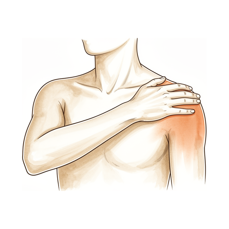
Phẫu thuật này nhằm loại bỏ một mảnh xương nhỏ ở đầu ngoài của xương đòn, tại khớp nơi xương đòn gặp đỉnh vai. Phẫu thuật được đề xuất cho những người bị đau do hao mòn tại khớp đó, hoặc do xương bị tiêu dần sau khi nâng vật nặng lặp đi lặp lại, và chỉ khi các dây chằng giữ vững khớp vẫn còn tốt. Việc loại bỏ mảnh xương nhỏ đó giúp các bề mặt xương thô ráp không còn cọ xát vào nhau. Mục tiêu là giảm đau và giúp vai vận động thoải mái hơn, và cả hình thức nội soi lẫn hình thức mổ mở của phẫu thuật này đều giúp giảm đau đáng kể sau 1 năm.
Trước khi phẫu thuật
Bác sĩ phẫu thuật sẽ lên kế hoạch phẫu thuật dựa trên các hình ảnh bạn đã chụp, chẳng hạn như chụp X-quang, MRI hoặc siêu âm vai. Các hình ảnh này cho thấy rõ khớp và giúp quyết định cần cắt bỏ bao nhiêu xương. Vào ngày phẫu thuật, hãy ngừng ăn và uống bảy tiếng trước ca phẫu thuật. Chúng tôi yêu cầu bảy tiếng thay vì sáu tiếng để ca phẫu thuật của bạn có thể được đẩy lên sớm hơn nếu lịch mổ diễn ra sớm hơn dự kiến. Bác sĩ phẫu thuật sẽ cho bạn biết nên bỏ qua loại thuốc thường dùng nào và nên tiếp tục dùng loại nào. Hãy mang theo một danh sách ghi đầy đủ tất cả các loại thuốc bạn đang dùng. Hãy sắp xếp người lái xe đưa bạn về nhà sau phẫu thuật. Hãy mặc quần áo rộng rãi, thoải mái và dễ mặc vào, cởi ra. Nếu bạn mắc các bệnh lý khác, có thể bạn sẽ cần làm xét nghiệm máu hoặc được bác sĩ gây mê thăm khám lại.
Vào ngày phẫu thuật
Bạn sẽ đến đơn vị tiếp nhận bệnh nhân phẫu thuật của bệnh viện, nơi bạn sẽ được làm thủ tục nhập viện và chuẩn bị cho ca mổ. Bác sĩ gây mê sẽ gặp bạn trước ca mổ và giải thích chi tiết về cả hai phương pháp gây mê. Ca phẫu thuật này được thực hiện dưới gây mê toàn thân kết hợp với kỹ thuật chặn dây thần kinh vùng. Sau đó, bạn được đưa vào phòng mổ để tiến hành phẫu thuật.
Bạn sẽ tỉnh dậy tại khu hồi sức, nơi các điều dưỡng theo dõi bạn trong lúc thuốc mê hết tác dụng. Khi sức khỏe ổn định, tùy thuộc vào loại phẫu thuật và tiến triển hồi phục, bạn sẽ được chuyển về phòng bệnh hoặc xuất viện về nhà.
Quy trình phẫu thuật
Đây là một ca phẫu thuật nội soi. Bác sĩ phẫu thuật sẽ thực hiện vài vết rạch nhỏ quanh khớp vai, trong đó có ít nhất một vết ở phía sau, và thao tác với một chiếc camera nhỏ đặt bên trong khớp. Camera hiển thị hình ảnh khớp trên màn hình, nên không cần một vết rạch lớn để quan sát khớp.
Qua những vết rạch nhỏ này, bác sĩ phẫu thuật cắt bỏ khoảng 5 mm xương ở đầu ngoài của xương đòn. Như vậy là đủ để các bề mặt thô ráp, bị hao mòn tại khớp không còn cọ xát vào nhau. Bác sĩ phẫu thuật cẩn trọng cắt bỏ một lượng xương có kiểm soát, đủ để giảm đau nhưng vẫn bảo vệ các dây chằng và bao khớp giữ vững khớp. Việc bảo vệ các cấu trúc này rất quan trọng, vì cắt bỏ quá nhiều xương có thể khiến xương đòn mất vững.
Các vết rạch nhỏ được khâu lại. Băng gạc được đặt lên trên, và bạn sẽ giữ băng gạc đó trong khoảng 10 ngày.
Sau khi phẫu thuật
Hầu hết bệnh nhân sẽ ở lại bệnh viện một đêm sau ca phẫu thuật này; tuy nhiên một số người có thể về nhà ngay trong ngày. Vui lòng sắp xếp để có người ở lại với bạn trong 24 giờ đầu sau khi bạn về nhà. Bạn sẽ bị đau trong một hai ngày đầu; chúng tôi sẽ cho bạn thuốc để giữ cho bạn dễ chịu, và vai có thể bị tê một thời gian do kỹ thuật chặn dây thần kinh. Tê và yếu trong 24 giờ đầu sau khi chặn dây thần kinh là điều bình thường, đã được dự kiến. Hãy gọi cho phòng khám nếu bạn không thể cảm nhận hoặc cử động cánh tay, bàn tay hoặc các ngón tay khi tác dụng chặn dây thần kinh đã hết, vào khoảng 24 giờ. Cánh tay của bạn sẽ được đặt trong chiếc đai đỡ đơn giản để giảm khó chịu; chiếc đai này sẽ được tháo ra khi bạn tập các bài tập hoặc tắm rửa. Chúng tôi sẽ giữ băng gạc trên vết mổ khoảng 10 ngày; vui lòng không tự ý tháo băng ra trước thời hạn đó trừ khi chúng tôi yêu cầu. Chúng tôi sẽ thay hoặc gỡ băng gạc khi khám lại cho bạn.
Quá trình hồi phục
Trong một hai ngày đầu, vai của bạn sẽ đau nhức và có thể có cảm giác sưng. Thuốc giảm đau chúng tôi cho sẽ giúp bạn dễ chịu, và tình trạng tê do chặn dây thần kinh cũng giúp ích trong thời gian đầu. Nghỉ ngơi với cánh tay được đỡ và tập các bài tập một cách nhẹ nhàng thường giúp giảm khó chịu. Hầu hết mọi người thấy cơn đau nhức giảm dần đều trong vài tuần đầu.
Để tăng sự thoải mái, cánh tay của bạn sẽ được đặt trong một chiếc đai treo đơn giản. Bạn tháo đai ra khi tập các bài tập và khi tắm rửa, và chuyên viên vật lý trị liệu sẽ chỉ cho bạn những động tác nên bắt đầu. Các động tác này bắt đầu nhẹ nhàng và tăng dần khi cơn đau giảm bớt. Bạn có thể dùng bàn tay cho các việc nhẹ ở nhà, chẳng hạn như ăn uống hoặc viết, khi bạn cảm thấy vững vàng. Tránh nâng bất kỳ vật nặng nào hoặc cố làm dù đang đau.
Khi khả năng vận động trở lại, các hoạt động hằng ngày sẽ dễ dàng hơn. Mặc quần áo, với tay lên cao và nằm ngủ nghiêng về bên vai đó đều cải thiện dần dần. Ngủ ở tư thế kê cao bằng thêm gối có thể dễ chịu hơn trong những ngày đầu. Khi bác sĩ phẫu thuật cho phép bạn lái xe, thường là trong buổi tái khám ở tuần thứ sáu, bạn có thể cầm lái trở lại; hãy xem Lái xe sau phẫu thuật chi trên để biết thêm chi tiết. Công việc văn phòng và các việc nhẹ hằng ngày thường trở lại trong vòng 2 đến 6 tuần, còn các hoạt động nặng hơn như thể thao hoặc tập gym mất nhiều thời gian hơn, từ 3 đến 6 tháng.
Mỗi người hồi phục theo tốc độ riêng, nên thời gian hồi phục của bạn có thể khác. Bác sĩ phẫu thuật và chuyên viên vật lý trị liệu sẽ hướng dẫn bạn ở mỗi lần tái khám.
Những biến chứng có thể xảy ra
Hầu hết bệnh nhân đều hồi phục tốt, nhưng đôi khi vẫn có thể gặp phải các vấn đề. Bác sĩ phẫu thuật và đội ngũ y tế sẽ theo dõi sát sao để phát hiện sớm bất kỳ dấu hiệu bất thường nào.
Đôi khi cơn đau vẫn tiếp diễn sau phẫu thuật. Đây là vấn đề phổ biến nhất của phẫu thuật này. Điều này có thể xảy ra nếu lượng xương được cắt bỏ hơi quá nhiều hoặc hơi quá ít, hoặc nếu một mảnh xương nhỏ mọc lại. Nếu vai bạn vẫn đau nhức như trước, hoặc cơn đau quay trở lại sau một thời gian đỡ đau, hãy nêu ra trong lần tái khám tiếp theo. Đôi khi cần phẫu thuật thêm để khắc phục.
Cắt bỏ quá nhiều xương có thể gây ra một vấn đề khác. Các dây chằng và bao khớp giữ vững xương đòn có thể bị ảnh hưởng, khiến khớp trở nên lỏng lẻo hoặc mất vững. Bạn có thể nhận thấy xương đòn có cảm giác như bị xê dịch hoặc kêu lục cục, hoặc cảm giác khớp không chắc chắn khi bạn cử động cánh tay. Nếu khớp đã lỏng lẻo từ trước phẫu thuật, nguy cơ này cao hơn. Khớp mất vững có thể cần phẫu thuật thêm để tái tạo các dây chằng. Hãy báo cho bác sĩ phẫu thuật trong lần tái khám tiếp theo nếu khớp có cảm giác lỏng lẻo hoặc cử động theo một cách mới lạ.
Các vấn đề khác ít gặp nhưng đáng để biết. Chúng bao gồm nhiễm trùng, cứng khớp, gãy xương, khớp tự dính lại, và một tình trạng đau kéo dài gọi là hội chứng đau vùng phức tạp. Các dấu hiệu nhiễm trùng cần theo dõi là sốt, vùng đỏ lan rộng quanh vết mổ, hoặc dịch hay mủ rỉ ra từ vết mổ. Hãy gọi cho phòng khám ngay trong ngày nếu bạn nhận thấy bất kỳ dấu hiệu nào trong số này. Cơn đau ngày càng nặng hơn dù đã dùng thuốc giảm đau cũng cần gọi cho phòng khám ngay trong ngày. Tình trạng trật khớp sau phẫu thuật nội soi cũng đã được ghi nhận. Nếu vai bạn đột ngột thay đổi hình dạng, sa xuống phía trước, hoặc rất đau và mất vững sau một cú va chạm hay té ngã, hãy liên hệ với phòng khám ngay lập tức.
Bảng liệt kê các biến chứng ở trang này cung cấp tỷ lệ xảy ra cụ thể nếu bạn muốn biết thêm chi tiết.
Khi nào nên gọi cho chúng tôi
Hầu hết các vấn đề được phát hiện sớm khi bạn báo cho chúng tôi. Hãy gọi cho phòng khám ngay trong ngày nếu bạn bị sốt, vùng đỏ lan rộng quanh vết mổ, hoặc dịch hay mủ rỉ ra từ vết mổ. Cũng hãy gọi cho phòng khám ngay trong ngày nếu cơn đau ngày càng nặng hơn dù đã dùng thuốc giảm đau. Hãy đến phòng cấp cứu nếu bạn bị sưng hoặc đau ở bắp chân, hoặc khó thở hay đau ngực. Đây có thể là dấu hiệu của cục máu đông. Cũng hãy đến phòng cấp cứu nếu các ngón tay hoặc bàn tay chuyển sang nhợt nhạt, lạnh, trắng, xanh tím hoặc sẫm màu. Hãy gọi cho phòng khám nếu bạn không thể cảm nhận hoặc cử động cánh tay, bàn tay hoặc các ngón tay khi tác dụng chặn dây thần kinh đã hết, vào khoảng 24 giờ. Nếu bạn không thể liên lạc với phòng khám, hãy đến phòng cấp cứu gần nhất.
Nơi để tìm đọc thêm thông tin về bệnh lý
Trang này nói về chính ca phẫu thuật. Hai bệnh lý mà phẫu thuật này điều trị có trang riêng, mỗi trang trình bày những bằng chứng cho thấy khi nào phẫu thuật mang lại hiệu quả và khi nào thì không: tình trạng hao mòn tại khớp được trình bày trên trang Viêm khớp thoái hóa khớp AC, còn dạng gặp ở người tập tạ, trong đó đầu xương đòn bị phá hủy dần, được trình bày trên trang Tiêu xương đầu ngoài xương đòn.
Evidence & references
This is the clinical evidence summary written for health professionals. It is technical, and it lists the research this page was built from. You do not need to read it to understand your treatment or to make a decision about it.
Overview
- Simple excision of the outer end of the clavicle has yielded satisfactory results in patients with complete dislocation and subluxation of the acromioclavicular joint, with no residual upward displacement disturbing the patients [2].
- Excision of the outer end of the clavicle is preferred for old dislocations, while open reduction and internal fixation are not recommended due to complications and poor functional results [16].
- Open distal clavicle resection yields good to excellent results in properly selected patients and does not create significant subjective or objective weakness [3].
- In appropriately selected patients, open or arthroscopic distal clavicle resection is necessary to relieve symptoms of painful conditions of the acromioclavicular joint [7].
- Arthroscopic distal clavicle resection has provided more 'good or excellent' results than the open procedure, but is comprised of low-level evidence [6].
- Arthroscopic and open distal clavicle excisions both provide significant pain reduction at 1 year with no significant difference in outcome measures between groups, except for VAS pain score improvement [8].
- Among patients undergoing distal clavicle excision for acromioclavicular joint pathology, those having an arthroscopic procedure, specifically through the direct approach, can expect a faster return to activities while obtaining similar long-term outcomes compared with the open procedure [9].
- A well-performed distal clavicle excision will likely perform better than a poorly performed one, regardless of whether an open or arthroscopic approach is chosen [5].
- In carefully selected patients with isolated ACJ pathology, arthroscopic distal clavicle excision results in statistically and clinically significant improvements in range of motion and patient-reported outcome measures [18].
- Both the direct superior approach and the indirect subacromial approach to the arthroscopic distal clavicle resection result in successful clinical outcome with clinically insignificant difference at final follow-up [19].
- Incomplete excision and regrowth of the distal clavicle are the most common causes of revision [12].
- Regeneration of the excised portion of clavicle occurs frequently and may produce an unacceptable result, and may be more likely to occur in the younger patient population [13].
- Horizontal instability of the clavicle is evident with distal clavicle resection of greater than 10 mm [15].
- Although distal clavicle excision with 2.5 mm of bone was successful in many specimens, a 5 mm resection guaranteed no bone-to-bone abutment [17].
- Routine distal clavicle resection in the setting of rotator cuff repair does not result in improved outcomes for patients with no difference being observed at 24 months post surgery [11].
- A records review found that 10 of 894 (1.1%) rotator cuff repairs underwent subsequent distal clavicle resection [40].
- Regardless of the technique chosen for distal clavicle resection, portal placement remains paramount in both facilitating surgery and avoiding injury to adjacent extra-articular structures [1].
- The use of intraoperative ultrasound and cannulated dilators allows surgeons to perform distal clavicle excisions in a more efficient, reproducible and safer manner [4].
- A novel anterior portal technique for arthroscopic distal clavicle excision yields a safe corridor for placement of instruments in a reliable manner that does not disrupt the important stabilizers of the glenohumeral or AC joints [20].
- Clavicular reconstruction may be the treatment of choice in patients with excess resection of the clavicle [14].
Anatomy & Pathophysiology
Bony Anatomy
- The clavicle is the first bone to ossify, occurring at the fifth week of gestation [59].
- The clavicle is the only long bone to ossify by intramembranous ossification [59, 65].
- The medial (sternal) epiphysis of the clavicle is the last ossification center to fuse, occurring at age 20 to 25 years [59].
- The primary blood supply to the clavicle is periosteal, and no nutrient artery is present [59, 65].
- The distal clavicle is flat in the anteroposterior plane [65].
- The scapula has only one true diarthrodial articulation, the acromioclavicular joint [59].
- The acromion has three ossification centers: the metacromion (base), the mesoacromion (middle), and the preacromion (tip) [59].
- Failure of fusion of the acromial ossification centers results in os acromiale [59].
- The coracoid process serves as an attachment site for the coracoacromial ligament, coracoclavicular ligaments, conjoined tendon, and pectoralis minor muscle [70].
Ligaments and Joint Structures
- The acromioclavicular joint is a small diarthrodial joint with an interposed fibrocartilaginous disk [59].
- The acromioclavicular joint is a planar diarthroidal joint that couples scapular and sternoclavicular motion [32].
- A fibrocartilaginous load-bearing disk cushions the high contact stress of the acromioclavicular joint when load is placed across the small articular surface area [32].
- The superior and posterior acromioclavicular ligaments are the primary stabilizers to anterior and posterior (horizontal) translation of the clavicle [59].
- The coracoclavicular ligaments consist of the conoid (medial) and trapezoid (lateral) components [59, 65].
- The coracoclavicular ligaments are the primary stabilizers to superior (vertical) translation of the distal clavicle [59, 65].
- The trapezoid ligament is located approximately 25 mm from the acromioclavicular joint [70].
- The conoid ligament is located approximately 45 mm from the acromioclavicular joint and is the stronger of the two coracoclavicular ligaments [70].
- The superior shoulder suspensory complex is composed of the glenoid, coracoid process, coracoclavicular ligaments, distal clavicle, acromioclavicular joint, and acromion [59].
- The superior shoulder suspensory complex provides a stable connection between the scapula and the axial skeleton [59].
Pathophysiology and Biomechanics
- The acromioclavicular joint articulation helps provide a stable base for the glenohumeral joint to position the arm in space [32].
- Distal clavicle resection does not address instability and may accentuate it when horizontal or vertical instability exists [23].
- Distal clavicle resection must be reserved for patients in whom the coracoclavicular ligaments are intact and there is no concomitant instability [23].
- Kinematic changes resulting from acromioclavicular joint dislocation could be a potential source of pain and dysfunction in the shoulder [87].
- A combined injury of the acromioclavicular and coracoclavicular ligaments significantly alters glenohumeral kinematics during abduction [117].
- Large kinematic differences exist between the intact state and a Rockwood V lesion during both humerothoracic and scapulothoracic movements [118, 126].
- The distal clavicle has a wide range of motion during shoulder abduction, which does not support the concept of synchronous motion with the scapula [123].
- The trapezoid and conoid ligaments have unique functions in normal shoulder kinematics due to their anatomic attachments [86].
- Scapular and clavicular kinematics are affected in acromioclavicular separation models [89].
- Type I and II acromioclavicular joint disruptions impair long-term shoulder function in about half of patients 10 years after injury [101].
- The normal coracoclavicular distance measures approximately 1.1 to 1.3 cm [150].
- Injury to the acromioclavicular ligaments in a type II injury causes an increase of 3.6 mm in anterior and 6.4 mm in posterior translation [150].
Classification
- The Rockwood classification system groups similar diagnoses, provides prognostic information, and guides treatment options [147].
- Studies have shown fair to moderate reliability in the use of the Rockwood classification for classification and surgical decision making [147].
- The ISAKOS Upper Extremity Committee suggests adding grade IIIA and grade IIIB injuries to a modified Rockwood classification to distinguish between stable type III injuries and unstable grade III injuries with therapy-resistant scapular dysfunction and overriding clavicle [154].
- In the Rockwood classification, type I is established when 0% to <10% superior displacement of the distal clavicle is found [144].
- In the Rockwood classification, type II is present when the affected side differs by 10% to ≤25% superior displacement of the distal clavicle [144].
- In the Rockwood classification, type III dislocation is defined as a coracoclavicular distance difference of >25% to ≤100% compared with the contralateral side [144].
- In the Rockwood classification, a coracoclavicular distance difference of >100% compared with the contralateral side indicates a type V separation [144].
- A new classification of AC joint instability defines Group 1 as having a coracoclavicular distance difference ≤30%, which includes all Rockwood type I, type II, and borderline low-grade type III patients [144].
- A new classification of AC joint instability defines Group 2 as having a coracoclavicular distance difference >30%, which represents high-grade AC joint dislocations including all Rockwood type V patients and the majority of Rockwood type III patients [144].
- The Rockwood type IV AC injury includes disruption of AC ligaments, disruption of coracoclavicular ligaments, and posterior translation of the clavicle [33].
- Methods to diagnose both superior and posterior translation of the clavicle need further debate [34].
- Fractures of the clavicle may be divided into three groups: Group I (middle third), Group II (distal to the coracoclavicular ligament), and Group III (proximal end) [24].
- Neer classified fractures of the distal end of the clavicle into two types [24].
- Robinson classified clavicular injuries and defined the lateral one-fifth of the clavicle as Type 3 [52].
Clinical Presentation
Indications and Selection
- In appropriately selected patients, open or arthroscopic distal clavicle resection is necessary to relieve symptoms [7].
- Distal clavicle excision must be reserved for patients in whom the coracoclavicular ligaments are intact and there is no concomitant instability [23].
- When horizontal or vertical instability exists, results of distal clavicle excision are compromised because the technique does not address instability and may accentuate it [23].
- Operation for acromioclavicular dislocation should be considered only in thin patients with a prominent clavicle, those doing heavy work, or those whose work requires frequent shoulder abduction and flexion [49].
- For chronic symptomatic acromioclavicular injuries, partial claviculectomy is believed to be the best procedure, offering negligible morbidity and rapid return to function [35].
- Clavicular resection reliably produced significant improvement in patients with persistent pain or posttraumatic arthritis following acromioclavicular joint dislocation [22].
- Simple excision of the outer end of the clavicle has yielded satisfactory results in patients with complete dislocation and subluxation, with no residual upward displacement disturbing the patients [2].
- Routine distal clavicle resection in the setting of rotator cuff repair does not result in improved outcomes for patients, with no difference observed at 24 months post-surgery [11].
Diagnostic Considerations
- There may be misdiagnosis due to associated or underlying superior labral detachment causing symptoms similar to those of acromioclavicular joint pathology [127].
- Clinical examination and surgical treatment should address anatomic restoration of individual structures to optimize the mechanical capability of the claviscapular segment [25].
Investigations
Radiographic Evaluation
- The Zanca view is a special radiographic view indicated for the acromioclavicular joint, performed as an AP with 10° cephalic tilt centered over the AC joint [83].
- The Zanca view technique requires the patient to be supine and uses only one-half the voltage of a routine shoulder AP view to create a soft-tissue view [83].
- Weighted stress radiographs significantly increase the measured elevation of the clavicle and the coracoclavicular distance compared to non-weighted views [155].
- The normal coracoclavicular distance is 1.1 to 1.3 cm [83].
- Methods to diagnose both superior and posterior translation of the clavicle require further debate [34].
- Plain radiographs can observe arthritis, calcific tendinitis, and osteolysis of the distal clavicle [82].
- The axillary view provides good visualization of the coracoid process, acromion, and distal clavicle [83].
Advanced Imaging
- CT imaging is frequently used to evaluate fractures of the shoulder, assess for bony lesions in recurrent instability cases, or for preoperative templating for shoulder arthritis [82].
- MRI is the modality of choice for evaluating the rotator cuff, biceps, and subacromial/subdeltoid bursa [82].
- Ultrasonography is a low-cost alternative to MRI and arthrography for evaluating both skeletal and soft-tissue structures of the shoulder [82].
- Ultrasonography can provide immediate, real-time visualization of the rotator cuff, biceps tendon, and calcific deposits [82].
- Ultrasonography is highly operator dependent and is not as useful for evaluating labral tears or rotator cuff tears that are very small or larger than 3 cm [82].
- Intraoperative ultrasound allows surgeons to safely identify and access the acromioclavicular joint for distal clavicle excision [4].
- The cross-sectional A-frame morphology of the superior cortex of the distal clavicle provides a reproducible landmark that is eliminated approximately 1.0 cm medial to the distal, lateral end of the clavicle, which can be used intraoperatively to determine when adequate resection has been completed [28].
Treatment
Indications and Contraindications
- Distal clavicle resection is indicated for symptomatic acromioclavicular joint pathology in appropriately selected patients [7].
- The procedure is reserved for patients with intact coracoclavicular ligaments and no concomitant instability [23].
- Results are compromised when horizontal or vertical instability exists because the technique does not address instability and may accentuate it [23].
- Clavicular resection reliably produced significant improvement in patients with persistent pain or posttraumatic arthritis following dislocation [22].
- Routine distal clavicle excision is not absolutely necessary, even in patients with symptomatic acromioclavicular joint osteoarthritis [136].
Outcomes and Efficacy
- Simple excision of the outer end of the clavicle has yielded satisfactory results with no residual upward displacement disturbing the patients [2].
- Patients undergoing arthroscopic distal clavicle excision through the direct approach can expect a faster return to activities while obtaining similar long-term outcomes compared with the open procedure [9].
- Both the direct superior approach and the indirect subacromial approach to arthroscopic distal clavicle resection result in successful clinical outcome with clinically insignificant difference at final follow-up [19].
Surgical Technique and Landmarks
- Portal placement remains paramount in both facilitating surgery and avoiding injury to adjacent extra-articular structures regardless of the technique chosen for distal clavicle resection [1].
- Intraoperative use of ultrasound and cannulated dilators allows surgeons to perform distal clavicle excisions in a more efficient, reproducible and safer manner [4].
- Beveling the inferior 20% to 25% of the clavicle to make it co-planar with the decompressed acromion is safe and is not an etiologic factor in acromioclavicular joint pain or instability for appropriate clinical indications [139].
Complications and Revision
- A 5-mm distal clavicle resection guaranteed no abutment but decreased joint stiffness [29].
- The early results of clavicular reconstruction after failure of partial excision of the lateral end of the clavicle are encouraging and may be the treatment of choice in patients with excess resection of the clavicle [14].
Complications
Resection-Related Complications
- Incomplete excision and regrowth of the distal clavicle are the most common causes of revision surgery [12].
- Regeneration of the excised portion of the clavicle occurs frequently and may produce an unacceptable result, with a higher likelihood in younger patients [13].
- The most common complication of distal clavicle resection is persistent pain, which can result from over- or under-resection [140].
- Incomplete resection can occur due to poor visualization [140].
- Overexuberant resection of the clavicle or disruption of the acromioclavicular and coracoclavicular ligamentous system can result in iatrogenic instability of the acromioclavicular joint [140].
- Posterior translation of the acromioclavicular joint is increased by 32% after a distal clavicle resection with an acromioclavicular capsular incision [140].
- Posterior translation can be reduced to 13% if the distal clavicle resection is completed with a coracoacromial ligament augmentation procedure [140].
- Postoperative iatrogenic instability may require revision surgery or coracoclavicular ligament reconstruction [140].
- Other complications of distal clavicle resection include infection, stiffness, fracture, spontaneous fusion, and complex regional pain syndrome [140].
- A 5-mm distal clavicle resection guaranteed no bone-to-bone abutment but decreased joint stiffness [29].
- Distal clavicle excision with 2.5 mm of bone was successful in many specimens, whereas a 5 mm resection guaranteed no bone-to-bone abutment [17].
- The high failure rate in patients with even subtle acromioclavicular instability is 42% [167].
- When horizontal or vertical instability exists, results of distal clavicle resection are compromised because the technique does not address instability and may accentuate it [23].
Instability and Dislocation
- Acromioclavicular dislocation can occur after arthroscopic distal clavicle resection [1].
- Portal placement is paramount in avoiding injury to adjacent extra-articular structures during distal clavicle resection [1].
- Distal clavicle resection should be reserved for patients with intact coracoclavicular ligaments and no concomitant instability [23].
- Clavicular resection reliably produced significant improvement in patients with persistent pain or posttraumatic arthritis following late loss of reduction [22].
- Simple excision of the outer end of the clavicle yielded satisfactory results with no residual upward displacement disturbing patients [2].
Fractures and Bony Complications
- Fracture of the distal clavicle or coracoid process after coracoclavicular ligament repair or reconstruction is a rare but serious complication that can occur independent of bone tunnels created during the index procedure [100].
- Clavicle and coracoid fractures occurred in 1.9 out of 100 cases in a review of operative acromioclavicular joint separations in an active population [161].
- Retaining the clavicular hook-plate indefinitely carries a potential risk of developing osteolysis and fracture around the implant [51].
- Subacromial osteolysis is a complication associated with hook plate fixation for acromioclavicular dislocation [163].
- Hook plates used for lateral clavicle fractures can have complications including rotator cuff tear, impingement pain, and need for removal [52].
- Clavicular reconstruction after failure of partial excision of the lateral end of the clavicle has encouraging early results and may be the treatment of choice in patients with excess resection [14].
Infection and Other Complications
- Older patients and females were more likely to experience postoperative complications requiring reoperations, including revision acromioclavicular joint reconstruction, distal clavicle excision, and irrigation and debridement [37].
- The incidence of complications in operative acromioclavicular joint separations in an active population was 1.35 per 100 person-years [161].
Recovery
Outcomes and Functional Improvement
- Arthroscopic distal clavicle excision results in statistically and clinically significant improvements in range of motion and patient-reported outcome measures in carefully selected patients with isolated ACJ pathology [18].
- For chronic symptomatic injuries, partial claviculectomy is believed to be the best procedure, offering negligible morbidity and rapid return to function [35].
- Clavicular resection reliably produced significant improvement in patients with persistent pain or posttraumatic arthritis [22].
- Open and arthroscopic distal clavicle excision are both effective surgeries to treat recalcitrant acromioclavicular joint pain, providing similarly good to excellent results regarding patient satisfaction and shoulder function at intermediate-term follow-up [166].
- Arthroscopic distal clavicle resection has provided more 'good or excellent' results than has the open procedure, but is comprised of low-level evidence [6].
- Less residual pain was found using the arthroscopic technique compared to the open technique [166].
Return to Activity
- More than 90% of patients manage to return to driving within 4 weeks and to work within 6 weeks following arthroscopic subacromial decompression and acromioclavicular joint excision [97].
Complications and Revision
- Older patients and females were more likely to experience postoperative complications requiring reoperations, including revision ACJR, distal clavicle excision, and irrigation and debridement [37].
Key Evidence
- [Case_report] Regardless of the technique chosen for distal clavicle resection, portal placement remains paramount in both facilitating surgery and avoiding injury to adjacent extra-articular structures. [1] (10.1016/j.jse.2010.08.032)
- [L4] Open distal clavicle resection yields good to excellent results in properly selected patients and does not create significant subjective or objective weakness. [3] (10.1016/s1058-2746(10)80006-0)
- [L5] The technique will allow surgeons to perform distal clavicle excisions in a more efficient, reproducible and safer manner. [4] (10.1016/j.eats.2024.103331)
- [L5] A well-performed distal clavicle excision will likely perform better than a poorly performed one, regardless of whether an open or arthroscopic approach is chosen. [5] (10.1016/j.arthro.2018.03.004)
- [L3] Arthroscopic distal clavicle resection has provided more 'good or excellent' results than has the open procedure, but is comprised of low-level evidence. [6] (10.1097/blo.0b013e31802f5450)
- [L5] In appropriately selected patients, open or arthroscopic distal clavicle resection is necessary to relieve symptoms. [7] (10.5435/00124635-199905000-00004)
- [L1] Arthroscopic and open distal clavicle excisions both provide significant pain reduction at 1 year with no significant difference in outcome measures between groups, except for VAS pain score improvement. [8] (10.1016/j.jse.2006.10.006)
- [L3] Among patients undergoing distal clavicle excision for acromioclavicular joint pathology, those having an arthroscopic procedure, specifically through the direct approach, can expect a faster return to activities while obtaining similar long-term outcomes compared with the open procedure. [9] (10.1016/j.arthro.2009.12.007)
- [L2] Routine distal clavicle resection in the setting of rotator cuff repair does not result in improved outcomes for patients with no difference being observed at 24 months post surgery. [11] (10.1177/1758573217741124)
- [L4] Incomplete excision and regrowth of the distal clavicle are the most common causes of revision. [12] (10.1016/j.arthro.2009.06.010)
- [L4] Regeneration of the excised portion of clavicle occurs frequently and may produce an unacceptable result, and may be more likely to occur in the younger patient population. [13] (10.1016/s1058-2746(05)80049-7)
- [L4] The early results are encouraging and may be the treatment of choice in patients with excess resection of the clavicle. [14] (10.1016/s1058-2746(95)80128-6)
- [L4] Horizontal instability of the clavicle is evident with distal clavicle resection of greater than 10 mm. [15] (10.1016/j.xrrt.2021.05.003)
- [L4] Excision of the outer end of the clavicle is preferred for old dislocations, while open reduction and internal fixation are not recommended due to complications and poor functional results. [16] (10.2106/00004623-196345080-00024)
- [Abstract] Although distal clavicle excision with 2.5 mm of bone was successful in many specimens, a 5 mm resection guaranteed no bone-to-bone abutment. [17] (10.1016/j.jse.2007.02.105)
- [L4] In carefully selected patients with isolated ACJ pathology, arthroscopic distal clavicle excision results in statistically and clinically significant improvements in range of motion and patient-reported outcome measures. [18] (10.1016/j.jseint.2023.07.014)
- [L2] Both the direct superior approach and the indirect subacromial approach to the arthroscopic distal clavicle resection result in successful clinical outcome with clinically insignificant difference at final follow-up. [19] (10.1177/0363546506294855)
- [L4] The technique was validated using cadaveric dissection and yields a method of establishing the anterior portal for distal clavicle excision with a safe corridor for placement of instruments in a reliable manner that does not disrupt the important stabilizers of the glenohumeral or AC joints. [20] (10.1097/bte.0000000000000033)
- [L3] Late loss of reduction was common, and clavicular resection reliably produced significant improvement in patients with persistent pain or posttraumatic arthritis. [22] (10.2106/00004623-198769070-00013)
- [L5] [23] (10.5435/00124635-200904000-00002)
- [L4] [24] (10.2106/00004623-196749040-00024)
- [L5] Clinical examination and surgical treatment should address anatomic restoration of individual structures to optimize the mechanical capability of the claviscapular segment. [25] (10.5435/jaaos-d-24-00360)
- [L5] The cross-sectional A-frame morphology of the superior cortex of the distal clavicle provides a reproducible landmark that is eliminated approximately 1.0 cm medial to the distal, lateral end of the clavicle, which can be used intraoperatively to determine when adequate resection has been completed. [28] (10.1016/j.jse.2021.10.013)
- [L5] A 5-mm distal clavicle resection guaranteed no abutment but decreased joint stiffness. [29] (10.1016/j.arthro.2007.07.004)
- [Paper] [32] (10.1097/bte.0000000000000044)
- [L4] [33] (10.1016/j.arthro.2016.06.013)
- [L4] Methods to diagnose both superior and posterior translation of the clavicle need further debate. [34] (10.1016/j.jseint.2019.11.006)
- [L4] Older patients and females were more likely to experience postoperative complications requiring reoperations, including revision ACJR, distal clavicle excision, and irrigation and debridement. [37] (10.1007/s00167-016-4206-y)
- [L3] This records review found that 10 of 894 (1.1%) rotator cuff repairs underwent subsequent distal clavicle resection. [40] (10.1177/2325967119844295)
- [L1] Operation should be considered only in thin patients with a prominent clavicle, those doing heavy work, or those whose work requires frequent shoulder abduction and flexion. [49] (10.2106/00004623-198668040-00011)
- [L5] Retaining the clavicular hook-plate indefinitely carries a potential risk of developing osteolysis and fracture around the implant. [51] (10.1016/j.injury.2004.08.010)
- [L4] [52] (10.1177/1758573214536535)
- [L5] The trapezoid and conoid ligaments have unique functions in normal shoulder kinematics because of their anatomic attachments. [86] (10.1016/j.arthro.2009.12.031)
- [L5] The kinematic changes could be a potential source of pain and dysfunction in the shoulder with AC joint dislocation. [87] (10.1177/0363546512458571)
- [L5] Scapular and clavicular kinematics were affected in AC separation models. [89] (10.1016/j.jse.2013.01.004)
- [L3] The results obtained in the present study suggest that more than 90% of the patients manage to return to driving within 4 weeks and to work within 6 weeks following arthroscopic subacromial decompression and acromio-clavicular joint excision. [97] (10.1111/j.1758-5740.2010.00048.x)
- [L4] Fracture of the distal clavicle or coracoid process after CC ligament repair or reconstruction is a rare but serious complication that can occur independent of bone tunnels created during the index procedure. [100] (10.1177/03635465211036713)
- [L4] Type I and II acromioclavicular joint disruptions impair long-term shoulder function in about half of patients 10 years after injury. [101] (10.1177/0363546508319047)
- [L5] A combined injury of the AC and CC ligaments significantly alters GH kinematics during abduction. [117] (10.1186/s12891-016-1330-2)
- [L5] There are large kinematic differences between the intact state and a Rockwood V lesion not only during humerothoracic, but also during scapulothoracic movements. [118] (10.1016/j.jse.2022.01.096)
- [L5] The distal clavicle had a wide range of motion during shoulder abduction, which did not support the concept of synchronous motion with the scapula. [123] (10.1016/j.arthro.2012.04.001)
- [L5] Large kinematic differences were seen between the intact state and a Rockwood V lesion not only during humerothoracic movements but also during scapulothoracic movements in the cadaveric model. [126] (10.1177/03635465211053016)
- [L4] The successful treatment of 15 patients by SLAP lesion repair after initial distal clavicle resection indicates that there may be misdiagnosis due to associated or underlying superior labral detachment causing similar symptoms. [127] (10.1016/s1058-2746(96)80319-3)
- [L2] Routine distal clavicle excision is not absolutely necessary, even in patients with symptomatic ACJ osteoarthritis. [136] (10.1007/s00167-020-06098-y)
- [L4] We conclude that for appropriate clinical indications, beveling the inferior 20% to 25% of the clavicle to make it co-planar with the decompressed acromion is safe and is not an etiologic factor in acromioclavicular joint pain or instability. [139] (10.1067/mse.2000.109560)
- [L5] [140] (10.1177/0363546513485359)
- [L1] [144] (10.1016/j.jse.2020.10.026)
- [L5] The Rockwood classification system groups similar diagnoses, provides prognostic information, and guides treatment options, though studies have shown fair to moderate reliability in its use for classification and surgical decision making. [147] (10.1007/s11999-016-5079-6)
- [L5] [150] (10.1016/j.jse.2010.10.030)
- [L5] The ISAKOS Upper Extremity Committee suggests adding grade IIIA and grade IIIB injuries to a modified Rockwood classification to distinguish between stable type III injuries and unstable grade III injuries with therapy-resistant scapular dysfunction and overriding clavicle. [154] (10.1016/j.arthro.2013.11.005)
- [L4] Weighted stress radiographs significantly increased the measured elevation of the clavicle and the coracoclavicular distance compared to non-weighted views. [155] (10.1016/j.jseint.2023.06.011)
- [L3] This review demonstrated an incidence of 1.35 complications per 100 person-years, with clavicle and coracoid fractures occurring in 1.9 out of 100 cases. [161] (10.1177/2325967121s00330)
- [L1] The current analysis suggests coracoclavicular ligament reconstruction as an effective surgical approach for decreasing the incidence of subacromial osteolysis. [163] (10.1016/j.jse.2024.03.018)
- [L3] Open and arthroscopic distal clavicle excision are both effective surgeries to treat recalcitrant acromioclavicular joint pain, providing similarly good to excellent results regarding patient satisfaction and shoulder function at intermediate-term follow-up, though less residual pain was found using the arthroscopic technique. [166] (10.1177/0363546511419633)
- [L4] The high failure rate in patients with even subtle acromioclavicular instability (42%) suggests that in these cases formal stabilization with ligament reconstruction should be considered in addition to resection of the distal clavicle. [167] (10.1016/s1058-2746(10)80007-2)
References
[1] Acromioclavicular dislocation after arthroscopic distal clavicle resection: a case report. Journal of Shoulder and Elbow Surgery. 2011. DOI: 10.1016/j.jse.2010.08.032
[2] Complete Dislocation and Subluxation of the Acromioclavicular Joint: End Result in Seventy-three Cases.. The Journal of Bone and Joint Surgery. American Volume. 1961.
[3] Surgical resection of the distal clavicle. Journal of Shoulder and Elbow Surgery. 1995. DOI: 10.1016/s1058-2746(10)80006-0
[4] Intraoperative Use of Ultrasound and Cannulated Dilators to Safely Identify and Access the Acromioclavicular Joint for Distal Clavicle Excision: A Technique Guide. Arthroscopy Techniques. 2024. DOI: 10.1016/j.eats.2024.103331
[5] Editorial Commentary: The “Mumford” & Sons: For Distal Clavicle Excisions, What Are Our Young Surgeons Doing, and How Well Are They Doing It?. Arthroscopy. 2018. DOI: 10.1016/j.arthro.2018.03.004
[6] Surgical Treatment of Symptomatic Acromioclavicular Joint Problems. Clinical Orthopaedics and Related Research. 2007. DOI: 10.1097/blo.0b013e31802f5450
[7] Painful Conditions of the Acromioclavicular Joint. Journal of the American Academy of Orthopaedic Surgeons. 1999. DOI: 10.5435/00124635-199905000-00004
[8] Arthroscopic versus open distal clavicle excision: Comparative results at six months and one year from a randomized, prospective clinical trial. Journal of Shoulder and Elbow Surgery. 2007. DOI: 10.1016/j.jse.2006.10.006
[9] Open Versus Arthroscopic Distal Clavicle Resection. Arthroscopy. 2010. DOI: 10.1016/j.arthro.2009.12.007
[11] Is routine distal clavicle resection necessary in rotator cuff repair surgery? A systematic review and meta-analysis. Shoulder & Elbow. 2017. DOI: 10.1177/1758573217741124
[12] Open Versus Arthroscopic Acromioclavicular Joint Resection: A Retrospective Comparison Study. Arthroscopy. 2009. DOI: 10.1016/j.arthro.2009.06.010
[13] Proximal clavicle excision: An analysis of results. Journal of Shoulder and Elbow Surgery. 1995. DOI: 10.1016/s1058-2746(05)80049-7
[14] Clavicular reconstruction after failure of partial excision of the lateral end of the clavicle. Journal of Shoulder and Elbow Surgery. 1995. DOI: 10.1016/s1058-2746(95)80128-6
[15] The reverse coracoacromial ligament transfer for “horizontal” acromioclavicular joint instability. JSES Reviews, Reports, and Techniques. 2021. DOI: 10.1016/j.xrrt.2021.05.003
[16] COMPLETE DISLOCATION OF THE ACROMIOCLAVICULAR JOINT. The Journal of Bone & Joint Surgery. 1963. DOI: 10.2106/00004623-196345080-00024
[17] Arthroscopic Distal Clavicle Resection: A Biomechanical Analysis In A Cadaver Model. Journal of Shoulder and Elbow Surgery. 2007. DOI: 10.1016/j.jse.2007.02.105
[18] Patient outcomes following arthroscopic distal clavicle excision: a prospective case series. JSES International. 2023. DOI: 10.1016/j.jseint.2023.07.014
[19] Arthroscopic Distal Clavicle Resection in Athletes. The American Journal of Sports Medicine. 2007. DOI: 10.1177/0363546506294855
[20] A Novel and Reproducible Anterior Portal for Arthroscopic Distal Clavicle Excision. Techniques in Shoulder & Elbow Surgery. 2014. DOI: 10.1097/bte.0000000000000033
[22] Dislocation of the acromioclavicular joint. An end-result study.. The Journal of Bone & Joint Surgery. 1987. DOI: 10.2106/00004623-198769070-00013
[23] Acromioclavicular Joint Injuries: Diagnosis and Management. Journal of the American Academy of Orthopaedic Surgeons. 2009. DOI: 10.5435/00124635-200904000-00002
[24] Fractures and Ligamentous Injuries of the Clavicle and Its Articulation. The Journal of Bone & Joint Surgery. 1967. DOI: 10.2106/00004623-196749040-00024
[25] Effect of Acromioclavicular Joint Injuries on the Acromioclavicular Joint Complex and Scapulohumeral Rhythm: A Functional and Mechanical Perspective. Journal of the American Academy of Orthopaedic Surgeons. 2025. DOI: 10.5435/jaaos-d-24-00360
[28] Distal clavicle “A-frame” morphology: a reliable intraoperative guide for arthroscopic distal clavicle excision. Journal of Shoulder and Elbow Surgery. 2022. DOI: 10.1016/j.jse.2021.10.013
[29] Arthroscopic Distal Clavicle Resection: A Biomechanical Analysis of Resection Length and Joint Compliance in a Cadaveric Model. Arthroscopy. 2007. DOI: 10.1016/j.arthro.2007.07.004
[32] How to Perform a Distal Clavicle Resection During Rotator Cuff Repair Surgery. Techniques in Shoulder & Elbow Surgery. 2015. DOI: 10.1097/bte.0000000000000044
[33] Posterior Distal Clavicle Beveling for Chronic Nonincarcerated Type IV Acromioclavicular Separations: Surgical Technique and Early Clinical Outcomes. Arthroscopy. 2016. DOI: 10.1016/j.arthro.2016.06.013
[34] Methods used to assess the severity of acromioclavicular joint separations in Japan: a survey. JSES International. 2020. DOI: 10.1016/j.jseint.2019.11.006
[35] Acromioclavicular-Joint Injury: AN END-RESULT STUDY.. The Journal of Bone and Joint Surgery. American Volume. 1966.
[37] Early complications of acromioclavicular joint reconstruction requiring reoperation. Knee Surgery, Sports Traumatology, Arthroscopy. 2016. DOI: 10.1007/s00167-016-4206-y
[40] Preoperative Factors Associated With Subsequent Distal Clavicle Resection After Rotator Cuff Repair. Orthopaedic Journal of Sports Medicine. 2019. DOI: 10.1177/2325967119844295
[49] Conservative or surgical treatment of acromioclavicular dislocation. A prospective, controlled, randomized study.. The Journal of Bone & Joint Surgery. 1986. DOI: 10.2106/00004623-198668040-00011
[51] Clavicular hook–plate: complications of retaining the implant. Injury. 2005. DOI: 10.1016/j.injury.2004.08.010
[52] Open reduction and fixation of displaced lateral clavicle fractures using the Minimally Invasive Acromioclavicular Joint Reconstruction (MINAR®) technique: a case series review. Shoulder & Elbow. 2014. DOI: 10.1177/1758573214536535
[59] Aaos Comprehensive Orthopaedic Review 3. Anatomy of the Shoulder, Arm, and Elbow > I. Shoulder.
[65] Aaos Comprehensive Orthopaedic Review 3. Fractures of the Clavicle, Scapula, and Glenoid > I. Clavicular Fractures.
[70] Miller S Review Of Orthopaedics. Genetics of musculoskeletal conditions and abnormalities are summarized in Table 1.27 > UPPER EXTREMITY > SHOULDER.
[82] Orthopaedic Knowledge Update 13 Ebook Without Multimedia. Shoulder Anatomy and Biomechanics, Clinical Evaluation, Imaging > Clinical Evaluation > Imaging.
[83] Aaos Comprehensive Orthopaedic Review 3. Imaging of the Shoulder and Elbow > I. Shoulder.
[86] A Biomechanical Analysis of the Native Coracoclavicular Ligaments and Their Influence on a New Reconstruction Using a Coracoid Tunnel and Free Tendon Graft. Arthroscopy. 2010. DOI: 10.1016/j.arthro.2009.12.031
[87] The Function of the Acromioclavicular and Coracoclavicular Ligaments in Shoulder Motion. The American Journal of Sports Medicine. 2012. DOI: 10.1177/0363546512458571
[89] Acromioclavicular joint ligamentous system contributing to clavicular strut function: a cadaveric study. Journal of Shoulder and Elbow Surgery. 2013. DOI: 10.1016/j.jse.2013.01.004
[97] Return to Work and Driving following Arthroscopic Subacromial Decompression and Acromioclavicular Joint Excision. Shoulder & Elbow. 2010. DOI: 10.1111/j.1758-5740.2010.00048.x
[100] Coracoid or Clavicle Fractures Associated With Coracoclavicular Ligament Reconstruction. The American Journal of Sports Medicine. 2021. DOI: 10.1177/03635465211036713
[101] Long-Term Shoulder Function after Type I and II Acromioclavicular Joint Disruption. The American Journal of Sports Medicine. 2008. DOI: 10.1177/0363546508319047
[117] Influence of disruption of the acromioclavicular and coracoclavicular ligaments on glenohumeral motion: a kinematic evaluation. BMC Musculoskeletal Disorders. 2016. DOI: 10.1186/s12891-016-1330-2
[118] Kinematic Alterations In The Shoulder Complex In Rockwood V Acromioclavicular Dislocations During Humerothoracic And Scapulothoracic Movements. Journal of Shoulder and Elbow Surgery. 2022. DOI: 10.1016/j.jse.2022.01.096
[123] Dynamic Function of Coracoclavicular Ligament at Different Shoulder Abduction Angles: A Study Using a 3‐Dimensional Finite Element Model. Arthroscopy. 2012. DOI: 10.1016/j.arthro.2012.04.001
[126] Kinematic Alterations in the Shoulder Complex in Rockwood V Acromioclavicular Injuries During Humerothoracic and Scapulothoracic Movements: A Whole-Cadaver Study. The American Journal of Sports Medicine. 2021. DOI: 10.1177/03635465211053016
[127] The slap lesion as a cause of continued symptoms and failure of distal clavicle resection. Journal of Shoulder and Elbow Surgery. 1996. DOI: 10.1016/s1058-2746(96)80319-3
[136] Acromioclavicular joint arthritis is not an indication for routine distal clavicle excision in arthroscopic rotator cuff repair. Knee Surgery, Sports Traumatology, Arthroscopy. 2020. DOI: 10.1007/s00167-020-06098-y
[139] Midterm results of arthroscopic co-planing of the acromioclavicular joint. Journal of Shoulder and Elbow Surgery. 2000. DOI: 10.1067/mse.2000.109560
[140] Degenerative Joint Disease of the Acromioclavicular Joint. The American Journal of Sports Medicine. 2013. DOI: 10.1177/0363546513485359
[144] The ligamentous injury pattern in acute acromioclavicular dislocations and its impact on clinical and radiographic parameters. Journal of Shoulder and Elbow Surgery. 2021. DOI: 10.1016/j.jse.2020.10.026
[147] Classifications in Brief: Rockwood Classification of Acromioclavicular Joint Separations. Clinical Orthopaedics & Related Research. 2017. DOI: 10.1007/s11999-016-5079-6
[150] Acromioclavicular joint injuries: indications for treatment and treatment options. Journal of Shoulder and Elbow Surgery. 2011. DOI: 10.1016/j.jse.2010.10.030
[154] ISAKOS Upper Extremity Committee Consensus Statement on the Need for Diversification of the Rockwood Classification for Acromioclavicular Joint Injuries. Arthroscopy. 2014. DOI: 10.1016/j.arthro.2013.11.005
[155] Position of scapula and clavicle in acute acromioclavicular joint dislocations: depressed scapula or elevated distal clavicle?. JSES International. 2023. DOI: 10.1016/j.jseint.2023.06.011
[161] Characteristics and Complications of Operative Acromioclavicular Joint Separations in an Active Population (222). Orthopaedic Journal of Sports Medicine. 2021. DOI: 10.1177/2325967121s00330
[163] Subacromial osteolysis following hook plate fixation for acromioclavicular dislocation: a systematic review and meta-analysis. Journal of Shoulder and Elbow Surgery. 2024. DOI: 10.1016/j.jse.2024.03.018
[166] Arthroscopic Versus Open Distal Clavicle Excision. The American Journal of Sports Medicine. 2011. DOI: 10.1177/0363546511419633
[167] Arthroscopic resection of the distal clavicle with a superior approach. Journal of Shoulder and Elbow Surgery. 1995. DOI: 10.1016/s1058-2746(10)80007-2