Patients › Hand
U tế bào khổng lồ bao gân (khối u ở ngón tay hoặc ngón cái)
A giant cell tumour of tendon sheath is a common, benign (non-cancerous) lump on a finger or thumb. What causes it, how it is diagnosed and removed, and how often it comes back.
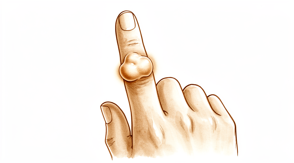
Những triệu chứng bạn đang gặp phải
Hầu hết mọi người nhận thấy một khối u không đau trên ngón tay hoặc ngón cái. Khối u thường chắc và phát triển chậm. Một số khối u nằm ngay sát một khớp, thường là khớp gần đầu ngón tay nhất, và chúng có thể xuất hiện ở mặt mu, mặt bên hoặc mặt lòng của ngón tay. Ngón trỏ là vị trí hay gặp nhất.
Bản thân khối u hầu như luôn không gây đau. Khi có cảm giác khó chịu, nguyên nhân thường là do khối u chèn ép vào các cấu trúc lân cận khi nó to lên. Một số ít người cảm thấy ngứa ran hoặc tê ở ngón tay nếu khối u chèn ép vào dây thần kinh. Một số người thấy ngón tay bị vướng hoặc kêu lách cách khi cử động, hơi giống với chứng ngón tay cò súng, khi ngón tay bị kẹt lại trong chốc lát rồi bật ra. Nếu khối u nằm gần một khớp, việc gập hoàn toàn ngón tay đó có thể khó khăn vì khối u gây cản trở.
Các sinh hoạt hằng ngày có dùng đến ngón tay đó có thể trở nên vướng víu. Nhặt những đồng xu nhỏ, cài cúc áo, cầm bút hoặc vặn nắp lọ có thể trở nên lóng ngóng nếu khối u nằm đúng chỗ bạn cầm nắm. Khối u cũng có thể bị vướng vào găng tay hoặc túi áo, túi quần.
Khối u thường đã có từ lâu trước khi nó gây phiền toái đến mức bạn đi hỏi bác sĩ. Nhiều người chờ hàng tháng hoặc hàng năm, theo dõi nó thay đổi từ từ.
Có một vài điều bạn nên biết về tình trạng này. Khối u lành tính, nghĩa là nó không lan sang các bộ phận khác của cơ thể. Tình trạng này gặp ở phụ nữ nhiều hơn nam giới một chút, và thường xuất hiện trong độ tuổi từ 32 đến 51. Hầu hết mọi người chỉ có một khối u, dù một số người có nhiều hơn một khối u ở các vị trí khác nhau.
Hãy để ý nếu ngón tay hoặc bàn tay trở nên nóng, đỏ, sưng và đau, đặc biệt là kèm theo sốt. Nếu điều đó xảy ra, hãy đến khoa cấp cứu ngay trong ngày. Nếu khối u tiếp tục to lên, bắt đầu gây đau, hoặc ngón tay không hoạt động bình thường, hãy đến gặp bác sĩ đa khoa hoặc đề nghị được bác sĩ chuyên khoa thăm khám.
Chuyện gì đang xảy ra thực sự
Gân là những sợi dây chắc khỏe nối cơ với xương và giúp các ngón tay gập lại. Mỗi gân chạy bên trong một lớp vỏ bọc mỏng gọi là bao gân, hoạt động hơi giống một vòng đệm bao quanh sợi dây thừng, giữ cho sợi dây trơn để nó có thể trượt khi ngón tay cử động.
Trong tình trạng này, các tế bào trong lớp vỏ bọc đó bắt đầu phát triển nhiều hơn mức bình thường. Kết quả là một khối u mô mềm nằm trên hoặc bên cạnh gân, thường ở gần một khớp. Khối u lành tính, nghĩa là nó không lan rộng, và phát triển chậm. Hầu hết các khối u đều nhỏ, trung bình khoảng 1.35 cm, dù một số có thể to hơn.
Khối u được tạo thành từ một vài thành phần tập trung lại với nhau: các tế bào viêm, các tế bào khổng lồ (đơn giản là những tế bào có nhiều nhân), và một sắc tố sẫm màu gọi là hemosiderin, cũng chính là chất tạo nên màu của một vết bầm tím cũ. Sắc tố đó là lý do những khối u này có thể trông hơi nâu nếu được nhìn thấy trong lúc phẫu thuật.
Các triệu chứng bạn nhận thấy ở phần trên là do khối u gây cản trở. Khi to lên, nó có thể chèn ép vào gân, khớp hoặc dây thần kinh, điều này giải thích hiện tượng vướng, cứng và đôi khi ngứa ran. Phần lớn thời gian khối u hoàn toàn không gây đau, và chỉ khoảng 16 trên 100 người cho biết có đau.
Một vài điểm bạn nên biết. Hầu hết mọi người chỉ có một khối u, nhưng khoảng 21 trên 100 người có từ hai khối u riêng biệt trở lên. Khối u thường nằm cạnh một khớp, và trong khoảng một phần ba các trường hợp ở ngón tay, nó nằm ở khớp gần đầu ngón tay nhất. Rất hiếm khi khối u có thể ấn lõm vào chính xương, và điều này sẽ thấy được trên phim X-quang.
Ngoài ra còn có một dạng hiếm gặp hơn, lan tỏa hơn của cùng tình trạng này, lan dọc theo gân thành những nhánh mỏng thay vì tạo thành một khối u gọn gàng. Dạng này ít gặp hơn nhiều ở ngón tay và có xu hướng diễn tiến khác, đó là lý do bác sĩ phẫu thuật sẽ muốn biết rõ chính xác đây là loại khối u nào trước khi lên bất kỳ kế hoạch nào.
Những biện pháp chúng tôi có thể áp dụng
Bác sĩ Kieran Hirpara, bác sĩ phẫu thuật chi trên tại Bệnh viện tư nhân Mater Rockhampton, sẽ bắt đầu bằng các phương pháp ít xâm lấn nhất phù hợp với tình trạng của bạn. Thông thường, bệnh nhân được bác sĩ đa khoa giới thiệu đến phòng khám chúng tôi; nếu nhà vật lý trị liệu khuyên bạn nên gặp chúng tôi, bạn vẫn cần có giấy giới thiệu từ bác sĩ đa khoa để được hưởng mức hoàn trả từ Medicare. Tại phòng khám, chúng tôi sẽ hỏi bệnh sử, khám bàn tay và chỉ định các phương pháp chẩn đoán hình ảnh khi cần thiết để xác định chính xác đây là loại khối u nào.
Vì khối u này lành tính và phát triển chậm, không làm gì cả cũng là một lựa chọn thực sự. Đối với một khối u nhỏ, không đau, chỉ cần theo dõi có thể là lựa chọn đúng. Nếu khối u gây cản trở, trị liệu tay có thể giúp cải thiện tình trạng cứng và vướng. Trị liệu nhằm giữ cho ngón tay tiếp tục cử động và giúp các sinh hoạt hằng ngày dễ dàng hơn. Thông thường, chúng tôi khuyên bạn nên thử phương pháp này trong một thời gian đầy đủ trước khi nghĩ đến phẫu thuật.
Không có loại thuốc nào làm khối u này teo nhỏ lại, vì vậy thuốc giảm đau và thuốc chống viêm chỉ làm dịu cảm giác khó chịu do khối u chèn ép vào mô lân cận. Chúng không làm khối u nhỏ đi.
Phẫu thuật là phương pháp điều trị thông thường khi khối u lớn, gây đau, hoặc khiến ngón tay không hoạt động bình thường. Mục tiêu là lấy bỏ toàn bộ khối u cùng với chỗ bám của nó trên vỏ bọc gân, vì chính việc để sót lại chỗ bám đó là điều khiến khối u có thể tái phát. Chúng tôi lên kế hoạch cẩn thận trước khi mổ, sử dụng kính phóng đại trong khi phẫu thuật, và lấy bỏ hoàn toàn khối u. Tái phát là nguy cơ chính của tình trạng này, và thường xảy ra trong vòng 36 tháng sau khi cắt bỏ. Trong một nhóm 605 người được cắt bỏ khối u ở ngón tay, 14.8% bị tái phát. Nói cách khác: hầu hết mọi người, khoảng 85 trên 100 người, không bị tái phát. Nếu khối u tái phát, việc cắt bỏ lại kèm theo các bước bổ sung để hạn chế khối u mọc lại vẫn có thể giải quyết được vấn đề.
Những điều bạn có thể mong đợi
Tiên lượng của khối u này ổn định chứ không có thay đổi đột ngột. Khối u lành tính, vì vậy nó không lan đến bất kỳ nơi nào khác trong cơ thể bạn. Nếu để yên, một khối u nhỏ không đau thường tiếp tục to lên từ từ, hoặc đơn giản là giữ nguyên như vậy. Nó hiếm khi tự biến mất, và không biến thành bất cứ thứ gì nguy hiểm.
Điều chính mà tình trạng này gây ra theo thời gian là tái phát sau khi cắt bỏ. Tái phát là nguy cơ hàng đầu của khối u này, và thường xảy ra trong vòng 36 tháng sau khi cắt bỏ. Một số khối u dễ tái phát hơn những khối u khác: những khối u gồm từ hai phần riêng biệt trở lên không dính liền với nhau, và những khối u đã phát triển vào chính gân hoặc lớp màng lót khớp. Khi toàn bộ khối u được lấy bỏ cùng với vị trí bám của nó, nguy cơ đó giảm đi rất nhiều.
Chức năng sau điều trị thường tốt. Những người đã được cắt bỏ một khối u loại này ở ngón tay đạt, tính trung bình, 92% chức năng chi bình thường theo một thang đo tiêu chuẩn đánh giá hoạt động của chi. Trong các trường hợp được báo cáo, người bệnh giữ được vận động hoàn toàn của ngón tay, và hình dạng xương ngón tay trên phim X-quang đã trở lại bình thường vào lần kiểm tra ở mốc hai năm.
Nếu khối u thuộc dạng hiếm gặp hơn, lan rộng hơn, bao quanh gân thành những nhánh, thì việc cắt bỏ hoàn toàn khó hơn và khối u dễ tái phát hơn. Trong những trường hợp đó, xạ trị nhắm vào vùng này có thể giúp kiểm soát khối u trong khi vẫn giữ cho bàn tay hoạt động.
Nếu bạn chọn chỉ theo dõi một khối u nhỏ, không đau, bức tranh thực tế là một khối u phát triển chậm hoặc giữ nguyên, và có thể dần dần khiến việc gập ngón tay hoặc dùng ngón tay cho các thao tác nhỏ trở nên vướng víu hơn. Nếu khối u bắt đầu gây đau, tiếp tục to lên, hoặc ngón tay không còn hoạt động bình thường, đó là lúc việc điều trị thường trở nên xứng đáng.
Khi nào nên đi khám bác sĩ
Hầu hết các khối u như thế này không khẩn cấp, và việc điều trị không bao giờ cần vội vàng. Hãy đến gặp bác sĩ đa khoa nếu khối u không thuyên giảm, to lên trong vài tuần, bắt đầu gây đau, hoặc khiến bạn không thể sử dụng ngón tay hay bàn tay bình thường. Hãy đề nghị được bác sĩ chuyên khoa thăm khám nếu ngón tay bị vướng hoặc kêu lách cách khi cử động, nếu bạn nhận thấy ngứa ran hoặc tê, hoặc nếu việc gập hoàn toàn ngón tay trở nên khó khăn vì khối u gây cản trở. Đây là những điều nên được kiểm tra sớm chứ đừng để muộn, vì một khối u đã phát triển vào gân hoặc lớp màng lót khớp sẽ khó cắt bỏ hoàn toàn hơn và dễ tái phát hơn.
Có một nhóm dấu hiệu cần được chăm sóc ngay trong ngày. Nếu ngón tay hoặc bàn tay của bạn trở nên nóng, đỏ, sưng và đau, đặc biệt là kèm theo sốt, hãy đến khoa cấp cứu. Bạn không cần giấy giới thiệu của bác sĩ đa khoa trước. Nếu bạn không thể liên lạc với phòng khám ngoài giờ làm việc hoặc vào cuối tuần, hãy đến khoa cấp cứu gần nhất.
Phân tích sâu hơn
Advanced reading: the deeper science (optional)
Phần này đi sâu hơn mức cần thiết cho việc đưa ra quyết định điều trị của bạn. U tế bào khổng lồ ở màng bao gân là một chủ đề đáng để tìm hiểu thêm, bởi vấn đề cốt lõi của nó là khả năng tái phát; các bằng chứng cho thấy việc tái phát này chịu ảnh hưởng nhiều hơn từ đặc tính sinh học của khối u ở từng bệnh nhân, chứ không phải từ cách thức cắt bỏ nó.
Tái phát là đặc tính của khối u, chứ không chỉ là hậu quả của phẫu thuật
Lý giải thông thường cho việc khối u tái phát là do vẫn còn sót lại một phần khối u sau phẫu thuật. Tuy nhiên, một phân tích hệ thống trên 605 ca bệnh lại cho kết quả ngược lại: các đặc tính sinh học nội tại của khối u dường như đóng vai trò quan trọng hơn trong việc gây tái phát so với vị trí khối u hay mức độ xâm lấn tại chỗ. Các tác giả cũng kêu gọi tiến hành thêm các nghiên cứu tiền cứu quy mô lớn nhằm xác định những loại khối u nào có nguy cơ tái phát [1].
Đây là thông tin vô cùng hữu ích mà bệnh nhân cần biết trước khi phẫu thuật. Việc khối u tái phát sau một ca phẫu thuật được thực hiện đúng kỹ thuật là đặc tính tự nhiên của loại khối u này; điều đó không đồng nghĩa với việc có điều gì đó đã diễn ra sai sót trong quá trình phẫu thuật.
Việc điều trị là tùy chọn; việc không điều trị cũng là một lựa chọn hợp lý
Khi đã lên kế hoạch phẫu thuật, người ta dễ dàng quên mất điều này. Nguyên tắc điều trị ưu tiên là cắt bỏ hoàn toàn khối u, nhưng việc điều trị không bao giờ mang tính cấp cứu; chỉ định điều trị cần được cân nhắc dựa trên các triệu chứng, tốc độ phát triển của khối u, vị trí và hoàn cảnh cá nhân của bệnh nhân [4].
Đối với những khối u nhỏ, không gây đau đớn và phát triển chậm, việc theo dõi là một lựa chọn hợp lý. Khối u này lành tính và không di căn; vì vậy lý do để phẫu thuật chỉ liên quan đến chức năng vận động, kích thước và mức độ gây phiền toái – chứ không phải nguy cơ nguy hiểm.
Tuy nhiên, hai yếu tố phẫu thuật vẫn có vai trò quan trọng
Yếu tố sinh học không phải là tất cả. Trên 941 bệnh nhân mắc u khổng lồ màng gân dạng khu trú, các yếu tố liên quan đến nguy cơ tái phát sau phẫu thuật cắt bỏ là kích thước khối u lớn và việc điều trị ban đầu bằng phương pháp nội soi khớp. Với tỷ lệ biến chứng tương đối thấp cùng kết quả chức năng tốt, các tác giả khuyến nghị nên áp dụng phương pháp mổ mở kết hợp cắt bỏ triệt để khi có thể nhằm giảm nguy cơ tái phát ở những ca có nguy cơ cao [2].
Tuy nhiên, theo một nghiên cứu tổng quan trên 1,448 bệnh nhân, phương pháp cắt bỏ qua nội soi khớp cũng cho hiệu quả tốt đối với dạng u khu trú ở bốn khớp khác nhau; còn đối với dạng u lan tỏa, phương pháp nội soi cắt bỏ màng khớp chỉ cho kết quả khả quan tại khớp gối mà thôi [3].
Để dung hòa hai quan điểm này: đối với các tổn thương nhỏ, khu trú và ranh giới rõ ràng, cả hai phương pháp đều có thể áp dụng. Khi kích thước khối u tăng lên hoặc ở dạng u lan tỏa, phương pháp cắt bỏ mở triệt để mới là lựa chọn phù hợp hơn. Lý do nằm ở yếu tố cơ học: khối u này phát triển thành các nhánh quanh gân, dây thần kinh và khớp; những vùng dễ bị bỏ sót nhất lại là những vùng nằm phía sau các cấu trúc cần được nâng lên và kiểm tra trực tiếp.
Tại sao việc đặt tên lại quan trọng
Hiện nay, tình trạng này được gọi là u tế bào khổng lồ màng gân, thuật ngữ này bao gồm cả dạng khu trú ở bàn tay và dạng lan tỏa trong khớp vốn trước đây được gọi là viêm màng hoạt dịch sắc tố nốt hạt ⟨[4]⟩. Đây là cùng một bệnh lý nhưng xuất hiện ở các vị trí và có kiểu phát triển khác nhau.
Bạn nên biết điều này khi tìm hiểu thêm về bệnh, vì các tài liệu tìm được thường nói về bệnh ở khớp gối và khớp háng – tức là dạng lan tỏa của bệnh – và tỷ lệ tái phát đối với dạng này cao hơn nhiều so với các tổn thương khu trú ở ngón tay. Việc áp dụng các con số liên quan đến khớp gối cho một khối u ở ngón tay sẽ làm phóng đại mức độ nguy cơ thực tế.
Vai trò của xạ trị
Đối với các trường hợp bệnh lan tỏa tái phát hoặc không thể cắt bỏ hoàn toàn, đôi khi người ta cân nhắc sử dụng xạ trị hỗ trợ. Một phân tích tổng hợp cho thấy rằng việc cắt bỏ màng hoạt dịch mở hoặc kết hợp cắt bỏ màng hoạt dịch với xạ trị trong giai đoạn phẫu thuật giúp giảm tỷ lệ tái phát ở bệnh nhân mắc viêm màng hoạt dịch sần sùi sắc tố lan tỏa; tuy nhiên cần có các nghiên cứu tiền cứu quy mô lớn, dài hạn để xác nhận điều này [5].
Đối với các khối u khu trú ở ngón tay – chiếm đa số các ca bệnh ở vùng tay – thì vấn đề này không xảy ra. Loại bệnh này thuộc nhóm bệnh lan tỏa, tái phát và liên quan đến khớp; chúng tôi chỉ đề cập đến nó ở đây vì việc tìm kiếm theo tên bệnh sẽ cho ra kết quả liên quan.
Điều này không phải là gì
Mặc dù có tên gọi như vậy, đây là một khối u lành tính. Nó không di căn sang các bộ phận khác của cơ thể. Từ “khối u” ở đây mang ý nghĩa nặng nề hơn thực tế; mối lo ngại về khả năng tái phát chỉ liên quan đến việc phải phẫu thuật nhiều lần tại chỗ, tình trạng cứng khớp và sự gần gũi giữa khối u với các dây thần kinh, chứ không phải là ung thư.
Tài liệu tham khảo
[1] Fotiadis E, Papadopoulos A, Svarnas T, Akritopoulos P, Sachinis NP, Chalidis BE. U hạt khổng lồ màng bao gân các ngón tay: Một bài tổng quan có hệ thống. Hand (N Y). 2011;6(3):244-9. https://doi.org/10.1007/s11552-011-9341-9
[2] Mastboom M, Staals E, Verspoor F, Rueten-Budde A, Stacchiotti S, Palmerini E, và cộng sự. Điều trị phẫu thuật u hạt khổng lồ màng bao gân dạng khu trú ở các khớp lớn: Nghiên cứu dựa trên cơ sở dữ liệu tổng hợp từ 31 trung tâm điều trị sarcoma quốc tế. J Bone Joint Surg Am. 2019;101(14):1309-18. https://doi.org/10.2106/JBJS.18.01147
[3] Noailles T, Brulefert K, Briand S, Longis P, Andrieu K, Chalopin A, và cộng sự. U hạt khổng lồ màng bao gân: Phẫu thuật mở hay nội soi cắt màng bao gân? Một bài tổng quan có hệ thống về tài liệu y học. Orthop Traumatol Surg Res. 2017;103(5):809-14. https://doi.org/10.1016/j.otsr.2017.03.016
[4] Gouin F, Noailles T. Các dạng khu trú và lan tỏa của u hạt khổng lồ màng bao gân (trước đây gọi là u hạt khổng lồ màng bao gân và viêm màng hoạt dịch sợi nốt sắc tố). Orthop Traumatol Surg Res. 2017;103(1):S91-S97. https://doi.org/10.1016/j.otsr.2016.11.002
[5] Mollon B, Lee A, Busse JW, Griffin AM, Ferguson PC, Wunder JS, và cộng sự. Ảnh hưởng của việc cắt màng bao gân qua phẫu thuật và xạ trị đối với tỷ lệ tái phát của viêm màng hoạt dịch sợi nốt sắc tố ở khớp gối. Bone Joint J. 2015;97-B(4):550-7. https://doi.org/10.1302/0301-620X.97B4.34907
Evidence & references
This is the clinical evidence summary written for health professionals. It is technical, and it lists the research this page was built from. You do not need to read it to understand your treatment or to make a decision about it.
Overview
- The diagnosis and treatment of suspected hand neoplasms should proceed in an orderly fashion similar to accepted methods applied to tumors in other locations of the extremities [1].
- Giant cell tumors of the tendon sheaths in the hand are benign lesions where recurrence is the primary risk [3, 4].
- Recurrence of giant cell tumors of the tendon sheaths typically occurs within 36 months of excision [3].
- The fundamental consideration in the management of hand tumours is to reconcile adequate tumour clearance with the maintenance of function and appearance [20].
- Complete surgical resection remains the treatment of choice for most patients with tenosynovial giant cell tumors [15].
- Diffuse tenosynovial giant cell tumor disease presents challenges due to high recurrence rates [15].
- Curettage, cryosurgery, and cementation performed by experienced surgeons appears to be a safe, effective, and reliable method for treating selected giant cell tumors of the hand [5].
- In cases of infiltrative giant cell tumor of the tendon sheath, radiation therapy may provide local tumor control with preservation of hand function [12].
- En bloc resection of the tumour and reconstruction with a fibular graft where necessary should be considered as the treatment of choice in giant cell tumour of the hand [10].
- En bloc resection of the tumour and reconstruction with an autograft should be considered in the treatment of recurrent giant cell tumour of the hand [8].
- En bloc resection and matched nonvascularized toe phalangeal transfer for Campanacci Grade 2 or 3 giant cell tumor of the phalanges resulted in a functional tumor-free digit with a low complication rate and no recurrences [35].
- Well-designed studies combining the recurrence rates of several hand surgery centers implementing a standardized treatment are needed to better demonstrate the associated risk factors for recurrence [28].
Anatomy & Pathophysiology
Clinical Presentation and Demographics
- Giant cell tumour of tendon sheath (GCTTS) is the second most common benign proliferative tumour in the upper extremities after ganglion cysts [102].
- GCTTS are usually slow-growing, painless, benign soft tissue tumours [102].
- GCTTS are most commonly found in the fingers and among women in their fourth and fifth decades [102].
- The male-to-female ratio for GCTTS of the digits is 1:1.47 [23].
- The mean age range for GCTTS of the digits is 32 to 51 years [23].
- Pain was reported in 15.7% of GCTTS cases and sensory disturbances in 4.57% of cases [23].
- A definite history of trauma was recorded in 5% of GCTTS lesions [23].
- The most frequent tumour location for GCTTS of the digits is the index finger (29.7%) [23].
- In a series of 64 cases, the most frequent location for GCTTS was the long finger (23.5%), followed by the thumb, index finger, and hand (20.3% each) [49].
- Patients with GCTTS arising in the fingers complained of a painless mass in almost every instance [24].
- The duration of symptoms for finger GCTTS ranged from two weeks to fifteen years, with an average duration of thirty-eight months [24].
- GCTTS are usually firm, lobulated, and non-tender masses [24].
- As the tumour grows, patients may present with swelling, pain, and limitation of movement [102].
Anatomical Distribution and Morphology
- Fifty-three xanthomas (GCTTS) arose adjacent to joints in the fingers, whereas twenty-nine bore no relation to joints [24].
- Thirty-one of ninety-one finger GCTTS occurred at the distal joints [24].
- Of the thirty-one distal joint GCTTS, eighteen were on the dorsal aspect and thirteen were distributed evenly on the radial, ulnar, and volar aspects [24].
- Type I GCTTS (single lesions) were more frequently detected (78.7%) than Type II tumours (two or more distinct tumours that were not joined together) (21.3%) [23].
- Type 1 GCTTS includes a nodular or multinodular lesion surrounded by a capsule, while Type 2 describes tumours with no connective tissue membrane and satellite, diffuse, or multicentric nodules [102].
- The average size of GCTTS was 1.35 cm, with a range of 0.3 cm to 5 cm [49].
- Bone erosion was found in 3 patients (4.7%) in a series of 64 GCTTS cases [49].
- Tendon involvement with flexors/extensors ratio of 4:3 was found in 7 cases (10.9%) in a series of 64 GCTTS cases [49].
- Involvement of the neurovascular bundle was presented in 7 patients (10.9%) in a series of 64 GCTTS cases [49].
- A case report describes the first patient with three separate GCTTS developing from both flexor and extensor tendons in a single digit [7].
Histology and Pathogenesis
- Microscopically, all GCTTS contained multinucleated giant cells, histiocytes, and haemosiderin deposits [49].
- GCTTS originates from the synovial membrane, tendon sheath, or synovial bursa [102].
- Inflammation resulting from reactive or regenerative hyperplasia is the generally accepted theory of pathogenesis for GCTTS [102].
- Genetic factors have been observed in previous studies regarding GCTTS pathogenesis [102].
- Fibroma of tendon sheath is histologically distinct from giant cell tumor of tendon sheath, a lesion with which it is commonly confused [16].
Diagnostic Imaging
- Radiography can be helpful in evaluating cortical destruction but is not helpful in the definitive diagnosis of GCTTS [102].
- Magnetic resonance imaging (MRI) is the most useful examination for the diagnosis and treatment planning of GCTTS [102].
- Al-Qattan classified GCTTS with MRI into two types according to the risk of recurrence [102].
Classification
- Giant cell tumors of the synovial sheaths in the hand are benign lesions in which recurrence is the primary risk [4].
- Recurrence for giant cell tumors of the tendon sheaths typically occurs within 36 months of excision [3].
- The Al-Qattan classification divides giant cell tumours of tendon sheath into two main types based on whether the entire tumour is surrounded by one pseudocapsule as assessed by the surgeon during surgery [90].
- Type I tumours are defined as those where the entire tumour is surrounded by one pseudocapsule [90].
- Type II tumours are defined as those where the entire tumour is not surrounded by one pseudocapsule [90].
- Type I tumours are sub-classified according to the thickness of the capsule, lobulation of the tumour, the presence of satellite lesions, and the diffuse or multicenteric nature of the tumour [90].
- Type II tumours are sub-classified according to the thickness of the capsule, lobulation of the tumour, the presence of satellite lesions, and the diffuse or multicenteric nature of the tumour [90].
- In a prospective study of 43 consecutive cases, none of the type I tumours (n=30) recurred [90].
- In a prospective study of 43 consecutive cases, recurrence occurred in five out of 13 type II tumours [90].
- Second recurrences were seen with type II B and C tumours, but not type II A tumours [90].
- Type I tumours (single lesions) were more frequently detected (78.7%) than type II tumours (two or more distinct tumours that were not joined together) (21.3%) in a systematic review of 605 patients [23].
- Type II tumours were associated with higher recurrence rates compared to Type I tumours in a systematic review of 605 patients [23].
- Direct involvement of the extensor tendons, flexor tendons, or joint capsule puts patients in a high-risk category with respect to recurrence [43].
- Preoperative diagnosis and meticulous surgical technique were found to be the only predictive factor of recurrence in a series of 14 cases [34].
- The overall recurrence rate in a systematic review of 605 patients was 14.8% [23].
Clinical Presentation
- Patients with fibrous xanthomas arising in the fingers complained of a painless mass in almost every instance [24].
- The duration of symptoms for finger tumors ranged from two weeks to fifteen years, with an average duration of thirty-eight months [24].
- The tumors were usually firm, lobulated, and non-tender [24].
- Fifty-three xanthomas arose adjacent to joints in the fingers, whereas twenty-nine bore no relation to joints [24].
- Thirty-one of the ninety-one tumors in the fingers occurred at the distal joints [24].
- Eighteen of the thirty-one distal joint tumors were on the dorsal aspect of the joint, and thirteen were distributed about evenly on the radial, ulnar, and volar aspects of the joint [24].
- The most frequent tumour location for giant cell tumour of tendon sheath of the digits was the index finger (29.7%) [23].
- Pain was reported in 15.7% of cases and sensory disturbances in 4.57% of cases [23].
- A definite history of trauma was recorded in 5% of lesions [23].
- Type I tumours (single lesions) were more frequently detected (78.7%) than type II tumours (two or more distinct tumours that were not joined together) (21.3%) [23].
- The male-to-female ratio for giant cell tumour of tendon sheath of the digits was 1:1.47 [23].
- The mean age for giant cell tumour of tendon sheath of the digits ranged from 32 to 51 years [23].
- A tendon tumour can present as a trigger finger [18].
- Trigger wrist caused by a giant cell tumour of tendon sheath has been reported [26].
- The trigger phenomenon in trigger wrist caused by a giant cell tumour of tendon sheath probably occurred when the tumour entered and left the distal carpal tunnel [26].
- Fibroma of tendon sheath is a benign soft tissue tumor that has a predilection for the hand [16].
- Fibroma of tendon sheath may be more common than has been previously recognized [16].
- The differential diagnosis of a soft tissue tumor in the digits and hand is usually limited to a ganglion, an inclusion cyst, or a giant cell tumor of tendon sheath (xanthofibroma) [16].
- Fibroma of tendon sheath should be included in the differential diagnosis of a soft tissue tumor in the digits and hand [16].
- A case of fibroma of tendon sheath presented as a 6-year history of a slowly enlarging palmar mass of the left index finger [16].
- Examination of a fibroma of tendon sheath case demonstrated a soft, compressible, lobulated, nontender mass extending from the second web space to the tip of the finger [16].
- Flexion of the digit was limited by the bulge in a case of fibroma of tendon sheath [16].
- The unusual location of a fibroma of the tendon sheath and atypical nature of the triggering phenomenon can lead to it being reported as a rare cause of median nerve compression at the wrist [50].
- Giant cell tumor of tendon sheath and pigmented villonodular synovitis are benign, locally invasive tumors involving synovium, tendon sheaths, and bursae [47].
- Patients with giant cell tumor of tendon sheath or pigmented villonodular synovitis may present with a discrete mass or with joint swelling, pain, or locking or catching [47].
- The knee and digits of the hand are the most common locations of lesions for giant cell tumor of tendon sheath and pigmented villonodular synovitis [47].
- Multifocal lesions of giant cell tumor of tendon sheath and pigmented villonodular synovitis are rare [47].
- The authors report the uniqueness of a case of multicentric giant cell tumor in the upper extremity [9].
- Radiological changes in the form of bony indentation was seen in only 2 cases of giant cell tumor of tendon sheath in a series of 12 patients [34].
- The most common presentation for giant cell tumor of tendon sheath in a series of 12 patients was with a mass over the hand, with a predilection to the thumb [34].
- A 65-year-old man presented with a slowly enlarging mass on the dorsal aspect of the left thumb consistent with a recurrent giant cell tumor of the tendon sheath [33].
Investigations
- The differential diagnosis of a soft tissue tumor in the digits and hand is usually limited to a ganglion, an inclusion cyst, or a giant cell tumor of tendon sheath [16].
- Fibroma of tendon sheath should be included in the differential diagnosis as it may be more common than has been previously recognized [16].
- Fibro-osseous pseudotumor of the digit should be considered in the differential diagnosis of rapidly enlarging digital lesions [64].
- Although MRI findings and location might help in the diagnosis of tenosynovial giant cell tumors, careful assessment is mandatory, especially in unusual locations [129].
Treatment
General Principles
Surgical Excision
- Complete surgical resection remains the treatment of choice for most patients with tenosynovial giant cell tumors, though diffuse disease presents challenges due to high recurrence rates [15].
- En bloc resection of metacarpal giant cell tumors with reconstruction by iliac graft appears to be an effective treatment [14].
- En bloc resection and matched nonvascularized toe phalangeal transfer resulted in a functional tumor-free digit with a low complication rate and no recurrences for Campanacci Grade 2 or 3 giant cell tumors of the phalanges [35].
- Total surgical excision ensuring removal of the attachment site (flexor sheath/palmar fascia) resulted in no local recurrences at a mean follow-up of 3.2 years for fibroma of tendon sheath [11].
- Local excision of the tumour appears to be a satisfactory method of treatment for Nora’s lesion [109].
- Preoperative planning aided by a tissue diagnosis with fine needle aspiration cytology, wide surgical exposure, and meticulous dissection with help of magnification are imperative for a successful outcome in giant cell tumor of tendon sheath [34].
- In all cases of giant cell tumor of tendon sheath, magnifying loupe or operating microscope was used during surgical excision [49].
- In cases of infiltrative giant cell tumor of tendon sheath, radiation therapy may provide local tumor control with preservation of hand function [12].
Recurrence and Adjuvant Therapy
- Giant cell tumors of the tendon sheaths in the hand are benign lesions where recurrence is the primary risk, typically occurring within 36 months of excision [3].
- In a study of 64 cases of giant cell tumor of tendon sheath, the recurrence rate was 4.7% (n=3) [49].
- In a systematic review of 605 patients with giant cell tumor of tendon sheath of the digits, 14.8% of patients had tumour recurrence [23].
- Repeated curettage with adjuvants eventually resulted in the cure for all patients and is therefore a reasonable treatment for both primary and recurrent giant cell tumor of the small bones of the hands and feet [105].
- Intralesional excision with cautery and methylmethacrylate provides a reliable method of treatment of giant cell tumors with good long-term functional results [71].
- Intralesional excision with local adjuvant therapy is recommended for the treatment of giant cell tumor of bone because it results in a good functional outcome compared to extralesional excision [98].
- Intralesional excision remains a viable, and likely the standard, mode of treatment for most giant cell tumors of the distal radius unless there is extensive bone loss [29].
- Based on data obtained from the number of studies available, intralesional excision appears to be more appropriate for the treatment of local lesions (eg, Grades 1 and 2) than Grade 3 giant cell tumors of the distal radius [52].
- Well-designed studies combining the recurrence rates of several hand surgery centers implementing a standardized treatment are needed to better demonstrate the associated risk factors for recurrence in giant cell tumor of tendon sheath [28].
Reconstruction and Resection Options
- Both curettage and resection/amputation are acceptable treatment options for the rare condition of giant cell tumour of bone in the hand, with a need to individualize treatment decisions based on the site and extent of disease to minimize treatment morbidity while maximizing disease control [6].
- Aggressive and malignant bone tumors of the second through fifth metacarpals generally require en bloc bone excision [17].
- Ray excision is usually best for lesions of the second and fifth metacarpals [17].
- If bone graft has been used for reconstruction, carpometacarpal arthrodesis and MCP ligament reconstruction or silicone arthroplasty may be practical [17].
- Whatever level is chosen for reconstruction, it is essential that an adequate, safe margin of normal tissue first be excised en bloc with the tumor [17].
- Aggressive tumors of the second through fifth metacarpals that have invaded soft tissue or sustained pathologic fractures often require excision of not only the involved ray but also the contiguous ray or rays radial and ulnar to the involved digit [17].
- If a malignant tumor has broken into the midpalm and extends across the metacarpals, removal of all digital rays may be needed to gain an adequate soft tissue margin [17].
- Retention of a sensate and relatively mobile thumb may be considerably more esthetic and functionally satisfactory than a forearm- or wrist-level amputation [17].
- If a tumor extends proximally from the metacarpal level, a more proximal level of hand, wrist, or forearm amputation is required for safe tumor management [17].
- Malignant soft tissue tumors in the palm or carpal tunnel often require at least partial hand amputation [17].
- If treated by only local or limited excision, the chance of recurrence is extremely high particularly in the setting of a positive margin for malignant soft tissue tumors in the palm or carpal tunnel [17].
- Below-elbow amputation is necessary to treat larger tumors of the palm or carpal tunnel [17].
- Wide en bloc excision of soft tissue sarcomas with negative margins is required to achieve local control of the lesion [17].
- At a minimum, aggressive soft tissue tumors, such as bone tumors, require ray resection or removal of multiple rays [17].
- Central palmar lesions more likely require sacrifice of three rays; those on the border are more likely than those in the center to be salvageable by removing just two rays [17].
- In the presence of proximal, broader, and larger lesions, all four digits or the entire hand may have to be sacrificed to save the patient [17].
- The use of a massive biocompatible bipolar unconstrained prosthesis is a viable treatment option for distal radius reconstruction after en-bloc resection of a giant cell tumour, offering rapid functional improvement without donor-site morbidity [68].
- Reconstruction after wide excision by nonvascularized fibular graft is a viable alternative for giant cell tumors of the lower end of radius though it is a challenging procedure and may be accompanied by major complications [111].
- En bloc resection is recommended for aggressive giant cell tumours (GCT) of the bone involving the distal end of the radius to minimize the risk of recurrence [91].
- The distal ulna may be widely resected with or without stabilization of the residual ulnar stump, yielding satisfactory local disease control and functional outcome [48].
- This is a simple and effective modality of reconstruction after resection of distal radial tumors using centralization of the ulna [30].
Complications
- Recurrence is the primary risk associated with giant cell tumors of the tendon sheaths in the hand [3].
- The overall recurrence rate for giant cell tumour of tendon sheath of the digits is 14.8% [23].
- Type II tumours (two or more distinct tumours that were not joined together) are associated with a higher risk of recurrence compared to Type I tumours [23].
- Direct involvement of the extensor tendons, flexor tendons, or joint capsule places patients in a high-risk category for recurrence [43].
- Surgical treatment of pigmented villonodular synovitis led to good functional results with an average Enneking score of 92% of normal limb function [32].
- A tendon tumour arising from the superficialis tendon can present as a trigger finger [18].
- A giant cell tumour of the tendon sheath can cause a trigger wrist phenomenon when the tumour enters and leaves the distal carpal tunnel [26].
- A fibroma of the tendon sheath in an unusual location can cause a triggering phenomenon and median nerve compression at the wrist [50].
- Total surgical excision ensuring removal of the attachment site (flexor sheath/palmar fascia) for fibroma of tendon sheath resulted in no local recurrences at a mean follow-up of 3.2 years [11].
- Patients with giant cell tumor of bone in the hand who are at higher risk of recurrence should be clinically followed more closely [41].
- Both curettage and resection/amputation are acceptable treatment options for giant cell tumour of bone in the hand, with a need to individualize treatment decisions based on the site and extent of disease to minimize treatment morbidity while maximizing disease control [6].
- Below-elbow amputation is necessary to treat larger malignant soft tissue tumors in the palm or carpal tunnel [17].
- Patients who received preoperative radiotherapy for hand tumors had a decrease in functional outcomes and grip strength that averaged 66% compared with the contralateral hand [55].
- None of the patients who underwent single-ray amputation for tumors of the hand, including giant cell tumors of bone, had local recurrences [55].
- A 29-year-old patient with recurrent giant-cell tumor of the digit was treated by phalangeal excision and toe phalanx transplant [54].
Recovery
- Recurrence of giant cell tumors of the tendon sheaths in the hand typically occurs within 36 months of excision [3].
- Total surgical excision ensuring removal of the attachment site (flexor sheath/palmar fascia) resulted in no local recurrences at a mean follow-up of 3.2 years [11].
- In cases of infiltrative GCTTS, radiation therapy may provide local tumor control with preservation of hand function [12].
- At 2-year final follow-up examination, no recurrence was noted, finger range of motion was full, and the x-ray film contour of the phalanx had returned to normal [13].
- Surgical treatment led to good functional results with an average Enneking score of 92% of normal limb function [32].
- En bloc resection and matched nonvascularized toe phalangeal transfer resulted in a functional tumor-free digit with a low complication rate and no recurrences [35].
Key Evidence
- [L5] The diagnosis and treatment of suspected hand neoplasms should proceed in an orderly fashion similar to accepted methods applied to tumors in other locations of the extremities. [1] (10.1016/s0363-5023(83)80277-9)
- [L4] Giant cell tumors of the tendon sheaths in the hand are benign lesions where recurrence is the primary risk, typically occurring within 36 months of excision. [3] (10.1016/j.otsr.2013.03.008)
- [L4] Giant cell tumors of the synovial sheaths in the hand are benign lesions in which recurrence is the primary risk. [4] (10.1016/j.jhsa.2013.08.051)
- [L4] Curettage, cryosurgery, and cementation performed by experienced surgeons appears to be a safe, effective, and reliable method for treating selected giant cell tumors of the hand. [5] (10.1053/jhsu.2001.22525)
- [L4] Both curettage and resection/amputation are acceptable treatment options for the rare condition of giant cell tumour of bone in the hand, with a need to individualize treatment decisions based on the site and extent of disease to minimize treatment morbidity while maximizing disease control. [6] (10.1177/17531934211007820)
- [L5] We believe this case report describes the first patient with three separate GCTTS developing from both flexor and extensor tendons in a single digit. [7] (10.1142/s2424835518720189)
- [L4] En bloc resection of the tumour and reconstruction with an autograft should be considered in the treatment of recurrent giant cell tumour of the hand. [8] (10.1016/s0266-7681(98)80197-6)
- [L5] The authors report the uniqueness of this case of multicentric giant cell tumor in the upper extremity. [9] (10.1016/j.main.2011.11.006)
- [L4] En bloc resection of the tumour and reconstruction with a fibular graft where necessary should be considered as the treatment of choice in giant cell tumour of the hand. [10] (10.1016/s0266-7681(96)80161-6)
- [L4] Total surgical excision ensuring removal of the attachment site (flexor sheath/palmar fascia) resulted in no local recurrences at a mean follow-up of 3.2 years. [11] (10.1177/1753193412469146)
- [L4] In cases of infiltrative GCTTS, radiation therapy may provide local tumor control with preservation of hand function. [12] (10.1016/j.jhsa.2012.01.011)
- [L5] At 2-year final follow-up examination, no recurrence was noted, finger range of motion was full, and the x-ray film contour of the phalanx had returned to normal. [13] (10.1016/s0363-5023(05)80151-0)
- [L5] En bloc resection of metacarpal giant cell tumors with reconstruction by iliac graft appears to be an effective treatment. [14] (10.1016/s1297-3203(03)00042-8)
- [L5] Complete surgical resection remains the treatment of choice for most patients with tenosynovial giant cell tumors, though diffuse disease presents challenges due to high recurrence rates. [15] (10.5435/jaaos-d-24-01255)
- [L5] [16] (10.1016/s0363-5023(84)80031-3)
- [L5] At operation a tumour arising from the superficials tendon was found and removed. [18] (10.1016/0266-7681(86)90283-4)
- [L5] The fundamental consideration in the management of hand tumours is to reconcile adequate tumour clearance with the maintenance of function and appearance. [20] (10.1016/0266-7681(91)90159-l)
- [L1] [23] (10.1007/s11552-011-9341-9)
- [L4] [24] (10.2106/00004623-196951010-00005)
- [L5] [26] (10.1016/s0266-7681(85)80038-3)
- [L3] Well-designed studies combining the recurrence rates of several hand surgery centers implementing a standardized treatment are needed to better demonstrate the associated risk factors for recurrence. [28] (10.1186/s12891-019-2866-8)
- [L3] Intralesional excision remains a viable, and likely the standard, mode of treatment for most giant cell tumors of the distal radius unless there is extensive bone loss. [29] (10.1007/s11999-014-4054-3)
- [L4] This is a simple and effective modality of reconstruction after resection of distal radial tumors. [30] (10.1016/j.jhsa.2022.05.011)
- [L4] Surgical treatment led to good functional results with an average Enneking score of 92% of normal limb function. [32] (10.1097/01.blo.0000224051.01873.fb)
- [L4] [33] (10.1016/j.jhsa.2012.11.001)
- [L4] [34] (10.1007/s12593-010-0020-9)
- [L4] En bloc resection and matched nonvascularized toe phalangeal transfer resulted in a functional tumor-free digit with a low complication rate and no recurrences. [35] (10.1016/j.jhsa.2024.06.013)
- [L4] Our observations suggest there are subsets of patients with giant cell tumor of bone who are at higher risk of recurrence and should be clinically followed more closely. [41] (10.1007/s11999-011-2172-8)
- [L3] Direct involvement of the extensor tendons, flexor tendons, or joint capsule puts patients in a high-risk category with respect to recurrence. [43] (10.1016/j.jhsa.2009.12.004)
- [Case_report] [47] (10.1016/j.jhsa.2014.11.010)
- [L3] The distal ulna may be widely resected with or without stabilization of the residual ulnar stump, yielding satisfactory local disease control and functional outcome. [48] (10.1177/1558944717743598)
- [L4] [49] (10.11138/gchir/2013.34.5.149)
- [L5] The unusual location of the fibroma of the tendon sheath (FGT) and the atypical nature of the triggering phenomenon led to the reporting of this observation as a rare cause of median nerve compression at the wrist. [50] (10.1016/j.main.2006.03.001)
- [L3] Based on data obtained from the number of studies available, intralesional excision appears to be more appropriate for the treatment of local lesions (eg, Grades 1 and 2) than Grade 3 GCTs of the distal radius. [52] (10.1007/s11999-012-2464-7)
- [L5] [54] (10.1016/s0363-5023(79)80134-3)
- [Paper] Fibro-osseous pseudotumor of the digit should be considered in the differential diagnosis of rapidly enlarging digital lesions. [64] (10.1177/15589447261481209)
- [Case_report] The use of a massive biocompatible bipolar unconstrained prosthesis is a viable treatment option for distal radius reconstruction after en-bloc resection of a giant cell tumour, offering rapid functional improvement without donor-site morbidity. [68] (10.1016/j.otsr.2013.04.001)
- [L4] Intralesional excision with cautery and methylmethacrylate provides a reliable method of treatment of giant cell tumors with good long-term functional results. [71] (10.1097/01.blo.0000128280.59965.e3)
- [L3] [90] (10.1054/jhsb.2000.0522)
- [L4] [91] (10.1177/17531934211068622)
- [L3] Intralesional excision with local adjuvant therapy is recommended for the treatment of giant cell tumor of bone because it results in a good functional outcome compared to extralesional excision. [98] (10.1007/s004020100317)
- [L4] [102] (10.1177/17531934231222401)
- [L3] Repeated curettage with adjuvants eventually resulted in the cure for all patients and is therefore a reasonable treatment for both primary and recurrent GCT of the small bones of the hands and feet. [105] (10.1302/0301-620x.95b6.30876)
- [L5] Local excision of the tumour appears to be a satisfactory method of treatment. [109] (10.1016/s0266-7681(97)80269-0)
- [L4] Reconstruction after wide excision by nonvascularized fibular graft is a viable alternative for giant cell tumors of the lower end of radius though it is a challenging procedure and may be accompanied by major complications. [111] (10.1007/s00402-010-1059-6)
- [L4] Although MRI findings and location might help in the diagnosis of a T-GCT, careful assessment is mandatory, especially in unusual locations. [129] (10.1186/s12891-016-1050-7)
References
[1] Tumors in the hand. The Journal of Hand Surgery. 1983. DOI: 10.1016/s0363-5023(83)80277-9
[3] Giant cell tumors of the tendon sheaths in the hand: Review of 96 patients with an average follow-up of 12 years. Orthopaedics & Traumatology: Surgery & Research. 2013. DOI: 10.1016/j.otsr.2013.03.008
[4] Giant Cell Tumors of the Tendon Sheaths in the Hand: Review of 96 Patients With an Average Follow-Up of 12 Years. The Journal of Hand Surgery. 2013. DOI: 10.1016/j.jhsa.2013.08.051
[5] Giant cell tumor of the hand: Superior results with curettage, cryosurgery, and cementation. The Journal of Hand Surgery. 2001. DOI: 10.1053/jhsu.2001.22525
[6] Giant cell tumour of hand bones: outcomes of treatment. Journal of Hand Surgery (European Volume). 2021. DOI: 10.1177/17531934211007820
[7] Multiple Giant Cell Tumor of Tendon Sheath Involving Both Flexor and Extensor Tendons in a Single Digit: A Case Report and Review of the Literatures. The Journal of Hand Surgery (Asian-Pacific Volume). 2018. DOI: 10.1142/s2424835518720189
[8] Recurrent Metacarpal Giant Cell Tumour Treated by En Bloc Resection and Metatarsal Transfer. Journal of Hand Surgery. 1998. DOI: 10.1016/s0266-7681(98)80197-6
[9] Tumeurs à cellules géantes à localisations multiples du membre supérieur. Chirurgie de la Main. 2012. DOI: 10.1016/j.main.2011.11.006
[10] Giant Cell Tumours of the Hand. Journal of Hand Surgery. 1996. DOI: 10.1016/s0266-7681(96)80161-6
[11] Fibroma of tendon sheath of the hand: a series of 20 patients with 23 tumours. Journal of Hand Surgery (European Volume). 2012. DOI: 10.1177/1753193412469146
[12] Radiation Therapy for Infiltrative Giant Cell Tumor of the Tendon Sheath. The Journal of Hand Surgery. 2012. DOI: 10.1016/j.jhsa.2012.01.011
[13] Vascular leiomyoma of the finger causing bone erosion. The Journal of Hand Surgery. 1995. DOI: 10.1016/s0363-5023(05)80151-0
[14] Tumeur à cellules géantes du troisième métacarpien : à propos d’un cas. Chirurgie de la Main. 2003. DOI: 10.1016/s1297-3203(03)00042-8
[15] Tenosynovial Giant Cell Tumor and Pigmented Villonodular Synovitis. Journal of the American Academy of Orthopaedic Surgeons. 2025. DOI: 10.5435/jaaos-d-24-01255
[16] Fibroma of tendon sheath. The Journal of Hand Surgery. 1984. DOI: 10.1016/s0363-5023(84)80031-3
[17] Green S Operative Hand Surgery. BOX 59.1 Ganglions of the Hand and Wrist > Finger Metacarpals.
[18] A tendon tumour presenting as a trigger finger. The Journal of Hand Surgery: Journal of the British Society for Surgery of the Hand. 1986. DOI: 10.1016/0266-7681(86)90283-4
[20] Soft Tissue Tumours of the Hand. Journal of Hand Surgery. 1991. DOI: 10.1016/0266-7681(91)90159-l
[23] Giant Cell Tumour of Tendon Sheath of the Digits. A Systematic Review. HAND. 2011. DOI: 10.1007/s11552-011-9341-9
[24] Fibrous Xanthoma of Synovium (Giant-Cell Tumor of Tendon Sheath, Pigmented Nodular Synovitis). The Journal of Bone & Joint Surgery. 1969. DOI: 10.2106/00004623-196951010-00005
[26] Trigger Wrist Caused by a Giant Cell Tumour of Tendon Sheath. Journal of Hand Surgery. 1985. DOI: 10.1016/s0266-7681(85)80038-3
[28] Giant cell tumor of tendon sheath in the hand: analysis of risk factors for recurrence in 50 cases. BMC Musculoskeletal Disorders. 2019. DOI: 10.1186/s12891-019-2866-8
[29] Is Intralesional Treatment of Giant Cell Tumor of the Distal Radius Comparable to Resection With Respect to Local Control and Functional Outcome?. Clinical Orthopaedics & Related Research. 2015. DOI: 10.1007/s11999-014-4054-3
[30] Functional Outcomes of Centralization of the Ulna as a Method of Reconstruction Following Resection of Campanacci Grade 3 Giant Cell Tumor of the Distal Radius. The Journal of Hand Surgery. 2024. DOI: 10.1016/j.jhsa.2022.05.011
[32] What Affects the Recurrence and Clinical Outcome of Pigmented Villonodular Synovitis?. Clinical Orthopaedics and Related Research. 2006. DOI: 10.1097/01.blo.0000224051.01873.fb
[33] Giant Cell Tumor of the Tendon Sheath. The Journal of Hand Surgery. 2013. DOI: 10.1016/j.jhsa.2012.11.001
[34] Giant Cell Tumor of Tendon Sheath: Case Series and Review of Literature. Journal of Hand and Microsurgery. 2010. DOI: 10.1007/s12593-010-0020-9
[35] Campanacci Grade 2 or 3 Giant Cell Tumor of the Phalanges: En Bloc Resection and Matched Nonvascularized Toe Phalangeal Transfer. The Journal of Hand Surgery. 2025. DOI: 10.1016/j.jhsa.2024.06.013
[41] Giant Cell Tumor of Bone: Are We Stratifying Results Appropriately?. Clinical Orthopaedics & Related Research. 2012. DOI: 10.1007/s11999-011-2172-8
[43] Recurrence of Giant Cell Tumors in the Hand: A Prospective Study. The Journal of Hand Surgery. 2010. DOI: 10.1016/j.jhsa.2009.12.004
[47] Recurrent Pigmented Villonodular Synovitis and Multifocal Giant Cell Tumor of the Tendon Sheath: Case Report. The Journal of Hand Surgery. 2015. DOI: 10.1016/j.jhsa.2014.11.010
[48] Extensor Carpi Ulnaris Tenodesis Versus No Stabilization After Wide Resection of Distal Ulna Giant Cell Tumors. HAND. 2017. DOI: 10.1177/1558944717743598
[49] Giant cell tumor of tendon sheath: study of 64 cases and review of literature. Giornale di Chirurgia - Journal of Surgery. 2013. DOI: 10.11138/gchir/2013.34.5.149
[50] Fibrome de la gaine tendineuse occasionnant un « poignet à ressaut » et un syndrome du canal carpien. Chirurgie de la Main. 2006. DOI: 10.1016/j.main.2006.03.001
[52] Which Treatment is the Best for Giant Cell Tumors of the Distal Radius? A Meta-analysis. Clinical Orthopaedics & Related Research. 2012. DOI: 10.1007/s11999-012-2464-7
[54] Treatment of recurrent giant-cell tumor of the digit by phalangeal excision and toe phalanx transplant: A case report. The Journal of Hand Surgery. 1979. DOI: 10.1016/s0363-5023(79)80134-3
[55] Green S Operative Hand Surgery. Index Finger Ray Amputation.
[64] Recurrent Fibro-Osseous Pseudotumour of the Digit Mimicking Infection: A Narrative Review and Case Report. HAND. 2026. DOI: 10.1177/15589447261481209
[68] Massive wrist prosthesis for giant cell tumour of the distal radius: A case report with a 3-year follow-up. Orthopaedics & Traumatology: Surgery & Research. 2013. DOI: 10.1016/j.otsr.2013.04.001
[71] Results of Giant Cell Tumor of Bone Treated With Intralesional Excision. Clinical Orthopaedics & Related Research. 2004. DOI: 10.1097/01.blo.0000128280.59965.e3
[90] Giant Cell Tumours of Tendon Sheath: Classification and Recurrence Rate. Journal of Hand Surgery. 2001. DOI: 10.1054/jhsb.2000.0522
[91] En bloc resection and vascularized ulnar pedicle graft reconstruction with plate fixation for giant cell tumour of the distal radius. Journal of Hand Surgery (European Volume). 2022. DOI: 10.1177/17531934211068622
[98] Oncologic and functional results after treatment of giant cell tumors of bone. Archives of Orthopaedic and Trauma Surgery. 2001. DOI: 10.1007/s004020100317
[102] The effect of surgical factors on recurrence of tendon sheath giant cell tumours. Journal of Hand Surgery (European Volume). 2024. DOI: 10.1177/17531934231222401
[105] Giant cell tumours of the small bones of the hands and feet. The Bone & Joint Journal. 2013. DOI: 10.1302/0301-620x.95b6.30876
[109] Nora’s Lesion. Journal of Hand Surgery. 1997. DOI: 10.1016/s0266-7681(97)80269-0
[111] Autogenous non-vascularized fibula for treatment of giant cell tumor of distal end radius. Archives of Orthopaedic and Trauma Surgery. 2010. DOI: 10.1007/s00402-010-1059-6
[129] Tenosynovial giant cell tumors in unusual locations detected by positron emission tomography imaging confused with malignant tumors: report of two cases. BMC Musculoskeletal Disorders. 2016. DOI: 10.1186/s12891-016-1050-7