Những triệu chứng bạn đang gặp phải
Viêm khớp ở khớp đầu ngón tay, tức là khớp gần móng tay nhất, gây đau ngay tại phần cuối của ngón tay. Khớp thường bị sưng và đau khi chạm vào, và những khối u xương chắc gọi là nốt Heberden có thể hình thành quanh khớp. Những khối u này là dấu hiệu thường gặp của viêm khớp ở khớp này.
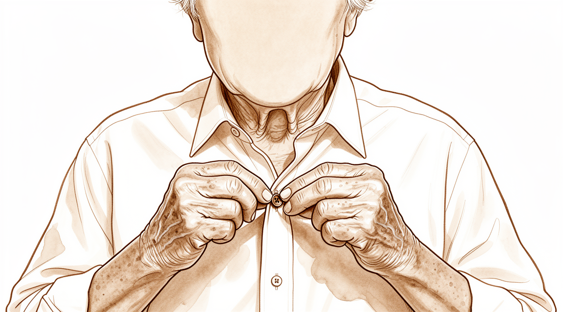
Cơn đau thường bùng phát sau khi bạn dùng ngón tay, và ngón tay có thể đau khi bạn thức dậy vào buổi sáng. Những việc tỉ mỉ cần dùng đến tận đầu ngón tay trở nên khó khăn: cài cúc áo sơ mi, nhặt những đồng xu nhỏ, xâu kim, hoặc cầm chìa khóa để xoay mở ổ khóa. Viết tay cũng có thể gây đau nhức. Theo thời gian, khớp có thể cứng lại và mất bớt khả năng gập, và ngón tay có thể dần dần bị lệch trục.
Một số người nhận thấy một khối nhỏ chứa dịch gần móng tay, gọi là nang dịch nhầy. Nang này phát triển từ khớp bị viêm nằm bên dưới, vì vậy nó thường tái phát ngay cả sau khi đã được hút dịch.
Nếu bệnh viêm khớp xuất hiện sau một chấn thương ngón tay, chẳng hạn như ngón tay dùi cui – khi gân duỗi bị rách hoặc bứt ra kèm theo một mảnh xương – khớp có thể bị hao mòn nhanh hơn so với bình thường. Sự hao mòn đó gây ra cùng kiểu đau và cứng khớp, và ngón tay có thể mất bớt khả năng cử động dù bản thân chấn thương đã lành.
Hãy để ý một vài dấu hiệu cảnh báo. Nếu ngón tay của bạn trở nên nóng, đỏ, sưng và đau, đặc biệt là khi kèm theo sốt, hãy đến khoa cấp cứu ngay trong ngày. Đây có thể là nhiễm trùng cần được chăm sóc ngay trong ngày, và bạn không cần có giấy giới thiệu của bác sĩ đa khoa trước. Nếu cơn đau không giảm sau nhiều tuần, ngày càng nặng hơn hoặc khiến bạn thức giấc vào ban đêm, hãy đến gặp bác sĩ đa khoa hoặc đề nghị được bác sĩ chuyên khoa khám.
Chuyện gì đang xảy ra thực sự
Mỗi ngón tay là một chuỗi các xương nhỏ. Khớp ở tận đầu ngón tay, ngay phía sau móng tay, là nơi xương cuối cùng tiếp giáp với xương phía trên nó. Một lớp trơn láng bao phủ đầu của cả hai xương và giúp chúng trượt lên nhau. Bạn có thể hình dung lớp này như một bộ giảm xóc có sẵn, đồng thời giữ cho bề mặt khớp luôn trơn tru.
Trong bệnh viêm khớp, lớp trơn láng đó mỏng dần hoặc bị hư hại. Khi đó các xương cọ xát vào nhau, và khớp phản ứng bằng cách tạo thêm xương quanh các mép khớp. Những gai xương này chính là các khối chắc mà bạn có thể sờ thấy gần móng tay, như đã mô tả ở phần trên. Sự cọ xát gây ra cảm giác đau nhức, còn bề mặt khớp bị mòn cùng các gai xương giải thích vì sao khớp bị sưng, cứng lại và dần dần lệch trục.
Khớp còn được giữ lại với nhau nhờ những dải mô nhỏ ở hai bên và một dải sợi gân dẹt chạy ngang qua mặt trên, bám ngay phía sau khớp. Khi viêm khớp làm khớp thay đổi, các dải mô và gân này có thể bị kéo mất cân bằng. Đó là một trong những lý do khiến ngón tay có thể bị gập ngược quá mức ở khớp giữa trong khi đầu ngón tay cong xuống – một sự thay đổi hình dạng thường xuất hiện khi viêm khớp tiến triển.
Đôi khi sự hao mòn xảy ra sau một chấn thương chứ không đơn thuần do hao mòn theo thời gian. Một cú va đập mạnh, một chấn thương dập nát, hoặc chấn thương gân như ngón tay dùi cui có thể làm hỏng bề mặt khớp. Khi đó khớp bị mòn sớm hơn so với bình thường, đó là lý do đau và cứng khớp có thể xuất hiện nhiều năm sau khi chấn thương tưởng như đã lành.
Viêm khớp ở khớp này cũng có thể tạo ra một khối nhỏ chứa dịch cạnh móng tay, chính là nang dịch nhầy đã nói ở trên. Nang phát triển ra từ chính khớp đang bị kích thích, đó là lý do nó thường tái phát sau khi được hút dịch.
Tin tốt là khớp này đảm nhận rất ít công việc cầm nắm nặng. Nhiệm vụ của nó là điều khiển các cử động tinh tế ở đầu ngón tay, đó là lý do việc điều trị tập trung vào làm dịu cơn đau và giữ cho khả năng điều khiển tinh tế đó tiếp tục hoạt động.
Những biện pháp chúng tôi có thể áp dụng
Bác sĩ Kieran Hirpara, bác sĩ phẫu thuật chi trên tại Bệnh viện tư nhân Mater Rockhampton, sẽ bắt đầu bằng các phương pháp ít xâm lấn nhất phù hợp với tình trạng của bạn. Thông thường, bệnh nhân được bác sĩ đa khoa giới thiệu đến phòng khám chúng tôi; nếu vật lý trị liệu viên khuyên bạn nên gặp chúng tôi, bạn vẫn cần có giấy giới thiệu từ bác sĩ đa khoa để được hưởng mức hoàn trả từ Medicare. Trong lần khám đầu tiên, chúng tôi sẽ hỏi bệnh sử, khám ngón tay và chỉ định chẩn đoán hình ảnh nếu cần. Vì đây là một vấn đề hao mòn kéo dài, chúng tôi thường thử điều trị không phẫu thuật trước và chỉ bàn đến phẫu thuật khi cách đó chưa mang lại đủ cải thiện.
Bước đầu tiên thường là một chiếc nẹp nhỏ giữ khớp đầu ngón tay ở tư thế thẳng. Đeo nẹp giúp giảm đau ở khớp và giúp ngón tay duỗi thẳng hơn. Cách này đơn giản và an toàn, và bạn có thể đeo nẹp trong các sinh hoạt bình thường hằng ngày. Chúng tôi thường kết hợp nẹp với thuốc chống viêm – loại thuốc viên giúp làm dịu sưng và đau. Liệu pháp trị liệu tay cũng có thể giúp bạn duy trì khả năng dùng đầu ngón tay cho các việc tỉ mỉ trong khi tình trạng kích thích lắng dịu. Hãy thử các biện pháp này một cách đầy đủ trước khi quyết định bước tiếp theo.
Nếu sau đó cơn đau và cứng khớp vẫn còn hạn chế sinh hoạt của bạn, phẫu thuật trở thành một lựa chọn. Phẫu thuật tiêu chuẩn là hợp nhất khớp, còn gọi là cố định khớp, trong đó các bề mặt khớp bị mòn được loại bỏ và hai xương được nối lại để liền thành một. Sau phẫu thuật, khớp không còn gập được nữa, nhưng khớp không đau và vững chắc, và ngón tay được đặt ở tư thế thuận tiện cho các công việc hằng ngày. Hợp nhất khớp cũng giúp làm thẳng một ngón tay đã bị lệch trục, và đây là phương pháp điều trị giúp ngăn nang dịch nhầy tái phát. Nếu việc giữ lại một phần cử động ở đầu ngón tay quan trọng với bạn hơn việc hết đau hoàn toàn, có một phẫu thuật khác giúp gọt bỏ các khối u xương và làm sạch khớp, đồng thời vẫn để khớp cử động được. Chúng tôi sẽ cùng bạn trao đổi xem lựa chọn nào phù hợp với ngón tay của bạn và những gì bạn muốn bàn tay mình làm được, và cùng nhau quyết định.
Những điều có thể xảy ra
Viêm khớp ở khớp đầu ngón tay thường tiến triển chậm qua nhiều năm. Cơn đau thường lúc có lúc không: bùng phát sau khi bạn dùng ngón tay, rồi lại dịu đi khi nghỉ ngơi. Nếu không điều trị, khớp thường cứng thêm và ngón tay có thể bị lệch trục nhiều hơn theo thời gian. Một số người thấy cơn đau tự giảm khi khớp cứng lại, nhưng với nhiều người, cơn đau vẫn âm ỉ dai dẳng, đặc biệt khi làm những việc tỉ mỉ như cài cúc áo sơ mi hoặc xoay chìa khóa.
Hầu hết mọi người giảm đau tốt nhờ các biện pháp đơn giản đã mô tả ở trên: đeo nẹp, thuốc chống viêm và liệu pháp trị liệu tay. Những biện pháp này làm dịu cơn đau trong khi bạn vẫn dùng ngón tay cho các công việc hằng ngày. Nếu các biện pháp đó không hiệu quả sau khi đã được thử đầy đủ, phẫu thuật thường giúp giảm đau tốt. Khớp đầu ngón tay sau khi được hợp nhất sẽ không đau và vững chắc, và nhìn chung mọi người trở lại sử dụng bàn tay bình thường, dù đầu ngón tay không còn gập được nữa. Nếu việc giữ lại một phần cử động quan trọng hơn với bạn, phẫu thuật làm sạch khớp giúp ngón tay vẫn gập được, với hiệu quả giảm đau tốt nhưng không phải lúc nào cũng hoàn toàn.
Quá trình hồi phục sau phẫu thuật kéo dài nhiều tuần đến nhiều tháng chứ không phải vài ngày. Lúc đầu ngón tay được giữ bất động trong khi các xương liền lại, và chuyên viên trị liệu tay sẽ hướng dẫn bạn khi bạn lấy lại dần sức cầm nắm và khả năng điều khiển tinh tế. Hầu hết mọi người thấy kết quả cuối cùng là một ngón tay làm tốt chức năng của nó mà không còn đau nhức, dù đầu ngón tay nằm thẳng hơn trước.
Một vài lưu ý thẳng thắn. Hợp nhất khớp không phải lúc nào cũng liền xương ngay ở lần phẫu thuật đầu tiên, và một số người cần phẫu thuật thêm để tháo dụng cụ kim loại hoặc làm lại việc hợp nhất khớp. Hút thuốc làm chậm quá trình liền xương, vì vậy bỏ thuốc trước khi phẫu thuật sẽ cho ngón tay của bạn cơ hội liền xương tốt nhất. Nếu cơn đau không giảm sau nhiều tuần, ngày càng nặng hơn hoặc khiến bạn thức giấc vào ban đêm, hãy đến gặp bác sĩ đa khoa hoặc đề nghị được bác sĩ chuyên khoa khám.
Khi nào nên gặp bác sĩ
Hầu hết các trường hợp viêm khớp ở khớp đầu ngón tay tiến triển chậm, vì vậy một lần khám thông thường với bác sĩ đa khoa là đủ để bắt đầu. Hãy đến gặp bác sĩ đa khoa nếu cơn đau không giảm sau nhiều tuần, ngày càng nặng hơn hoặc khiến bạn thức giấc vào ban đêm. Bạn cũng nên đi khám nếu ngón tay khiến bạn không thể làm việc hoặc không làm được những việc tỉ mỉ như cài cúc áo sơ mi.
Một số dấu hiệu cần được chăm sóc nhanh hơn. Hãy đến khoa cấp cứu ngay trong ngày nếu ngón tay của bạn trở nên nóng, đỏ, sưng và đau, đặc biệt là khi kèm theo sốt. Biểu hiện này gợi ý tình trạng nhiễm trùng, và bạn không cần có giấy giới thiệu của bác sĩ đa khoa trước.
Hãy đến khoa cấp cứu sau chấn thương nếu da ở vùng khớp bị rách, lộ xương, hoặc ngón tay trông rõ ràng bị biến dạng. Bạn cũng phải đến khoa cấp cứu nếu ngón tay chuyển sang nhợt nhạt, lạnh, trắng hoặc xanh tím, hoặc nếu bạn đột ngột mất cảm giác hoặc mất khả năng cử động ngón tay sau một cú va đập.
Nang dịch nhầy cứ tái phát, hoặc ngón tay đang bị lệch trục, là những tình trạng nên được bác sĩ chuyên khoa khám.
Phân tích chi tiết hơn
Advanced reading: the deeper science (optional)
Phần này đi sâu hơn mức cần thiết để bạn tự đưa ra quyết định điều trị. Viêm khớp ở khớp ngón tay xứng đáng được nghiên cứu kỹ hơn, bởi phương pháp nối cốt – ca phẫu thuật tiêu chuẩn thường được mô tả là đơn giản – lại có tỷ lệ biến chứng cao hơn mức mà người ta vẫn nghĩ; đồng thời còn có một phương pháp thay thế giúp bảo tồn vận động mà ít được đề cập đến.
Việc cố định khớp không hề đơn giản như vẻ bề ngoài
Việc cố định khớp đầu ngón tay được coi là một phẫu thuật đáng tin cậy; về mặt giảm đau thì đúng là như vậy. Tuy nhiên, các biến chứng có thể xảy ra lại không hề ít. Một nghiên cứu đánh giá các yếu tố nguy cơ trên 173 bệnh nhân đã nêu rõ: việc cố định khớp liên đốt ngón tay thứ hai thường dẫn đến nhiều biến chứng, trong đó viêm khớp thoái hóa, phải thực hiện lại ca phẫu thuật cố định khớp và việc hút thuốc được xác định là các yếu tố nguy cơ [1].
Việc hút thuốc nằm trong danh sách này là điều đáng lưu ý, bởi đây là yếu tố mà bệnh nhân hoàn toàn có thể kiểm soát được. Sự liền xương phụ thuộc vào nguồn cung cấp máu; đây là một khớp nhỏ, được bao bọc bởi lớp mô mềm mỏng manh, nằm ở cuối vùng cung cấp máu của ngón tay.
Implant giúp xương liền tốt hơn nhưng cũng gây ra những biến chứng mà các loại rẻ hơn không có
Trong cuộc tranh luận về phương pháp cố định xương, có một câu trả lời khá rõ ràng: đó là sự đánh đổi chứ không phải là việc chọn ra phương pháp “vượt trội”. Trên 1.125 bệnh nhân, các vít nén không có đầu vít dường như giúp tăng tỷ lệ liền xương; tuy nhiên lại gây ra những biến chứng mà các kỹ thuật rẻ hơn và đã được kiểm chứng khác không gây ra. Ngoài yếu tố liền xương ra, hiện vẫn chưa có đủ bằng chứng chứng minh loại vít này vượt trội hơn [2].
Những biến chứng đặc thù liên quan đến loại vít này xuất phát từ cấu trúc giải phẫu. Vít đi dọc theo trục đầu ngón tay nên đi gần với móng tay, có thể gây biến dạng móng; ở những ngón tay có đốt ngón tay xa rất nhỏ thì có thể không đủ xương để giữ vít lại. Các dây kim loại thì rẻ hơn và tránh được những vấn đề trên, dù hiệu quả giúp xương liền hơi kém hơn một chút.
Trong những trường hợp chỉ cần ưu tiên việc xương liền, khi cần phẫu thuật hợp nhất xương lại lần nữa, hoặc ở những bệnh nhân hút thuốc, ưu điểm của loại vít này mới thực sự thể hiện rõ. Còn ở những trường hợp xương nhỏ và móng tay có vai trò quan trọng, thì việc chọn loại vít này vẫn chưa chắc đã là lựa chọn tối ưu.
Phương án thay thế giúp duy trì vận động
Liệu pháp hợp nhất khớp không phải là lựa chọn duy nhất; phương án thay thế này cũng chưa được bàn luận nhiều. Đối với những bệnh nhân chủ yếu phàn nàn về các khối xương mọc ra và cơn đau do chúng gây ra chứ không phải tình trạng viêm khớp toàn bộ, có thể loại bỏ những gai xương này mà vẫn giữ nguyên cấu trúc khớp.
Phẫu thuật cắt bỏ gai xương ở khớp đầu ngón tay được xem là phương pháp thay thế an toàn và hiệu quả so với việc hợp nhất khớp ở những bệnh nhân bị viêm khớp thoái hóa có triệu chứng, những người muốn duy trì khả năng vận động của khớp, với 78 bệnh nhân tham gia nghiên cứu [3].
Điều này rất quan trọng vì khớp đầu ngón tay đóng góp rất ít vào sức nắm tay nhưng lại có vai trò lớn trong các thao tác tinh tế và thẩm mỹ của bàn tay. Đối với những người có triệu chứng xuất phát từ chính các nốt Heberden, việc phải chấp nhận tình trạng khớp đầu ngón tay bị cứng vĩnh viễn là một sự hy sinh lớn hơn nhiều so với vẻ bề ngoài, và thực tế vẫn còn một phương án trung gian.
Nang dịch thường xuất hiện kèm theo
Viêm khớp ở khớp này thường gây ra nang dịch nhầy – một khối sưng nhỏ chứa đầy dịch, hình thành từ khớp bị viêm, thường nằm cạnh móng tay. Vì được tạo thành do hoạt động của khớp, nang này có đặc tính tương tự như nang ganlia ở cổ tay: việc chọc hút dịch chỉ giúp giảm sưng chứ không giải quyết được nguyên nhân gốc rễ. Nang này được đề cập riêng trong một mục khác, nhưng việc hiểu rõ mối liên hệ giữa nó và viêm khớp là rất quan trọng; bởi một nang tái phát là dấu hiệu cho thấy tình trạng viêm khớp đang tiến triển chứ không phải là vấn đề tách biệt.
Tài liệu tham khảo
[1] Runkel A, Bonaventura B, Sundermann B, Zajonc H, Eisenhardt S, Leibig N. Các yếu tố nguy cơ trong phẫu thuật cố định khớp liên đốt xa ở bàn tay: một nghiên cứu hồi cứu. J Hand Surg Eur Vol. 2022;47(9):907-14. https://doi.org/10.1177/17531934221111641
[2] Dickson D, Mehta S, Nuttall D, Ng C. Tổng quan có hệ thống về phẫu thuật cố định khớp liên đốt xa. J Hand Microsurg. 2014;6(2):74-84. https://doi.org/10.1007/s12593-014-0163-1
[3] Lin EA, Papatheodorou LK, Sotereanos DG. Phẫu thuật cắt bỏ gai xương trong điều trị viêm khớp thoái hóa khớp liên đốt xa có triệu chứng: tổng quan trên 78 ca bệnh. J Hand Surg Am. 2017;42(11):889-93. https://doi.org/10.1016/j.jhsa.2017.07.006
Evidence & references
This is the clinical evidence summary written for health professionals. It is technical, and it lists the research this page was built from. You do not need to read it to understand your treatment or to make a decision about it.
Overview
- Osteoarthritis is the most common indication for distal interphalangeal joint arthrodesis [19].
- In a cohort of 149 cases, postoperative complications for distal interphalangeal and thumb interphalangeal joint arthrodesis occurred at a rate similar to that reported in existing literature [19].
- Finger distal interphalangeal and thumb interphalangeal joint arthrodesis generally results in favorable outcomes in terms of bony union regardless of underlying medical condition or technical details of the surgical operation [6].
- The X-fuse implant provides excellent stability with minimal hardware problems and a high rate of union in patients with finger distal interphalangeal and thumb interphalangeal joint arthritis [1].
- In select patients, percutaneous distal interphalangeal joint arthrodesis is advantageous in comparison with open fusion techniques [2].
- Reamed percutaneous distal interphalangeal joint arthrodesis is associated with higher union rates achieved in a shorter time and reduced postoperative pain compared with the non-reamed percutaneous technique [8].
- Successful fusions of the distal interphalangeal joint at an angle up to 35 degrees can be achieved using small diameter headless compression screws [14].
- The Mini-Acutrak screw is suitable for distal interphalangeal joint fusion in all fingers with the exception of the small finger [22].
- The nonaxial multiple small screws technique is a feasible option for distal interphalangeal and thumb interphalangeal joint arthrodesis, especially when a small finger is indicated and a significant flexion angle is required [24].
- Given the lack of difference in biomechanical performance between K-wires and compression screws, consideration should be given to other factors such as cost and complication profiles when choosing an implant for distal interphalangeal joint fusion [34].
- The smile incision and reverse shotgun approach may be a good surgical option for distal interphalangeal joint arthrodesis when more volar part joint preparation and more volar implant insertion sites are necessary [17].
- Results from a lateral approach and plate fixation for distal interphalangeal joint arthrodesis are equivalent to traditional methods but with fewer major complications [11].
- For persistently symptomatic, unstable distal interphalangeal joints, arthrodesis should be considered [4].
- In the distal interphalangeal joint, there is probably good reason to proceed to arthrodesis immediately for synovial chondromatosis [3].
- The swan neck deformity can progress significantly with time because of increasing distal interphalangeal joint flexion contracture [5].
- Silicone interpositional arthroplasty of the distal interphalangeal joint is an acceptable alternative to arthrodesis, achieving excellent pain relief and a range of movement of 30–40 degrees with a low overall complication rate of 5% [13].
- Swanson hinge implant arthroplasty can be the surgical treatment of choice for the patient who desires continued mobility, albeit limited, of the involved osteoarthritic distal interphalangeal joint [9].
- Open distal interphalangeal joint cheilectomy is a safe and effective alternative to distal interphalangeal joint arthrodesis in patients with symptomatic osteoarthritis who wish to preserve joint motion [28].
- Denervation with cheilectomy of the distal interphalangeal joint presents a compelling motion-preserving alternative to arthrodesis for symptomatic distal interphalangeal joint osteoarthritis [10].
- A surgical technique for treating symptomatic distal interphalangeal joint arthritis reduces pain while preserving distal interphalangeal joint motion [18].
- The authors recommend simultaneous surgical intervention in case of severe painful osteoarthritis of the proximal interphalangeal and distal interphalangeal joints of the same digit [7].
- The combination of distal interphalangeal arthrodesis and proximal interphalangeal Swanson arthroplasty resulted in a favorable outcome in terms of simultaneous bony union and flexibility [12].
- Combined distal interphalangeal arthrodesis and proximal interphalangeal procedures present unique challenges regarding hardware conflict, where K-wires offer the easiest compatibility and headless screws must ideally not reach proximal to the midpoint of the middle phalanx [90].
- A customized structural bone graft addresses issues of bone stock loss and medullary absence in failed distal interphalangeal joint silicone arthroplasty, achieving reliable union rates and high patient satisfaction [21].
Anatomy & Pathophysiology
Bony Anatomy & Morphology
- The skeleton of the hand consists of 27 bones, of which 19 are long bones [47].
- The skeleton is divided into five rays, each ray making up a polyarticulated chain comprising the metacarpals and phalanges [47].
- The thumb ray is made up of only three bones—a metacarpal and two phalanges [47].
- The other four digital rays are formed by four skeletal segments—a metacarpal and three phalanges [47].
- The thumb metacarpal is the shortest, and the index metacarpal is by far the longest [47].
- The proximal and middle phalanges of the long and ring fingers are longer than those of the index finger [47].
- The epiphyseal plates are located at the proximal ends of the phalanges and the first metacarpal [47].
- An examination of 100 specimens of the terminal joints of human fingers was described in a 1982 study [75].
- A substantial number of distal phalanges are too small to accommodate commonly available headless compression screws, particularly in females and the small finger [39].
- Understanding of DIP joint morphology may lend insight into the biomechanics and disease progression within the DIP joints [23].
Soft Tissue Anatomy & Extensor Mechanism
- Distal interphalangeal joint extension is achieved through the conjoined lateral bands that are composed of tendinous slips from the extrinsic and intrinsic tendons [46].
- The principal bony insertion of the extrinsic digital extensors is on the dorsal proximal aspect of the middle phalanx [46].
- The deep head of each dorsal interosseous muscle forms a lateral tendon, or lateral band, at the level of the MP joint [59].
- The lateral bands are joined by the lateral slips of the extensor tendon to form the conjoined lateral band [59].
- The two conjoined lateral bands to each finger unite at the distal third of the middle phalanx to form the terminal tendon [59].
- The terminal tendon inserts at the base of the distal phalanx to extend it [59].
- The flexor digitorum profundus inserts on the proximal volar aspect of the distal phalanx, flexing the distal interphalangeal joint as well as the proximal interphalangeal and metacarpophalangeal joints [46].
- The flexor digitorum profundus provides digital flexion at both the proximal and distal interphalangeal joints [66].
- The A5 annular pulley is located over the distal interphalangeal joint [66].
- The vinculum breve of the flexor digitorum profundus and check-rein ligaments may account for the greater amount of passive hyperextension that can be achieved at the distal than the proximal interphalangeal joint [80].
Biomechanics & Kinematics
- The wrist influences the position of the metacarpophalangeal joint; the metacarpophalangeal joint also affects the position of the proximal interphalangeal joint, which in turn affects the distal interphalangeal joint [67].
- Almost all movements in the hand are around oblique and variable axes, resulting in combined movements permitting optimal orientation of the phalanges at the time of prehension [67].
- The axes of flexion are so arranged that flexion of all the metacarpophalangeal and proximal interphalangeal joints causes the fingers to converge toward the scaphoid [70].
- In a combinatorial relationship, intrinsic muscles produce steep inclination of extensor forces at the DIP joint [73].
- There were no significant differences in comparisons among loads (200, 400, 600, and 800 g) regarding the inclination slope of intrinsic extensor forces at the DIP joint [73].
- Immobilization of the distal interphalangeal joint of any finger reduces the overall grip strength of the hand, with the effect becoming progressively more pronounced from the index to the little fingers [37].
- Positioning the middle finger DIP joint in either extension or 20° of flexion did not significantly affect grip strength or dexterity [49].
- Biomechanically, dynamic tenodesis for the DIP joint using the remaining FDP tendon results in a flexion angle greater than 30 degrees [76].
Pathophysiology & Etiology
- Osteoarthritis occurs with the highest prevalence in the distal interphalangeal joint of the hand and has been divided into an erosive and a nonerosive form [26].
- The pathogenesis of the early stages of osteoarthritis is poorly understood, but considerable emphasis has been placed on the role of cartilage and subchondral bone as well as soft tissue structures such as collateral ligaments and tendons [26].
- Osteoarthritis is a complex disease resulting in the failure of articular cartilage due to a combination of genetic, metabolic, biochemical, and biomechanical factors [26].
- Cartilage loss and reactive new bone formation at joint margins is accompanied by the proliferation of osteoarticular tissue in the capsule [26].
- Heberden's nodes are more common in women who engage in cooking in school and the incidence increases with the number of tasks involved [25].
- Cooking as an occupation is an aetiological factor in the pathogenesis of Heberden's nodes [25].
- Work-load, sports, and repeated minimal trauma contribute to the development of Heberden's nodes [25].
- Repetitive injuries to a joint lead to an increased risk of developing posttraumatic arthritis [48].
- Athletes have a higher incidence of degenerative joint changes compared to the general population [48].
- Mallet finger is caused by forced flexion of an extended fingertip [51].
- Less common hyperextension or hyperextension/axial loading tends to cause the larger fractures and subluxation in mallet injuries [51].
- One-third of all mallet fingers are associated with a fracture [51].
- Mallet fractures associated with large fragments may result in volar subluxation of the distal phalanx as the collateral ligaments remain attached to the fracture fragment [51].
- Fracture fragments that are less than 43% of the distal phalanx articular surface usually do not allow volar subluxation [51].
- Fracture fragments involving larger than 52% of the joint surface consistently allow subluxation [51].
- Palmar subluxation of a DIP joint without preexisting arthritic deformity is expected when more than one half of the dorsal articular surface is injured [20].
- The mechanism of displaced intra-articular osteochondral fracture causing irreducible dislocation of the DIP joint probably involved hyperextension of the DIP joint [15].
- The palmar plate was torn at the attachment to the distal phalanx in cases of displaced intra-articular osteochondral fracture [15].
- Attempts to perform a closed reduction of displaced intra-articular osteochondral fractures can lead to further displacement of the fracture, with folding of the fragment on the articular cartilage hinge [15].
- The cause of irreducible palmar dislocation of the DIP joint can be an entrapment of the extensor tendon in front of the head of the middle phalanx [41].
- Irreducibility was more commonly seen in dorsal than in volar dislocations of the DIP joint [85].
- Volar dislocations of the DIP joint carried a higher risk of instability immediately after reduction compared to dorsal dislocations [85].
- Some loss of motion in small joints of the fingers after hyperflexion injuries would be expected [79].
Classification
- Osteoarthritis of the distal interphalangeal joint has been divided into an erosive and a nonerosive form [26].
- Radiographic evaluation represents the most standardized method to quantify disease progression in distal interphalangeal joint osteoarthritis, with different systems developed for defining and grading radiographic features [26].
- The interrater reliability of the Kellgren & Lawrence and OARSI classification systems for post-traumatic osteoarthritis in the distal interphalangeal joint after mallet finger fractures is considerably lower than initially assumed [86].
Clinical Presentation
- Osteoarthritis occurs with the highest prevalence in the distal interphalangeal joint of the hand [26].
- Heberden's nodes are more common in women who engage in cooking in school, with incidence increasing with the number of tasks involved [25].
- There is no confirmed difference in the incidence of disease between right and left hands in elementary school cooks [25].
- Radiographic evaluation represents the most standardized method to quantify disease progression in distal interphalangeal joint osteoarthritis [26].
- The pathogenesis of the early stages of distal interphalangeal joint osteoarthritis is poorly understood [26].
- Considerable emphasis has been placed on the role of cartilage and subchondral bone in the pathogenesis of distal interphalangeal joint osteoarthritis [26].
- Soft tissue structures such as collateral ligaments and tendons play a role in the pathogenesis of distal interphalangeal joint osteoarthritis [26].
- Cartilage loss and reactive new bone formation at joint margins are accompanied by the proliferation of osteoarticular tissue in the capsule [26].
- Radiological osteoarthritis after a mallet finger fracture is similar to the natural degenerative process in the distal interphalangeal joint [27].
- Radiological osteoarthritis after a mallet finger fracture is accompanied by a decrease in range of motion of the distal interphalangeal joint [27].
- The decrease in range of motion following mallet finger fracture does not clinically affect patient-reported outcomes [27].
- Palmar subluxation of a distal interphalangeal joint without preexisting arthritic deformity is expected when more than one half of the dorsal articular surface is injured [20].
- Primary synovial chondromatosis of the distal interphalangeal joint is an extremely rare entity that requires accurate diagnosis to distinguish from other arthropathies [50].
- Adnexal tumours may masquerade as a distal interphalangeal joint ganglion and should enter the differential diagnosis when examining tumours of the digits [89].
- Floating distal interphalangeal joint injuries can be misdiagnosed initially due to minimal deformity [33].
- Osteoarthritis may develop following floating distal interphalangeal joint injuries [33].
- The mechanism of displaced intra-articular osteochondral fracture causing irreducible dislocation of the distal interphalangeal joint probably involved hyperextension of the joint [15].
- The palmar plate is torn at the attachment to the distal phalanx in displaced intra-articular osteochondral fractures of the distal interphalangeal joint [15].
- Attempts to perform a closed reduction of a displaced intra-articular osteochondral fracture can lead to further displacement of the fracture with folding of the fragment on the articular cartilage hinge [15].
- The cause of irreducibility in palmar dislocation of the distal interphalangeal joint can be entrapment of the extensor tendon in front of the head of the middle phalanx [41].
- Interrater reliability for measurements of the distal interphalangeal joint motions of the index and long digits is high [16].
- Interrater reliability for distal interphalangeal joint motion measurements is slightly higher for the dorsal method of placement than for the lateral method [16].
Investigations
- Radiographic evaluation is the most standardized method to quantify disease progression in distal interphalangeal joint osteoarthritis [26].
- Different systems have been developed for defining and grading radiographic features of distal interphalangeal joint osteoarthritis [26].
- Interrater reliability was high for measurements of the proximal and distal interphalangeal joint motions of the index and long digits [16].
- Interrater reliability was slightly higher for the dorsal method of goniometer placement than for the lateral method [16].
- Primary synovial chondromatosis of the distal interphalangeal joint requires accurate diagnosis to distinguish from other arthropathies [50].
- A distinct collagen septum exists between the extensor tendon and skin at the distal interphalangeal joint [53].
- The existence of a distinct collagen septum between the extensor tendon and skin at the distal interphalangeal joint was confirmed using MRI and histology [53].
Treatment
Non-Operative
- Splinting of the distal interphalangeal joint reduces pain and improves extension at the joint [29].
- Splinting for DIP joint arthritis does not give rise to non-compliance, increased stiffness, or restriction of range of motion [29].
- Splinting and anti-inflammatory medications are the mainstay of nonoperative treatment for DIP joint arthritis [48].
- Injection with Collagenase Clostridium histolyticum is an option for the treatment of DIP joint contractures in Dupuytren disease, though the potential risk for recurrence should be carefully weighed prior to its use [93].
Operative: Arthrodesis
- Arthrodesis is generally the most accepted surgical option for treatment of degenerative and traumatic conditions involving the distal interphalangeal joint of the fingers or the interphalangeal joint of the thumb [38].
- The ideal position of DIP arthrodesis is thought to be slightly flexed to improve power, fine pinch, and grip [38].
- In select patients, percutaneous DIP joint arthrodesis is advantageous in comparison with open fusion techniques [2].
- Reamed percutaneous DIPJ arthrodesis is associated with higher union rates achieved in a shorter time and reduced postoperative pain compared with the non-reamed percutaneous technique [8].
- The X-fuse implant provides excellent stability with minimal hardware problems and a high rate of union in patients with finger DIPJ and thumb IPJ arthritis [1].
- Finger DIP and thumb IP joint arthrodesis generally resulted in favourable outcome in terms of bony union regardless of the underlying medical condition or technical details of the surgical operation [6].
- Osteoarthritis was the most common indication for DIP and thumb IP arthrodesis, and postoperative complications occurred at a rate similar to that reported in existing literature [19].
- Successful fusions of the DIP joints at an angle up to 35 degrees were achieved using small diameter headless compression screws, providing benefits including early mobilization and favourable functional outcome scores [14].
- The Mini-Acutrak screw is suitable for DIPJ fusion in all fingers with the exception of the small finger [22].
- The nonaxial multiple small screws technique could be used as a feasible option in DIPJ and thumb IPJ arthrodesis, especially when a small finger is indicated and a significant flexion angle is required [24].
- Given the lack of difference in biomechanical performance between K-wires and compression screws, consideration should be given to other factors such as cost and complication profiles when choosing an implant for DIPJ fusion [34].
- DIP joint preparation is not necessary before performing arthrodesis in fingers with stage IV chondropathy [44].
- The use of a buried break-away compression screw avoids complications related to bulky hardware in DIP arthrodesis [44].
- Dorsal plate fixation allows for the performance of a distal finger joint arthrodesis in slight flexion [38].
- The smile incision and reverse shotgun approach may be a good surgical option for DIPJ arthrodesis when more volar part joint preparation and more volar implant insertion sites are necessary [17].
- Lateral approach and plate fixation for DIP joint arthrodesis yields results equivalent to traditional methods but with fewer major complications [11].
- For persistently symptomatic, unstable DIP joints, arthrodesis should be considered [4].
- In the DIP joint, there is probably good reason to proceed to arthrodesis immediately in cases of synovial chondromatosis [3].
- A customized structural bone graft addresses issues of bone stock loss and medullary absence in failed DIPJ silicone arthroplasty, achieving reliable union rates and high patient satisfaction [21].
- All joints had fused radiologically in a series of proximodistal interphalangeal arthrodesis of the little finger [32].
- Arthrodesis of the DIP joint provides stability and pain relief and can correct deformity [48].
- A painless, stable, distal interphalangeal joint that is in a functional position is more important than the ability to move the joint [48].
- The authors recommend simultaneous surgical intervention in case of severe painful OA of the PIP and DIP joints of the same digit [7].
- The combination of DIP arthrodesis and PIP Swanson arthroplasty resulted in a favourable outcome in terms of simultaneous bony union and flexibility [12].
- For patients prioritizing hand aesthetics or with unstable joints, distal interphalangeal joint arthrodesis is preferable [31].
Operative: Arthroplasty and Motion-Preserving
- Silicone interpositional arthroplasty of the DIP joint is an acceptable alternative to arthrodesis, achieving excellent pain relief and a range of movement of 30–40 degrees with a low overall complication rate of 5% [13].
- Swanson hinge implant arthroplasty can be the surgical treatment of choice for the patient who desires continued mobility, albeit limited, of the involved osteoarthritic DIP joint [9].
- Open DIP joint cheilectomy is a safe and effective alternative to DIP joint arthrodesis in patients with symptomatic osteoarthritis who wish to preserve joint motion [28].
- Denervation with cheilectomy of the distal interphalangeal joint presents a compelling motion-preserving alternative to arthrodesis for symptomatic DIP joint osteoarthritis [10].
- A surgical technique for DIP joint arthritis reduces pain while preserving DIP joint motion [18].
- Volar plate arthroplasty of the distal interphalangeal joint allows for a painless functional arc of motion without residual joint subluxation [40].
Complications
- The X-fuse implant provides excellent stability with minimal hardware problems [1].
- The X-fuse implant has a high rate of union in patients with finger DIPJ and thumb IPJ arthritis [1].
- Reamed percutaneous DIPJ arthrodesis is associated with reduced postoperative pain compared with the non-reamed percutaneous technique [8].
- The swan neck deformity progressed significantly with time because of increasing DIPJ flexion contracture [5].
- Postoperative complications occurred at a rate similar to that reported in the existing literature in a retrospective cohort of 149 cases of DIP and thumb IP joint arthrodesis [19].
- Arthrodesis of the distal interphalangeal joint often leads to complications [30].
- Silicone interpositional arthroplasty of the DIP joint has a low overall complication rate of 5% [13].
- Bony union of the joint was delayed in a series using the Herbert screw for DIPJ arthrodesis [56].
- Follow-up to 5 years of DIP joint flexible implant arthroplasty showed only one complication [92].
Recovery
Arthrodesis Outcomes and Complications
- Postoperative complications in DIP and thumb IP joint arthrodesis occurred at a rate similar to that reported in existing literature [19].
- Finger DIP and thumb IP joint arthrodesis generally resulted in favourable outcomes in terms of bony union regardless of underlying medical condition or technical details of the surgical operation [6].
- Reamed percutaneous DIPJ arthrodesis is associated with higher union rates achieved in a shorter time compared with the non-reamed percutaneous technique [8].
- Successful fusions of the DIP joints at an angle up to 35 degrees were achieved using small diameter headless compression screws [14].
- Small diameter headless compression screws for DIP arthrodesis provide benefits including early mobilization and favourable functional outcome scores [14].
- Although bony union of the joint was delayed in a minimally invasive technique using the Herbert Screw, firm bone union was ultimately obtained in all joints [56].
Arthroplasty Outcomes
- Silicone interpositional arthroplasty of the DIP joint achieves a range of movement of 30–40 degrees [13].
Non-Operative Management
- Splinting of the DIP joint does not give rise to non-compliance, increased stiffness, or restriction of range of motion [29].
Key Evidence
- [L4] The X-fuse implant seems to provide excellent stability with minimal hardware problems with a high rate of union in a wide array of patients with finger DIPJ and thumb IPJ arthritis. [1] (10.1097/bth.0b013e31829ba688)
- [L4] In select patients, this percutaneous DIP joint arthrodesis is advantageous in comparison with open fusion techniques. [2] (10.1007/s11552-010-9265-9)
- [L5] In the DIP joint there is probably good reason to proceed to arthrodesis immediately. [3] (10.1016/s0363-5023(09)91115-7)
- [L4] For persistently symptomatic, unstable DIP joints, arthrodesis should be considered. [4] (10.1016/j.jhsb.2005.09.003)
- [L5] The swan neck deformity in this individual progressed significantly with time because of increasing DIPJ flexion contracture. [5] (10.1016/j.jht.2009.11.005)
- [L3] In our cohort finger DIP and thumb IP joint arthrodesis generally resulted in favourable outcome in terms of bony union regardless of the underlying medical condition or technical details of the surgical operation. [6] (10.1142/s2424835520500216)
- [L3] The authors recommend simultaneous surgical intervention in case of severe painful OA of the PIP and DIP joints of the same digit. [7] (10.1177/17531934231191255)
- [L3] Reamed percutaneous DIPJ arthrodesis is associated with higher union rates achieved in a shorter time and reduced postoperative pain compared with the non-reamed percutaneous technique. [8] (10.1177/15589447261487482)
- [L5] Swanson hinge implant arthroplasty can be the surgical treatment of choice for the patient who desires continued mobility, albeit limited, of the involved osteoarthritic DIP joint. [9] (10.1016/s0894-1130(98)80061-6)
- [L4] It presents a compelling motion-preserving alternative to arthrodesis for symptomatic DIP joint osteoarthritis. [10] (10.1016/j.jhsa.2026.01.027)
- [L4] The results obtained in this small series are equivalent to the traditional methods of DIP joint arthrodesis but with fewer major complications. [11] (10.1016/j.jhsa.2007.09.004)
- [L4] The combination of DIP arthrodesis and PIP Swanson arthroplasty resulted in a favourable outcome in terms of simultaneous bony union and flexibility. [12] (10.1177/17531934231215790)
- [L4] The study confirms that silicone interpositional arthroplasty of the DIP joint is an acceptable alternative to arthrodesis, achieving excellent pain relief and a range of movement of 30–40 degrees with a low overall complication rate of 5%. [13] (10.1177/1753193411422679)
- [L4] In this series, successful fusions of the DIP joints, at an angle up to 35 degrees were achieved using small diameter headless compression screws, which provided benefits including early mobilization and favourable functional outcome scores. [14] (10.1142/s2424835518500406)
- [L5] [15] (10.1016/s0363-5023(82)80018-x)
- [L5] Interrater reliability was high for measurements of the PIP and DIP joint motions of the index and long digits and slightly higher for the dorsal method of placement than for the lateral method. [16] (10.1016/s0894-1130(01)80021-1)
- [L4] This technique may be a good surgical option for DIPJ arthrodesis when more volar part joint preparation and more volar implant insertion sites are necessary. [17] (10.1186/s12891-024-08016-6)
- [L4] This surgical technique reduces pain while preserving DIP joint motion. [18] (10.1177/1558944716660555i)
- [L3] Osteoarthritis was the most common indication for arthrodesis and postoperative complications occurred at a rate similar to that reported in the existing literature. [19] (10.1186/s12891-024-07361-w)
- [L5] Palmar subluxation of a DIP joint without preexisting arthritic deformity is expected when more than one half of the dorsal articular surface is injured. [20] (10.1016/j.jhsa.2007.09.006)
- [L4] A customized structural bone graft using the described technique addresses issues of bone stock loss and medullary absence in failed DIPJ silicone arthroplasty, achieving reliable union rates and high patient satisfaction. [21] (10.1177/17531934231151217)
- [L4] The authors conclude that the Mini-Acutrak screw is suitable for DIPJ fusion in all fingers with the exception of the small finger. [22] (10.1016/j.jhsa.2005.09.009)
- [L5] Our understanding of morphology may lend insight into the biomechanics and disease progression within the DIP joints. [23] (10.1007/s11552-014-9605-2)
- [L4] Thus, the NMSS technique could be used as a feasible option in DIPJ and thumb IPJ arthrodesis, especially when a small finger is indicated and a significant flexion angle is required. [24] (10.1186/s12891-022-05473-9)
- [L4] [25] (10.1016/0266-7681(93)90167-e)
- [L5] [26] (10.1016/j.jhsa.2010.09.003)
- [L4] Radiological OA after an MFF is similar to the natural degenerative process in the DIP joint and is accompanied by a decrease in range of motion of the DIP joint, which does not clinically affect PROMs. [27] (10.1016/j.jhsa.2023.03.027)
- [L4] Open DIP joint cheilectomy is a safe and effective alternative to DIP joint arthrodesis in patients with symptomatic osteoarthritis who wish to preserve joint motion. [28] (10.1016/j.jhsa.2017.07.006)
- [L2] It does not give rise to non-compliance, increased stiffness or restriction of range of motion. [29] (10.1016/j.jht.2013.08.004)
- [L3] Arthrodesis of the distal interphalangeal joint often leads to complications. [30] (10.1177/17531934221111641)
- [L3] For patients prioritizing hand aesthetics or with unstable joints, distal interphalangeal joint arthrodesis is preferable. [31] (10.1177/1753193420917818)
- [L4] All joints had fused radiologically. [32] (10.1016/j.hansur.2016.06.003)
- [Case_report] Floating DIP joint injuries can be misdiagnosed initially due to minimal deformity; open reduction and internal fixation is a viable treatment option for chronic cases, though osteoarthritis may develop. [33] (10.1016/j.jhsa.2010.05.025)
- [L5] Given the lack of difference in biomechanical performance between K-wires and compression screws, consideration should be given to other factors such as cost and complication profiles when choosing an implant for DIPJ fusion. [34] (10.1177/1558944715627211)
- [L4] Immobilization of the distal interphalangeal joint of any finger reduces the overall grip strength of the hand, with the effect becoming progressively more pronounced from the index to the little fingers. [37] (10.1177/1753193418765068)
- [L4] [38] (10.1016/j.jhsa.2018.03.049)
- [L4] A substantial number of distal phalanges are too small to accommodate commonly available headless compression screws, particularly in females and the small finger. [39] (10.1007/s11552-014-9679-x)
- [L4] The technique allows for a painless functional arc of motion without residual joint subluxation. [40] (10.1053/jhsu.2001.26325)
- [L5] The cause of irreducibility was an entrapment of the extensor tendon in front of the head of the middle phalanx. [41] (10.1016/s0363-5023(87)80116-8)
- [L4] Our results show that DIP joint preparation is not necessary before performing arthrodesis in fingers with stage IV chondropathy and that the use of a buried break-away compression screw avoids complications related to bulky hardware. [44] (10.1016/j.main.2015.03.002)
- [L4] [48] (10.1142/s0218810417500149)
- [L2] Positioning the middle finger DIP joint in either extension or 20° of flexion did not significantly affect grip strength or dexterity, allowing other considerations such as appearance to be prioritized. [49] (10.1016/j.jhsa.2014.06.021)
- [Case_report] Primary synovial chondromatosis of the distal interphalangeal joint is an extremely rare entity that requires accurate diagnosis to distinguish from other arthropathies. [50] (10.1177/15589447211049520)
- [L5] [51] (10.1177/1753193414554772)
- [L5] We confirmed the existence of a distinct collagen septum between the extensor tendon and skin at the DIP joint using MRI and histology. [53] (10.1016/j.jhsa.2008.11.030)
- [L4] Although bony union of the joint was delayed, firm bone union was ultimately obtained in all joints. [56] (10.1097/00130911-200212000-00008)
- [L5] [73] (10.1002/jor.22021)
- [L5] An examination of 100 specimens of the terminal joints of human fingers is described. [75] (10.1016/s0363-5023(82)80084-1)
- [L5] Biomechanically, dynamic tenodesis for the DIP joint using the remaining FDP tendon is a valuable procedure because it results in a flexion angle greater than 30 degrees. [76] (10.1016/j.jhsg.2020.08.007)
- [L5] Therefore, some loss of motion in small joints of the fingers after hyperflexion injuries would be expected. [79] (10.1142/s2424835516720206)
- [L5] This may account for the greater amount of passive hyperextension that can be achieved at the distal than the proximal interphalangeal joint. [80] (10.1016/0266-7681(91)90058-v)
- [L4] Irreducibility was more commonly seen in dorsal than in volar dislocations, while volar dislocations carried a higher risk of instability immediately after reduction. [85] (10.1177/1753193415616957)
- [L4] The interrater reliability of the Kellgren & Lawrence and OARSI classification systems for post-traumatic osteoarthritis in the distal interphalangeal joint after mallet finger fractures is considerably lower than initially assumed. [86] (10.1016/j.jhsa.2024.03.012)
- [L5] Adnexal tumours may masquerade as a DIPJ ganglion, and should enter the differential diagnosis of the hand surgeon when examining tumours of the digits. [89] (10.1177/1753193415620179)
- [L5] Combined DIP arthrodesis and PIP procedures present unique challenges regarding hardware conflict; K-wires offer the easiest compatibility, while headless screws must ideally not reach proximal to the midpoint of the middle phalanx. [90] (10.1016/j.jhsa.2024.08.006)
- [L4] Follow-up to 5 years showed results that were good to excellent, with only one complication, and a high degree of patient satisfaction. [92] (10.1016/0363-5023(89)90184-6)
- [L4] Injection with CCH is an option for the treatment of DIP joint contractures in Dupuytren disease, though the potential risk for recurrence should be carefully weighed prior to its use. [93] (10.1016/j.jhsa.2018.07.004)
References
[1] Distal Interphalangeal Joint Arthrodesis Using Nitinol Intramedullary Fixation Implants. Techniques in Hand & Upper Extremity Surgery. 2013. DOI: 10.1097/bth.0b013e31829ba688
[2] Treatment of Symptomatic Distal Interphalangeal Joint Arthritis with Percutaneous Arthrodesis: A Novel Technique in Select Patients. HAND. 2010. DOI: 10.1007/s11552-010-9265-9
[3] Synovial chondromatosis in the distal interphalangeal joint. The Journal of Hand Surgery. 1990. DOI: 10.1016/s0363-5023(09)91115-7
[4] Management of Tophaceous Gout of the Distal Interphalangeal Joint. Journal of Hand Surgery. 2006. DOI: 10.1016/j.jhsb.2005.09.003
[5] Swan Neck Deformity after Distal Interphalangeal Joint Flexion Contractures: A Biomechanical Analysis. Journal of Hand Therapy. 2010. DOI: 10.1016/j.jht.2009.11.005
[6] Factors Influencing Bone Union in Finger Distal Interphalangeal and Thumb Interphalangeal Joint Arthrodesis. The Journal of Hand Surgery (Asian-Pacific Volume). 2020. DOI: 10.1142/s2424835520500216
[7] Does distal interphalangeal joint arthrodesis affect proximal interphalangeal joint arthroplasty outcomes in the same finger?. Journal of Hand Surgery (European Volume). 2023. DOI: 10.1177/17531934231191255
[8] Reamed Percutaneous Arthrodesis for Distal Interphalangeal Joint Arthritis Improves Bone Fusion and Pain Compared With a Non-Reamed Technique. HAND. 2026. DOI: 10.1177/15589447261487482
[9] Distal interphalangeal joint implant arthroplasty in a musician. Journal of Hand Therapy. 1998. DOI: 10.1016/s0894-1130(98)80061-6
[10] Denervation with Cheilectomy of the Distal Interphalangeal Joint: Technique and Medium-Term Results. The Journal of Hand Surgery. 2026. DOI: 10.1016/j.jhsa.2026.01.027
[11] Alternative to the Distal Interphalangeal Joint Arthrodesis: Lateral Approach and Plate Fixation. The Journal of Hand Surgery. 2008. DOI: 10.1016/j.jhsa.2007.09.004
[12] Simultaneous anterograde screw arthrodesis of distal interphalangeal joint and silastic proximal interphalangeal joint replacement for osteoarthritis. Journal of Hand Surgery (European Volume). 2023. DOI: 10.1177/17531934231215790
[13] Joint replacement in 131 painful osteoarthritic and post-traumatic distal interphalangeal joints. Journal of Hand Surgery (European Volume). 2011. DOI: 10.1177/1753193411422679
[14] A Functional Angle of Up to 35° at the Distal Interphalangeal Joint Can Be Achieved with Headless Compression Screw Fusion. The Journal of Hand Surgery (Asian-Pacific Volume). 2018. DOI: 10.1142/s2424835518500406
[15] Displaced intra-articular osteochondral fracture—Cause for irreducible dislocation of the distal interphalangeal joint. The Journal of Hand Surgery. 1982. DOI: 10.1016/s0363-5023(82)80018-x
[16] Goniometry of the proximal and distal interphalangeal joints, part II: Placement preferences, interrater reliability, and concurrent validity. Journal of Hand Therapy. 2001. DOI: 10.1016/s0894-1130(01)80021-1
[17] Smile incision and reverse shotgun approach in distal interphalangeal joint arthrodesis. BMC Musculoskeletal Disorders. 2024. DOI: 10.1186/s12891-024-08016-6
[18] Treatment of Symptomatic Distal Interphalangeal Joint Arthritis. HAND. 2016. DOI: 10.1177/1558944716660555i
[19] Arthrodesis of distal interphalangeal and thumb interphalangeal joint: a retrospective cohort study of 149 cases. BMC Musculoskeletal Disorders. 2024. DOI: 10.1186/s12891-024-07361-w
[20] A Biomechanical Study of Distal Interphalangeal Joint Subluxation After Mallet Fracture Injury. The Journal of Hand Surgery. 2008. DOI: 10.1016/j.jhsa.2007.09.006
[21] Salvage of failed Swanson’s arthroplasty of the distal interphalangeal joint. Journal of Hand Surgery (European Volume). 2023. DOI: 10.1177/17531934231151217
[22] Use of a Headless Compressive Screw for Distal Interphalangeal Joint Arthrodesis in Digits: Clinical Outcome and Review of Complications. The Journal of Hand Surgery. 2006. DOI: 10.1016/j.jhsa.2005.09.009
[23] Curvatures of the DIP Joints of the Hand. HAND. 2014. DOI: 10.1007/s11552-014-9605-2
[24] Distal interphalangeal joint arthrodesis with nonaxial multiple small screws: a biomechanical analysis with axial headless compression screw and clinical result of 15 consecutive cases. BMC Musculoskeletal Disorders. 2022. DOI: 10.1186/s12891-022-05473-9
[25] The Aetiological Significance of Work-Load in the Development of Osteoarthritis of the Distal Interphalangeal Joint. Journal of Hand Surgery. 1993. DOI: 10.1016/0266-7681(93)90167-e
[26] Osteoarthritis of the Distal Interphalangeal Joint. The Journal of Hand Surgery. 2010. DOI: 10.1016/j.jhsa.2010.09.003
[27] Posttraumatic Osteoarthritis of the Distal Interphalangeal Joint: A Follow-Up Study of 12 Years After Nonsurgical Treatment of Mallet Finger Fractures. The Journal of Hand Surgery. 2023. DOI: 10.1016/j.jhsa.2023.03.027
[28] Cheilectomy for Treatment of Symptomatic Distal Interphalangeal Joint Osteoarthritis: A Review of 78 Patients. The Journal of Hand Surgery. 2017. DOI: 10.1016/j.jhsa.2017.07.006
[29] Splinting of the Distal Interphalangeal Joint Reduces Pain and Improves Extension at the Joint; Results Front the Splint-OA Study. Journal of Hand Therapy. 2014. DOI: 10.1016/j.jht.2013.08.004
[30] Risk factors in distal interphalangeal joint arthrodesis in the hand: a retrospective study of 173 cases. Journal of Hand Surgery (European Volume). 2022. DOI: 10.1177/17531934221111641
[31] Silicone arthroplasty versus screw arthrodesis in distal interphalangeal joint osteoarthritis. Journal of Hand Surgery (European Volume). 2020. DOI: 10.1177/1753193420917818
[32] Proximodistal interphalangeal arthrodesis of the little finger: A series of 7 cases. Hand Surgery and Rehabilitation. 2016. DOI: 10.1016/j.hansur.2016.06.003
[33] Floating Distal Interphalangeal Joint Injury: Case Report. The Journal of Hand Surgery. 2010. DOI: 10.1016/j.jhsa.2010.05.025
[34] Biomechanical Analysis of Internal Fixation Methods for Distal Interphalangeal Joint Arthrodesis. HAND. 2016. DOI: 10.1177/1558944715627211
[37] Effect of immobilization of the distal interphalangeal joint of fingers on grip strength. Journal of Hand Surgery (European Volume). 2018. DOI: 10.1177/1753193418765068
[38] Dorsal Plate Fixation for Distal Interphalangeal Joint Arthrodesis of the Fingers and Thumb. The Journal of Hand Surgery. 2018. DOI: 10.1016/j.jhsa.2018.03.049
[39] Dimensional Analysis of the Distal Phalanx with Consideration of Distal Interphalangeal Joint Arthrodesis Using a Headless Compression Screw. HAND. 2014. DOI: 10.1007/s11552-014-9679-x
[40] Volar plate arthroplasty of the distal interphalangeal joint. The Journal of Hand Surgery. 2001. DOI: 10.1053/jhsu.2001.26325
[41] Irreducible palmar dislocation of the distal interphalangeal joint of the finger. The Journal of Hand Surgery. 1987. DOI: 10.1016/s0363-5023(87)80116-8
[44] Comparison of distal interphalangeal fusion with and without joint preparation in cases of stage IV chondropathy. Chirurgie de la Main. 2015. DOI: 10.1016/j.main.2015.03.002
[46] A Lange Medical Book Current Diagnosis Treatment In Orthopedics Fifth Edition. 9Hand Surgery > Image DISORDERS OF THE MUSCULATURE OF THE HAND.
[47] Exam Of The Hand Wrist 2Ed. 1.1 SKELETON OF THE HAND > The osseous skeleton.
[48] Arthrodesis of Little Finger Distal Interphalangeal Joint in Flexion to Regain Sporting Ability. The Journal of Hand Surgery (Asian-Pacific Volume). 2017. DOI: 10.1142/s0218810417500149
[49] Simulated Distal Interphalangeal Joint Fusion of the Index and Middle Fingers in 0° and 20° of Flexion: A Comparison of Grip Strength and Dexterity. The Journal of Hand Surgery. 2014. DOI: 10.1016/j.jhsa.2014.06.021
[50] Primary Distal Interphalangeal Joint Tenosynovial Chondromatosis of the Small Finger: A Case Report With Literature Review. HAND. 2022. DOI: 10.1177/15589447211049520
[51] Mallet fingers with bone avulsion and DIP joint subluxation. Journal of Hand Surgery (European Volume). 2014. DOI: 10.1177/1753193414554772
[53] Dorsal Digital Septum of the Distal Interphalangeal Joint. The Journal of Hand Surgery. 2009. DOI: 10.1016/j.jhsa.2008.11.030
[56] Distal Interphalangeal Joint Arthrodesis Using a Minimally Invasive Technique with the Herbert Screw. Techniques in Hand and Upper Extremity Surgery. 2002. DOI: 10.1097/00130911-200212000-00008
[59] Green S Operative Hand Surgery. Interosseous and Hypothenar Muscles.
[66] A Lange Medical Book Current Diagnosis Treatment In Orthopedics Fifth Edition. 9Hand Surgery > FLEXOR TENDON INJURY.
[67] Exam Of The Hand Wrist 2Ed. 1.3 MOVEMENTS OF THE HAND AND WRIST > Gliding mechanisms.
[70] Exam Of The Hand Wrist 2Ed. The digital rays.
[73] Combinatorial roles of extrinsic and intrinsic muscles in extension strength of the distal interphalangeal joint. Journal of Orthopaedic Research. 2011. DOI: 10.1002/jor.22021
[75] The distal interphalangeal joints of human fingers. The Journal of Hand Surgery. 1982. DOI: 10.1016/s0363-5023(82)80084-1
[76] The Effect of Flexor Digitorum Profundus Dynamic Tenodesis on the Distal Interphalangeal Joint: A Cadaver Study. Journal of Hand Surgery Global Online. 2020. DOI: 10.1016/j.jhsg.2020.08.007
[79] Simultaneous Volar Dislocation of Distal Interphalangeal Joint and Volar Fracture-Subluxation of Proximal Interphalangeal Joint of Little Finger: A New Mechanism of Injury. The Journal of Hand Surgery (Asian-Pacific Volume). 2016. DOI: 10.1142/s2424835516720206
[80] Accessory Roles of the Vinculum Breve of the Flexor Digitorum Profundus and Check-Rein Ligaments at the Distal Interphalangeal Joint. Journal of Hand Surgery. 1991. DOI: 10.1016/0266-7681(91)90058-v
[85] Differences between dorsal and volar dislocations of the distal interphalangeal joint of fingers: a report of 30 cases. Journal of Hand Surgery (European Volume). 2016. DOI: 10.1177/1753193415616957
[86] Rater Agreement of Post-Traumatic Osteoarthritis of the Distal Interphalangeal Joint 12 Years After a Mallet Finger Fracture. The Journal of Hand Surgery. 2024. DOI: 10.1016/j.jhsa.2024.03.012
[89] An adnexal tumour masquerading as a distal interphalangeal joint ganglion. Journal of Hand Surgery (European Volume). 2016. DOI: 10.1177/1753193415620179
[90] Combined Distal Interphalangeal Joint Arthrodesis With Proximal Interphalangeal Joint Arthroplasty or Arthrodesis: Technical Considerations. The Journal of Hand Surgery. 2025. DOI: 10.1016/j.jhsa.2024.08.006
[92] Distal interphalangeal joint flexible implant arthroplasty. The Journal of Hand Surgery. 1989. DOI: 10.1016/0363-5023(89)90184-6
[93] Collagenase Clostridium histolyticum for the Treatment of Distal Interphalangeal Joint Contractures in Dupuytren Disease. The Journal of Hand Surgery. 2019. DOI: 10.1016/j.jhsa.2018.07.004