Patients › Shoulder
Thoái hóa khớp cùng đòn (khớp AC)
AC joint osteoarthritis causes localized shoulder pain with cross-body movements; treatment ranges from activity modification to surgery.
Những cảm giác mà bạn đang trải qua
Cơn đau xuất hiện ngay ở phía trên vai, tại khớp nhỏ nơi xương đòn gặp phần cao nhất của xương bả vai. Cơn đau thường lan ra phía trước vai hoặc lên cơ chạy từ cổ đến vai. Nâng bất cứ vật gì nặng đều gây đau, và nằm nghiêng về bên đó vào ban đêm có thể làm bạn thức giấc hoặc khiến bạn khó tìm được tư thế thoải mái.
Ấn trực tiếp vào điểm đó thường thấy đau. Vươn tay ngang qua ngực, hoặc nâng tay lên cao hết mức và dang ra sang bên, thường gây đau tại khớp. Những công việc hằng ngày làm phần trên của vai phải chịu lực trở nên khó khăn: xách túi đồ đi chợ bên người, nhấc một chiếc chảo nặng lên bếp, với tay lấy đồ trên kệ cao, hoặc kéo dây an toàn ngang qua người.
Cơn đau âm ỉ thường bùng lên sau khi hoạt động chứ không phải trong lúc hoạt động, vì vậy một ngày nâng vật nặng hoặc làm việc với tay giơ cao qua đầu có thể khiến bạn bị đau vào buổi tối hôm đó và sáng hôm sau. Đau về đêm ở bên bị ảnh hưởng là điều thường gặp, và một số người nhận thấy họ phải nằm ngửa hoặc nằm nghiêng sang bên kia.
Có một điều bạn nên biết: viêm khớp ở khớp này xuất hiện trên phim chụp của rất nhiều người hoàn toàn không cảm thấy gì. Hầu hết những khớp như thế này trông có vẻ bị mòn trên phim X-quang vẫn không gây triệu chứng, và một bên vai bị đau với khớp trông có vẻ bị mòn không phải lúc nào cũng có nghĩa khớp đó là nguyên nhân. Đó là lý do bác sĩ phẫu thuật có thể đề nghị tiêm thuốc gây tê vào chính khớp này. Nếu cơn đau dịu đi trong một thời gian ngắn sau đó, rất có thể khớp này là nguồn gốc vấn đề của bạn. Nếu không, tình trạng mòn thấy trên phim chụp có lẽ không phải là thứ gây đau cho bạn, và việc điều trị nó khó có thể giúp ích.
Nếu cơn đau vai không dịu đi sau nhiều tuần, đang nặng dần lên, làm bạn thức giấc về đêm, hoặc khiến bạn không thể sử dụng cánh tay hay làm việc, hãy đến gặp bác sĩ đa khoa hoặc đề nghị được bác sĩ chuyên khoa đánh giá.
Chuyện gì đang thực sự xảy ra
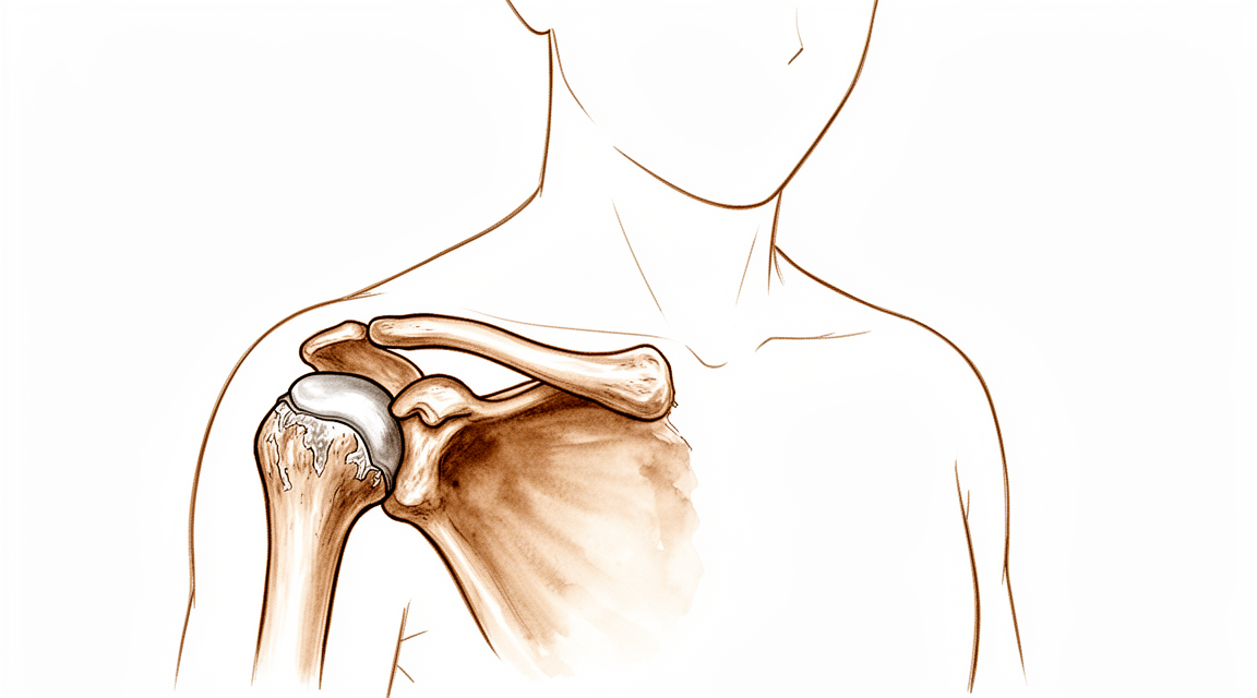
Vai của bạn có một khớp nhỏ nơi đầu ngoài của xương đòn gặp đỉnh của xương bả vai. Đây là khớp AC, viết tắt của khớp cùng vai đòn (acromioclavicular). Đây là một khớp có cử động, không phải một mối hàn cứng nhắc. Nó dịch chuyển vài milimét khi xương bả vai xoay và trượt trong lúc bạn sử dụng cánh tay bình thường, và giúp xương bả vai và xương đòn hoạt động như một khối liên kết thống nhất. Các dây chằng chắc khỏe giữ khớp lại với nhau, cùng với sự hỗ trợ của các cơ phủ phía trên vai.
Bên trong khớp có một đĩa đệm mỏng, hơi giống một miếng gioăng nằm giữa hai mẩu xương. Từ đầu tuổi trung niên trở đi, đĩa đệm này bắt đầu mòn, và các mặt khớp phía sau nó cũng mòn theo. Đó là viêm khớp do hao mòn, và ở khớp này nó rất phổ biến. Quá trình mòn thường âm thầm bắt đầu ở độ tuổi hai mươi và ba mươi, rồi tiến triển chậm theo năm tháng. Nâng vật nặng hoặc nâng lặp đi lặp lại, nhất là công việc giơ tay qua đầu, làm quá trình này diễn ra nhanh hơn. Một lần bong gân hoặc trật khớp này trước đây do ngã cũng vậy, kể cả một lần nhẹ tưởng như đã lành.
Điều đáng lưu ý là một khớp trông có vẻ bị mòn không phải lúc nào cũng gây đau. Nhiều người có dấu hiệu mòn trên phim chụp mà hoàn toàn không có triệu chứng, và hầu hết các khớp trông có vẻ bị mòn vẫn không gây triệu chứng. Vì vậy, cơn đau bạn cảm thấy chỉ xuất phát từ chính khớp này khi tình trạng mòn, các bề mặt khớp bị trầy trợt, hoặc tình trạng viêm xung quanh thực sự kích thích các mô tại đó. Đó là lý do triệu chứng của bạn nằm ngay ở phía trên vai và bùng lên sau khi vai phải chịu lực.
Một dạng ít gặp hơn của vấn đề này được gọi là tiêu xương đầu ngoài xương đòn. Trong trường hợp này, chính đầu ngoài của xương đòn bị tổn hại, với tình trạng viêm, các vết gãy nhỏ trong xương và mất xương. Tình trạng này chủ yếu gặp ở nam giới trẻ tuổi thường nâng vật nặng hoặc nâng lặp đi lặp lại, và nó dẫn đến cùng một dạng viêm khớp ở khớp này.
Nếu tình trạng mòn trở nên nặng, gai xương có thể mọc gần khớp và đè lên gân chạy bên dưới mái vai, làm vấn đề thêm trầm trọng.
Những biện pháp chúng tôi có thể áp dụng
Bác sĩ Kieran Hirpara, bác sĩ phẫu thuật chi trên tại Bệnh viện tư nhân Mater Rockhampton, sẽ bắt đầu với các phương pháp ít xâm lấn nhất phù hợp với tình trạng của bạn. Thông thường, bệnh nhân được bác sĩ đa khoa giới thiệu đến phòng khám chúng tôi; nếu nhà vật lý trị liệu khuyên bạn nên đến gặp chúng tôi, bạn vẫn cần có giấy giới thiệu từ bác sĩ đa khoa để được hưởng mức hoàn trả từ chương trình Medicare. Chúng tôi xác định tình trạng của bạn qua việc hỏi bệnh sử cẩn thận, thăm khám và chụp chiếu khi cần. Đối với một vấn đề kéo dài như thế này, chúng tôi thường thử điều trị không phẫu thuật trước và chỉ cân nhắc phẫu thuật khi cách đó không mang lại cải thiện đầy đủ.
Những bước đầu tiên là những việc bạn có thể tự thực hiện. Cho khớp nghỉ ngơi nghĩa là giảm bớt việc nâng vật nặng và công việc giơ tay qua đầu, là những việc làm phần trên của vai phải chịu lực. Chườm đá có thể làm dịu một đợt đau bùng phát sau một ngày bận rộn. Vật lý trị liệu nhằm làm dịu khớp đang bị kích ứng và tăng cường sức mạnh quanh vai để các công việc hằng ngày ít gây áp lực lên khớp hơn. Hãy thực hiện những thay đổi này một thời gian đủ dài trước khi đánh giá hiệu quả, vì cơn đau âm ỉ thường bùng lên sau khi hoạt động chứ không phải trong lúc hoạt động.
Nếu việc tự chăm sóc chưa đủ, thuốc giảm đau và thuốc chống viêm có thể giúp ích. Thuốc chống viêm là thuốc viên giúp làm dịu tình trạng viêm trong khớp đồng thời giảm đau. Chúng tôi cũng tiêm cortisone, một loại thuốc chống viêm mạnh, vào chính khớp này. Mũi tiêm có thể có hai mục đích: giúp xác nhận rằng khớp này là nguồn gốc cơn đau của bạn, và có thể giảm đau như một biện pháp điều trị. Khoảng 47 trên 100 người được tiêm vẫn có kết quả tốt sau một năm.
Phẫu thuật được cân nhắc khi cơn đau vẫn kéo dài dù đã áp dụng các phương pháp điều trị trên. Ca mổ cắt bỏ vài milimét xương ở đầu ngoài của xương đòn, tức là nửa khớp bị mòn nằm ở phía xương đòn. Khi bề mặt bị mòn đó được loại bỏ, hai đầu xương trầy trợt không còn cọ xát vào nhau nữa. Ca mổ có thể được thực hiện qua các vết rạch nhỏ bằng camera, hoặc qua một vết mổ mở nhỏ. Nếu trước đây bạn từng bị trật khớp này và khớp vẫn còn lỏng lẻo kéo dài, chúng tôi sẽ trao đổi kỹ với bạn về điều đó, vì nó ảnh hưởng đến mức độ hiệu quả của ca mổ này. Việc phẫu thuật có phù hợp với bạn hay không là quyết định mà chúng ta cùng nhau đưa ra, cân nhắc giữa những gì bạn muốn vai mình làm được và những gì ca mổ đòi hỏi.
Những điều bạn có thể mong đợi
Đối với hầu hết mọi người, tình trạng mòn ở khớp này xuất hiện trên phim chụp nhưng không gây đau thì vẫn cứ yên lặng như vậy. Trong thời gian bảy năm, 90% những khớp như thế này vốn không đau ngay từ đầu chưa bao giờ xuất hiện triệu chứng. Vì vậy, nếu khớp của bạn trông có vẻ bị mòn nhưng không đau, nhiều khả năng nó sẽ tiếp tục ổn định như vậy.
Khi khớp thực sự gây đau, diễn biến có thể khác nhau. Một số người thấy cơn đau âm ỉ dịu đi khi nghỉ ngơi, giảm bớt việc nâng vật nặng và tập vật lý trị liệu. Những người khác thấy cơn đau lúc có lúc không, bùng lên sau một ngày bận rộn và dịu đi khi bạn nghỉ ngơi trở lại. Nếu không điều trị, cơn đau thường kéo dài hoặc liên tục tái phát mỗi khi phần trên của vai phải chịu lực, vì tình trạng mòn trong khớp không tự hồi phục.
Nếu điều trị không phẫu thuật không giúp giảm đau đầy đủ, tiêm cortisone vào khớp có ích cho một số người. Khoảng 47 trên 100 người được tiêm vẫn có kết quả tốt sau một năm. Điều đó có nghĩa là với khoảng một nửa số người, hiệu quả giảm đau hết đi trong vòng một năm, và có thể cần tiêm lại hoặc cân nhắc các bước khác.
Nếu cơn đau vẫn kéo dài dù đã áp dụng các phương pháp điều trị này, cắt bỏ vài milimét xương ở đầu ngoài của xương đòn là một lựa chọn mà bác sĩ phẫu thuật có thể trao đổi với bạn. Mục đích là để hai bề mặt bị mòn, trầy trợt không còn cọ xát vào nhau. Quá trình hồi phục sau ca mổ đó mất vài tuần đến vài tháng, với sự hướng dẫn của nhà vật lý trị liệu, và hầu hết mọi người thấy cơn đau âm ỉ ở phía trên vai dịu đi khi khớp ổn định trở lại.
Những gì bạn có thể kỳ vọng một cách thực tế trong những tháng tới tùy thuộc vào hướng điều trị bạn chọn. Với việc tự chăm sóc hợp lý và vật lý trị liệu, nhiều người trở lại các hoạt động bình thường với cơn đau âm ỉ đã dịu xuống ở mức họ có thể chung sống được. Nếu khớp vẫn liên tục bùng phát cơn đau dù đã thử các bước này một thời gian hợp lý, bạn nên quay lại gặp bác sĩ đa khoa hoặc đề nghị được bác sĩ chuyên khoa đánh giá thay vì cố chịu đựng cơn đau.
Khi nào nên đi khám
Đây là vấn đề do hao mòn, không phải tình trạng cấp cứu, vì vậy không có dấu hiệu nguy hiểm nào cần xử lý ngay trong ngày đối với bệnh lý này. Điều quan trọng là cơn đau có dịu đi hay không. Hãy đến gặp bác sĩ đa khoa nếu cơn đau ở phía trên vai không cải thiện sau vài tuần ngừng nâng vật nặng, hoặc nếu nó liên tục làm bạn thức giấc khi nằm nghiêng về bên đó. Hãy đề nghị được bác sĩ chuyên khoa đánh giá nếu cơn đau âm ỉ nặng dần lên qua nhiều tuần, khiến bạn không thể làm việc hoặc sử dụng cánh tay bình thường, hoặc nếu việc nâng vật và vươn tay ngang qua thân mình vẫn gây đau dù bạn đã giảm bớt hoạt động. Nếu vai của bạn trở nên nóng, đỏ, sưng và đau, đặc biệt là kèm theo sốt, đó không phải là viêm khớp và bạn cần đến khoa cấp cứu để được chăm sóc ngay trong ngày.
Phân tích sâu hơn
Advanced reading: the deeper science (optional)
Phần này đi sâu hơn mức cần thiết để bạn đưa ra các quyết định điều trị. Việc tìm hiểu về viêm khớp khớp AC là điều đáng làm, dù với một lý do không mấy dễ chịu: tình trạng này rất phổ biến khi chụp hình ảnh y tế, thường bị coi là nguyên nhân gây đau vai; tuy nhiên các bằng chứng cho thấy việc điều trị có hiệu quả lại ít hơn hẳn so với hầu hết các vấn đề khác được đề cập trên trang web này.
Các bằng chứng hiện có không chứng minh được rằng bất kỳ phương pháp điều trị nào có hiệu quả
Một phân tích hệ thống về các phương pháp điều trị viêm khớp vai nguyên phát đã tổng hợp dữ liệu từ 1.902 bệnh nhân và đi đến kết luận hiếm khi được nêu ra một cách rõ ràng như vậy: các nghiên cứu có sự khác biệt về chỉ định, phương pháp can thiệp và chất lượng; do đó không cung cấp bằng chứng cho thấy các biện pháp điều trị không phẫu thuật hay phẫu thuật đều có hiệu quả [1].
Điều này không có nghĩa là không có phương pháp nào giúp ích. Điều này chỉ có nghĩa là các thử nghiệm cần thiết để chứng minh điều đó vẫn chưa được tiến hành theo tiêu chuẩn đủ để đưa ra kết luận. Phẫu thuật cắt bỏ đầu xương đòn là một thủ thuật đã được áp dụng từ lâu; lý do thực hiện là dựa trên cơ sở cơ học rằng việc loại bỏ bề mặt khớp bị tổn thương sẽ giúp giảm đau. Tuy nhiên, “đã được áp dụng từ lâu và có cơ sở cơ học hợp lý” không đồng nghĩa với việc đó là bằng chứng khoa học; việc hiểu rõ sự khác biệt này là rất quan trọng khi đưa ra quyết định điều trị.
Việc thực hiện thủ thuật này cùng với các phẫu thuật khác không mang lại hiệu quả tốt hơn
Bằng chứng rõ ràng nhất đến từ những bệnh nhân được thực hiện thêm các thủ thuật khác trong cùng một ca mổ. Trong số 208 bệnh nhân bị rách gân cơ chóp xoay, việc thêm thủ thuật cắt bỏ phần xa của xương đòn không giúp cải thiện điểm số đánh giá lâm sàng hay phạm vi vận động của bệnh nhân [2].
Điều này rất quan trọng vì đây chính là tình huống thường gặp nhất khi cần cắt bỏ phần xương này: bác sĩ phẫu thuật đã ở trong vùng vai, hình ảnh chụp cho thấy khớp AC đã bị thoái hóa, và việc cắt bỏ vài milimét xương cũng rất nhanh chóng. Tuy nhiên, các bằng chứng cho thấy bệnh nhân không nhận được lợi ích đáng kể nào từ việc này. Nếu có đề xuất thực hiện thủ thuật này như một bước bổ sung cho ca phẫu thuật sửa chữa gân cơ chóp xoay, thì việc đặt câu hỏi về hiệu quả của nó là hoàn toàn hợp lý.
Kỹ thuật không phải là vấn đề đáng quan tâm
Việc so sánh giữa phương pháp mổ mở và phẫu thuật nội soi đã được thực hiện nhiều lần, và kết quả luôn nhất quán: kết quả chức năng và lâm sàng tương đương giữa hai phương pháp trên 319 bệnh nhân [3]. Một nghiên cứu trước đó với 429 bệnh nhân cũng không cho thấy sự vượt trội rõ rệt của phương pháp nào [4].
Khi hai phương pháp kỹ thuật hoàn toàn khác nhau lại cho ra cùng một kết quả, kết luận hợp lý là kỹ thuật không phải yếu tố quyết định kết quả; điều quan trọng là việc lựa chọn bệnh nhân.
Tại sao việc chẩn đoán lại khó khăn đến vậy
Gần như mọi người đều bị thoái hóa khớp AC theo tuổi tác. Đây là một khớp nhỏ nhưng chịu nhiều lực; đĩa khớp mỏng nên dễ bị mòn sớm. Vì vậy, việc phát hiện dấu hiệu thoái hóa khớp AC trên phim chụp X-quang ở người trung niên gần như là điều dễ hiểu chứ không phải là một chẩn đoán bệnh lý.
Có một mối tương quan cấu trúc có thể đo lường được: ở những người có triệu chứng thoái hóa khớp AC, cả mỏm dưới đòn và mỏm cùng vai đều to ra; ngược lại, ở những người không có triệu chứng thì tỷ lệ giữa hai bộ phận này vẫn không thay đổi [5]. Đây là manh mối hữu ích cho thấy sự khác biệt về cấu trúc giữa tình trạng thoái hóa có triệu chứng và tình trạng thoái hóa ngẫu nhiên; tuy nhiên đây chỉ là quan sát ở cấp độ nhóm chứ không phải là xét nghiệm có thể áp dụng riêng cho từng bệnh nhân.
Trên thực tế, đó là lý do tại sao việc tiêm thuốc gây tê vào khớp AC có ý nghĩa quyết định trong chẩn đoán. Nếu việc tiêm thuốc gây tê vào đúng vị trí khớp AC giúp giảm đau, thì khớp này rất có thể là nguyên nhân gây bệnh. Ngược lại, nếu không có hiệu quả, thì dấu hiệu thoái hóa trên phim chụp chỉ là hiện tượng phụ; việc cắt bỏ các tổn thương này hầu như không mang lại lợi ích gì – điều mà các bằng chứng nêu trên đang cảnh báo.
Tài liệu tham khảo
[1] Welch M, Rankin S, How Saw Keng M, Woods D. Tổng quan có hệ thống về các phương pháp điều trị viêm khớp vai mức độ nặng. Shoulder Elbow. 2023;16(2):129-44. https://doi.org/10.1177/17585732231157090
[2] Wang J, Ma J, Zhu S, Jia H, Ma X. Việc cắt bỏ phần xa xương đòn có giúp giảm đau hoặc cải thiện chức năng vai ở bệnh nhân bị rách gân cơ chóp xoay không? Phân tích tổng hợp. Clin Orthop Relat Res. 2018;476(12):2402-14. https://doi.org/10.1097/CORR.0000000000000424
[3] Hohmann E, Tetsworth K, Glatt V. Phẫu thuật mở so với phẫu thuật nội soi trong việc cắt bỏ khớp vai: Tổng quan có hệ thống và phân tích tổng hợp. Arch Orthop Trauma Surg. 2019;139(5):685-94. https://doi.org/10.1007/s00402-019-03114-w
[4] Pensak M, Grumet RC, Slabaugh MA, Bach BR. Phẫu thuật mở so với phẫu thuật nội soi trong việc cắt bỏ phần xa xương đòn. Arthroscopy. 2010;26(5):697-704. https://doi.org/10.1016/j.arthro.2009.12.007
[5] Bulkmans K, Peeters I, De Wilde L, Van Tongel A. Mối liên hệ giữa mỏm vai và phần xa xương đòn ở những người bình thường cũng như bệnh nhân bị thoái hóa khớp vai. Arch Orthop Trauma Surg. 2019;140(4):465-72. https://doi.org/10.1007/s00402-019-03258-9
Evidence & references
This is the clinical evidence summary written for health professionals. It is technical, and it lists the research this page was built from. You do not need to read it to understand your treatment or to make a decision about it.
Overview
- Asymptomatic acromioclavicular osteoarthritis remained asymptomatic in 90% of cases over a seven-year course [1].
- Treatment choice for acromioclavicular dislocation should not be influenced by the potential development of acromioclavicular osteoarthritis [2].
- Further characterisation is needed to identify patients in whom mild arthroscopic findings of acromioclavicular joint osteoarthritis are clinically significant and warrant resection [3].
- Persistent pain and osteoarthritis progression remain concerns following revision of failed open anterior stabilization of the shoulder [4].
- Both open and arthroscopic resection arthroplasty techniques provide predictable pain relief for symptomatic acromioclavicular osteoarthritis [5].
- Symptomatic acromioclavicular joint osteoarthritis occurred in 15.9% of patients after total anatomic shoulder replacement with follow-up of up to 12 years [6].
- Untreated acromioclavicular joint osteoarthritis, symptomatic or not, encountered during arthroscopic rotator cuff repair is associated with a low percentage of failure [8].
- Biplanar reconstruction with pectoralis minor tendon and coracoacromial ligament transfer aims to preserve the lateral portion of the clavicle whenever possible, limiting resection exclusively to cases of acromioclavicular joint osteoarthritis or irreducible joints [9].
- Radiographic acromioclavicular joint osteoarthritis is common in patients undergoing reverse shoulder arthroplasty [10].
- Clinicians should consider overreduction of the acromioclavicular joint because it may lead to favorable radiological results [11].
- A distinction between symptomatic and asymptomatic radiographic acromioclavicular osteoarthritis is unnecessary, as all patients were equally satisfied with the outcome following preoperative acromioclavicular injection [12].
- Open and arthroscopic acromioclavicular joint reconstruction techniques have no differences in loss of reduction, the complication rate, and the revision rate [13].
- Arthroscopic debridement of the acromioclavicular joint can produce a favorable result in young patients presenting with isolated acromioclavicular joint-related pain but with normal imaging [15].
- Limited distal clavicle excision of patients with acromioclavicular joint osteoarthritis resistant to conservative treatment reduced pain and improved shoulder function at midterm follow-up [17].
- All patients undergoing free tissue graft reconstruction of the acromioclavicular joint are satisfied with their surgery and demonstrate good reduction of the acromioclavicular joint on radiographs, although follow-up periods are less than 1 year [21].
- Additional research is needed to determine the main cause of pain and compare clinical outcomes of intra-articular versus extra-articular injections for isolated acromioclavicular osteoarthritis [23].
- Nanoscopic distal clavicle resection offers advantages including decreased morbidity, less risk to the acromioclavicular joint ligaments, and decreased loss and need for fluid [44].
Anatomy & Pathophysiology
Bony Anatomy and Joint Structure
- The acromioclavicular (AC) joint is a diarthrodial joint that supports the shoulder girdle through the clavicular “strut” [36].
- The AC joint serves as a primary link between the axial skeleton and the upper extremity [122].
- The AC joint is movable in all planes and is not a rigid structure [122].
- The normal AC joint is capable of translating 4 to 6 mm in the anterior, posterior, and superior planes under 70-N loads [122].
- The AC joint accommodates rotary motion of 5° to 8° during scapulothoracic motion and 40° to 45° with shoulder abduction and elevation [122].
- The scapula and clavicle are linked into a single segment referred to as the “claviscapula” [112].
Ligamentous Anatomy and Stability
- The AC and coracoclavicular (CC) ligaments are the static stabilizers of the AC joint, whereas the deltoid and trapezius muscles are the dynamic stabilizers [122].
- The horizontal plane stability of the clavicle is provided by the AC ligaments, specifically the posterior and superior portions [25].
- The AC and CC ligaments play a role in torque transduction and horizontal and vertical stability [112].
- Injury to the AC joint results in progressive disruption of the ligamentous support, beginning with the capsular ligaments and progressing to the CC ligaments [30].
Pathophysiology of Degeneration
- AC joint osteoarthritis is more common with advanced age following degeneration of the intra-articular disk [25].
- Arthritic deterioration of the AC joint starts in early middle age [25].
- AC joint osteoarthritis is more common in patients engaged in repetitive overhead or lifting activities [25].
- Previous low-grade AC joint separations can result in painful arthritis [25].
- The radiographic severity of AC joint arthritis does not always correlate with patient symptoms [25].
- Asymptomatic AC joint degeneration is frequent and does not always correlate with the presence of symptoms [36].
- Degeneration of the AC joint is classified as primary osteoarthritis, posttraumatic osteoarthritis, or distal clavicle osteolysis [123].
- The process of AC joint degeneration begins in the second decade of life and can progress to complete obliteration of the joint [123].
- Complete obliteration of the AC joint can lead to subclavicular spurring, which contributes to subacromial impingement of the supraspinatus [123].
- Asymptomatic AC osteoarthritis remained asymptomatic in 90% of cases over a seven-year course [1].
- Patients with loss of immediate postoperative reduction after AC joint dislocation more often developed radiologic and symptomatic AC osteoarthritis than those in whom the AC joint was anatomically restored [7].
- Kinematic changes associated with AC joint dislocation could be a potential source of pain and dysfunction in the shoulder [59].
- Scapular and clavicular kinematics are affected in AC separation models [60].
- Decoupling the claviscapular segment has deleterious effects on the functional integrity of the AC joint complex within scapulohumeral rhythm [112].
Distal Clavicle Osteolysis
- Distal clavicle osteolysis is characterized by localized hyperemia of the distal clavicle, resulting in inflammation, bone resorption, microfractures, and secondary arthritis of the AC joint [25].
- Distal clavicle osteolysis is more common in males and seen in younger patients [25].
- Distal clavicle osteolysis is associated with heavy lifting or repetitive motions [25].
- Osteolysis of the distal clavicle was first reported in conjunction with acute trauma [121].
- Intraarticular distal clavicle fractures and AC separations can lead to osteolysis [121].
- Other causes of distal clavicle osteolysis include rheumatoid arthritis, hyperparathyroidism, and repetitive microtrauma [121].
- The most common cause of distal clavicle osteolysis is repetitive microtrauma [121].
- Radiographic findings indicative of distal clavicle osteolysis include loss of subchondral bone detail, cystic reabsorption of the distal clavicle, and generalized osteopenia of the distal clavicle [121].
Classification
- Asymptomatic acromioclavicular osteoarthritis diagnosed by MRI remained asymptomatic in 90% of cases over a seven-year course [1].
- Treatment choice for acromioclavicular dislocation should not be influenced by the potential development of acromioclavicular osteoarthritis based on available evidence [2].
- Patients with loss of immediate postoperative reduction following arthroscopically assisted reduction of acute acromioclavicular joint dislocation more often developed radiologic and symptomatic acromioclavicular osteoarthritis than those in whom the joint was anatomically restored [7].
- Untreated acromioclavicular joint osteoarthritis, whether symptomatic or not, encountered during arthroscopic rotator cuff repair is associated with a low percentage of failure [8].
- Biplanar reconstruction with pectoralis minor tendon and coracoacromial ligament transfer aims to preserve the lateral portion of the clavicle, limiting resection exclusively to cases of acromioclavicular joint osteoarthritis or irreducible joints [9].
- Open and arthroscopic acromioclavicular joint reconstruction techniques have no differences in loss of reduction, complication rate, and revision rate based on available literature [13].
- A study evaluated three groups of patients: 84 acromioclavicular joints without clinical or radiological pathology, 39 patients with signs of osteoarthritis on CT without symptoms, and 30 patients with acromioclavicular joint pain [14].
- In the study of acromion-clavicle relationships, Group 2 included patients with signs of osteoarthritis on CT (joint space narrowing, osteophytes, and/or bone cyst) who were asymptomatic based on physical examination and negative cross-body adduction test [14].
- In the study of acromion-clavicle relationships, Group 3 included patients with acromioclavicular joint pain confirmed by tenderness and temporary pain relief with intra-articular injection, who were treated with open distal clavicle resection after failure of non-operative treatment [14].
- The occurrence of acromioclavicular joint osteoarthrosis was surprisingly frequent in a study of shoulder pain [18].
- Anatomic techniques that address both coracoclavicular ligaments and the acromioclavicular capsule are recommended to restore horizontal and vertical stability while allowing physiological rotation [19].
- The acromial center line to dorsal clavicle radiographic measurement and use of the Alexander view provides a more realistic appreciation of true acromioclavicular joint displacement, especially in defining watershed cases (IIIA/IIB/IV) [22].
- Osteolytic changes seemed to be associated with incongruity of the acromioclavicular joint but did not correlate with clinical results in a review of surgical treatment for acromioclavicular dislocation [24].
- A procedure for exact anatomical acromioclavicular joint reconstruction using acromioclavicular and coracoclavicular FiberWire cerclage shows good objective results with high Constant and Rowe scores for shoulder function [27].
- Preoperative radiographs were evaluated to determine whether there was any osteolysis of the distal clavicle in a study of preoperative factors associated with subsequent distal clavicle resection after rotator cuff repair [31].
- Preoperative radiographs were graded for severity of arthritis per the Petersson classification by two attending orthopaedic surgeons blinded to each other's grade in a study of preoperative factors associated with subsequent distal clavicle resection after rotator cuff repair [31].
- Type V acromioclavicular dislocations may be given a trial of conservative therapy [35].
- Non-operative reduction and stabilization of high-grade acromioclavicular joint separations seems to be a valuable treatment option [45].
- Evaluating the integrity of the capsuloligamentous structures stabilizing the acromioclavicular joint is reproducible and gives additional information on the severity of the injury, which might also influence the treatment decision [49].
- Severe chronic symptomatic acromioclavicular joint separations (Rockwood types III through V) can be repaired entirely by arthroscopy safely and effectively by transferring the coracoacromial ligament with a bone block in the distal clavicle [50].
- The treatment of Type III acromioclavicular joint separation has remained controversial, with many studies demonstrating good results with nonoperative management and most series failing to document the superiority of either operative or nonoperative management [51].
- Surgical management of acromioclavicular joint injuries is associated with a relatively high complication profile and common loss of reduction, though the revision surgery rate is low [53].
- Arthroscopically assisted treatment of acute acromioclavicular joint dislocation provides good clinical results and few complications, though the rate of recurrence and postoperative loss of reduction requires better definition of indications and improvement of surgical implants and technique [55].
- In every coracoclavicular loop repair, regardless of hole placement, complete acromioclavicular joint congruity is not restored [86].
- Given that more than 100 surgical stabilization techniques have been proposed for acromioclavicular joint injuries, a deeper understanding of the underlying anatomy integrated with biomechanical evidence may help guide the development of a standardized, evidence-based approach to treatment [88].
- Early and delayed surgical interventions of high-grade acromioclavicular joint dislocation provide equivalent clinical scores when combined coracoclavicular and acromioclavicular joint fixation is used for stabilization [95].
- Acromioclavicular joint osteoarthritis was graded using the Kellgren-Lawrence classification system in a retrospective cohort study of concomitant open distal clavicle excision after reverse total shoulder arthroplasty [101].
Clinical Presentation
History and Symptoms
- Patients report activity-related pain localized to the AC joint [25].
- Pain may radiate anteriorly or along the trapezius [25].
- Pain is reported with heavy lifting or when sleeping on the affected side [25].
- AC joint osteoarthrosis is a frequent cause of shoulder pain [18].
- In patients with rheumatoid arthritis, clinical symptoms and radiologic appearances are often poor indicators of the source of pain in the shoulder joint complex [64].
Physical Examination
- Point tenderness is seen at the AC joint [25].
- Horizontal stability of the AC joint should be assessed [25].
- Pain at the AC joint with terminal elevation and cross-body motion is often seen [25].
- The adduction sign, where pain is produced in the AC joint during maximal adduction of the internally rotated arm, was positive in all 20 patients with AC joint arthritis in one study [63].
- An average decrease of 5 cm in the distance from the ipsilateral antecubital fossa to the contralateral anterior shoulder was noted during cross-body adduction in patients with AC joint arthritis [63].
- Selective injection of anesthetic into the AC joint can confirm the diagnosis [25].
- Patients with ACJ pain showed clinical signs including AC joint tenderness to palpation and temporary pain relief with intra-articular injection [14].
Imaging
- An AP view and/or a Zanca view of the shoulder provides good visualization of the AC joint [25].
- Osteophyte formation, sclerotic reaction, and bone cysts are commonly seen on radiographs of the AC joint [25].
- Bone and joint edema on MRI correlate with AC joint pain [25].
- The radiographic severity of arthritis does not always correlate with patient symptoms [25].
- Radiographic ACJ osteoarthritis is common in patients undergoing reverse shoulder arthroplasty [10].
- Asymptomatic AC-OA remained asymptomatic in 90% of patients over a seven-year course [1].
- A distinction between symptomatic and asymptomatic radiographic AC OA may be unnecessary, as all patients were equally satisfied with the outcome in one study [12].
- Osteolytic changes seemed to be associated with incongruity of the AC joint but did not correlate with clinical results [24].
Investigations
Clinical Evaluation
- Patients with AC joint osteoarthritis report activity-related pain localized to the AC joint, with occasional radiation anteriorly or along the trapezius [25].
- Pain with heavy lifting or when sleeping on the affected side is reported in patients with AC joint osteoarthritis [25].
- Physical examination of the AC joint reveals point tenderness at the joint [25].
- Horizontal stability of the AC joint should be assessed during physical examination [25].
- Pain at the AC joint with terminal elevation and cross-body motion is often seen in patients with AC joint osteoarthritis [25].
- Selective injection of anesthetic into the AC joint can confirm the diagnosis of symptomatic AC osteoarthritis [25].
- A distinction between symptomatic and asymptomatic radiographic AC OA is unnecessary, as all patients were equally satisfied with the outcome following preoperative injection [12].
Imaging
- Patients with edema on MRI were more likely to present pain than patients without edema [92].
- Subchondral bone edema on histologic examination was more frequent in patients with pain [92].
- The AC-DC measurement and use of the Alexander view provides a more realistic appreciation of true AC joint displacement, especially in defining watershed cases (ie, IIIA/IIB/IV) [22].
- Preoperative radiographs are evaluated to determine whether there is any osteolysis of the distal clavicle [31].
- The Petersson classification is used to grade the severity of arthritis on preoperative radiographs [31].
- In a study of AC joint osteoarthrosis, the occurrence was surprisingly frequent [18].
Treatment
Non-Operative Management
- Rest, ice, and NSAIDs are used initially for AC joint osteoarthritis [25].
- Corticosteroid injections can be used for diagnostic or therapeutic purposes in AC joint osteoarthritis [25].
- AC injections offer a 1-year success rate of 47% [33].
- Conservative and surgical treatments are both effective in acromioclavicular joint osteoarthritis management [38].
- A distinction between symptomatic and asymptomatic radiographic AC OA is unnecessary, as all patients were equally satisfied with the outcome following injection [12].
- Additional research is needed to determine the main cause of pain and compare clinical outcomes of intra-articular versus extra-articular injections [23].
Operative Management: Distal Clavicle Resection
- Surgical indications for AC joint osteoarthritis include persistent pain and failure of nonsurgical treatment [25].
- Both open and arthroscopic resection arthroplasty techniques provide predictable pain relief for symptomatic AC osteoarthritis [5].
- Arthroscopic distal clavicle excision provides reliable pain relief in >90% of patients in the absence of instability [25].
- Previous traumatic instability is associated with persistent pain in 30% to 40% of cases following distal clavicle excision [25].
- One systematic review showed slightly better results with arthroscopic excision than with open distal clavicle excision [25].
- Direct comparison studies have shown similar or better results with arthroscopic excision than with open techniques [25].
- Biomechanical evidence suggests a resection of 5 mm is needed to prevent contact between the clavicle and the acromion in the absence of instability [25].
- Care should be taken to preserve the posterior and superior AC ligaments during arthroscopic distal clavicle excision [25].
- Between 5 and 10 mm of the distal clavicle should be resected during an open Mumford procedure [25].
- Meticulous repair of the deltotrapezial fascia is important during open distal clavicle resection [25].
- Limited distal clavicle excision of patients with AC joint osteoarthritis resistant to conservative treatment reduced pain and improved shoulder function at midterm follow-up [17].
- Arthroscopic debridement of the AC joint can produce a favorable result in young patients presenting with isolated AC joint-related pain but with normal imaging [15].
- The nanoscopic distal clavicle resection technique offers advantages including decreased morbidity, less risk to the AC joint ligaments, and decreased loss and need for fluid [44].
- Patients with AC joint instability have poor results after distal clavicle resection [36].
- The optimal amount of distal clavicle resection remains elusive [36].
Operative Management: Concomitant Procedures and Specific Populations
- Preventive arthroscopic distal clavicle resection is not recommended in patients with radiological but asymptomatic ACJ arthritis and rotator cuff tears [34].
- Untreated ACJ osteoarthritis, symptomatic or not, encountered during arthroscopic rotator cuff repair is associated with a low percentage of failure [8].
- Further characterisation of patients in whom mild arthroscopic findings of OA of AC joint are clinically significant and warrant resection is needed [3].
- Symptomatic ACJ OA occurred in 15.9% of patients after total anatomic shoulder replacement with follow-up of up to 12 years [6].
- Osteoarthritis is associated with poorer final clinical outcomes after rotator cuff repair, and an unhealed or re-torn cuff increases the risk of osteoarthritis [26].
Operative Management: Reconstruction and Stabilization
- Open and arthroscopic AC joint reconstruction techniques have no differences in loss of reduction, the complication rate, and the revision rate based on the available literature [13].
- Treatment choice for AC dislocation should not be influenced by the potential development of AC OA [2].
- Achieving and maintaining satisfactory joint reduction is a factor of good prognosis, both radiologically and functionally, in AC dislocation treatment [7].
- Patients with loss of immediate postoperative reduction more often developed radiologic and symptomatic AC osteoarthritis than those in whom the AC joint was anatomically restored [7].
- Clinicians should consider overreduction of the AC joint because it may lead to favorable radiological results [11].
- Ensuring good initial recovery of the coracoclavicular distance and precise placement of the AC joints was important in maintaining the proper AC position at the final follow-up [46].
- Combined stabilization of the AC capsule and CC ligaments demonstrated the greatest capacity to restore native stability against translational and rotational loads [81].
- The biplanar reconstruction procedure aims to preserve the lateral portion of the clavicle whenever possible, limiting resection exclusively to cases of AC joint osteoarthritis or irreducible joints [9].
- Some persistent pain and osteoarthritis progression remain concerns following revision of failed open anterior stabilization of the shoulder [4].
Complications
Natural History and Progression
- Patients who experienced loss of immediate postoperative reduction after acute acromioclavicular dislocation more often developed radiologic and symptomatic acromioclavicular osteoarthritis than those in whom the joint was anatomically restored [7].
- Osteoarthritis is associated with poorer final clinical outcomes following rotator cuff repair [26].
- An unhealed or re-torn rotator cuff increases the risk of developing osteoarthritis [26].
- Severe acromioclavicular joint osteoarthritis is associated with acromial stress fractures after reverse shoulder arthroplasty [10].
Surgical Complications and Outcomes
- Treatment of acromioclavicular dislocation by synthetic ligament reconstruction carries a risk of significant early osteolysis [56].
- Complications such as anterior shoulder pain, acromioclavicular joint asymmetry, and activity-related weakness are common sequelae of coracoclavicular ligament reconstruction [43].
- Three patients with old acromioclavicular separations had prominence of the distal clavicle or symptoms due to residual hypermobility of the clavicle despite concurrent coracoclavicular stabilization [114, 115, 116, 117].
- A patient who did not undergo anterior acromioplasty despite subacromial impingement had unchanged impingement symptoms after distal clavicle resection [114, 115, 116, 117].
- Open and arthroscopic resection arthroplasty techniques for symptomatic acromioclavicular osteoarthritis each have a unique set of potential complications that may be minimized with improved understanding of anatomy, biomechanics, and meticulous surgical technique [5].
Recovery
Natural History and Prognosis
- Asymptomatic acromioclavicular osteoarthritis diagnosed by MRI remained asymptomatic in 90% of patients over a seven-year course [1].
- Osteolytic changes associated with incongruity of the acromioclavicular joint did not correlate with clinical results in a review of 39 patients [24].
- An unhealed or re-torn rotator cuff increases the risk of osteoarthritis following rotator cuff repair [26].
Impact of Joint Reduction and Stability
- Patients with loss of immediate postoperative reduction after arthroscopically assisted reduction of acute acromioclavicular joint dislocation more often developed radiologic and symptomatic acromioclavicular osteoarthritis than those in whom the joint was anatomically restored [7].
- Achieving and maintaining satisfactory joint reduction is a factor of good prognosis, both radiologically and functionally [7].
- Ensuring good initial recovery of the coracoclavicular distance and precise placement of the acromioclavicular joints was important in maintaining the proper acromioclavicular position at final follow-up [46].
Surgical Considerations Regarding Osteoarthritis
- Further characterisation of patients in whom mild arthroscopic findings of acromioclavicular joint osteoarthritis are clinically significant and warrant resection is needed [3].
- Biplanar reconstruction for chronic acromioclavicular joint dislocations aims to preserve the lateral portion of the clavicle, limiting resection exclusively to cases of acromioclavicular joint osteoarthritis or irreducible joints [9].
- The average angle from the central portion of the acromioclavicular joint anteriorly for a novel anterior portal for arthroscopic distal clavicle excision was 6 degrees, with a range of 0 to 15 degrees [29].
Non-Operative Management and Diagnostic Implications
- Acromioclavicular injections offer a 1-year success rate of 47% in patients with acromioclavicular osteoarthritis [33].
Key Evidence
- [L2] Asymptomatic AC-OA remained asymptomatic in 90% over 7 years. [1] (10.1016/j.jse.2019.04.004)
- [L4] Based on the available evidence, treatment choice for AC dislocation should not be influenced by the potential development of AC OA. [2] (10.2106/jbjs.rvw.24.00085)
- [L2] Further characterisation of patients in whom mild arthroscopic findings of OA of AC joint are clinically significant and warrant resection is needed. [3] (10.1007/s00167-014-3114-2)
- [L4] Some persistent pain and osteoarthritis progression remain concerns. [4] (10.1016/j.arthro.2009.04.073)
- [L5] Both open and arthroscopic resection arthroplasty techniques provide predictable pain relief for symptomatic AC osteoarthritis, though each has a unique set of potential complications that may be minimized with improved understanding of anatomy, biomechanics, and meticulous surgical technique. [5] (10.1177/0363546513485359)
- [L4] Symptomatic ACJ OA occurred in 15.9% of patients after total anatomic shoulder replacement with follow-up of up to 12 years. [6] (10.1177/17585732221114796)
- [L4] [7] (10.1016/j.otsr.2017.11.001)
- [L2] Untreated ACJ osteoarthritis, symptomatic or not, encountered during arthroscopic RCR is associated with a low percentage of failure. [8] (10.1007/s00167-020-06098-y)
- [L5] The procedure aims to preserve the lateral portion of the clavicle whenever possible, limiting resection exclusively to cases of AC joint osteoarthritis or irreducible joints. [9] (10.1016/j.eats.2024.103104)
- [L3] Radiographic ACJ osteoarthritis is common in patients undergoing RSA. [10] (10.1016/j.jseint.2021.11.008)
- [L3] Clinicians should consider overreduction of the AC joint because it may lead to favorable radiological results. [11] (10.1177/0363546519862850)
- [L4] The outcomes of this study seem to suggest that a distinction between symptomatic and asymptomatic radiographic AC OA is unnecessary, as all patients were equally satisfied with the outcome. [12] (10.5397/cise.2023.00073)
- [L1] Open and arthroscopic AC joint reconstruction techniques have no differences in loss of reduction, the complication rate, and the revision rate based on the available literature. [13] (10.1177/0363546518795147)
- [L3] [14] (10.1007/s00402-019-03258-9)
- [L4] Arthroscopic debridement of the AC joint can produce a favorable result in this difficult group of young patients presenting with isolated AC joint-related pain but with normal imaging. [15] (10.1097/bte.0b013e31816276cb)
- [L4] Limited distal clavicle excision of patients with AC joint osteoarthritis resistant to conservative treatment reduced pain and improved shoulder function at midterm follow-up. [17] (10.1016/j.otsr.2016.01.008)
- [L3] In this study the occurrence of AC joint osteoarthrosis was surprisingly frequent. [18] (10.1016/s1058-2746(96)80122-4)
- [L5] Anatomic techniques that address both coracoclavicular ligaments and the AC capsule are recommended to restore horizontal and vertical stability while allowing physiological rotation. [19] (10.1016/j.arthro.2019.01.038)
- [L4] Although the follow-up periods are less than 1 year, all patients are satisfied with their surgery and demonstrate good reduction of the AC joint on radiographs. [21] (10.1097/bte.0b013e31818a5dd9)
- [L4] The AC-DC measurement and use of the Alexander view provides the clinician with a more realistic appreciation of true AC joint displacement, especially in defining watershed cases (ie, IIIA/IIB/IV) and may better inform the decision-making process regarding management options and recommendations. [22] (10.1016/j.jse.2019.12.014)
- [L4] Additional research is needed to determine the main cause of pain and compare clinical outcomes of intra-articular versus extra-articular injections. [23] (10.5397/cise.2023.00311)
- [L4] Osteolytic changes seemed to be associated with incongruity of the AC joint, but did not correlate with clinical results. [24] (10.1016/0020-1383(83)90092-x)
- [L4] Osteoarthritis is associated with poorer final clinical outcomes, and an unhealed or re-torn cuff increases the risk of osteoarthritis. [26] (10.1016/j.otsr.2017.03.007)
- [L4] This procedure for an exact anatomical AC joint reconstruction shows good objective results with a high Constant and Rowe scores for shoulder function. [27] (10.1055/s-2008-1038577)
- [L4] The average angle from the central portion of the AC joint anteriorly was 6 degrees (range, 0 to 15 degrees). [29] (10.1097/bte.0000000000000033)
- [L3] [31] (10.1177/2325967119844295)
- [L4] AC injections offer a 1-year success rate of 47%. [33] (10.5397/cise.2023.00031)
- [L1] Preventive arthroscopic DCR is not recommended in patients with radiological but asymptomatic ACJ arthritis. [34] (10.1177/0363546514547254)
- [L4] This suggests that Type V AC dislocations may be given a trial of conservative therapy. [35] (10.1177/2325967115s00017)
- [L5] [36] (10.5435/00124635-199905000-00004)
- [L4] Conservative and surgical treatments are both effective in acromioclavicular joint osteoarthritis management. [38] (10.1007/s00167-020-06377-8)
- [L4] Most patients were ultimately treated surgically, although complications such as anterior shoulder pain, AC joint asymmetry, and activity-related weakness were common sequelae resulting in physical limitations and separation from military service. [43] (10.1177/03635465211036713)
- [L5] The technique offers advantages including decreased morbidity, less risk to the AC joint ligaments, and decreased loss and need for fluid. [44] (10.1016/j.eats.2021.12.006)
- [L4] Non-operative reduction and stabilization of high-grade AC joint separations seems to be a valuable treatment option. [45] (10.1007/s00402-020-03630-0)
- [L4] Ensuring good initial recovery of the CCD and precise placement of the AC joints was important in maintaining the proper AC position at the final follow-up. [46] (10.1016/j.arthro.2018.07.007)
- [L1] Evaluating the integrity of the capsuloligamentous structures stabilizing the AC joint is reproducible and gives additional information on the severity of the injury, which might also influence the treatment decision. [49] (10.1016/j.jse.2020.10.026)
- [L4] Severe chronic symptomatic AC joint separations (Rockwood types III through V) can be repaired entirely by arthroscopy safely and effectively by transferring the coracoacromial ligament with a bone block in the distal clavicle. [50] (10.1016/j.arthro.2009.08.008)
- [L5] [51] (10.1097/00132589-200603000-00004)
- [L4] Surgical management of AC joint injuries is associated with a relatively high complication profile and common loss of reduction, though the revision surgery rate is low. [53] (10.1016/j.jse.2023.03.019)
- [L4] [55] (10.1016/j.otsr.2010.10.004)
- [L4] Treatment of ACD by synthetic ligament reconstruction gave satisfactory results, notably in terms of recovery of strength, but evolution is not risk-free with onset of significant early osteolysis. [56] (10.1016/j.otsr.2010.06.004)
- [L5] The kinematic changes could be a potential source of pain and dysfunction in the shoulder with AC joint dislocation. [59] (10.1177/0363546512458571)
- [L5] Scapular and clavicular kinematics were affected in AC separation models. [60] (10.1016/j.jse.2013.01.004)
- [L4] [63] (10.1177/036354659302100113)
- [L4] Clinical symptoms and radiologic appearances are often poor indicators of the source of pain in the shoulder joint complex in patients with rheumatoid arthritis. [64] (10.1016/s1058-2746(09)80111-0)
- [L5] Combined stabilization of the AC capsule and CC ligaments demonstrated the greatest capacity to restore native stability against translational and rotational loads. [81] (10.1177/0363546518807908)
- [L5] In every CC loop repair, regardless of hole placement, complete AC joint congruity is not restored. [86] (10.1016/s1058-2746(03)00050-8)
- [L5] Given that more than 100 surgical stabilization techniques have been proposed for AC-joint injuries, a deeper understanding of the underlying anatomy, integrated with biomechanical evidence, may help to guide the development of a standardized, evidence-based approach to the treatment of AC-joint instability. [88] (10.2106/jbjs.25.01392)
- [L4] Patients with edema on MRI were more likely to present pain than patients without edema, and subchondral bone edema on histologic examination was more frequent in patients with pain. [92] (10.1016/j.jseint.2020.03.007)
- [L3] Early and delayed surgical interventions of high-grade AC joint dislocation provide equivalent clinical scores when combined CC and AC joint fixation is used for stabilization. [95] (10.1016/j.jse.2020.06.026)
- [L3] [101] (10.5397/cise.2023.00465)
- [L5] [112] (10.5435/jaaos-d-24-00360)
- [L4] [114] (10.1016/s1058-2746(96)80521-0)
- [L4] [115] (10.1016/s1058-2746(96)80520-9)
- [L4] [116] (10.1016/s1058-2746(96)80523-4)
- [L3] [117] (10.1016/s1058-2746(96)80522-2)
- [L4] [121] (10.1016/s1058-2746(10)80007-2)
- [L4] [122] (10.1302/2058-5241.3.170027)
- [L5] [123] (10.1016/s0278-5919(02)00102-3)
References
[1] Seven-year course of asymptomatic acromioclavicular osteoarthritis diagnosed by MRI. Journal of Shoulder and Elbow Surgery. 2019. DOI: 10.1016/j.jse.2019.04.004
[2] Choice of Acromioclavicular Dislocation Treatment Should Not Be Influenced by Risk of Development of Acromioclavicular Osteoarthritis. JBJS Reviews. 2024. DOI: 10.2106/jbjs.rvw.24.00085
[3] Outcome of distal clavicle resection in patients with acromioclavicular joint osteoarthritis and full‐thickness rotator cuff tear. Knee Surgery, Sports Traumatology, Arthroscopy. 2014. DOI: 10.1007/s00167-014-3114-2
[4] The Role of Arthroscopy in Revision of Failed Open Anterior Stabilization of the Shoulder. Arthroscopy. 2009. DOI: 10.1016/j.arthro.2009.04.073
[5] Degenerative Joint Disease of the Acromioclavicular Joint. The American Journal of Sports Medicine. 2013. DOI: 10.1177/0363546513485359
[6] The incidence and treatment of symptomatic acromioclavicular joint osteoarthritis following total shoulder arthroplasty. Shoulder & Elbow. 2022. DOI: 10.1177/17585732221114796
[7] Arthroscopically assisted reduction of acute acromioclavicular joint dislocation using a single double-button device: Medium-term clinical and radiological outcomes. Orthopaedics & Traumatology: Surgery & Research. 2018. DOI: 10.1016/j.otsr.2017.11.001
[8] Acromioclavicular joint arthritis is not an indication for routine distal clavicle excision in arthroscopic rotator cuff repair. Knee Surgery, Sports Traumatology, Arthroscopy. 2020. DOI: 10.1007/s00167-020-06098-y
[9] Biplanar Reconstruction With Pectoralis Minor Tendon and Coracoacromial Ligament Transfer for Chronic Acromioclavicular Joint Dislocations. Arthroscopy Techniques. 2024. DOI: 10.1016/j.eats.2024.103104
[10] Severe acromioclavicular joint osteoarthritis is associated with acromial stress fractures after reverse shoulder arthroplasty. JSES International. 2022. DOI: 10.1016/j.jseint.2021.11.008
[11] Factors Predicting the Outcome After Arthroscopically Assisted Stabilization of Acute High-Grade Acromioclavicular Joint Dislocations. The American Journal of Sports Medicine. 2019. DOI: 10.1177/0363546519862850
[12] Diagnostic value of a preoperative acromioclavicular injection for symptomatic acromioclavicular osteoarthritis: a retrospective study of cross-sectional midterm outcomes. Clinics in Shoulder and Elbow. 2024. DOI: 10.5397/cise.2023.00073
[13] Current Concepts in the Operative Management of Acromioclavicular Dislocations: A Systematic Review and Meta-analysis of Operative Techniques. The American Journal of Sports Medicine. 2018. DOI: 10.1177/0363546518795147
[14] The relationship of the acromion to the distal clavicle in normal and symptomatic degenerated acromioclavicular joints. Archives of Orthopaedic and Trauma Surgery. 2019. DOI: 10.1007/s00402-019-03258-9
[15] Acromioclavicular Joint Arthroscopy and Debridement. Techniques in Shoulder & Elbow Surgery. 2008. DOI: 10.1097/bte.0b013e31816276cb
[17] Limited distal clavicle excision of acromioclavicular joint osteoarthritis. Orthopaedics & Traumatology: Surgery & Research. 2016. DOI: 10.1016/j.otsr.2016.01.008
[18] Osteoarthrosis of the acromioclavicular joint, an underestimated cause of shoulder pain. Journal of Shoulder and Elbow Surgery. 1996. DOI: 10.1016/s1058-2746(96)80122-4
[19] Editorial Commentary: Why We Have To Respect The Anatomy In Acromioclavicular Joint Surgery And Why Clinical Shoulder Scores Might Not Give Us The Information We Need!. Arthroscopy. 2019. DOI: 10.1016/j.arthro.2019.01.038
[21] Free Tissue Graft Reconstruction of the Acromioclavicular Joint. Techniques in Shoulder & Elbow Surgery. 2008. DOI: 10.1097/bte.0b013e31818a5dd9
[22] Improved identification of unstable acromioclavicular joint injuries in a clinical population using the acromial center line to dorsal clavicle radiographic measurement. Journal of Shoulder and Elbow Surgery. 2020. DOI: 10.1016/j.jse.2019.12.014
[23] Isolated acromioclavicular osteoarthritis and steroid injection. Clinics in Shoulder and Elbow. 2023. DOI: 10.5397/cise.2023.00311
[24] Surgical treatment of acromioclavicular dislocation: A review of 39 patients. Injury. 1983. DOI: 10.1016/0020-1383(83)90092-x
[25] Aaos Comprehensive Orthopaedic Review 3. Disorders of the Acromioclavicular Joint > III. Atraumatic and Degenerative Conditions of the AC Joint.
[26] Osteoarthritis after rotator cuff repair: A 10-year follow-up study. Orthopaedics & Traumatology: Surgery & Research. 2017. DOI: 10.1016/j.otsr.2017.03.007
[27] Versorgung der Schultereckgelenksprengung mittels akromioklavikulärer und korakoklavikulärer FiberWire®-Cerclage. Zeitschrift für Orthopädie und Unfallchirurgie. 2008. DOI: 10.1055/s-2008-1038577
[29] A Novel and Reproducible Anterior Portal for Arthroscopic Distal Clavicle Excision. Techniques in Shoulder & Elbow Surgery. 2014. DOI: 10.1097/bte.0000000000000033
[30] Aaos Comprehensive Orthopaedic Review 3. Disorders of the Acromioclavicular Joint > II. Traumatic Conditions of the AC Joint.
[31] Preoperative Factors Associated With Subsequent Distal Clavicle Resection After Rotator Cuff Repair. Orthopaedic Journal of Sports Medicine. 2019. DOI: 10.1177/2325967119844295
[33] Mid- to long-term success rate and functional outcomes of acromioclavicular injections in patients with acromioclavicular osteoarthritis. Clinics in Shoulder and Elbow. 2023. DOI: 10.5397/cise.2023.00031
[34] Is Arthroscopic Distal Clavicle Resection Necessary for Patients With Radiological Acromioclavicular Joint Arthritis and Rotator Cuff Tears?. The American Journal of Sports Medicine. 2014. DOI: 10.1177/0363546514547254
[35] Successful Conservative Therapy in Rockwood Type V Acromioclavicular Dislocations. Orthopaedic Journal of Sports Medicine. 2015. DOI: 10.1177/2325967115s00017
[36] Painful Conditions of the Acromioclavicular Joint. Journal of the American Academy of Orthopaedic Surgeons. 1999. DOI: 10.5435/00124635-199905000-00004
[38] No differences between conservative and surgical management of acromioclavicular joint osteoarthritis: a scoping review. Knee Surgery, Sports Traumatology, Arthroscopy. 2021. DOI: 10.1007/s00167-020-06377-8
[43] Coracoid or Clavicle Fractures Associated With Coracoclavicular Ligament Reconstruction. The American Journal of Sports Medicine. 2021. DOI: 10.1177/03635465211036713
[44] Nanoscopic Distal Clavicle Resection. Arthroscopy Techniques. 2022. DOI: 10.1016/j.eats.2021.12.006
[45] Can an acute high-grade acromioclavicular joint separation be reduced and stabilized without surgery? A surgeon’s experience. Archives of Orthopaedic and Trauma Surgery. 2020. DOI: 10.1007/s00402-020-03630-0
[46] Arthroscopic Coracoclavicular Fixation Using Multiple Low‐Profile Devices in Acute Acromioclavicular Joint Dislocation. Arthroscopy. 2018. DOI: 10.1016/j.arthro.2018.07.007
[49] The ligamentous injury pattern in acute acromioclavicular dislocations and its impact on clinical and radiographic parameters. Journal of Shoulder and Elbow Surgery. 2021. DOI: 10.1016/j.jse.2020.10.026
[50] All‐Arthroscopic Weaver‐Dunn‐Chuinard Procedure With Double‐Button Fixation for Chronic Acromioclavicular Joint Dislocation. Arthroscopy. 2009. DOI: 10.1016/j.arthro.2009.08.008
[51] Primary and Revision Acromioclavicular Joint Reconstruction. Techniques in Shoulder and Elbow Surgery. 2006. DOI: 10.1097/00132589-200603000-00004
[53] Complications after operative treatment of high-grade acromioclavicular injuries. Journal of Shoulder and Elbow Surgery. 2023. DOI: 10.1016/j.jse.2023.03.019
[55] Endoscopically assisted reconstruction of acute acromioclavicular joint dislocation using a synthetic ligament. Outcomes at 12 months. Orthopaedics & Traumatology: Surgery & Research. 2011. DOI: 10.1016/j.otsr.2010.10.004
[56] Acute grade III and IV acromioclavicular dislocations: Outcomes and pitfalls of reconstruction procedures using a synthetic ligament. Orthopaedics & Traumatology: Surgery & Research. 2010. DOI: 10.1016/j.otsr.2010.06.004
[59] The Function of the Acromioclavicular and Coracoclavicular Ligaments in Shoulder Motion. The American Journal of Sports Medicine. 2012. DOI: 10.1177/0363546512458571
[60] Acromioclavicular joint ligamentous system contributing to clavicular strut function: a cadaveric study. Journal of Shoulder and Elbow Surgery. 2013. DOI: 10.1016/j.jse.2013.01.004
[63] Arthroscopic resection of the acromioclavicular joint. The American Journal of Sports Medicine. 1993. DOI: 10.1177/036354659302100113
[64] The source of shoulder pain in rheumatoid arthritis: Usefulness of local anesthetic injections. Journal of Shoulder and Elbow Surgery. 1994. DOI: 10.1016/s1058-2746(09)80111-0
[81] Primary Stability of an Acromioclavicular Joint Repair Is Affected by the Type of Additional Reconstruction of the Acromioclavicular Capsule. The American Journal of Sports Medicine. 2018. DOI: 10.1177/0363546518807908
[86] A cadaveric study examining acromioclavicular joint congruity after different methods of coracoclavicular loop repair. Journal of Shoulder and Elbow Surgery. 2003. DOI: 10.1016/s1058-2746(03)00050-8
[88] Anatomical Nuance and Relevance: Reflections on the Trapezius and Acromioclavicular Joint Stability. Journal of Bone and Joint Surgery. 2026. DOI: 10.2106/jbjs.25.01392
[92] Histologic and magnetic resonance image evaluation in acromioclavicular joint osteoarthritis. JSES International. 2020. DOI: 10.1016/j.jseint.2020.03.007
[95] Early and delayed acromioclavicular joint reconstruction provide equivalent outcomes. Journal of Shoulder and Elbow Surgery. 2021. DOI: 10.1016/j.jse.2020.06.026
[101] Concomitant open distal clavicle excision is associated with greater improvement in range of motion without increased risk of acromial stress fracture after reverse total shoulder arthroplasty: a retrospective cohort study. Clinics in Shoulder and Elbow. 2023. DOI: 10.5397/cise.2023.00465
[112] Effect of Acromioclavicular Joint Injuries on the Acromioclavicular Joint Complex and Scapulohumeral Rhythm: A Functional and Mechanical Perspective. Journal of the American Academy of Orthopaedic Surgeons. 2025. DOI: 10.5435/jaaos-d-24-00360
[114] Arthroscopic surgery for the throwing injury of the shoulder department of orthopaedic surgery school of medicine, fukuoka university. Journal of Shoulder and Elbow Surgery. 1996. DOI: 10.1016/s1058-2746(96)80521-0
[115] The usefulness of distal clavicle resection for acromioclavicular joint disease. Journal of Shoulder and Elbow Surgery. 1996. DOI: 10.1016/s1058-2746(96)80520-9
[116] Open reduction and internal fixation for fractures of the proximal humerus by rush pin and wire. Journal of Shoulder and Elbow Surgery. 1996. DOI: 10.1016/s1058-2746(96)80523-4
[117] Natural history of rotator cuff tear using the evaluation of MR imaging. Journal of Shoulder and Elbow Surgery. 1996. DOI: 10.1016/s1058-2746(96)80522-2
[121] Arthroscopic resection of the distal clavicle with a superior approach. Journal of Shoulder and Elbow Surgery. 1995. DOI: 10.1016/s1058-2746(10)80007-2
[122] Acromioclavicular joint injuries: diagnosis, classification and ligamentoplasty procedures. EFORT Open Reviews. 2018. DOI: 10.1302/2058-5241.3.170027
[123] Complications of the treatment of the acromioclavicular and sternoclavicular joint injuries, including instability. Clinics in Sports Medicine. 2003. DOI: 10.1016/s0278-5919(02)00102-3