Patients › Wrist
Phẫu thuật hợp nhất cổ tay một phần
Patient-facing topic for four-corner and capitolunate (two-corner) fusion of the wrist — joint-preserving alternative to total wrist fusion that retains some bending motion.
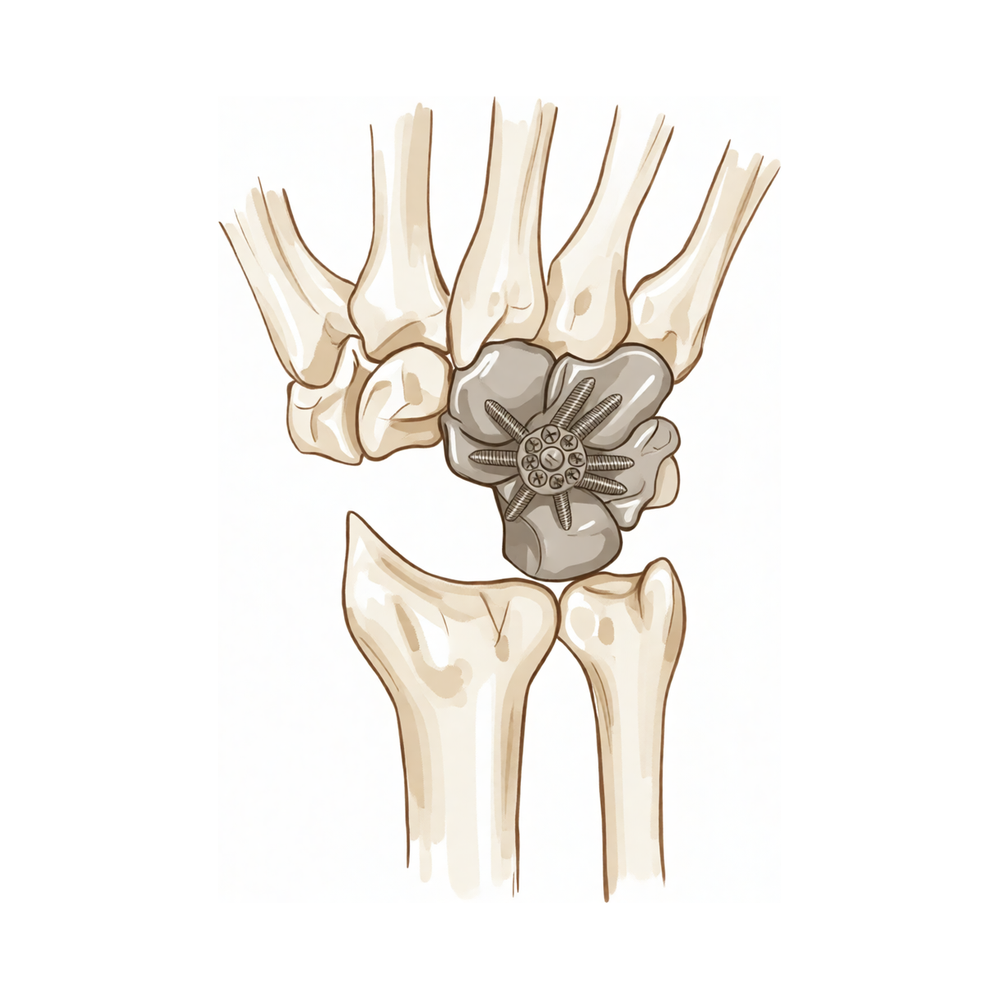
Lý do phẫu thuật này được đề xuất
Bác sĩ Kieran Hirpara, bác sĩ phẫu thuật chi trên tại Bệnh viện tư nhân Mater Rockhampton, sẽ bắt đầu bằng các phương pháp ít xâm lấn nhất phù hợp với tình trạng của bạn. Thông thường, bệnh nhân được bác sĩ đa khoa giới thiệu đến phòng khám chúng tôi; nếu vật lý trị liệu viên khuyên bạn nên gặp chúng tôi, bạn vẫn cần có giấy giới thiệu từ bác sĩ đa khoa để được hưởng mức hoàn trả từ Medicare. Việc đánh giá tại phòng khám, bao gồm hỏi tiền sử bệnh, thăm khám và chụp hình ảnh nếu cần thiết, sẽ giúp xác định nguyên nhân gây đau cổ tay của bạn.
Phẫu thuật hợp nhất một phần cổ tay chỉ kết nối các xương cổ tay bị tổn thương với nhau, để các khớp khỏe mạnh vẫn tiếp tục vận động. Chúng tôi thường đề xuất phẫu thuật này khi viêm khớp do thoái hóa chỉ giới hạn ở một phần của cổ tay, và thường là sau khi các phương pháp điều trị khác như trị liệu tay, đeo nẹp, thuốc chống viêm hoặc tiêm thuốc chưa giúp giảm đau đủ. Mục đích là loại bỏ các bề mặt khớp bị mòn gây đau, đồng thời duy trì tối đa khả năng vận động cổ tay, sức nắm và độ ổn định mà chúng tôi có thể giữ được. Phẫu thuật hợp nhất toàn bộ cổ tay chỉ được dành cho những trường hợp đặc biệt. Ca mổ này có phù hợp với bạn hay không phụ thuộc vào những bề mặt khớp nào bị ảnh hưởng, và chúng tôi sẽ cùng bạn thảo luận kỹ điều này để đi đến một quyết định chung.
Trước khi phẫu thuật
Trước ca phẫu thuật, bạn sẽ cần chụp một số hình ảnh để chúng tôi có thể lên kế hoạch phẫu thuật. Việc này thường bao gồm chụp X-quang, và đôi khi chụp MRI, phương pháp chụp sử dụng nam châm mạnh để cho thấy chi tiết các xương, dây chằng và bề mặt khớp ở cổ tay. Vào ngày phẫu thuật, hãy nhịn ăn và uống bảy giờ trước ca phẫu thuật. Chúng tôi yêu cầu bảy giờ thay vì sáu giờ để ca mổ của bạn có thể được đưa lên sớm hơn nếu lịch mổ diễn ra sớm hơn dự kiến. Hãy mang theo danh sách viết sẵn các loại thuốc bạn đang dùng và mặc quần áo thoải mái có tay áo rộng. Hãy sắp xếp người đưa bạn về nhà sau phẫu thuật. Nếu bạn mắc các bệnh lý khác, có thể bạn sẽ cần làm xét nghiệm máu hoặc gặp bác sĩ gây mê, tức là bác sĩ thực hiện gây mê cho bạn.
Vào ngày phẫu thuật
Bạn sẽ đến khoa tiếp nhận bệnh nhân phẫu thuật của bệnh viện, tại đó bạn sẽ được làm thủ tục nhập viện và chuẩn bị cho ca mổ. Bạn sẽ gặp bác sĩ gây mê, tức là bác sĩ thực hiện gây mê cho bạn. Ca mổ này được thực hiện dưới gây mê toàn thân. Bạn sẽ hoàn toàn ngủ say trong suốt ca phẫu thuật. Một số bệnh nhân có thể được gây tê vùng để giảm đau sau mổ; quyết định này do bác sĩ gây mê đưa ra vào ngày phẫu thuật tùy theo tình trạng cụ thể của từng người.
Sau đó, bạn sẽ được đưa vào phòng mổ để tiến hành ca phẫu thuật. Khi ca mổ kết thúc, bạn sẽ tỉnh dậy tại khu vực hồi sức, nơi các điều dưỡng theo dõi bạn trong khi thuốc mê hết tác dụng. Khi tình trạng ổn định, bạn sẽ được chuyển về phòng bệnh hoặc về nhà, tùy thuộc vào loại phẫu thuật và quá trình hồi phục của bạn.
Quy trình phẫu thuật
Phẫu thuật hợp nhất một phần cổ tay được thực hiện qua một đường rạch ở mặt sau cổ tay. Qua đường rạch này, bác sĩ phẫu thuật sẽ tiếp cận các xương cổ tay nhỏ tạo nên khớp bị thoái hóa. Các bề mặt khớp bị tổn thương sẽ được làm sạch để xương khỏe mạnh có thể liền lại với nhau.
Sau đó, các xương đang gây đau cho bạn sẽ được nối lại với nhau. Các đinh vít kim loại nhỏ giữ chúng ở đúng vị trí trong khi chúng liền lại, và bác sĩ phẫu thuật sẽ chèn xương ghép quanh khớp để giúp khớp liền. Những xương nào được nối lại tùy thuộc vào vị trí viêm khớp của bạn. Nếu một xương bị tổn thương quá nặng không thể sửa chữa, xương đó có thể được lấy ra và các xương còn lại được nối với nhau thay vào chỗ của nó. Các khớp khỏe mạnh bên cạnh vùng được hợp nhất sẽ được giữ nguyên để cổ tay của bạn vẫn có thể cử động được phần nào.
Khi các xương đã được giữ chắc chắn, vết mổ sẽ được khâu lại và băng bằng gạc. Băng gạc được giữ nguyên khoảng 10 ngày, và bác sĩ phẫu thuật sẽ cho bạn biết khi nào tháo băng.
Sau phẫu thuật
Bạn sẽ tỉnh dậy tại khu vực hồi sức, nơi các điều dưỡng theo dõi sát bạn trong khi thuốc mê hết tác dụng. Hầu hết bệnh nhân sẽ ở lại bệnh viện một đêm sau ca phẫu thuật này, tuy nhiên một số người có thể về nhà ngay trong ngày. Cổ tay của bạn sẽ được cố định bằng nẹp hoặc bó bột, và chúng tôi sẽ cho bạn dùng thuốc để bạn cảm thấy dễ chịu. Nếu bạn được gây tê vùng, bàn tay của bạn có thể có cảm giác tê hoặc nặng trong 24 giờ đầu; đây là điều bình thường. Khi thuốc tê đã hết tác dụng, hãy gọi cho phòng khám nếu bạn bị tê hoặc không thể cử động các ngón tay. Chúng tôi sẽ giữ băng gạc trên vết thương khoảng 10 ngày; xin đừng tháo nó ra trước thời hạn đó trừ khi chúng tôi yêu cầu. Chúng tôi sẽ thay hoặc gỡ băng gạc khi khám lại cho bạn. Vui lòng nhờ ai đó ở bên cạnh bạn trong 24 giờ đầu tiên.
Quá trình hồi phục
Trong vài ngày đầu, cổ tay của bạn sẽ đau và sưng, và bàn tay có thể cảm thấy cứng. Tình trạng này sẽ dần cải thiện. Việc kê tay cao trên gối, kể cả khi ngủ, giúp giảm sưng. Thuốc mà chúng tôi cho bạn dùng sẽ giúp bạn cảm thấy dễ chịu trong khi giai đoạn đau nhiều nhất qua đi.
Cổ tay của bạn sẽ được cố định bằng nẹp hoặc bó bột trong khi các xương liền lại với nhau. Việc trị liệu tay sau phẫu thuật của bạn sẽ do Ruby Doolan tại trung tâm Extend Rehabilitation đảm nhận. Ruby là chuyên gia trị liệu tay: cô ấy sẽ hướng dẫn các bài tập và làm nẹp cho bạn nếu cần. Giai đoạn đầu, trọng tâm là cử động nhẹ nhàng các ngón tay, khuỷu tay và vai để chúng không bị cứng trong khi cổ tay được nghỉ ngơi. Theo thời gian, chuyên gia trị liệu sẽ bổ sung các bài tập để tăng sức nắm và, khi vùng hợp nhất đã liền, để lấy lại mức cử động cổ tay an toàn với bạn.
Lúc đầu, các công việc hằng ngày cần được sắp xếp trước. Bạn sẽ cần người giúp đi chợ, nấu ăn và làm bất cứ việc gì cần dùng cả hai tay hoặc cần nắm chặt. Hầu hết mọi người thấy dễ ngủ nhất khi kê cánh tay trên gối thay vì nằm đè lên tay. Bạn không được lái xe cho đến khi đã tháo bột và được bác sĩ phẫu thuật cho phép; vui lòng xem trang thông tin của chúng tôi về lái xe sau phẫu thuật chi trên.
Quá trình hồi phục có thể khác nhau tùy từng người. Có cổ tay hồi phục nhanh, có cổ tay cần nhiều thời gian hơn để cảm thấy khỏe và dễ chịu. Bác sĩ phẫu thuật và chuyên gia trị liệu tay sẽ hướng dẫn bạn ở mỗi lần khám và cho bạn biết điều gì sẽ diễn ra tiếp theo.
Những biến chứng có thể xảy ra
Hầu hết bệnh nhân đều hồi phục tốt, nhưng đôi khi vẫn có thể gặp phải các vấn đề. Bác sĩ phẫu thuật và đội ngũ y tế sẽ theo dõi sát sao để phát hiện sớm bất kỳ dấu hiệu bất thường nào.
Đôi khi các xương không liền lại với nhau như dự kiến. Bạn có thể cảm thấy đau âm ỉ, sâu trong cổ tay mà không thuyên giảm, hoặc cảm giác cọ xát khi cử động. Nếu cơn đau ngày càng nặng hơn dù đã dùng thuốc giảm đau, hãy gọi cho phòng khám ngay trong ngày. Nếu không, hãy báo lại với bác sĩ tại lần tái khám tiếp theo. Nếu các xương vẫn không liền lại, có thể cần phẫu thuật thêm để giúp xương liền.
Các đinh vít kim loại dùng để giữ xương đôi khi có thể gây ra vấn đề. Một đinh vít có thể bị xê dịch vị trí, hoặc đè lên vùng da hay gân lân cận. Bạn có thể cảm thấy có cạnh sắc dưới da, cảm giác lục cục, hoặc đau ở chỗ có dụng cụ kim loại. Hầu hết dụng cụ kim loại không bao giờ cần phải tháo ra, nhưng nếu tiếp tục gây đau, một ca mổ nhỏ để tháo bỏ có thể giúp ích. Hãy nói về điều này khi tái khám.
Trong những trường hợp hiếm hoi, cổ tay có thể bị hội chứng ống cổ tay sau phẫu thuật. Biểu hiện là cảm giác kiến bò, tê hoặc nóng rát ở các ngón tay, nặng hơn vào ban đêm. Hãy báo cho bác sĩ phẫu thuật nếu điều này xảy ra, vì một ca mổ nhỏ giải phóng ống cổ tay có thể điều trị được tình trạng này.
Nhiễm trùng là tình trạng hiếm gặp nhưng cần được xử lý ngay. Hãy gọi cho phòng khám ngay trong ngày nếu bạn bị sốt, vùng đỏ lan rộng quanh vết mổ, hoặc có dịch hay mủ rỉ ra từ vết mổ.
Một số cổ tay vẫn đau ngay cả khi các xương đã liền tốt. Một vài người thấy cổ tay vẫn đau nhức khi hoạt động, hoặc cần dùng thuốc giảm đau thường xuyên. Nếu điều này xảy ra, bác sĩ phẫu thuật sẽ thảo luận với bạn về các lựa chọn, có thể bao gồm phẫu thuật thêm.
Rất hiếm khi, phẫu thuật hợp nhất một phần không giúp giảm đau và sau đó toàn bộ cổ tay được hợp nhất thay thế. Đây là một ca mổ lớn hơn, làm mất toàn bộ khả năng cử động cổ tay, nhưng thường giúp hết đau.
Bảng liệt kê các biến chứng ở trang này nêu rõ tỷ lệ xảy ra của từng vấn đề nếu bạn muốn biết chi tiết.
Khi nào nên gọi cho chúng tôi
Chúng tôi muốn nghe bạn báo hơn là để bạn lo lắng ở nhà. Hãy gọi cho phòng khám ngay trong ngày nếu bạn bị sốt, vùng đỏ lan rộng quanh vết mổ, hoặc có dịch hay mủ rỉ ra từ vết mổ, hoặc nếu cơn đau ngày càng nặng hơn dù đã dùng thuốc giảm đau. Hãy đến phòng cấp cứu nếu bạn bị sưng hoặc đau ở bắp chân, khó thở hoặc đau ngực, vì đây có thể là dấu hiệu của cục máu đông. Hãy đến phòng cấp cứu nếu các ngón tay hoặc bàn tay của bạn trở nên nhợt nhạt, lạnh, trắng bệch, xanh tím hoặc thâm đen. Khi thuốc gây tê vùng đã hết tác dụng, sau khoảng 24 giờ, hãy gọi cho phòng khám nếu bạn bị tê hoặc không thể cử động cánh tay, bàn tay hoặc các ngón tay. Nếu bạn không thể liên lạc với chúng tôi ngoài giờ làm việc hoặc vào cuối tuần, hãy đến phòng cấp cứu gần nhất.
Nơi để tìm đọc thêm thông tin về bệnh lý
Trang này nói về chính ca phẫu thuật. Các thông tin liên quan đến bệnh lý được điều trị, bao gồm cả những bằng chứng cho thấy khi nào phẫu thuật mang lại hiệu quả và khi nào thì không, đều được trình bày chi tiết hơn trên trang Viêm khớp cổ tay.
Evidence & references
This is the clinical evidence summary written for health professionals. It is technical, and it lists the research this page was built from. You do not need to read it to understand your treatment or to make a decision about it.
Overview
- Total wrist fusion should only be used in exceptional circumstances [1].
- Total wrist fusion should only be used for exceptional circumstances [2].
- Arthroscopic partial wrist fusion is a viable option for patients who have posttraumatic or nonprogressive wrist arthritis who would like to preserve useful wrist motion with good aesthetic outcome [3].
- A series of 28 selective limited wrist arthrodeses was presented, with most being intercarpal fusions and some including the radius or index metacarpal [4].
- Four of the 24 patients developed non-unions, two of which had successful revision to limited wrist arthrodesis, while the other two required pan arthrodesis of the wrist [5].
- Good and excellent clinical results in the majority of the patients following radiolunate fusion do not depend on the fixation device [6].
- Total wrist arthrodesis combined with PRC provides reliable and reproducible benefits [7].
- PRC and FCA demonstrated similarly low rates of conversion to total wrist arthrodesis [8].
- There is a low rate of conversion to total wrist arthrodesis [9].
- Nonspanning arthrodesis is advocated as an alternative method for total wrist fusion with a high union rate and minimal risk of complications at the carpometacarpal joint [11].
- Complete or total wrist arthrodesis is a well established reconstructive surgical procedure that results in predictable pain relief and satisfactory function in patients who have various inflammatory, degenerative, and post-traumatic conditions [12].
- The timing, primary or conversion, of total wrist arthrodesis could influence patient outcomes [14].
- Although TWA and partial wrist arthroplasty are attractive treatment options for the painful arthritic wrist, there remains a noteworthy potential for complications requiring additional surgery [15].
- RSL fusion is a palliative procedure that preserves some of the wrist's mobility but has a significant nonunion rate [16].
- Additional studies are required to confirm findings and investigate who may benefit from partial wrist denervation [17].
- Patients are generally satisfied after total wrist arthrodesis, although they are not always pain-free despite limited or absent wrist motion [18].
- Total wrist arthrodesis provides reliable pain relief and good functional outcomes with high patient satisfaction, particularly for end-stage arthritis and as a salvage technique [19].
- For stage II SLAC wrist with a preserved capitolunate joint, proximal row carpectomy is preferred because it is technically less demanding and yields durable results [29].
- Proximal row carpectomy is a motion-preserving salvage procedure with a low rate of conversion to wrist arthrodesis [30].
- The use of decellularized dermal allograft adds another surgical option for the treatment of wrist arthritis and expands the indications for PRC to include select patients with degeneration of the capitate head [31].
- Radioscapholunate arthrodesis with compression screws and local autograft is an effective method in appropriately selected patients with a preserved midcarpal joint, achieving a 100% union rate at mean follow-up of 12 months with no complications [40].
- Total wrist arthroplasty and total wrist arthrodesis are both extremely cost-effective procedures [45].
- Limited wrist arthrodesis techniques are reliable and effective for a wide range of wrist disorders, with low complication rates and high patient satisfaction [46].
Anatomy & Pathophysiology
Bony Anatomy
- The wrist includes the distal radioulnar, radiocarpal, and ulnocarpal joints and the eight carpal bones and their proximal and distal articulations and attached ligaments [82].
- The eight carpal bones include the scaphoid, lunate, triquetrum, and pisiform in the proximal row and the trapezium, trapezoid, capitate, and hamate in the distal row [82].
- The radiocarpal joints are formed by the articulation of the distal radius with the scaphoid and lunate through their respective concave facets on the distal radius and the triquetrum on the triangular fibrocartilage [82].
- The distal concave articular surfaces of the proximal carpal row form the midcarpal articulations with the distal row [82].
- The distal radius articular surface has two concave facets, the scaphoid and lunate facets, separated by the scapholunate, or anterior-posterior, ridge [89].
- The sigmoid notch, along the ulnar border of the distal radius, is a shallow concavity for the articulating ulnar head at the distal radioulnar joint [89].
- The distal ulna is covered with hyaline cartilage on its dorsal, lateral, palmar, and distal surfaces [89].
- The ulnar styloid projects distally; at its base, the fovea is the insertion for the triangular fibrocartilaginous complex (TFCC) [89].
- The scaphoid's primary vascular supply is a branch of the radial artery at the dorsal ridge, with a group of smaller vessels entering the palmar tubercle and supplying the distal 30% [89].
- A dorsal and a palmar vascular supply are found in 80% of wrists for the lunate; in 20% of wrists, only a palmar supply is found [89].
- The lunate is broader palmarly than dorsally [89].
- The triquetrum articulates with the hamate distally, the lunate radially, and the pisiform volarly [89].
- The triquetrum is stabilized to the fovea of the ulna through the ulnotriquetral ligament [89].
- The hamate consists of the body and the hook (hamulus) of the hamate, which serves as an attachment for the transverse carpal ligament and for the origins of the flexor digiti minimi and opponens digiti minimi [89].
- The head of the capitate often relies on a retrograde vascular supply [89].
- Two ridges separate the distal articular surface of the capitate into three facets for articulation with the metacarpals of the index, long, and ring fingers [89].
- The trapezoid has two distal facets, which articulate with the metacarpal of the index finger [89].
- The trapezium has a saddle-shaped articulation with the base of the thumb metacarpal [89].
- The trapezium has a palmar groove for the flexor carpi radialis (FCR), bordered laterally by a palmar tuberosity and the attachment for the transverse carpal ligament [89].
- The pisiform is a sesamoid bone within the flexor carpi ulnaris (FCU) tendon and is the origin for the abductor digiti minimi [89].
- The concave elliptical distal radius is oriented in the sagittal plane with an average of 11 degrees of volar tilt [86].
- In the frontal plane, the average radial inclination of the distal radius is 23 degrees [86].
- Radial length is measured from the tip of the radial styloid to the ulnar articular surface and averages 13 mm [86].
- The distal ulna itself has an ulnar styloid, which contains attachments to the triangular fibrocartilage complex, including the meniscus homolog, the volar and dorsal ulnar carpal ligaments, and the ulnar collateral ligament at the wrist [86].
- The radius has a lateral bow that is crucial to the maintenance of full pronation and supination [86].
Ligaments
- Extrinsic carpal ligaments connect the radius or the ulna to the carpus [87].
- In general, the volar ligaments are stronger than the dorsal ligaments [87].
- The radioscaphocapitate (RSC) ligament connects to the waist of the scaphoid, around which the scaphoid rotates, and limits ulnar translation of carpus [87].
- The long radiolunate ligament helps to limit ulnar translocation of the carpus [87].
- The short radiolunate ligament helps control lunate position [87].
- The radioscapholunate ligament is a vascular conduit, not a true ligament, also known as the ligament of Testut [87].
- The ulnolunate ligament attaches to the palmar radioulnar ligament and lunate [87].
- The ulnocapitate ligament attaches to the ulnar head, originates from the volar margin of the ulnar fovea, and is the most superficial or palmar [87].
- The ulnotriquetral ligament attaches to the palmar radioulnar ligament and triquetrum [87].
- The dorsal radiocarpal ligament (DRC) or dorsal radiotriquetral ligament has a trapezoidal shape and passes from the dorsal rim of the distal radius to the lunate and the triquetrum [87].
- Fibers of the dorsal radiocarpal ligament insert onto the dorsal lunotriquetral interosseous ligament [87].
- Damage to the dorsal radiocarpal ligament, when in conjunction with other intrinsic ligament injuries, confers further carpal instability [87].
- The scapholunate interosseous ligament (SLIL) is the major stabilizer of the wrist and the most commonly injured wrist ligament [87].
- The scapholunate interosseous ligament is C-shaped, consisting of dorsal, palmar, and interosseous portions with the dorsal portion being the strongest/thickest [87].
- The scapholunate interosseous ligament provides a flexion force on the lunate given its attachment to the scaphoid [87].
- The lunotriquetral interosseous ligament (LTIL) is C-shaped, where the volar portion is the thickest/strongest [87].
- The lunotriquetral interosseous ligament provides an extension moment on the lunate given its attachment to the triquetrum [87].
- The capitohamate ligament is a thick ligament, 5 × 5 mm in cross section, with extensions to the third or fourth metacarpals [87].
- The dorsal intercarpal ligament (DIC) passes from the dorsal tubercle of the triquetrum to the distal pole of the scaphoid [87].
- With the DRC ligament, the DIC ligament reinforces the elastic dorsal wrist capsule and helps stabilize the scapholunate articulation with a contribution to the dorsal SLIL from its deep fibers [87].
- The space of Poirier is an area adjacent to the proximal capitate without ligamentous attachment, situated ulnar to the RSC ligament and radial to the long radiolunate in the floor of the carpal tunnel [87].
- The space of Poirier is a weak area that is vulnerable to instability; the distal carpal row separates from the lunate through this space during a perilunate dislocation [87].
- The scapholunate interosseous ligament is C-shaped in the sagittal plane, with the dorsal third of the ligament being the thickest, strongest portion [89].
- The volar portion of the lunotriquetral ligament is the thickest [89].
- The TFCC is formed by the central meniscus homolog, the dorsal and volar radioulnar ligaments, the floor of the extensor carpi ulnaris (ECU) tendon sheath, and the volar ulnocarpal ligaments [89].
- The TFCC arises from the radial border of the distal radius and inserts into the base of the ulnar styloid and distal ulna through the ligamentum subcruentum [89].
- The dorsal and volar radioulnar ligaments are the primary stabilizers of the distal radioulnar joint [89].
- Only the peripheral 10% to 40% of the volar, ulnar, and dorsal TFCC has a vascular supply [89].
- The radial collateral ligament originates from the radius (0 mm from RS) and inserts on the scaphoid waist and distal palmar trapezium [89].
- The radioscaphocapitate ligament originates from the radius (4 mm from RS) and inserts on the scaphoid waist and midpalmar capitate [89].
- The radiolunatotriquetral ligament originates from the radius (10 mm from RS) and inserts on the lunate ± triquetrum [89].
- The radioscapholunate ligament originates from the mesocapsule with termination of AIN and AIA and inserts on the ligament of Testut and Kuenz [89].
- The short radiolunate ligament originates from the volar-ulnar margin of radius and inserts on the lunate [89].
- The ulnotriquetral ligament originates from the volar radioulnar ligament and inserts on the triquetrum [89].
- The ulnolunate ligament originates from the volar radioulnar ligament and inserts on the lunate [89].
- The ulnocapitate ligament originates from the volar margin of the ulnar head and inserts on the capitate [89].
- The dorsal radiocarpal ligament originates from the dorsal radius at the Lister tubercle and inserts on the lunate and triquetrum [89].
- The dorsal intercarpal ligament originates from the triquetrum and inserts on the scaphoid and trapezoid and capitate [89].
- The chondroligamentous supports attaching the distal radius and ulnar side of the carpus to the distal ulna are designated as the triangular fibrocartilage complex (TFCC) [82].
- Attaching to the ulnar margin of the lunate fossa of the radius, these supports include the ulnar collateral ligament, the dorsal and volar radioulnar ligaments, the articular disc, the meniscal homologue, the extensor carpi ulnaris sheath, and the ulnolunate and ulnotriquetral ligament [82].
- The interosseous ligaments include the scapholunate and lunotriquetral interosseous ligaments connecting the proximal carpal row and the ligaments connecting the trapezium to the trapezoid, the trapezoid to the capitate, and the capitate to the hamate in the distal carpal row [82].
- The extrinsic or crossing ligaments include the radial collateral ligament from the radial styloid to the scaphoid waist, the ulnar collateral ligament from the base of the ulnar styloid attaching to the pisiform, and the transverse carpal ligament [82].
- The volar extrinsic or crossing ligaments also include the radioscapocapitate ligament, the radiolunotriquetral ligament, and the radioscapolunate ligament on the radial side and the ulnolunate and ulnotriquetral components of the TFCC on the ulnar side [82].
- On the palmar side of the carpus, between the radiolunotriquetral ligament and the radioscapocapitate ligament, is a relatively thin area, the space of Poirier, overlying the palmar surface of the lunate [82].
- The trapezoidal dorsal radiocarpal ligament attaches along the dorsal radial articular margin of the lunate fossa, from the Lister tubercle to the lesser sigmoid notch [82].
- The trapezoidal dorsal radiocarpal ligament spans the lunotriquetral joint and inserts on the dorsal surface of the triquetrum [82].
- The dorsal intercarpal ligament, which is attached to the distal, dorsal surface of the triquetrum, passes across the midcarpal joint to attach to the dorsal surfaces of the scaphoid waist and the trapezoid [82].
Vascular Anatomy
- The terminal branches of the radial, ulnar, and anterior interosseous arteries provide extraosseous blood supply to the carpus through three dorsal and three palmar transverse arterial arches with longitudinal connections [92].
- The dorsal arches are (1) the dorsal radiocarpal at the radiocarpal joint, supplying the lunate and triquetrum; (2) the dorsal intercarpal (the largest) between the proximal and distal carpal rows, supplying the distal carpal row and, through anastomoses with the radiocarpal arch, the lunate and triquetrum; and (3) the basal metacarpal arch at the base of the metacarpals (the most variable) to supply the distal carpal row [92].
- The palmar arches are (1) the palmar radiocarpal at the level of the radiocarpal joint on the palmar surfaces of the lunate and triquetrum, (2) the intercarpal arch between the proximal and distal carpal rows, which is the most variable and does not contribute to nutrient vessels in the carpus, and (3) the deep palmar arch at the level of the metacarpal bases, which is consistent and communicates with the dorsal basal metacarpal arch and the palmar metacarpal arteries [92].
Biomechanics and Kinematics
- The wrist can essentially be considered to be a two-joint system linking the hand (described as the distal carpal row and the metacarpals) to the forearm (radius and ulna) around the highly mobile bones of the proximal carpal row [90].
- The two principle articulations are the radiocarpal and midcarpal joints, situated proximal and distal to the mobile proximal carpal row [90].
- The wrist joint’s motion planes include flexion, extension, radial deviation, ulnar deviation, and circumduction; there is minimal carpal motion with pronosupination [87].
- Approximately 62° of wrist extension occurs through the RC joint and 62% of wrist flexion occurs through the MC joint [87].
- The MC joint is mostly responsible for 20° and 40° of radial and ulnar deviation, respectively, and is responsible for the “dart thrower’s motion” which involves moving from radial extension into ulnar flexion positioning of the wrist [87].
- The radius bears 80% of the axial load transmitted through the RC joint, while the ulna bears 20% in neutral ulnar variance [87].
- The proximal row of carpal bones form an intercalated segment between the distal carpal row and the distal radius and are bound into a functional unit by the SLIL and LTIL [87].
- The distal row is rigid, with little motion between its bones due to stout intercarpal ligaments, and thus they act as a functional unit with the scaphoid bridging both rows [87].
- During wrist flexion from neutral, the distal row flexes and ulnarly deviates slightly while the scaphoid also pronates [87].
- During wrist flexion from neutral, the proximal row flexes differentially, with more rotation through the scaphoid, followed by the triquetrum and the lunate [87].
- During wrist flexion from neutral, the proximal row translates dorsally [87].
- During wrist extension from neutral, the distal row extends and radially deviates slightly while the scaphoid also supinates [87].
- During wrist extension from neutral, the proximal row extends differentially, with more motion in the scaphoid, followed by the triquetrum and then the lunate [87].
- During wrist extension from neutral, the proximal row translates palmarly [87].
- The proximal carpal row has no muscular or tendinous attachments and is an intercalary segment [89].
- With ulnar deviation, the proximal row extends relative to the forearm/distal row [89].
- With radial deviation, the proximal row flexes relative to the forearm/distal row [89].
- With axial loading through the neutral wrist, approximately 80% of forces are transmitted through the distal radius (60% scaphoid facet, 40% lunate facet) and 20% through the distal ulna [89].
- With wrist flexion, 60% of the motion is midcarpal and 40% is radiocarpal [89].
- With wrist extension, 33% of the motion is midcarpal and 66% is radiocarpal [89].
- Biomechanical studies have determined that between 30% and 50% of sagittal motion at the wrist occurs through the midcarpal joint, with the remainder through the radiocarpal joint [25].
- The wrist forms the junction between the forearm and the hand, providing a mechanism that allows wrist flexion, extension, radial deviation, ulnar deviation, and a limited amount of axial rotation [32].
- The eight carpal bones
Classification
- The position of 10° to 30° of extension and 0° to 15° of ulnar deviation is the most commonly accepted position for maximum hand function [13].
- In the assessment of prognostic factors for good clinical outcome after radioscapholunate fusion, a good outcome corresponds to the Cooney et al. classification's definition of good and excellent results after fracture-dislocation of the wrist [37].
- In the assessment of prognostic factors for good clinical outcome after radioscapholunate fusion, a poor clinical outcome is defined as patients who underwent total wrist fusion [37].
- In the assessment of prognostic factors for good clinical outcome after radioscapholunate fusion, a good global pain result is defined as none or slight pain with a Visual Analog Scale score of 3 or less [37].
- In the assessment of prognostic factors for good clinical outcome after radioscapholunate fusion, a poor global pain result is defined as moderate or severe pain with a Visual Analog Scale score greater than 3 [37].
- In the assessment of prognostic factors for good clinical outcome after radioscapholunate fusion, a good wrist motion result is defined as functional motion in all needed directions, specifically flexion of at least 5 degrees, extension of at least 30 degrees, radial deviation of at least 10 degrees, and ulnar deviation of at least 15 degrees [37].
- In the assessment of prognostic factors for good clinical outcome after radioscapholunate fusion, a poor wrist motion result is defined as nonfunctional motion in at least one direction [37].
- In the assessment of prognostic factors for good clinical outcome after radioscapholunate fusion, a good grip strength result is defined as 50% of the contralateral side [37].
- In the assessment of prognostic factors for good clinical outcome after radioscapholunate fusion, a poor grip strength result is defined as less than 50% of the contralateral side [37].
- Signs of postoperative radio-carpal osteoarthritis in limited intercarpal fusion versus proximal row carpectomy were graded in five categories: none, doubtful, mild, moderate, and severe arthritis [123].
- Signs of postoperative radio-carpal osteoarthritis in limited intercarpal fusion versus proximal row carpectomy were graded according to the Kellgren-Lawrence Classification System [123].
- Signs of postoperative radio-carpal osteoarthritis in limited intercarpal fusion versus proximal row carpectomy were graded as grade 0-IV [123].
- In the arthroscopic assessment and classification of Kienbock's disease, Grade 0 is defined as all articular surfaces being functional [24].
- In the arthroscopic assessment and classification of Kienbock's disease, Grade 1 is defined as a non-functional proximal lunate articular surface [24].
- In the arthroscopic assessment and classification of Kienbock's disease, Grade 2 is defined as two non-functional articular surfaces [24].
- In the arthroscopic assessment and classification of Kienbock's disease, Grade 2A is defined as the proximal articular surface of the lunate and the lunate fossa both being non-functional [24].
- In the arthroscopic assessment and classification of Kienbock's disease, Grade 2B is defined as the proximal and distal articular surfaces of the lunate being non-functional [24].
- In the arthroscopic assessment and classification of Kienbock's disease, Grade 3 is defined as three articular surfaces being non-functional [24].
Clinical Presentation
- The choice of procedure depends on the pattern of wrist destruction and stability, with radiolunate arthrodesis being effective for unstable wrists with preserved midcarpal joint space [10].
- RSL arthrodesis is a viable alternative to complete wrist arthrodesis for young patients with posttraumatic or rheumatoid arthritis limited to the radiocarpal joint if the midcarpal joint is normal, though limited wrist flexion and extension is expected [109].
- Arthritis due to scapholunate advanced collapse follows a predictable pattern in the wrist [39].
- Although the simultaneous occurrence of arthritis of the wrist and the base of the thumb is rare, it is nonetheless imperative to carry out a preoperative clinical and radiological assessment of the wrist when managing trapeziometacarpal osteoarthritis [43].
- Patients experienced good pain relief with preservation of wrist motion following proximal row carpectomy [22].
- Both proximal row carpectomy and four-corner fusion provide improvements in pain and subjective outcome measures for patients with symptomatic and appropriately staged SLAC or SNAC wrists [27].
- The principles of treatment for Kienbock's disease are to perform a synovectomy as part of the arthroscopic procedure and to perform a reconstructive procedure that addresses the non-functional articular surface and utilizes remaining functional articular surfaces [24].
- For Grade 1 Kienbock's disease with a non-functional proximal lunate articular surface, proximal row carpectomy or a radioscapholunate fusion can be performed [24].
- For Grade 2A Kienbock's disease where the proximal articular surface of the lunate and the lunate fossa are both non-functional, radioscapholunate fusion removes both non-functional articular surfaces and enables the wrist to articulate through the normal mid-carpal joint [24].
- For Grade 2B Kienbock's disease where the proximal and distal articular surfaces of the lunate are non-functional, typically due to a coronal fracture, the condition is best managed with a proximal row carpectomy [24].
- For Grade 3 Kienbock's disease where three articular surfaces are non-functional and usually only a functional capitate articular surface remains, the condition could theoretically be managed with a hemiarthroplasty [24].
- The resulting range of wrist motion after three-corner fusion is relatively better than that obtained with 4 corner fusions, while grip strength and other clinical parameters are comparable [26].
- Good and excellent clinical results in the majority of patients following radiolunate fusion do not depend on the fixation device [6].
- Total wrist arthrodesis with Wrist Fusion Rod is expected to provide long-lasting pain relief and stability with severe wrist deterioration and ulnar carpal shift [20].
- All patients regained full finger movements and rotation of the forearm following arthrodesis of the wrist with bone autograft and Hoffmann external fixation, though complete pain relief was rare and patients had significant limitations in activities of daily living [44].
- Fusion was successful in all but two patients in a series of rheumatoid wrist arthrodeses, and all patients benefited by increased strength and function in the hand [60].
- Post-operative motion and grip strength values following proximal row carpectomy appear to remain stable over time [59].
- Many patients following proximal row carpectomy continued to complain of pain requiring daily medication and were unable to return to manual labor [59].
- Surgical failure rates with conversion to wrist fusion after proximal row carpectomy occurred early within the post-operative follow-up [59].
- Radiographic follow-up beyond 2 years after proximal row carpectomy revealed joint narrowing and arthritic changes within the radiocapitate joint [59].
- Forty-six patients (74%) were not satisfied with the results of their proximal row carpectomy surgery due to persistent pain or inability to return to previous occupational activities [59].
- Fifty-two patients required daily pain medication for wrist pain following proximal row carpectomy [59].
- Twelve patients had undergone a wrist arthrodesis following proximal row carpectomy [59].
- After an average follow-up time of 14.7 years following midcarpal fusion, the mean DASH score was 20.4 and the mean VAS pain score at rest was 1.4 [62].
- Pain with daily activity after midcarpal fusion had a mean VAS pain score of 3.3 [62].
- An incomplete fusion was detected in 9 cases (15%) involving either the hamate alone or other sites after midcarpal fusion [62].
- Regenerated bone in the scaphoid fossa was detected in over a half of the cases (53.3%) after midcarpal fusion [62].
- Patients with a dominant hand affected by midcarpal fusion had less pain, better DASH-scores, and greater active range of wrist motion compared to those with a non-dominant hand affected [62].
- The position of 10° to 30° of extension and 0° to 15° of ulnar deviation is the most commonly accepted position for maximum hand function in wrist arthrodesis [13].
- Total wrist arthrodesis should only be used in exceptional circumstances [1, 2].
- There is a low rate of conversion to total wrist arthrodesis following scaphoid excision and four-corner arthrodesis for advanced carpal collapse [9].
- Our findings suggest an approximate 10% risk of secondary wrist arthrodesis in patients with persistent or progressive ulnar wrist pain following partial wrist denervation [21].
- Wrist arthrodesis with the AO/ASIF Titanium wrist fusion plate is an excellent option for treatment of various painful disorders of the wrist [36].
- Resection followed by wrist arthrodesis and structural iliac bone graft achieved satisfactory oncologic and functional results, albeit with one-third of all patients experiencing some complications at a minimum of 10 years of follow-up [68].
- The results of 4-corner arthrodesis using headless compression screws were comparable to or better than previously published techniques in terms of fusion rates, alleviation of pain, grip strength, range of motion, Mayo wrist score, and Disabilities of the Arm, Shoulder, and Hand questionnaire score [71].
- Union was observed in 22 of 23 (96%) patients at a mean time of three months from surgery following four-corner arthrodesis with a locking, dorsal circular polyether-ether-ketone (PEEK-Optima) plate [101].
- Only 2 patients did not show a complete fusion at the CT scan evaluation following four-corner fusion using a specialized circular locking plate [47].
- Four of the 24 patients developed non-unions following limited wrist arthrodesis for the salvage of SLAC wrist, two of which had successful revision to limited wrist arthrodesis, while the other two required pan arthrodesis of the wrist [5].
- Intercarpal arthrodesis is performed most commonly for the treatment of arthritis and carpal instability [35].
- Minimizing or eliminating pain, while maximizing motion and function, is the objective of intercarpal arthrodesis surgeries [35].
- Some intercarpal arthrodesis procedures have good, predictable outcomes with maintenance of results over time, while others are infrequently used or are of primarily historical interest owing to unpredictable results and high rates of complications [35].
- The goals of limited wrist arthrodesis are similar to those of total wrist arthrodesis in providing pain relief and improved function by fusing across arthritic or unstable joints, with the added benefit of motion preservation [25].
- Wrist arthrodesis has a role in the treatment of midcarpal instability, severe spastic deformity, paralysis of the hand, and bone loss from tumor or trauma [25].
- The first report of total wrist arthrodesis dates back to 1910 in treatment of tuberculosis [25].
- Thornton reported on the first limited wrist fusion in 1924 when he successfully fused the scaphoid, lunate, capitate, and hamate [25].
- The most common intercarpal arthrodesis procedure is the scaphoidectomy with fusion of the capitate, lunate, hamate and triquetrum, also known as a 4-corner fusion (4CF) [35].
- Compared with the intact specimen, simple 4-corner fusion with scaphoid retention led to a significant decrease in extension, radial deviation, and ulnar deviation, but no change in radiolunate contact characteristics [67].
- The wrist arthroscope can be used as an adjunct to proximal row carpectomy as a preoperative staging procedure to assess the condition of the proximal capitate and the distal radius [65].
- Wrist arthroscopy can be used to identify supplemental techniques that may be appropriate in performing a subsequent proximal row carpectomy [65].
- Wrist arthroscopy can be used to aid the selection of an alternative wrist salvage procedure if the surgeon chooses not to use adjunctive procedures during proximal row carpectomy [65].
- In situations where a proximal row carpectomy has been performed and there may be some residual pain, arthroscopy can be used to evaluate the status of the proximal capitate, the distal radial articular surface, and the condition of any interposition material [65].
Investigations
Imaging Modalities
- CT is more useful than plain X-rays for evaluating partial carpal arthrodesis [115].
- A properly performed, high-resolution MRI aids in the evaluation of ligament injuries of the wrist [94].
- A static magnetic field strength of at least 1.5 T using a dedicated wrist coil is recommended for analyzing the interosseous, intrinsic, and extrinsic ligament insertions [94].
- The volar extrinsic, SL interosseous, dorsal intercarpal, and LT ligaments are best visualized using 1 mm slices (with no interslice gap) in the coronal plane [94].
- The DRC and intercarpal ligaments are best viewed on both the coronal and sagittal images [94].
- Oblique axial views along the longitudinal axes of these ligaments allow further analysis, especially when an injury is suspected [94].
- Concomitant cartilage-sensitive imaging is integrative to influence the assessment/surgical management, as the cartilage integrity will influence the clinical/surgical management, especially in the setting of SLAC wrist [94].
- Dynamic fluoroscopy shows abnormal motion between the scaphoid and lunate and changes in the kinematics of the midcarpal joint [94].
- In SLD patients with DISI, the triquetrum-hamate relationship remains permanently engaged on dynamic fluoroscopy [94].
- Live imaging shows whether the DISI is reducible, giving the physician valuable information as the treatment is planned [94].
- CT scanning enables the 3D analysis of carpal dysfunction [94].
- By adding motion in real time (4D CT), this may hold promise in the future to potentially quantify the location and degree of injury noninvasively and help surgeons plan their surgical treatment [94].
- Arthroscopy is considered by many to be the diagnostic intervention of choice for determining the degree of injury to the wrist and can assess the condition of the cartilage (normal or degenerative), ability to reduce the carpus and any other associated injuries [94].
- The degree of intrinsic and extrinsic ligament injury can be identified from the arthroscopic evaluation [94].
- Geissler grade II SLIL injuries tend to be isolated, whereas grade IV injuries often involve complete dorsal extrinsic ligament disruption [94].
- MRI is the modality of choice for imaging radiographically occult fractures of the hand and wrist [88].
- The primary advantages of MRI compared with CT and radiography are the improved tissue characterization, especially of soft tissues such as the ligamentous structures in the wrist and synovium in the hand, and the lack of ionizing radiation [88].
- 3T is much preferred for hand and wrist imaging, especially for imaging small fields of view [88].
- MRI with contrast enhancement is most commonly used to determine whether soft-tissue lesions are solid or cystic or, in the case of rheumatologic imaging, to better visualize erosions and synovial burden [88].
- Dynamic contrast enhancement has been used with inconsistent results to assess for the presence of avascular necrosis in the lunate or scaphoid after injury [88].
- MR arthrography can be performed for evaluation of the triangular fibrocartilage and intercarpal ligament tears, but this is generally unnecessary with the increasing availability of high field MRI [88].
- With proper technique, injuries to the triangular fibrocartilage complex (TFCC) can be demonstrated with MRI [96].
- The TFCC is composed of signal-poor fibrocartilage, and perforations in the TFCC appear as linear defects or gaps filled with hyperintense fluid on coronal gradient-echo or T2-weighted pulse sequences [96].
- Although evaluation of the scapholunate and lunotriquetral ligaments is more challenging, with optimal technique and equipment the integrity of these structures can be consistently assessed [96].
- The addition of arthrographic contrast improves the visualization of these ligaments on MR images [96].
- The extrinsic carpal ligaments can be identified with three-dimensional volumetric scanning and subsequent reconstruction; however, at present, the MRI assessment of these ligaments has less impact on treatment [96].
- Not infrequently, bone marrow edema may reveal fractures of the carpal bones or distal radius that are radiographically occult [96].
- MRI is useful in detecting additional marrow abnormalities in osteonecrosis, as seen in the lunate in Kienböck disease or in the scaphoid after fracture [96].
- Asymmetry of marrow signal in proximal and distal fragments of a fractured scaphoid is suggestive of proximal pole ischemia [96].
- MRI currently has a limited role in the evaluation of carpal tunnel syndrome [96].
- Axial imaging with T2 weighting can clearly display masses within the confines of the carpal tunnel, as well as edema and swelling of the median nerve [96].
- Tenosynovitis and tendon injuries in the wrist and hand can be assessed with MRI [96].
- MRI provides earlier detection of synovitis and erosive bone changes associated with rheumatoid arthritis than do radiographs [96].
Clinical Assessment
- It is imperative to carry out a preoperative clinical and radiological assessment of the wrist when managing trapeziometacarpal osteoarthritis [43].
Treatment
Indications and General Principles
- The principles of treatment for Kienbock's disease are to perform a synovectomy as part of the arthroscopic procedure and to perform a reconstructive procedure that addresses the non-functional articular surface while utilizing remaining functional articular surfaces [24].
- In Kienbock's disease Grade 1, where the proximal lunate articular surface is non-functional, proximal row carpectomy or a radioscapholunate fusion can be performed [24].
- In Kienbock's disease Grade 2A, where the proximal articular surface of the lunate and the lunate fossa are both non-functional, radioscapholunate fusion removes both non-functional articular surfaces and enables the wrist to articulate through the normal mid-carpal joint [24].
- In Kienbock's disease Grade 2B, where the proximal and distal articular surfaces of the lunate are non-functional, the condition is best managed with a proximal row carpectomy [24].
- Capitolunate arthrodesis via a lateral approach is indicated for severe radioscaphoidal degenerative changes not amenable to reconstruction, provided the scaphoid is resected as part of the operation [28].
- Capitolunate fusion is contraindicated if severe degenerative changes are present in the radiolunate joint because it increases pressure and exacerbates pain [28].
- Scaphotrapeziotrapezoid arthrodesis is recommended in selected patients with advanced Kienböck's disease who have a fragmented lunate [120].
Proximal Row Carpectomy (PRC)
- Proximal row carpectomy, when performed for static scapholunate dissociation, results in a stiffened, weakened wrist [72].
- The use of decellularized dermal allograft in proximal row carpectomy expands the indications to include select patients with degeneration of the capitate head [31].
- Wrist arthroscopy can be used as an adjunct to proximal row carpectomy for preoperative staging of the proximal capitate and distal radius or to evaluate residual pain and interposition material status postoperatively [65].
- Total wrist arthrodesis combined with proximal row carpectomy provides reliable and reproducible benefits in the rheumatoid wrist [7].
- A technique of wrist arthrodesis combining proximal row carpectomy and rigid internal fixation has proved to be a highly predictable operation with much less morbidity and fewer complications than older techniques using distant bone graft [107].
Four-Corner Arthrodesis (FCA)
- Four-corner fusion with scaphoidectomy is a partial motion-preserving salvage procedure for treating wrists with symptomatic appropriately staged scapholunate advanced collapse (SLAC) or scaphoid nonunion advanced collapse (SNAC) [122].
- At a mean follow-up of 6 years, pain was significantly reduced and wrist function was significantly improved compared with preoperative status following four-corner arthrodesis with a dorsal locking plate [80].
- The combination of four-corner fusion with scaphoidectomy has been routinely performed since 1992 at the reporting institution [122].
- A case illustration of cerclage fusion technique for 4-corner arthrodesis demonstrated excellent alignment, fusion consolidation, and excellent pain relief at 4 to 5 months [129].
- Although it is still early, the authors anticipate that scaphoid excision and 4-corner fusion using retrograde headless compression screws will result in similar fusion rates to other forms of fixation [50].
- Parallel K-wire placement in scaphoid excision four-corner fusion involves inserting wires antegrade through the capitate and hamate and retrograde into the lunate and triquetrum to achieve fusion and midcarpal settling [74].
- In four-corner arthrodesis employing the native scaphoid as the principal donor graft, a broad cancellous trough is created spanning the entire midcarpal articulation to maximize restoration of carpal height [75].
- In four-corner bone arthrodesis with a dorsal rectangular plate, a cancellous bone graft taken from the scaphoid is placed between the capitate and lunate bones, especially on the palmar side [77].
- Major complications rates were 48% with plate fixation versus 6% with traditional fusion in a comparative study of four-corner arthrodesis [34].
- Revision surgery was performed on 8 wrists with plates in a comparative study of four-corner arthrodesis, including 4 for symptomatic impingement, 3 for nonunion, and 1 for persistent pain [34].
Radioscapholunate (RSL) and Radiolunate Arthrodesis
- Radioscapholunate fusion is a palliative procedure that preserves some of the wrist's mobility but has a significant nonunion rate [16].
- Various techniques for radiolunate arthrodesis require 6 to 12 weeks of postoperative cast immobilization to achieve rigid consolidation [70].
- A technique using compression screws and local autograft for radioscapholunate arthrodesis in appropriately selected patients with a preserved midcarpal joint achieved a 100% union rate at mean follow-up of 12 months with no complications [40].
Midcarpal Arthrodesis
- Final range of motion averaged 38° of wrist extension and 44° of wrist flexion following scapholunocapitate arthrodesis using the mini-spider plate [23].
- After an average follow-up time of 14.7 years, the mean DASH score was 20.4 and pain at rest was infrequent with a mean VAS pain score of 1.4 following midcarpal fusion [62].
- An incomplete fusion was detected in 9 cases (15%) involving either the hamate alone or other carpal bones in a long-term midcarpal fusion study [62].
- Regenerated bone in the scaphoid fossa was detected in over a half of the cases (53.3%) following midcarpal fusion [62].
- Computed tomography is the most reliable radiographic method of evaluating osseous union for midcarpal fusions, as plain films are unreliable due to curvature and overlap of carpal interfaces [56].
- A prospective protocol selected 8 weeks postoperative as the point in time to obtain the single determining CT scan for union of a midcarpal fusion [56].
Total Wrist Arthrodesis (TWA)
- Wrist arthrodesis is a well-established procedure that predictably relieves pain and provides a stable wrist for power grip [69].
- Total wrist arthrodesis with a Wrist Fusion Rod is expected to provide long-lasting pain relief and stability with severe wrist deterioration and ulnar carpal shift [20].
- Wrist arthrodesis is an effective procedure for the surgical reconstruction of the rheumatoid hand, providing a stable lasting solution to pain, dislocation, and instability [125].
- All patients regained full finger movements and rotation of the forearm following wrist arthrodesis with bone autograft and Hoffmann external fixation, though complete pain relief was rare and patients had significant limitations in activities of daily living [44].
- Although total wrist arthrodesis and partial wrist arthroplasty are attractive treatment options for the painful arthritic wrist, there remains a noteworthy potential for complications requiring additional surgery [15].
- Total wrist arthroplasty and total wrist arthrodesis are both extremely cost-effective procedures in rheumatoid arthritis [45].
- The lateral or radial approach for arthrodesis of the wrist avoids the distal radio-ulnar joint, preserves pronation and supination, and avoids involvement of the extensor mechanism [61].
- Between 10 to 15 degrees of dorsiflexion of the wrist with slight ulnar deviation is the optimum position desired for wrist arthrodesis [61].
- It is recommended that the second and third carpometacarpal joints always be included in the arthrodesis to prevent abnormal painful motion developing in the relatively immobile joint [61].
Partial Wrist Denervation
- Partial denervation of the wrist reduces pain stemming from intra-articular pathology by resecting peripheral nerves whose terminal fibers innervate only the joint capsule [32].
- The anterior and posterior interosseous nerves have been demonstrated to innervate the central two-thirds of the anterior and posterior carpal joint capsule, respectively [32].
- Simple PIN wrist denervation offers comparable pain relief and functional benefits to wrist arthrodesis and remains an appealing surgical intervention in the painful rheumatoid wrist [73].
Comparative Outcomes and Conversion Risks
- Proximal row carpectomy and four-corner arthrodesis demonstrated similarly low rates of conversion to total wrist arthrodesis for Stage-II SLAC/SNAC arthritis [8].
- There is a low rate of conversion to total wrist arthrodesis following scaphoid excision and four-corner arthrodesis for advanced carpal collapse at a minimum of ten years [9].
- Total wrist arthroplasty as an alternative to arthrodesis for the treatment of pancarpal posttraumatic arthritis may offer improved functional outcomes [113].
- The newer fourth-generation wrist implants appear to be performing better than earlier designs [41].
Complications
Nonunion and Hardware Complications
- Four of 24 patients undergoing limited wrist arthrodesis for SLAC wrist developed non-unions, with two requiring pan arthrodesis of the wrist [5].
- Major complications rates were 48% with plate fixation versus 6% with traditional fusion techniques for four-corner arthrodesis [34].
- Revision surgery was performed on 8 wrists with plates, including 4 for symptomatic impingement, 3 for nonunion, and 1 for persistent pain [34].
- In a series of radiolunate arthrodesis in the rheumatoid wrist, seven complications occurred, including one superficial wound problem, two fractures, and four displacements or malplacements of osteosynthetic material [58].
- Fracture of a plate in radiolunate arthrodesis led to secondary osseous union in ulnar translation [58].
- In three patients undergoing radiolunate arthrodesis, a screw protruded into the midcarpal joint space and induced arthrosis [58].
- Hardware complications were common in wrist arthrodesis for cerebral palsy, leading to a recommendation for routine hardware removal [137].
- The most common reason for reoperation after 4-corner arthrodesis was implant removal [138].
- In a series of 93 modified Clayton-Mannerfelt arthrodeses, the complication rate was low and hardware did not have to be removed in most cases [134].
- In a small cohort of scaphocapitate fusion for Kienböck disease, radiographic scaphoid waist index was numerically lower in wrists with nonunion [38].
Conversion to Total Wrist Arthrodesis
- The conversion rate to wrist arthrodesis after proximal row carpectomy (PRC) was 9% and after four-corner arthrodesis (FCA) was 4% [119].
- Smoking was independently associated with conversion to wrist arthrodesis following PRC or FCA, with an odds ratio of 4.9 [119].
- Anterior interosseous nerve and/or posterior interosseous nerve neurectomy was associated with a lower rate of conversion to wrist arthrodesis, with an odds ratio of 0.18 [119].
- Conversion rates to total wrist arthrodesis are significantly higher with partial wrist arthroplasty (19.2%) than with proximal row carpectomy (4.9%) [139].
- Proximal row carpectomy and four-corner arthrodesis demonstrated similarly low rates of conversion to total wrist arthrodesis [8].
- In a long-term follow-up of proximal row carpectomy, 12 patients had undergone a wrist arthrodesis [59].
- Total wrist arthrodesis was performed in six patients for secondary arthrosis or progressive arthritic destruction following radiolunate arthrodesis [58].
- An approximate 10% risk of secondary wrist arthrodesis exists in patients with persistent or progressive ulnar wrist pain following partial wrist denervation [21].
Infection and Other Complications
- Infection following wrist arthroplasty and arthrodesis is relatively uncommon in a nationally representative Medicare database cohort [136].
- Although total wrist arthrodesis and partial wrist arthroplasty are attractive treatment options, there remains a noteworthy potential for complications requiring additional surgery [15].
- Cautious patient selection and consideration of potential complications are crucial for good outcomes in wrist arthrodesis for the spastic hand [135].
- Wrist, first carpometacarpal joint, and thumb interphalangeal joint arthrodesis in patients with brachial plexus injuries had high union rates with minimal complications [140].
- Radioscapholunate arthrodesis with compression screws and local autograft achieved a 100% union rate at mean follow-up of 12 months with no complications [40].
Recovery
- Patients are immobilised in a short arm cast for 4 weeks following four-corner arthrodesis with a dorsal rectangular plate [77].
- Following the initial 4-week cast period, patients are asked to wear a removable splint for 2 further weeks [77].
- Range of motion exercise with traction has the potential to be applied to an advanced rehabilitation program targeting the radiolunate joint under specific pathological conditions [110].
Key Evidence
- [L4] Total wrist fusion should only be used in exceptional circumstances. [1] (10.1097/01.sap.0000194245.94684.54)
- [L4] Total wrist fusion should only be used for exceptional circumstances. [2] (10.1054/jhsb.2000.0434)
- [L4] Arthroscopic partial wrist fusion is a viable option for patients who have posttraumatic or nonprogressive wrist arthritis who would like to preserve useful wrist motion with good aesthetic outcome. [3] (10.1097/bth.0b013e318190244b)
- [L4] A series of 28 selective limited wrist arthrodeses was presented, with most being intercarpal fusions and some including the radius or index metacarpal. [4] (10.1016/s0363-5023(81)80074-3)
- [L4] Four of the 24 patients developed non-unions, two of which had successful revision to limited wrist arthrodesis, while the other two required pan arthrodesis of the wrist. [5] (10.1016/s0266-7681(97)80267-7)
- [L4] Good and excellent clinical results in the majority of the patients following radiolunate fusion do not depend on the fixation device. [6] (10.1177/1753193409342054)
- [Paper] Total wrist arthrodesis combined with PRC provides reliable and reproducible benefits. [7] (10.1016/j.otsr.2015.09.032)
- [L3] PRC and FCA demonstrated similarly low rates of conversion to total wrist arthrodesis. [8] (10.2106/jbjs.19.00965)
- [L4] There is a low rate of conversion to total wrist arthrodesis. [9] (10.1016/j.jhsa.2010.01.025)
- [L4] The choice of procedure depends on the pattern of wrist destruction and stability, with radiolunate arthrodesis being effective for unstable wrists with preserved midcarpal joint space. [10] (10.1016/j.hcl.2005.08.005)
- [L4] They advocate nonspanning arthrodesis as an alternative method for total wrist fusion with a high union rate and minimal risk of complications at the carpometacarpal joint. [11] (10.1055/s-0037-1606257)
- [L5] Complete or total wrist arthrodesis is a well established reconstructive surgical procedure that results in predictable pain relief and satisfactory function in patients who have various inflammatory, degenerative, and post-traumatic conditions. [12] (10.1016/j.hcl.2005.08.004)
- [L5] The position of 10° to 30° of extension and 0° to 15° of ulnar deviation is the most commonly accepted position for maximum hand function. [13] (10.1016/s0363-5023(86)80223-4)
- [L3] Therefore, we conclude that the timing, primary or conversion, of total wrist arthrodesis could influence patient outcomes. [14] (10.1177/17531934211057389)
- [L4] Although TWA and partial wrist arthroplasty are attractive treatment options for the painful arthritic wrist, there remains a noteworthy potential for complications requiring additional surgery. [15] (10.1016/j.jhsa.2015.10.021)
- [L4] RSL fusion is a palliative procedure that preserves some of the wrist's mobility but has a significant nonunion rate. [16] (10.1016/j.otsr.2017.07.012)
- [L3] Additional studies are required to confirm these findings and investigate who may benefit from partial wrist denervation. [17] (10.1177/17531934261425490)
- [L5] Patients are generally satisfied after total wrist arthrodesis, although they are not always pain-free despite limited or absent wrist motion. [18] (10.1177/17531934241295343)
- [L4] Total wrist arthrodesis provides reliable pain relief and good functional outcomes with high patient satisfaction, particularly for end-stage arthritis and as a salvage technique. [19] (10.5435/jaaos-d-15-00424)
- [L4] Total wrist arthrodesis with Wrist Fusion Rod is expected to provide long-lasting pain relief and stability with severe wrist deterioration and ulnar carpal shift. [20] (10.1016/j.jhsg.2026.101024)
- [L4] Our findings suggest an approximate 10% risk of secondary wrist arthrodesis in patients with persistent or progressive ulnar wrist pain. [21] (10.1007/s10067-019-04645-8)
- [L3] Patients experienced good pain relief with preservation of wrist motion. [22] (10.1177/1753193415597096)
- [L4] Final range of motion averaged 38° of wrist extension and 44° of wrist flexion. [23] (10.1097/00130911-200309000-00003)
- [L4] [24] (10.1097/00130911-200603000-00003)
- [L4] [25] (10.1016/j.hcl.2009.11.003)
- [L4] The resulted range of wrist motion is relatively better than that obtained with 4 corner fusions, while grip strength and other clinical parameters are yet comparable. [26] (10.1177/1558944716660555hq)
- [L4] Both procedures provide improvements in pain and subjective outcome measures for patients with symptomatic and appropriately staged SLAC or SNAC wrists. [27] (10.1177/1753193408100954)
- [L4] [28] (10.1097/00130911-200112000-00005)
- [L4] For stage II SLAC wrist with a preserved capitolunate joint, proximal row carpectomy is preferred because it is technically less demanding and yields durable results. [29] (10.5435/00124635-200307000-00007)
- [L3] Proximal row carpectomy is a motion-preserving salvage procedure with a low rate of conversion to wrist arthrodesis. [30] (10.1016/j.jhsa.2021.09.031)
- [L4] This technique adds another surgical option for the treatment of wrist arthritis and expands the indications for PRC to include select patients with degeneration of the capitate head. [31] (10.1016/j.jhsa.2018.01.012)
- [L4] [32] (10.1097/00130911-199803000-00004)
- [L3] [34] (10.1016/j.jhsa.2005.08.007)
- [L5] [35] (10.1016/j.jhsa.2013.09.014)
- [L4] Wrist arthrodesis with the AO/ASIF Titanium wrist fusion plate is an excellent option for treatment of various painful disorders of the wrist. [36] (10.1054/jhsb.2001.0600)
- [L4] [37] (10.1055/s-0039-1688939)
- [L4] In this small, selected cohort, rater-averaged radiographic scaphoid WI was numerically lower in wrists with nonunion after scaphocapitate fusion for Kienböck disease, suggesting a preliminary, hypothesis-generating WI-nonunion signal. [38] (10.1016/j.jhsa.2026.07.002)
- [L5] Arthritis due to scapholunate advanced collapse follows a predictable pattern in the wrist. [39] (10.1097/00130911-199712000-00003)
- [L4] This technique is an effective method to perform radioscapholunate arthrodesis in appropriately selected patients with a preserved midcarpal joint, achieving a 100% union rate at mean follow-up of 12 months with no complications. [40] (10.1016/j.jhsa.2013.01.026)
- [L1] The newer fourth-generation wrist implants appear to be performing better than earlier designs. [41] (10.1055/s-0038-1646956)
- [L5] Although the simultaneous occurrence of arthritis of the wrist and the base of the thumb is rare, it is nonetheless imperative to carry out a preoperative clinical and radiological assessment of the wrist when managing trapeziometacarpal osteoarthritis. [43] (10.1016/j.hansur.2020.08.013)
- [L4] All patients regained full finger movements and rotation of the forearm, though complete pain relief was rare and patients had significant limitations in activities of daily living. [44] (10.1177/1753193411416565)
- [L2] Total wrist arthroplasty and total wrist arthrodesis are both extremely cost-effective procedures. [45] (10.1016/j.jhsa.2009.12.013)
- [L4] The study demonstrates that limited wrist arthrodesis techniques are reliable and effective for a wide range of wrist disorders, with low complication rates and high patient satisfaction. [46] (10.1054/jhsb.1999.0066)
- [L4] Only 2 patients did not show a complete fusion at the CT scan evaluation. [47] (10.1016/j.hansur.2016.10.107)
- [L5] Although it is still early, the authors anticipate that this procedure will result in similar fusion rates to other forms of fixation. [50] (10.1097/bth.0b013e3182688c6a)
- [Paper] [56] (10.1007/s12593-011-0030-2)
- [L4] [58] (10.1054/jhsb.2001.0681)
- [L3] [59] (10.1007/s11552-011-9368-y)
- [L4] [62] (10.1055/s-0032-1329616)
- [L4] [65] (10.1016/j.hcl.2005.08.006)
- [L5] Compared with the intact specimen, simple 4-corner fusion with scaphoid retention led to a significant decrease in extension, radial deviation, and ulnar deviation, but no change in radiolunate contact characteristics. [67] (10.1016/s0363-5023(03)00372-1)
- [L2] Resection followed by wrist arthrodesis and structural iliac bone graft achieved satisfactory oncologic and functional results, albeit with one-third of all patients experiencing some complications at a minimum of 10 years of follow-up. [68] (10.1097/corr.0000000000003738)
- [L4] Wrist arthrodesis is a well-established procedure that predictably relieves pain and provides a stable wrist for power grip. [69] (10.5435/00124635-200101000-00006)
- [L4] Various techniques have been described, all of which require 6 to 12 weeks of postoperative cast immobilization to achieve rigid consolidation of the radiolunate arthrodesis. [70] (10.1097/00130911-200006000-00006)
- [L4] The results were comparable to or better than previously published techniques in terms of fusion rates, alleviation of pain, grip strength, range of motion, Mayo wrist score, and Disabilities of the Arm, Shoulder, and Hand questionnaire score. [71] (10.1016/j.jhsa.2011.12.022)
- [L4] Proximal row carpectomy, when performed for static scapholunate dissociation, results in a stiffened, weakened wrist. [72] (10.1177/1753193410382719)
- [L3] Simple PIN wrist denervation offers comparable pain relief and functional benefits to wrist arthrodesis and remains an appealing surgical intervention in the painful rheumatoid wrist. [73] (10.1007/s00402-014-2018-4)
- [L4] [74] (10.1177/15589447211057302)
- [L4] [75] (10.1177/1753193416676663)
- [L4] [77] (10.1177/1753193409105684)
- [L4] At a mean follow-up of 6 years, pain was significantly reduced and wrist function was significantly improved compared with preoperative status. [80] (10.1177/1753193420930587)
- [L4] [101] (10.1177/1753193413475962)
- [L4] This technique of wrist arthrodesis combining proximal row carpectomy and rigid internal fixation has proved to be a highly predictable operation with much less morbidity and fewer complications than with older techniques using distant bone graft. [107] (10.1016/j.jhsa.2012.11.010)
- [L5] RSL arthrodesis is a viable alternative to complete wrist arthrodesis for young patients with posttraumatic or rheumatoid arthritis limited to the radiocarpal joint if the midcarpal joint is normal, though limited wrist flexion and extension is expected. [109] (10.1016/j.hcl.2005.08.003)
- [L4] Range of motion exercise with traction has the potential to be applied to an advanced rehabilitation program targeting the radiolunate joint under specific pathological conditions. [110] (10.1177/1758998315580274)
- [L3] Total wrist arthroplasty as an alternative to arthrodesis for the treatment of pancarpal posttraumatic arthritis may offer improved functional outcomes. [113] (10.1016/j.jhsa.2013.02.013)
- [L4] We conclude that CT is more useful than plain X-rays for evaluating partial carpal arthrodesis. [115] (10.1016/s0266-7681(97)80367-1)
- [L4] [119] (10.1016/j.jhsa.2019.10.023)
- [L4] We recommend scaphotrapeziotrapezoid arthrodesis in selected patients with advanced Kienböck's disease who have a fragmented lunate. [120] (10.1016/s0266-7681(98)80087-9)
- [L4] [122] (10.1055/s-0035-1549277)
- [L3] [123] (10.1186/s13018-023-04177-7)
- [L4] Wrist arthrodesis is an effective procedure for the surgical reconstruction of the rheumatoid hand, providing a stable lasting solution to pain, dislocation, and instability. [125] (10.2106/00004623-197153070-00012)
- [L4] The case illustration demonstrated excellent alignment, fusion consolidation, and excellent pain relief at 4 to 5 months. [129] (10.1016/j.jhsa.2019.02.016)
- [L4] The complication rate was low, and the hardware did not have to be removed in most cases. [134] (10.1016/j.jhsa.2013.02.029)
- [L4] Cautious patient selection and consideration of potential complications are crucial for good outcomes. [135] (10.1177/17531934231205548)
- [L3] Infection following wrist arthroplasty and arthrodesis is relatively uncommon in a nationally representative Medicare database cohort. [136] (10.1177/1558944719890036)
- [L4] Hardware complications were common, and consequently, the authors now routinely recommend hardware removal. [137] (10.1016/j.jhsa.2009.03.006)
- [L3] The most common reason for reoperation after 4-corner arthrodesis was implant removal. [138] (10.1016/j.jhsg.2022.10.015)
- [L3] Conversion rates to total wrist arthrodesis are significantly higher with PWA (19.2%) than with PRC (4.9%) and have a greater associated direct cost. [139] (10.1016/j.jhsa.2017.07.032)
- [L4] Wrist, first carpometacarpal joint, and thumb interphalangeal joint arthrodesis had high union rates with minimal complications. [140] (10.1016/j.jhsa.2012.09.017)
References
[1] Long-Term Results of Midcarpal Arthrodesis in the Treatment of Scaphoid Nonunion Advanced Collapse (SNAC-Wrist) and Scapholunate Advanced Collapse (SLAC-Wrist). Annals of Plastic Surgery. 2006. DOI: 10.1097/01.sap.0000194245.94684.54
[2] Midcarpal Arthrodesis with Complete Scaphoid Excision and Interposition Bone Graft in the Treatment of Advanced Carpal Collapse (SNAC/SLAC Wrist): Operative Technique and Outcome Assessment. Journal of Hand Surgery. 2000. DOI: 10.1054/jhsb.2000.0434
[3] Arthroscopic Partial Wrist Fusion. Techniques in Hand & Upper Extremity Surgery. 2008. DOI: 10.1097/bth.0b013e318190244b
[4] Limited wrist arthrodesis. Part II: Intercarpal and radiocarpal combinations. The Journal of Hand Surgery. 1981. DOI: 10.1016/s0363-5023(81)80074-3
[5] Limited Wrist Arthrodesis for the Salvage of SLAC Wrist. Journal of Hand Surgery. 1997. DOI: 10.1016/s0266-7681(97)80267-7
[6] Radiolunate fusion in the rheumatoid wrist with Shapiro staples: clinical and radiological results of 22 cases. Journal of Hand Surgery (European Volume). 2009. DOI: 10.1177/1753193409342054
[7] Proximal row carpectomy in total arthrodesis of the rheumatoid wrist. Orthopaedics & Traumatology: Surgery & Research. 2015. DOI: 10.1016/j.otsr.2015.09.032
[8] Risk of Total Wrist Arthrodesis or Reoperation Following 4-Corner Arthrodesis or Proximal Row Carpectomy for Stage-II SLAC/SNAC Arthritis. Journal of Bone and Joint Surgery. 2020. DOI: 10.2106/jbjs.19.00965
[9] The Outcome of Scaphoid Excision and Four-Corner Arthrodesis for Advanced Carpal Collapse at a Minimum of Ten Years. The Journal of Hand Surgery. 2010. DOI: 10.1016/j.jhsa.2010.01.025
[10] Partial Arthrodesis for the Rheumatoid Wrist. Hand Clinics. 2005. DOI: 10.1016/j.hcl.2005.08.005
[11] Nonspanning Total Wrist Arthrodesis with a Low-Profile Locking Plate. Journal of Wrist Surgery. 2017. DOI: 10.1055/s-0037-1606257
[12] Wrist Arthrodesis. Hand Clinics. 2005. DOI: 10.1016/j.hcl.2005.08.004
[13] Arthrodesis of the wrist: Past and present. The Journal of Hand Surgery. 1986. DOI: 10.1016/s0363-5023(86)80223-4
[14] Comparative outcomes of total wrist arthrodesis for salvage of failed total wrist arthroplasty and primary wrist arthrodesis. Journal of Hand Surgery (European Volume). 2021. DOI: 10.1177/17531934211057389
[15] Complications Following Partial and Total Wrist Arthroplasty: A Single-Center Retrospective Review. The Journal of Hand Surgery. 2016. DOI: 10.1016/j.jhsa.2015.10.021
[16] Clinical and radiological outcomes following radioscapholunate fusion. Orthopaedics & Traumatology: Surgery & Research. 2017. DOI: 10.1016/j.otsr.2017.07.012
[17] Revision rate and long-term outcome after partial wrist denervation in wrist osteoarthritis. Journal of Hand Surgery (European Volume). 2026. DOI: 10.1177/17531934261425490
[18] Total wrist arthrodesis in patients with advanced osteoarthritis: current implants and outcomes. Journal of Hand Surgery (European Volume). 2025. DOI: 10.1177/17531934241295343
[19] Total Wrist Arthrodesis: Indications and Clinical Outcomes. Journal of the American Academy of Orthopaedic Surgeons. 2017. DOI: 10.5435/jaaos-d-15-00424
[20] Total Wrist Arthrodesis Using a Wrist Fusion Rod for the Dislocated Rheumatoid Wrist. Journal of Hand Surgery Global Online. 2026. DOI: 10.1016/j.jhsg.2026.101024
[21] Patient satisfaction and outcomes of partial wrist denervation in inflammatory arthritis. Clinical Rheumatology. 2019. DOI: 10.1007/s10067-019-04645-8
[22] Factors associated with improved outcomes following proximal row carpectomy: a long-term outcome study of 144 patients. Journal of Hand Surgery (European Volume). 2015. DOI: 10.1177/1753193415597096
[23] Scapholunocapitate Arthrodesis Using the Mini-Spider Plate. Techniques in Hand and Upper Extremity Surgery. 2003. DOI: 10.1097/00130911-200309000-00003
[24] Arthroscopic Assessment and Classification of Kienbock's Disease. Techniques in Hand & Upper Extremity Surgery. 2006. DOI: 10.1097/00130911-200603000-00003
[25] Complications of Limited and Total Wrist Arthrodesis. Hand Clinics. 2010. DOI: 10.1016/j.hcl.2009.11.003
[26] Three-Corner Fusion for Management of SNAC and SLAC Wrists. HAND. 2016. DOI: 10.1177/1558944716660555hq
[27] Proximal Row Carpectomy vs Four Corner Fusion for Scapholunate (Slac) or Scaphoid Nonunion Advanced Collapse (Snac) Wrists: A Systematic Review of Outcomes. Journal of Hand Surgery (European Volume). 2009. DOI: 10.1177/1753193408100954
[28] Capitolunate Arthrodesis: The Lateral Approach. Techniques in Hand and Upper Extremity Surgery. 2001. DOI: 10.1097/00130911-200112000-00005
[29] Proximal Row Carpectomy and Intercarpal Arthrodesis for the Management of Wrist Arthritis. Journal of the American Academy of Orthopaedic Surgeons. 2003. DOI: 10.5435/00124635-200307000-00007
[30] Risk of Total Wrist Arthrodesis Following Proximal Row Carpectomy: An Analysis of 1,070 Patients. The Journal of Hand Surgery. 2023. DOI: 10.1016/j.jhsa.2021.09.031
[31] Proximal Row Carpectomy Using Decellularized Dermal Allograft. The Journal of Hand Surgery. 2018. DOI: 10.1016/j.jhsa.2018.01.012
[32] Partial Denervation of the Wrist. Techniques in Hand & Upper Extremity Surgery. 1998. DOI: 10.1097/00130911-199803000-00004
[34] Complications and Outcome of Four-Corner Arthrodesis: Circular Plate Fixation Versus Traditional Techniques. The Journal of Hand Surgery. 2005. DOI: 10.1016/j.jhsa.2005.08.007
[35] Intercarpal Arthrodeses. The Journal of Hand Surgery. 2014. DOI: 10.1016/j.jhsa.2013.09.014
[36] Wrist Arthrodesis with the AO Titanium Wrist Fusion Plate: A Consecutive Series of 42 Cases. Journal of Hand Surgery. 2001. DOI: 10.1054/jhsb.2001.0600
[37] Radioscapholunate Fusion for Radiocarpal Osteoarthritis: Prognostic Factors of Clinical and Radiographic Outcomes. Journal of Wrist Surgery. 2019. DOI: 10.1055/s-0039-1688939
[38] Radiographic Scaphoid Waist Index and Nonunion After Scaphocapitate Fusion for Kienböck Disease. The Journal of Hand Surgery. 2026. DOI: 10.1016/j.jhsa.2026.07.002
[39] Midcarpal Arthrodesis. Techniques in Hand & Upper Extremity Surgery. 1997. DOI: 10.1097/00130911-199712000-00003
[40] Radioscapholunate Arthrodesis With Compression Screws and Local Autograft. The Journal of Hand Surgery. 2013. DOI: 10.1016/j.jhsa.2013.01.026
[41] Systematic Review of Total Wrist Arthroplasty and Arthrodesis in Wrist Arthritis. Journal of Wrist Surgery. 2018. DOI: 10.1055/s-0038-1646956
[43] Trapeziometacarpal osteoarthritis and arthritis of the wrist. Hand Surgery and Rehabilitation. 2021. DOI: 10.1016/j.hansur.2020.08.013
[44] Arthrodesis of the wrist with bone autograft and Hoffmann external fixation. Journal of Hand Surgery (European Volume). 2012. DOI: 10.1177/1753193411416565
[45] A Cost-Utility Analysis of Nonsurgical Management, Total Wrist Arthroplasty, and Total Wrist Arthrodesis in Rheumatoid Arthritis. The Journal of Hand Surgery. 2010. DOI: 10.1016/j.jhsa.2009.12.013
[46] One Thousand Intercarpal Arthrodeses. Journal of Hand Surgery. 1999. DOI: 10.1054/jhsb.1999.0066
[47] Four-corner fusion using a specialized circular locking plate. Hand Surgery and Rehabilitation. 2016. DOI: 10.1016/j.hansur.2016.10.107
[50] Scaphoid Excision and 4-Corner Fusion Using Retrograde Headless Compression Screws. Techniques in Hand & Upper Extremity Surgery. 2012. DOI: 10.1097/bth.0b013e3182688c6a
[56] Reliability of the 8 Week Time Point for Single Assessment of Midcarpal Fusion by CT Scan. Journal of Hand and Microsurgery. 2011. DOI: 10.1007/s12593-011-0030-2
[58] Radiolunate Arthrodesis in the Rheumatoid Wrist: A Retrospective Clinical and Radiological Longterm Follow-Up. Journal of Hand Surgery. 2002. DOI: 10.1054/jhsb.2001.0681
[59] Long-Term Outcomes of Proximal Row Carpectomy: A Minimum of 15-Year Follow-up. HAND. 2011. DOI: 10.1007/s11552-011-9368-y
[60] Arthrodesis of the Rheumatoid Wrist: AN EVALUATION OF SIXTY PATIENTS AND A DESCRIPTION OF A DIFFERENT SURGICAL TECHNIQUE.. The Journal of Bone and Joint Surgery. American Volume. 1973.
[61] Arthrodesis of the Wrist: A SURGICAL TECHNIQUE.. The Journal of Bone and Joint Surgery. American Volume. 1967.
[62] Long-Term Results after Midcarpal Arthrodesis. Journal of Wrist Surgery. 2012. DOI: 10.1055/s-0032-1329616
[65] Proximal Row Carpectomy. Hand Clinics. 2005. DOI: 10.1016/j.hcl.2005.08.006
[67] Management of the scaphoid during four-corner fusion—a cadaveric study. The Journal of Hand Surgery. 2003. DOI: 10.1016/s0363-5023(03)00372-1
[68] What Are the Long-term Outcomes of Wrist Arthrodesis Using Structural Iliac Bone Graft After Resection of the Distal Radius for Giant Cell Tumor of Bone? A Minimum 10-year Follow-up. Clinical Orthopaedics & Related Research. 2025. DOI: 10.1097/corr.0000000000003738
[69] Wrist Arthrodesis: Review of Current Techniques. Journal of the American Academy of Orthopaedic Surgeons. 2001. DOI: 10.5435/00124635-200101000-00006
[70] A New Method for Radiolunate Arthrodesis in Ulnar Translocation of the Carpus in the Rheumatoid Wrist. Techniques in Hand & Upper Extremity Surgery. 2000. DOI: 10.1097/00130911-200006000-00006
[71] Results of a Method of 4-Corner Arthrodesis Using Headless Compression Screws. The Journal of Hand Surgery. 2012. DOI: 10.1016/j.jhsa.2011.12.022
[72] Proximal row carpectomy for scapholunate dissociation. Journal of Hand Surgery (European Volume). 2010. DOI: 10.1177/1753193410382719
[73] Functional and patient-reported outcome of partial wrist denervation versus the Mannerfelt wrist arthrodesis in the rheumatoid wrist. Archives of Orthopaedic and Trauma Surgery. 2014. DOI: 10.1007/s00402-014-2018-4
[74] Parallel K-Wire Placement Leads to Fusion and Midcarpal Settling in Scaphoid Excision Four-Corner Fusion. HAND. 2021. DOI: 10.1177/15589447211057302
[75] Four-corner arthrodesis employing the native scaphoid as the principal donor graft for advanced collapse deformity of the wrist: technique and outcomes. Journal of Hand Surgery (European Volume). 2016. DOI: 10.1177/1753193416676663
[77] Four-corner bone arthrodesis with dorsal rectangular plate: series and personal technique. Journal of Hand Surgery (European Volume). 2009. DOI: 10.1177/1753193409105684
[80] Four-corner arthrodesis with a dorsal locking plate: 4–9-year follow-up. Journal of Hand Surgery (European Volume). 2020. DOI: 10.1177/1753193420930587
[82] Campbell S Operative Orthopaedics 4 Volume Set. NERVE INJURIES AT THE LEVEL OF THE HAND AND WRIST > ANATOMY.
[86] A Lange Medical Book Current Diagnosis Treatment In Orthopedics Fifth Edition. 2Musculoskeletal Trauma Surgery > FRACTURES AND DISLOCATIONS OF THE DISTAL AND MID-FOREARM.
[87] Aaos Comprehensive Orthopaedic Review 3. Carpal Instability* > II. Anatomy and Biomechanics (See Chapter 92).
[88] Orthopaedic Knowledge Update 13 Ebook Without Multimedia. Anatomy, Evaluation, Clinical Examination, and Imaging > Imaging: Advances in Imaging of the Hand and Upper Extremity > Magnetic Resonance Imaging.
[89] Aaos Comprehensive Orthopaedic Review 3. Anatomy of the Hand and Wrist > VII. The Wrist.
[90] Green S Operative Hand Surgery. WRIST BIOMECHANICS > Carpal Kinematics.
[92] Campbell S Operative Orthopaedics 4 Volume Set. NERVE INJURIES AT THE LEVEL OF THE HAND AND WRIST > CIRCULATION.
[94] Green S Operative Hand Surgery. Advanced Imaging.
[96] Campbell S Operative Orthopaedics 4 Volume Set. WRIST AND ELBOW.
[101] The rate of successful four-corner arthrodesis with a locking, dorsal circular polyether-ether-ketone (PEEK-Optima) plate. Journal of Hand Surgery (European Volume). 2013. DOI: 10.1177/1753193413475962
[107] Modified AO Arthrodesis of the Wrist (With Proximal Row Carpectomy). The Journal of Hand Surgery. 2013. DOI: 10.1016/j.jhsa.2012.11.010
[109] Radioscapholunate Arthrodesis. Hand Clinics. 2005. DOI: 10.1016/j.hcl.2005.08.003
[110] An in vivo study of dynamic effects of wrist traction on the radiolunate and capitolunate joints. Hand Therapy. 2015. DOI: 10.1177/1758998315580274
[113] Clinical Outcomes of Arthrodesis and Arthroplasty for the Treatment of Posttraumatic Wrist Arthritis. The Journal of Hand Surgery. 2013. DOI: 10.1016/j.jhsa.2013.02.013
[115] Computed Tomography in Partial Carpal Arthrodesis. Journal of Hand Surgery. 1997. DOI: 10.1016/s0266-7681(97)80367-1
[119] Factors Associated With Reoperation and Conversion to Wrist Fusion After Proximal Row Carpectomy or 4-Corner Arthrodesis. The Journal of Hand Surgery. 2020. DOI: 10.1016/j.jhsa.2019.10.023
[120] Proximal Row Carpectomy Versus Limited Wrist Arthrodesis for Advanced Kienböck’s Disease. Journal of Hand Surgery. 1998. DOI: 10.1016/s0266-7681(98)80087-9
[122] The Long-Term Outcome of Four-Corner Fusion. Journal of Wrist Surgery. 2015. DOI: 10.1055/s-0035-1549277
[123] Limited intercarpal fusion versus proximal row carpectomy in the treatment of SLAC or SNAC wrist, results after 3.5 years. Journal of Orthopaedic Surgery and Research. 2023. DOI: 10.1186/s13018-023-04177-7
[125] Arthrodesis of the Wrist for Rheumatoid Arthritis. The Journal of Bone & Joint Surgery. 1971. DOI: 10.2106/00004623-197153070-00012
[129] Cerclage Fusion Technique for 4-Corner Arthrodesis. The Journal of Hand Surgery. 2019. DOI: 10.1016/j.jhsa.2019.02.016
[134] The Modified Clayton-Mannerfelt Arthrodesis of the Wrist in Rheumatoid Arthritis: Operative Technique and Report on 93 Cases. The Journal of Hand Surgery. 2013. DOI: 10.1016/j.jhsa.2013.02.029
[135] Wrist arthrodesis and soft tissue rebalancing in the spastic hand. Journal of Hand Surgery (European Volume). 2023. DOI: 10.1177/17531934231205548
[136] Risk Factors for Infection Following Total Wrist Arthroplasty and Arthrodesis: An Analysis of 6641 Patients. HAND. 2019. DOI: 10.1177/1558944719890036
[137] Wrist Arthrodesis in Cerebral Palsy. The Journal of Hand Surgery. 2009. DOI: 10.1016/j.jhsa.2009.03.006
[138] Factors Associated With Unplanned Reoperation After 4-Corner Arthrodesis: A Study of 478 Wrists. Journal of Hand Surgery Global Online. 2023. DOI: 10.1016/j.jhsg.2022.10.015
[139] A Comparative Analysis of Resource Utilization Between Proximal Row Carpectomy and Partial Wrist Fusion: A Population Study. The Journal of Hand Surgery. 2017. DOI: 10.1016/j.jhsa.2017.07.032
[140] Wrist, First Carpometacarpal Joint, and Thumb Interphalangeal Joint Arthrodesis in Patients With Brachial Plexus Injuries. The Journal of Hand Surgery. 2012. DOI: 10.1016/j.jhsa.2012.09.017