Patients › Wrist
U nang cổ tay
Wrist ganglia are common, fluid-filled lumps – often painless – and this page covers observation, aspiration, and excision.
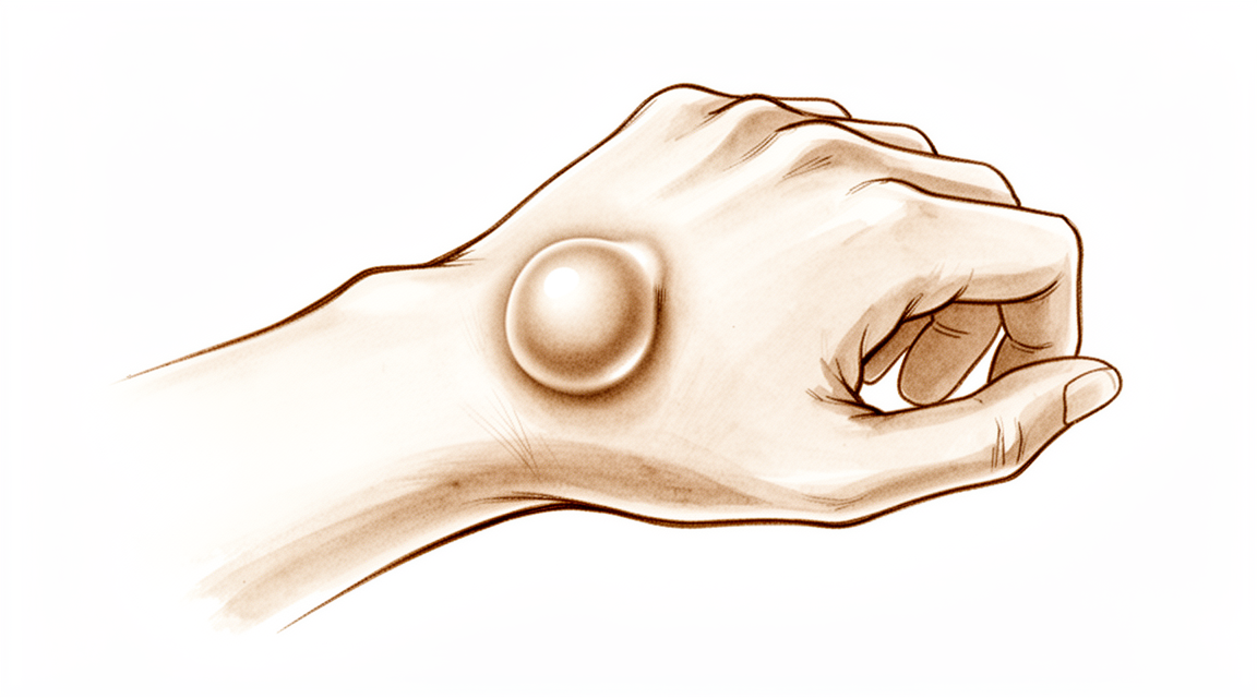
Những triệu chứng bạn đang gặp phải
U nang cổ tay là một khối chứa đầy dịch, hình thành từ khớp cổ tay hoặc từ lớp màng lót của bao gân. Đây không phải là một khối u có thể biến thành bất cứ thứ gì nguy hại. Nhiều người nhận thấy nó lần đầu dưới dạng một khối nhô lên, bề mặt nhẵn, ở mặt sau cổ tay, mặc dù nó cũng có thể xuất hiện ở phía lòng bàn tay. Khối có thể có cảm giác chắc, và kích thước có thể thay đổi theo thời gian.
Bản thân khối thường không gây đau. Khi có đau, cơn đau thường nằm sâu trong cổ tay, gần khối, và thường nặng hơn khi hoạt động. Những việc làm cổ tay phải gập hoặc chịu lực, chẳng hạn như chống tay đẩy người đứng dậy khỏi ghế, xách túi đồ đi chợ, hoặc cầm một chiếc chảo nặng, có thể gây đau âm ỉ. Một số người thấy cổ tay yếu hơn hoặc cứng hơn bình thường, điều này có thể khiến việc gõ bàn phím, viết hoặc mở nắp lọ trở nên khó khăn hơn.
Vì khối nằm gần các dây thần kinh ở cổ tay, đôi khi u nang có thể chèn ép lên chúng. Điều này có thể gây cảm giác châm chích, tê hoặc yếu ở bàn tay. Nếu bạn nhận thấy bất kỳ triệu chứng thần kinh nào trong số này, bạn nên đi khám cổ tay sớm thay vì chờ đợi, vì dây thần kinh cần được giải phóng khỏi sự chèn ép đó.
Nếu khối hoặc cơn đau không thuyên giảm, nặng dần lên trong nhiều tuần, hoặc khiến bạn không thể sử dụng bàn tay hay làm việc, hãy đến gặp bác sĩ đa khoa hoặc yêu cầu được bác sĩ chuyên khoa thăm khám.
Chuyện gì đang xảy ra thực sự
U nang phát triển từ một van nhỏ, một chiều nằm trong lớp màng lót của khớp cổ tay. Dịch vốn bôi trơn khớp bị đẩy ra ngoài qua van này nhưng không thể chảy ngược trở lại. Dịch tụ lại trong một chiếc túi dưới da, và chiếc túi đó chính là khối mà bạn có thể nhìn thấy và sờ thấy. Khối được nối với khớp bằng một cuống mảnh, đó là lý do kích thước của khối có thể thay đổi khi khớp tiết ra nhiều hoặc ít dịch hơn.
Hầu hết u nang phát triển từ dây chằng thuyền-nguyệt, một dải mô ngắn giữ hai xương nhỏ của cổ tay lại với nhau. Nó hoạt động hơi giống một miếng đệm kín giữa hai xương này. Lớp màng lót quanh dải mô này bị kích thích, thường sau một lần bong gân nhẹ hoặc căng giãn lặp đi lặp lại, và đó là nơi dịch rò ra. Điều này cũng giải thích vì sao cơn đau âm ỉ nằm sâu trong cổ tay chứ không ở ngoài da: vấn đề nằm ở chính khớp, không phải ở khối.
Vị trí của khối giải thích các triệu chứng còn lại của bạn. Ở mặt sau cổ tay, khối thường nằm ngay trên dây chằng đó, gần các gân làm duỗi thẳng các ngón tay. Ở phía lòng bàn tay, khối thường phát triển gần động mạch chính và các dây thần kinh chạy vào bàn tay, đó là lý do có thể xảy ra cảm giác châm chích hoặc tê khi u nang chèn ép lên chúng. Một số khối nhỏ và nằm ẩn, chỉ xuất hiện khi cổ tay gập về phía trước, nhưng vẫn có thể gây đau sâu.
Bạn nên biết rằng u nang là tình trạng phổ biến. Nhiều người có u nang mà không hề có triệu chứng nào, và u nang được phát hiện trên phim chụp của những cổ tay không đau thường xuyên hơn bạn nghĩ. Có u nang không có nghĩa là bản thân cấu trúc khớp cổ tay của bạn có vấn đề gì.
Những biện pháp chúng tôi có thể áp dụng
Bác sĩ Kieran Hirpara, bác sĩ phẫu thuật chi trên tại Bệnh viện Mater Private Rockhampton, thường bắt đầu bằng các phương pháp ít xâm lấn nhất phù hợp với tình trạng của bạn. Thông thường, bệnh nhân được bác sĩ đa khoa giới thiệu đến phòng khám chúng tôi; nếu nhà vật lý trị liệu khuyên bạn nên đến gặp chúng tôi, bạn vẫn cần có giấy giới thiệu từ bác sĩ đa khoa để được hưởng mức hoàn trả chi phí từ chương trình Medicare. Việc khám tại phòng khám, gồm hỏi bệnh sử, thăm khám và chụp chiếu khi cần, sẽ giúp xác định chẩn đoán.
Vì u nang vô hại, không làm gì cả là một bước đầu hợp lý. Nhiều u nang tự biến mất. Từ 28% đến 58% u nang cổ tay tự ổn định mà không cần điều trị. Ở trẻ em dưới 10 tuổi, u nang thường nằm ở phía lòng bàn tay của cổ tay, và từ 69% đến 79% tự nhỏ lại và biến mất trong vòng 12 đến 18 tháng. Theo dõi và chờ đợi, đôi khi kèm đeo nẹp cho dễ chịu, phù hợp với hầu hết trẻ em có u nang ở bàn tay hoặc cổ tay.
Nếu khối gây phiền toái, chúng tôi có thể hút dịch khối bằng kim. Thủ thuật này gọi là chọc hút. Rút dịch ra loại bỏ được tới 85% u nang khi được thực hiện một, hai hoặc ba lần. Dịch thường tích tụ lại một phần sau đó, vì việc hút dịch làm rỗng khối nhưng vẫn để lại chiếc van đã tạo ra nó. Dù vậy, 74% người bệnh hài lòng sau khi được chọc hút kèm lời khuyên đơn giản về khối của mình. Chọc hút trước cũng giúp giữ phẫu thuật là một lựa chọn về sau thay vì là bước đầu tiên. Một u nang ẩn ở mặt sau cổ tay, loại gây đau sâu nhưng không thể nhìn thấy, thường được xử trí trước tiên mà không phẫu thuật, bằng cách giữ cố định cổ tay và tiêm steroid vào lớp màng lót khớp.
Khi các bước này không giúp giảm đủ triệu chứng, phẫu thuật có thể loại bỏ u nang. Ca phẫu thuật lấy bỏ khối và cuống của nó, tức là phần nối mảnh với khớp cổ tay, vì để lại cuống sẽ khiến khối dễ tái phát hơn. Ca phẫu thuật có thể được thực hiện bằng mổ mở hoặc phẫu thuật nội soi qua các lỗ nhỏ. Chúng tôi sẽ cùng bạn trao đổi phương pháp nào phù hợp với cổ tay của bạn, và bạn sẽ cùng chúng tôi quyết định liệu phẫu thuật có đáng thực hiện đối với bạn hay không.
Những điều bạn có thể mong đợi
Nhiều u nang cổ tay thay đổi theo thời gian thay vì giữ nguyên. Chúng có thể nhỏ lại, sưng to lên, hoặc lúc xuất hiện lúc không. Một số tự ổn định: khoảng 40% u nang cổ tay giảm kích thước trong 6 năm đầu kể từ lần được đánh giá đầu tiên, và từ 39% đến 42% u nang ở mặt sau cổ tay tự biến mất mà không cần bất kỳ điều trị nào. Ở trẻ nhỏ, hầu hết các u nang ở bàn tay và cổ tay tự khỏi, thường trong vòng một năm. Nếu u nang của bạn không gây nhiều phiền toái, chờ đợi là một lựa chọn hợp lý, và khối sẽ không biến thành bất cứ thứ gì nguy hại nếu bạn để yên.
Điều trị làm thay đổi khả năng này. Chọc hút khối bằng kim làm rỗng khối, nhưng hầu hết u nang tái phát sau khi chọc hút, vì chiếc van đã tạo ra khối vẫn còn đó. Phẫu thuật loại bỏ khối cùng cuống của nó có khả năng tái phát thấp hơn nhiều: khoảng 10% u nang cổ tay tái phát sau phẫu thuật, và từ 3% đến 12% tái phát sau phẫu thuật mở cắt bỏ u nang ở mặt sau cổ tay. Hầu hết những người được phẫu thuật thấy triệu chứng dịu đi. Tuy nhiên, một số cơn đau có thể kéo dài sau đó, đặc biệt nếu công việc hoặc môn thể thao của bạn đòi hỏi gập cổ tay ra sau một cách mạnh mẽ.
Nếu bạn chọn phẫu thuật, quá trình hồi phục thường thuận lợi. Ban đầu bàn tay của bạn được quấn một lớp băng dày và kê cao để giữ dễ chịu, và cổ tay được bắt đầu cử động sớm, trong vòng 2 tuần đầu sau phẫu thuật u nang ở phía lòng bàn tay. Cử động cổ tay sớm rất quan trọng: cứng khớp không thường gặp nhưng có thể xảy ra nếu không khuyến khích cử động sớm. Trị liệu bàn tay sau phẫu thuật được thực hiện với Ruby Doolan tại Extend Rehabilitation, và cô ấy sẽ hướng dẫn các bài tập và làm bất kỳ loại nẹp nào bạn cần. Một điểm chắc hoặc đau khi chạm ở vị trí khối trước đây có thể tồn tại tới 3 tuần sau phẫu thuật nội soi ở phía lòng bàn tay, và xoa bóp vùng đó theo hướng dẫn giúp nó dịu đi. Sẹo là một phần của bất kỳ ca phẫu thuật nào, và các đường rạch cong gần các nếp gấp cổ tay thường lành với vết sẹo đẹp hơn.
Nếu khối đã được điều trị hoặc các triệu chứng của bạn không thuyên giảm, hoặc nặng dần lên trong nhiều tuần, hãy đến gặp bác sĩ đa khoa hoặc yêu cầu được bác sĩ chuyên khoa thăm khám.
Khi nào nên đi khám bác sĩ
Hầu hết các u nang cổ tay đều vô hại, và nhiều trường hợp tự ổn định, vì vậy chỉ riêng một khối thì không phải là tình trạng cấp cứu. Hãy đến gặp bác sĩ đa khoa hoặc yêu cầu được bác sĩ chuyên khoa thăm khám nếu khối hoặc cơn đau không thuyên giảm trong nhiều tuần, đang nặng dần lên, hoặc khiến bạn không thể làm việc hay sử dụng bàn tay. Hãy đi khám sớm hơn nếu bạn bị châm chích, tê hoặc yếu ở bàn tay, vì điều đó cho thấy u nang đang chèn ép lên một dây thần kinh. Hãy đến khoa cấp cứu nếu bàn tay hoặc các ngón tay của bạn trở nên nhợt nhạt, lạnh, trắng bệch hoặc tím tái, hoặc nếu bạn đột ngột mất cảm giác hay mất khả năng cử động ở bàn tay sau chấn thương. Những tình trạng này hiếm gặp với u nang, nhưng cần được chăm sóc ngay trong ngày.
Phân tích sâu hơn
Advanced reading: the deeper science (optional)
Phần này đi sâu hơn mức cần thiết để bạn tự đưa ra quyết định điều trị. Các nang ở cổ tay là chủ đề đáng để tìm hiểu thêm, bởi lựa chọn thực sự nằm giữa việc chấp nhận nguy cơ tái phát và chấp nhận việc phẫu thuật; các con số liên quan đến sự đánh đổi này cũng đủ rõ ràng để giúp bạn đưa ra quyết định.
Chích hút thường tái phát; cắt bỏ thì không, như dự đoán
Hai phương pháp điều trị chủ yếu là chích hút nang bằng kim và cắt bỏ nang cùng cuống nang qua phẫu thuật. Dựa trên dữ liệu từ 2.239 bệnh nhân, phẫu thuật cắt bỏ mở cho tỷ lệ tái phát thấp hơn đáng kể so với phương pháp chích hút. Phẫu thuật cắt bỏ nang qua nội soi cũng cho kết quả khả quan, nhưng dữ liệu từ các thử nghiệm so sánh còn hạn chế và chưa chứng minh được ưu thế vượt trội của phương pháp này [1].
Cơ chế sinh lý giải thích sự khác biệt này. Nang thần kinh không phải là túi chứa dịch tự do; nó được nối với màng khớp bên dưới bằng một cuống nang, và khớp vẫn tiếp tục tiết ra dịch. Chích hút chỉ làm rỗng túi dịch mà không loại bỏ nguồn gốc hình thành nang, nên việc nang tái hình thành là điều thường xảy ra chứ không phải là thất bại trong điều trị. Phương pháp cắt bỏ nhằm truy tìm và loại bỏ cuống nang ngay tại điểm xuất phát của nó.
Phương pháp nội soi và phẫu thuật mở cho kết quả tương đương
Khi lựa chọn phương pháp cắt bỏ, hai cách tiếp cận này đã được so sánh trực tiếp. Trên 910 bệnh nhân, cả phương pháp nội soi và phẫu thuật mở đều cho kết quả tương đương về tỷ lệ tái phát và biến chứng. Các tác giả kêu gọi tiến hành các nghiên cứu có quy mô đủ lớn và tiêu chuẩn hóa [2].
Do đó, việc lựa chọn phương pháp phẫu thuật nên dựa trên các yếu tố thứ yếu như vết sẹo, mức độ quen thuộc của bác sĩ phẫu thuật với từng kỹ thuật, cũng như việc có cần kiểm tra các tổn thương khác trong khớp hay không; chứ không phải dựa trên nguy cơ tái phát.
Không ai thống nhất được nên làm gì tiếp theo; thực ra điều đó cũng chẳng quan trọng lắm
Đây là một phát hiện nhỏ nhưng chân thực. Một phân tích hệ thống và khảo sát các bác sĩ phẫu thuật tay cho thấy họ phân chia thành hai phe về việc có nên cố định cổ tay sau khi cắt bỏ u nang vùng mu tay hay không; về mặt kết quả chức năng, không có dữ liệu thuyết phục nào cho thấy phương pháp nào vượt trội hơn [3].
Cần biết rằng sự khác biệt trong hướng dẫn điều trị giữa các bác sĩ ở đây là do thực sự tồn tại sự bất đồng ý kiến, chứ không phải vì một người nào đó sai.
Lập luận mạnh mẽ nhất thường là không cần can thiệp gì cả
Không có lý do nào trong số trên chứng minh rằng cần điều trị u nang gân. Đây là những khối u lành tính. Kích thước của chúng thường thay đổi, và một tỷ lệ nhất định tự biến mất mà không cần can thiệp. Chúng cũng không chuyển thành bất kỳ dạng bệnh lý nào khác.
Điều này làm thay đổi cách nhìn về quyết định điều trị. Các lý do để điều trị bao gồm đau, cản trở vận động cổ tay hoặc khả năng nắm chặt, áp lực lên dây thần kinh lân cận, hoặc kích thước khối u gây phiền toái cho người bệnh – chứ không phải chỉ vì sự hiện diện của khối u. Xét rằng thủ thuật chọc hút có tỷ lệ tái phát đáng kể, còn phẫu thuật cắt bỏ lại tiềm ẩn rủi ro đối với màng khớp, việc theo dõi tình trạng bệnh là một lựa chọn hợp lý làm bước đầu tiên; điều này cần được nêu rõ thay vì coi đó là sự thiếu hành động.
Ngoại lệ duy nhất là trường hợp u nang gân gây ra các triệu chứng thần kinh như tê, yếu cơ hoặc đau lan xuống tay; khi đó khối u đang gây chèn ép lên cấu trúc không thể chịu đựng áp lực này mãi mãi, và việc theo dõi nữa không còn là phương án ít rủi ro nữa.
Tài liệu tham khảo
[1] Head L, Gencarelli JR, Allen M, Boyd KU. Điều trị u nang cổ tay: tổng quan có hệ thống và phân tích tổng hợp. J Hand Surg Am. 2015;40(3):546-553.e8. https://doi.org/10.1016/j.jhsa.2014.12.014
[2] Crawford C, Keswani A, Lovy AJ, Levy I, Lutz K, Kim J, và cộng sự. Phẫu thuật nội soi so với phẫu thuật mở để cắt bỏ u nang vùng mu cổ tay: tổng quan có hệ thống và phân tích tổng hợp. J Hand Surg Eur Vol. 2017;43(6):659-64. https://doi.org/10.1177/1753193417734428
[3] Wong CR, Karpinski M, Hatchell AC, McRae MH, Murphy J, McRae MC. Việc cố định cổ tay sau khi cắt bỏ u nang vùng mu cổ tay: tổng quan có hệ thống và khảo sát. Hand (N Y). 2021;18(2):254-63. https://doi.org/10.1177/15589447211014631
Evidence & references
This is the clinical evidence summary written for health professionals. It is technical, and it lists the research this page was built from. You do not need to read it to understand your treatment or to make a decision about it.
Overview
Epidemiology and Natural History
- The dorsal wrist ganglion is the prototype of all hand and wrist ganglions, accounting for 60% to 70% of all hand and wrist ganglions [1].
- Volar wrist ganglia are the second most common ganglion of the hand and wrist, accounting for 18% to 20% of cases [11].
- In an asymptomatic population, 51% of 103 wrists scanned demonstrated ganglia on MRI [7].
- In an asymptomatic population evaluated by MRI, volar wrist ganglia are more common than dorsal wrist ganglia [2].
- Neither surgical excision nor aspiration provides a clear long-term benefit over the natural history of the dorsal wrist ganglion [6].
- Pediatric ganglions of the hand have a greater rate of resolution than ganglions of the wrist [4].
Clinical Presentation and Diagnosis
- The main cyst of a dorsal wrist ganglion is usually located directly over the scapholunate ligament [1].
- Dorsal wrist ganglions may occur anywhere between the extensor tendons and can be connected to the scapholunate ligament through an elongated pedicle [1].
- Careful preoperative palpation of a dorsal wrist ganglion with digital compression often reveals its extent and the direction of the pedicle [1].
- Transillumination or aspiration confirms the diagnosis of a wrist ganglion preoperatively [1].
- In patients with recalcitrant dorsal wrist pain in the area of the scapholunate interval, occult dorsal wrist ganglions had a prevalence of 11 out of 14 [3].
- Routine wrist radiography is not cost-effective in the evaluation and treatment decision-making process for patients with a wrist ganglion due to a low prevalence of therapeutically significant findings [5].
- A systematic X-ray should be performed for painful dorsal wrist ganglia [13].
- Preoperative x-rays should be obtained as part of the routine workup for a dorsal wrist ganglion [22].
- Volar wrist ganglions may appear small clinically but can be surprisingly extensive at surgery, with multiloculated cysts extending under the thenar muscles, along the flexor carpi radialis tendon, into the carpal canal, and under the first extensor compartment [11].
- The Allen test should be performed routinely to assess the patency of the radial and ulnar arteries in patients with volar wrist ganglions [11].
Operative Considerations
- Failure to identify and excise the pedicle attachment to the scapholunate ligament increases the likelihood of recurrence for dorsal wrist ganglions [1].
- A small, mucin-filled duct is invariably seen piercing the transverse fibers of the scapholunate ligament, connecting the underlying scapholunate joint with the main cyst [1].
- The radial artery is frequently intimately attached to the wall of a volar wrist ganglion and may be completely encircled by the ganglion [11].
- Some authors recommend leaving a portion of the volar ganglion cyst wall attached to the radial artery to avoid arterial injury [11].
- The best indication for arthroscopic treatment of a volar wrist ganglion is a sessile type arising from the radiocarpal joint and centered on the proximal wrist crease [12].
- Mobile volar ganglia with a long pedicle are poor candidates for arthroscopic surgery [12].
- Open excision of dorsal wrist ganglia leads to a lower recurrence rate than arthroscopic excision [25].
- Routine midcarpal joint exploration during arthroscopic excision of dorsal wrist ganglions appeared to reduce recurrence at 1 year without negatively impacting patient outcomes [35].
- Arthroscopic resection of dorsal wrist ganglia has low morbidity, with unnoticeable scars and wrist mobility and strength close to normal by three months [10].
- Arthroscopic resection of radiocarpal volar ganglia is a reasonable alternative to open excision with decreased postoperative morbidity, easy technique, and a high rate of good results [56].
- Arthroscopic ganglionectomy through an intrafocal cystic portal is a safe and efficacious option for the treatment of painful wrist ganglia [39].
- Surgical excision of primary wrist ganglia significantly reduces patient symptoms with low recurrence rates and high patient satisfaction [18].
- Open surgical excision offers a significantly lower chance of recurrence compared with aspiration in the treatment of wrist ganglions [59].
- Female patients who have preoperative pain around dorsal wrist ganglia are the most likely to have residual pain after surgery [8].
- Patients whose occupation or activities require forceful wrist extension should be counseled on the considerable risk of residual pain and functional limitations that may occur after open dorsal wrist ganglion excision [26].
- Hand surgeons are divided regarding the need to immobilize the wrist after dorsal wrist ganglion excision [63].
Pediatric Management
- There is no consensus within the literature regarding the best management of pediatric wrist ganglia, and no single treatment modality confers a particular advantage or disadvantage over another [14].
- Surgical excision of pediatric hand and wrist ganglions is indicated for those that are symptomatic, do not resolve after approximately 2 months of observation and/or splinting, or recur [31].
Anatomy & Pathophysiology
Dorsal Wrist Ganglion
- Dorsal wrist ganglions may occur anywhere else between the extensor tendons, connected to the scapholunate ligament through an elongated pedicle [1].
- Failure to identify and excise the pedicle attachment to the scapholunate ligament increases the likelihood of recurrence [1].
- Although ganglions have been reported in other carpal joints, they are rare, and attachments to the scapholunate joint must be ruled out before a dissection is considered complete [1].
- Dorsal wrist ganglions can be confused with carpal bosses, which are osteoarthritic spurs or prominences that develop at the base of the second or third carpometacarpal joints [23].
- A small ganglion is associated with a carpal boss in 30% of cases [23].
- Unlike protruding dorsal ganglions, smaller, occult dorsal ganglions are easily overlooked and can often only be palpated with the involved wrist in marked volar flexion [64].
- An occult ganglion may be the cause of unexplained wrist pain and is disproportionately tender [64].
- Dorsal ganglions occasionally occur in association with an underlying scapholunate diastasis [64].
- Dorsal prominence of the proximal pole of the scaphoid secondary to intercarpal instability may be confused with a painful occult ganglion [64].
- Chronic tenosynovitis of the extensor tendons can be confused with a dorsal ganglion but is distinguished by the diffuse nature of the swelling and the puckering seen with digital extension, known as the tuck sign [64].
- Ganglions arising on or within extensor tendons typically occur over the metacarpals and are distinguished by their proximal motion with the fingers in extension [125].
- Two anomalous muscles, the anomalous extensor indicis proprius and the extensor digitorum brevis manus, may be found in the area of the dorsal wrist ganglion and can complicate management [151].
Volar Wrist Ganglion
- The volar wrist ganglion is the second most common ganglion of the hand and wrist, accounting for 18% to 20% of cases [11].
- The majority of volar ganglions occur either directly over the distal edge of the radius or slightly more distally over the scaphoid tubercle [11].
- Volar ganglions arising from the distal edge of the radius originate from the capsular and ligamentous fibers of the radiocarpal joint and occur under the volar wrist crease between the flexor carpi radialis and abductor pollicis longus tendons [11].
- The main cyst of a volar ganglion may be intertwined with bifurcating branches of the radial artery [11].
- Another type of volar ganglion arises from the capsule of the scaphotrapezial joint [11].
- Volar ganglions may appear small clinically but can be surprisingly extensive at surgery, with multiloculated cysts extending under the thenar muscles, along the flexor carpi radialis tendon, into the carpal canal, and under the first extensor compartment [11].
- The radial artery is frequently intimately attached to the wall of a volar ganglion and may even be completely encircled by the ganglion [11].
- The radioscaphocapitate and long radiolunate ligament interval may be the site of origin for the volar wrist ganglion [27].
- Ganglia arising from the interval between the radioscapocapitate (RSC) and long radiolunate (LRL) ligament appear at the more lateral aspect of the distal radius on arthrogram [12].
- Ganglia arising from the interval between the long radiolunate (LRL) and short radiolunate (SRL) ligaments appear at a more central position of the distal radius on arthrogram [12].
- Ganglia arising from the midcarpal joint or scaphotrapeziotrapezoid (STT) joint will not be revealed by an arthrogram of the radiocarpal joint unless an interosseous ligament at the proximal carpal row is ruptured [12].
Intraneural Ganglion
- Involvement of the peripheral nerves of the upper extremity by intraneural ganglion is rare [42].
- The ulnar nerve is the most commonly involved nerve in the upper extremity for intraneural ganglions [42].
- Most patients with intraneural ganglions are aged 30 to 50 years, though occurrence in children has also been reported [42].
- Most patients with intraneural ganglions present with a painless mass, while some may present with symptoms of nerve irritation or entrapment neuropathy [42].
- The articular (synovial) theory has gained worldwide acceptance as a scientifically proven explanation for the formation of intraneural ganglions [42].
Wrist Anatomy & Biomechanics
- The wrist is the anatomic region between the forearm and the hand, including the distal radioulnar, radiocarpal, and ulnocarpal joints and the eight carpal bones [69].
- The eight carpal bones include the scaphoid, lunate, triquetrum, and pisiform in the proximal row and the trapezium, trapezoid, capitate, and hamate in the distal row [69].
- The radiocarpal joints are formed by the articulation of the distal radius with the scaphoid and lunate through their respective concave facets on the distal radius and the triquetrum on the triangular fibrocartilage [69].
- The distal concave articular surfaces of the proximal carpal row form the midcarpal articulations with the distal row [69].
- The triangular fibrocartilage complex (TFCC) attaches to the base of the ulnar styloid and separates the hyaline cartilage–covered ulnar head from the styloid [69].
- The scapholunate interosseous ligament is C-shaped in the sagittal plane, with the dorsal third being the thickest and strongest portion [70].
- The volar portion of the lunotriquetral ligament is the thickest [70].
- The proximal carpal row has no muscular or tendinous attachments and is an intercalary segment [70].
- With axial loading through the neutral wrist, approximately 80% of forces are transmitted through the distal radius (60% scaphoid facet, 40% lunate facet) and 20% through the distal ulna [70].
- With wrist flexion, 60% of the motion is midcarpal and 40% is radiocarpal [70].
- With wrist extension, 33% of the motion is midcarpal and 66% is radiocarpal [70].
- The wrist can essentially be considered to be a two-joint system linking the hand to the forearm around the highly mobile bones of the proximal carpal row [71].
- The joints within the proximal carpal row govern 35% of the wrist extension-flexion arc and 33% of the wrist radial-ulnar deviation arc [94].
- Wrist ligaments co-ordinate the positioning of the bones in the mid-range of carpal motions and restrict further motion in extreme positions of the wrist joint [97].
- The terminal branches of the radial, ulnar, and anterior interosseous arteries provide extraosseous blood supply to the carpus through three dorsal and three palmar transverse arterial arches with longitudinal connections [72].
- The dorsal intercarpal arch is the largest of the dorsal arches and supplies the distal carpal row and, through anastomoses with the radiocarpal arch, the lunate and triquetrum [72].
- The deep palmar arch at the level of the metacarpal bases is consistent and communicates with the dorsal basal metacarpal arch and the palmar metacarpal arteries [72].
- The distal radial articular surface has a double obliquity of 12–15 degrees in the lateral view and 15–20 degrees in the anteroposterior view [78].
- The carpus is more stable in flexion than extension due to its anterior concavity [78].
- The stability of the carpal bones relies largely on interosseous ligaments and bony configuration rather than tendon insertions, with the exception of the pisiform [78].
- The scaphoid presents a long axis inclined by 45 degrees to the long axis of the radius [78].
- The lunate sits on the capitate and has anterior and posterior horns, with a line drawn between them lying perpendicular to the long axis of the wrist in neutral position [78].
- The ulnar head sits proximal to the distal radius and has only an indirect effect on stability of the wrist [78].
- Extension of the wrist is dependent on the extensor carpi radialis longus (ECRL), extensor carpi radialis brevis (ECRB), and extensor carpi ulnaris (ECU) [79].
- The ECRB is the primary wrist extensor because it has the greatest tension and the most favorable moment arm [79].
- The ECU is an extensor of the wrist in supination and primarily causes ulnar deviation of the wrist in pronation [79].
- The moment arms for extension of the wrist are 16.30 mm for the ECRB and 12.50 mm for the ECRL [79].
- The moment arm for extension of the wrist by the ECU is 6.3 mm in supination and becomes zero when the wrist is in complete pronation [79].
- The most frequently used wrist movements are not in the axis of flexion–extension but in semipronation, with an oblique axis between the ECRL/ECRB and the flexor carpi ulnaris [79].
- The superficial branch of the radial nerve averages 16 mm (range 5 to 22 mm) from the 3-4 arthroscopic portal [27].
- The dorsal sensory branch of the ulnar nerve averages 8 mm (range 0 to 14 mm) from the 6R arthroscopic portal [27].
- The 1-2 arthroscopic portal carries a high risk of injury to the superficial branch of the radial nerve [27].
- The 6U arthroscopic portal carries a high risk of injury to the dorsal sensory branch of the ulnar nerve [27].
- The ideal working portal for arthroscopic excision of a dorsal wrist ganglion is the 3-4 portal with visualization from the 4-5 or 6R portals [27].
Pathophysiology & Diagnostic Associations
- Preoperative x-rays should be obtained as part of the routine workup for a dorsal wrist ganglion to rule out interosseous components or instability [22].
- MRI is useful in confirming the diagnosis of an occult dorsal ganglion and differentiating pain related to scapholunate ligament injury [64].
- Excising a dorsal ganglion alone might not alleviate all the patient’s preoperative pain if other causes of wrist pain and tenderness, especially directly over the scapholunate ligament, are present [64].
- An occult dorsal ganglion is best initially treated conservatively by immobilization and steroid injections directly into the dorsal capsule if other causes of wrist pain are excluded [64].
- Excision of the posterior interosseous nerve at the level of the radiocarpal joint may help alleviate pain and add to the patient’s postoperative comfort in cases of dorsal ganglion [64].
- Arthroscopic resection was an effective treatment method for well-selected volar wrist ganglion arising from the radiocarpal joint in the long run [20].
- Abnormal synovitis at the interligamentous sulcus is a hallmark of a volar ganglion from the radiocarpal joint [47].
- Pressing the ganglion externally should demonstrate bulging of the origin of the ganglion as viewed intraarticularly during arthroscopy [47].
- The complex nature of the wrist has plagued clinicians and hampered the ability to formulate concise, yet thorough, algorithmic approaches to evaluation and treatment [45].
- A wrist joint should be considered biomechanically unstable when it is not able to bear loads and does not exhibit normal kinematics throughout its arc of motion [67].
- The longitudinal 'columnar' concept of wrist kinematics does not fit in with many of the recent findings related to carpal instabilities [68].
- Traction (distraction) changes normal carpal kinematics and motion of the wrist combined with traction induces abnormal patterns of motion [73].
- Distal radial fractures that result in a loss of normal palmar tilt lead to progressive load on the ulnocarpal and radioscaphoid articulations [103].
- The most significant effect on radiocarpal joint contact characteristics occurs with a depression of the scaphoid side of the joint [110].
- Findings of abnormalities in the scapholunate joint were observed in 10 of 16 wrists with painful dorsal ganglia based on arthroscopic assessment [149].
- In most cases of dorsal wrist ganglion, there is a mild chronic 'sprain' of the scapholunate ligament which does not give rise to the well-recognised radiological and clinical features associated with scapholunate instability [149].
- Only one of 16 wrist radiographs showed radiological scapholunate widening in a study of painful dorsal ganglia [149].
- The anatomical configuration of ulnar nerve compression by a synovial cyst should be investigated if clinical signs do not improve following surgical decompression of the ulnar nerve at the wrist [32].
Classification
- In a selected patient group with recalcitrant dorsal wrist pain in the area of the scapholunate interval, occult dorsal wrist ganglions had a high prevalence of 11 of 14 [3].
- There is a significant prevalence of asymptomatic wrist ganglia, with 51% of 103 wrists scanned demonstrating ganglia [7].
- In a series of 543 ganglions of the hand and wrist, 57% occurred over the dorsal aspect of the wrist [65].
- In a series of 543 ganglions of the hand and wrist, 17% occurred over the volar aspect of the wrist [65].
- In a series of 543 ganglions of the hand and wrist, 26% occurred on the fingers [65].
- In a series of 543 ganglions of the hand and wrist, 7% were mucous cysts [65].
- Approximately 65% of anterior wrist ganglia arise from the radioscaphoid/scapholunate interval [48].
- Approximately 34% of anterior wrist ganglia arise from the scaphotrapezial joint [48].
- Ganglions in pediatric populations most commonly affect the dorsal wrist [28].
- In a longitudinal follow-up of 14 children less than 10 years old, 79% of all hand and wrist cysts spontaneously resolved, with the majority resolving within a year [37].
- The incidence of dorsal wrist ganglia was higher in the military population compared with the civilian population [40].
- In a series of 543 ganglions, 80% of patients were between twenty and fifty years of age [65].
- In a series of 543 ganglions, there were 363 females and 180 males, an almost 2 to 1 ratio [65].
- In a series of 543 ganglions, only one-fourth of patients complained of pain when first seen [65].
- In a series of 543 ganglions, the left and right hands were equally involved (242/257) [65].
- In a series of 543 ganglions, 90% were less than two centimeters in diameter [65].
- In a series of 543 ganglions, 50% were between 0.5 and 2.0 centimeters in diameter [65].
- In a series of 543 ganglions, the majority had been present for less than two years when patients sought attention [65].
- In a series of 543 ganglions, specific injury related to the onset of the ganglion occurred in only fifty patients [65].
- Ganglions may arise from a variety of additional sites over the dorsal wrist capsule, particularly in the region of the capitate, although a scapholunate origin is usual [15].
- Ganglions arising from wrist joints can present as painless masses in the center of the palm without signs or symptoms of median or ulnar nerve compression [17].
- The ulnar nerve is the most commonly involved nerve in the upper extremity by intraneural ganglion [42].
- Most patients with intraneural ganglions are aged 30 to 50 years, but occurrence in children has also been reported [42].
- Most patients with intraneural ganglions present with a painless mass, though some may present with symptoms of nerve irritation or entrapment neuropathy [42].
Clinical Presentation
Dorsal Wrist Ganglion
- The dorsal wrist ganglion accounts for 60% to 70% of all hand and wrist ganglions [1].
- Dorsal wrist ganglions may occur anywhere else between the extensor tendons, connected to the scapholunate ligament by an elongated pedicle [1].
- Transillumination or aspiration confirms the diagnosis of a dorsal wrist ganglion preoperatively [1].
- Review of preoperative radiographs is wise to rule out an interosseous component in dorsal wrist ganglions [1].
- MRI scans provide relatively good reliability in establishing the diagnosis of an occult dorsal wrist ganglion, with a sensitivity of 83% when using intra-operative findings as the standard [38].
- Female patients who have preoperative pain around dorsal wrist ganglia were the most likely to have residual pain after surgery [8].
- Although a scapholunate origin is usual, ganglia may also arise from a variety of additional sites over the dorsal wrist capsule, particularly in the region of the capitate [15, 16].
- A dorsal wrist ganglion can present preoperatively as an anterior wrist ganglion [19].
- The incidence of dorsal wrist ganglia was higher in the military compared with the civilian population [40].
Volar Wrist Ganglion
- The volar wrist ganglion is the second most common ganglion of the hand and wrist, accounting for 18% to 20% [11].
- Volar ganglions arising from the distal edge of the radius occur under the volar wrist crease between the flexor carpi radialis and abductor pollicis longus tendons [11].
- Volar ganglions arising from the scaphotrapezial joint capsule are a distinct type from those arising from the radiocarpal joint [11].
- Although volar ganglions may appear small clinically, they can be surprisingly extensive at surgery, with multiloculated cysts extending under the thenar muscles, along the flexor carpi radialis tendon, into the carpal canal, and under the first extensor compartment [11].
- Extensions of volar ganglions can often be appreciated preoperatively by careful palpation and digital compression [11].
- Unlike previous surgical and pathological series, an MRI study showed volar wrist ganglia are more common than dorsal wrist ganglia in an asymptomatic population [2].
- Wrist joint ganglions can present as painless masses in the center of the palm without signs or symptoms of median or ulnar nerve compression [17].
General Characteristics and Epidemiology
- There is a significant prevalence of asymptomatic wrist ganglia, with 51% of the 103 wrists scanned demonstrating ganglia [7].
- Ganglions in pediatric populations, which most commonly affect the dorsal wrist, demonstrate a female predilection [28].
- In children aged <10 years, ganglions mainly occur on the volar wrist [33].
- In children aged <10 years, 69% to 79% of ganglions display spontaneous regression within a span of 12-18 months [33].
- In a cohort of 14 children less than 10 years old, 79% of all cysts spontaneously resolved, the majority within a year [37].
- Ganglia are the most common benign soft tissue tumours of the hand [44].
- The majority of ganglia resolve spontaneously over 1 to 2 years [44].
- In a study of 50 patients with ganglia, 38% were concerned about the cosmetic appearance and 28% were concerned that their ganglion was a malignant growth [44].
- A minority of patients sought advice and treatment for ganglia because of pain [44].
Differential Diagnosis and Imaging
- The study concludes that routinely performing wrist radiography is not cost-effective in the evaluation and treatment decision-making process for patients with a wrist ganglion due to a low prevalence of therapeutically significant findings [5].
- It is suggested that preoperative x-rays should be obtained as part of the routine workup for a dorsal wrist ganglion [22].
- Sonography can localize occult ganglia [66].
- Preoperative ultrasound scanning helps to confirm the articular origin and nature of the cystic mass for volar wrist ganglions [12].
Investigations
Clinical Examination and Physical Diagnosis
- Careful preoperative palpation of the cyst with digital compression often reveals its extent and the direction of the pedicle [1].
- Transillumination or aspiration confirms the diagnosis preoperatively [1].
- A firm, bony, nonmobile, tender mass visible and palpable at the base of the carpometacarpal joints, especially when the wrist is flexed, is characteristic of a carpal boss [23].
- Dorsal wrist ganglions can be confused with carpal bosses [23].
Radiography
- Review of the patient's preoperative radiographs to rule out an interosseous component is wise [1].
- The mass of a carpal boss is best visualized radiologically with the hand in 30 to 40 degrees of supination and 20 to 30 degrees of ulnar deviation ("carpal boss view") [23].
Magnetic Resonance Imaging (MRI)
- In a selected patient group with recalcitrant dorsal wrist pain in the area of the scapholunate interval, occult dorsal wrist ganglions had a high prevalence (11 of 14) [3].
- MRI is an excellent diagnostic modality to evaluate rapidly growing upper extremity masses and distinguish ganglions from malignant processes [109].
- Convolutional neural networks (CNNs) can detect ganglion cysts in wrist MRI [136].
- Unlike previous surgical and pathological series, an MRI study of an asymptomatic population showed volar wrist ganglia are more common than dorsal wrist ganglia [2].
Arthrography and Arthroscopy
- A wrist arthrogram can be performed immediately prior to arthroscopic intervention or as a separate investigative procedure for volar wrist ganglions [12].
- Intraoperative arthrogram of a ganglion arising from the radiocarpal joint involves injecting 3 to 5 cc of nonionic contrast solution admixed with 2% lidocaine into the radiocarpal joint at the 3-4 portal site under fluoroscopic guidance [12].
- The relative position of the contrast-filling stalk on arthrogram can aid the identification of the true stalk during the actual arthroscopic procedure [12].
- Ganglia arising from the midcarpal joint or scaphotrapeziotrapezoid (STT) joint will not be revealed by arthrogram of the radiocarpal joint unless an interosseous ligament at the proximal carpal row is ruptured and creates a free communication between the radiocarpal and midcarpal joints [12].
- Arthroscopy is not the gold standard for the diagnosis of carpal ganglia because the majority of the cyst is found either subcutaneously or at the point of the capsular attachment to the proximal carpal row, external to the wrist joint [62].
- Sonography-assisted arthroscopic resection is a safer and more reliable method for treating volar wrist ganglia [21].
Treatment
Non-Operative Management
- The spontaneous resolution rate for wrist ganglia is reported as 28% to 58% [106].
- Aspiration treatment of ganglion cysts of the wrist and hand can safely remove 85% of these tumors if one, two, or three separate treatments are administered [95].
- Aspiration with or without cortisone injection has an average success rate of 35% to 50% [106].
- In a study of 50 patients, 74% were satisfied with aspiration of the ganglion and general advice [44].
- Although aspiration with triamcinolone acetonide injection plus wrist immobilization is an alternative method, surgery was the most successful form of treatment when considering the cure rate of dorsal wrist ganglion [84].
- In children aged <10 years, ganglions mainly occur on the volar wrist and can be treated expectantly, with 69% to 79% displaying spontaneous regression within a span of 12-18 months [33].
- While observation and/or splinting will likely be helpful in resolution of a majority of pediatric hand and wrist ganglions, surgical excision is indicated for those that are symptomatic, do not resolve after approximately 2 months of observation and/or splinting, or recur [31].
- An occult dorsal ganglion is best initially treated conservatively by immobilization and steroid injections directly into the dorsal capsule, which can also aid in diagnosis [64].
Operative Management: Dorsal Wrist Ganglion
- Failure to identify the pedicle and excise its attachment to the scapholunate ligament increases the likelihood of recurrence [1].
- Most dorsal ganglions can be approached through a transverse incision over the proximal carpal row [1].
- The diagnosis of ganglion cyst should be made before commitment to a transverse incision because this type of incision is not readily incorporated into a limb-sparing incision in the event of a subsequent diagnosis of a malignant soft tissue tumor [1].
- Typically, a dorsal ganglion appears between the extensor pollicis longus and extensor digitorum communis tendons [1].
- The main cyst and its pedicle are mobilized down to the underlying joint capsule [1].
- With the wrist in volar flexion, the joint capsule is opened along the border of the radius and scaphoid's proximal pole [1].
- The capsular incision is extended more laterally if any capsular ducts, which can be identified by small amounts of mucin drainage, are encountered during the dissection [1].
- The ganglion and its capsular attachments are tangentially excised off the scapholunate ligament [1].
- A small, mucin-filled duct is invariably seen piercing the transverse fibers of the scapholunate ligament [1].
- Synovial and capsular attachments along the distal margin of the scapholunate ligament are excised to give an unobstructed view of the head and neck of the capitate [1].
- The key to successful open treatment is excision of the ganglion stalk based at the scapholunate ligament and its associated dorsal-capsular attachments [106].
- Open ganglionectomy creates a defect in the capsule of 1 to 1.5 cm, which is left open [106].
- The rate of recurrence for open ganglionectomy can be as high as 40% [106].
- Stiffness has been reported from prolonged immobilization following open ganglionectomy [106].
- Excision of the posterior interosseous nerve at the level of the radiocarpal joint may help alleviate the pain and add to the patient’s postoperative comfort in cases of occult dorsal ganglion [64].
- Excising the ganglion alone might not alleviate all the patient’s preoperative pain if an occult dorsal ganglion is associated with underlying scapholunate diastasis [64].
- Dorsal ganglions occasionally occur in association with an underlying scapholunate diastasis, and they may be blamed for the carpal instability after their excision [64].
- In the treatment of dorsal wrist ganglion, surgery was the most successful form of treatment when considering the cure rate compared to aspiration with triamcinolone acetonide injection plus wrist immobilization [84].
- The quality of care would not be compromised by abandoning the practice of routine submission of surgical specimens for pathological examination after excision of the ganglion cyst in patients with the clinical diagnosis of wrist ganglion cyst [137].
- Following a dorsal incision distal to Lister’s tubercle, the extensor retinaculum between the tendons of the second and fourth extensor compartments is incised, the ganglion identified and its root followed to the dorsal capsule, and a small arthrotomy is performed over the scapholunate ligament [131].
- Complete excision of the ganglion along with its capsule is performed with local synovectomy [131].
- The capsule is either fenestrated or sutured, depending on the surgeon [131].
- After skin closure, a compressive dressing is applied and left in place for 3 days [131].
- Active mobilization of the wrist starts after the dressing is changed by the hand therapist [131].
- Sutures are removed 10 to 14 days postoperatively [131].
- Splints are not used on a regular basis after dorsal wrist ganglion excision [131].
- After 3 weeks, patients start with a stabilizing and strength programme under the guidance of the hand therapist [131].
Operative Management: Volar Wrist Ganglion
- The main cyst of a volar ganglion arising from the radiocarpal joint may be intertwined with bifurcating branches of the radial artery [11].
- The Allen test should be performed routinely and ulnar artery occlusion excluded before volar ganglion excision [11].
- The surgeon must be aware of the importance of preserving the radial artery, particularly in patients with a radial-dominant circulation [11].
- Longitudinal incisions allow for optimal visualization during volar ganglion excision [11].
- The radial artery is frequently intimately attached to the wall of the volar ganglion and may even be completely encircled by the ganglion [11].
- Loupe magnification aids in the dissection of the radial artery from the volar ganglion [11].
- The pedicle is traced to the volar joint capsule, usually the scaphotrapezial or radiocarpal ligament [11].
- The joint is opened and explored and the ganglion attachments are excised approximately 3 ± 4 mm [11].
- Once the ganglion has been excised, the surrounding tissues can be compressed digitally to rule out further mucin-filled pockets [11].
- Capsular closure is unnecessary and only delays early mobilization after volar ganglion excision [11].
- Motion of the wrist should begin within the first 2 weeks after volar ganglion surgery [11].
- Unexpected branches of the radial sensory or lateral antebrachial cutaneous nerves may be injured during volar ganglion excision and lead to troublesome neuromas [11].
- Extensions of the routine incision into the carpal canal must avoid injury to the palmar cutaneous branch of the median nerve [11].
- Injuries to the radial artery can be repaired microscopically [11].
- Some authors recommend leaving a portion of the cyst wall attached to the radial artery to avoid arterial injury [11].
- Stiffness of the wrist is less common than with dorsal ganglions but can occur if early motion is not encouraged [11].
- Curved incisions appear to consistently provide more attractive scars, especially near the volar wrist creases [11].
- Arthroscopic resection of radiocarpal volar ganglia is a reasonable alternative to open excision with decreased postoperative morbidity, easy technique, and high rate of good results [56].
- The best indication for arthroscopic volar wrist ganglionotomy is a sessile type of volar wrist ganglion arising from the radiocarpal joint and centered on the proximal wrist crease [12].
- Mobile ganglia with long pedicle are poor candidates for arthroscopic surgery [12].
- Preoperative ultrasound scanning helps to confirm the articular origin and nature of the cystic mass for volar ganglions [12].
- Arthroscopic drainage of a ganglion from the STT joint is feasible with the addition of new portals [12].
- A wrist arthrogram can be performed immediately prior to the arthroscopic intervention or as a separate investigative procedure to confirm the connection of the ganglion with the wrist joint [12].
- Ganglia arising from the interval between the RSC and the LRL ligament will show up at the more lateral aspect of the distal radius during arthrogram [12].
- Ganglia arising from the interval between the LRL and SRL ligaments will show up at a more central position of the distal radius during arthrogram [12].
- Ganglia arising from the midcarpal joint or STT joint will not be revealed by arthrogram of the radiocarpal joint unless one of the interosseous ligaments at the proximal carpal row is ruptured [12].
- For a right-handed surgeon operating on the right wrist, the scope entry site is typically 1-2 and the working portal is 3-4 [12].
- The author prefers to perform arthroscopic volar wrist ganglionotomy under PSLA without the use of a tourniquet to monitor any possible iatrogenic damage to the radial artery [12].
- Pressing the ganglion externally should demonstrate bulging of the origin of the ganglion as viewed intraarticularly [47].
- The ligament interval with abnormal synovitis should be shaved, but not the ligament itself, to abolish the abnormal check-valve of the ganglion [47].
- The cyst wall should not be shaved during arthroscopic volar ganglionotomy as the radial artery is at risk [47].
- The tip of the shaver should be palpated through the volar surface of the wrist to avoid accidental damage to the flexor tendons and radial artery [47].
- If the surgery is performed under PSLA without tourniquet, the radial pulse is checked intermittently to ensure its integrity [47].
- Overaggressive shaving may cause injury to the volar radiocarpal ligaments [47].
- Attempted shaving of the cyst wall may cause radial artery damage and bleeding [47].
- Inadequate instruction to the patient on massaging the ganglionotomy site at the postoperative period may lead to persistence of pseudorecurrence of the ganglion [47].
- No stitch is required for wound closure to optimize the scar appearance after arthroscopic volar ganglionotomy [47].
- Compressive bandaging is applied for 2 days to reduce saline swelling [47].
- The patient should be informed about the possibility of pseudorecurrence phenomenon of up to 3 weeks postoperative and be instructed to do daily regular massage at the ganglionotomy site [47].
- Further shaving of the stalk until a capsular defect of 5 to 10 mm is made, with an aim to abolish the pathologic valve phenomenon [47].
- One should avoid passing the shaver too anteriorly to endanger the superficial branch of the radial artery during STT ganglion decompression [47].
- The decompression of a ganglion arising from the STT interval is generally more difficult compared with the standard volar ganglion [47].
- A small arthroscope of 1.9 mm and small shaver of 2 mm are mandatory for STT interval ganglion decompression [47].
Operative Management: Arthroscopic Dorsal Wrist Ganglion
- Arthroscopic excision of dorsal wrist ganglia leads to a higher recurrence rate than open excision [25].
- The outcomes, recurrence, and complications rates after 4 years of follow-up support the use of arthroscopy as a treatment for dorsal wrist ganglion [29].
- The proposed classification of ganglia helps minimize the area of resection required in arthroscopic treatment [36].
- Arthroscopic treatment of a dorsal wrist ganglion is a good alternative to open surgery, though it is a difficult procedure requiring adequate experience [99].
- Arthroscopic resection of wrist ganglia is a safe technique allowing a satisfactory resection of the cyst and the adjoining joint capsule [101].
- Advancements in wrist arthroscopy have lowered the recurrence rates of dorsal wrist ganglions [124].
- The superficial branch of the radial nerve averages 16 mm (5 to 22 mm) from the 3-4 portal [27].
- The dorsal sensory branch of the ulnar nerve averages 8 mm (0 to 14 mm) from the 6R portal [27].
- The 1-2 portal carries a high risk of injury to the superficial branch of the radial nerve [27].
- The 6U portal carries a high risk of injury to the dorsal sensory branch of the ulnar nerve [27].
- Arthroscopy of the wrist typically is safe, with minor and transient complications [27].
- Nerve injury is a complication related to portal placement or suture of the TFCC, typically affecting the dorsal sensory branch of the radial or ulnar nerve [27].
- Infection is an uncommon complication of wrist arthroscopy [27].
- ECU tendinitis may be related to portal placement or the suture knot after TFCC repair [27].
- Tendon injury may result from improper portal placement [27].
- Metacarpophalangeal joint pain is typically caused by overdistraction and is transient [27].
- Wrist stiffness is an uncommon complication of uncertain etiology [27].
- The author prefers arthroscopic dorsal wrist ganglionotomy to be performed under PSLA as it minimizes the anesthetic risk to the patient [127].
- Tourniquet is not essential for arthroscopic dorsal wrist ganglionotomy as bleeding is rarely a problem [127].
- A 1.9 mm arthroscope is preferred to improve scar appearance in arthroscopic dorsal wrist ganglionotomy [127].
- Two portals are made at 1-2 and 6R for arthroscopic dorsal wrist ganglionotomy [127].
- Lidocaine is infiltrated around the stalk of the ganglion near 3-
Complications
Dorsal Wrist Ganglion Excision
- Patients whose occupation or activities require forceful wrist extension should be counseled on the considerable risk of residual pain and functional limitations after open dorsal wrist ganglion excision [26].
- Female patients with preoperative pain around dorsal wrist ganglia are the most likely to have residual pain after surgery [8].
- Surgical intervention for wrist ganglions has about a 10% recurrence rate, leaves scars, and carries some risk for adverse events [114].
Volar Wrist Ganglion Excision
- Unexpected branches of the radial sensory or lateral antebrachial cutaneous nerves may be injured during volar wrist ganglion excision, leading to troublesome neuromas [11].
- Injuries to the radial artery during volar wrist ganglion excision can be repaired microscopically [11].
- Wrist stiffness is less common with volar ganglion excision than with dorsal ganglion excision but can occur if early motion is not encouraged [11].
- Unpleasant scars are not an uncommon problem following volar wrist ganglion excision, particularly on the curved volar aspect of the wrist [11].
- Operation-related complications after arthroscopic volar wrist ganglionectomy are associated with the anatomical location of the ganglion being distal to the bifurcation of the radial artery and concurrently penetrated up to the superficial fascia layer [120].
Arthroscopic Complications
- Nerve injury during wrist arthroscopy is related to portal placement or suture of the TFCC and typically affects the dorsal sensory branch of the radial or ulnar nerve [27].
- Improper portal placement during wrist arthroscopy may result in tendon injury [27].
- Wrist stiffness is an uncommon complication of wrist arthroscopy of uncertain etiology [27].
- Overaggressive shaving during arthroscopic volar wrist ganglionotomy may cause injury to the volar radiocarpal ligaments [47].
- Attempted shaving of the cyst wall during arthroscopic volar wrist ganglionotomy may cause radial artery damage and bleeding [47].
- Inadequate instruction to the patient on massaging the ganglionotomy site postoperatively may lead to persistence of pseudorecurrence of the ganglion [47].
General and Pediatric Considerations
- In children aged <10 years, 69% to 79% of volar wrist ganglions display spontaneous regression within a span of 12-18 months [33].
Recovery
Postoperative Care and Mobilization
- A bulky bandage and elevation of the hand are used to ensure early postoperative comfort following volar wrist ganglion excision [11].
- Wrist motion should begin within the first 2 weeks after volar wrist ganglion surgery [11].
- Capsular closure is unnecessary after volar wrist ganglion excision and only delays early mobilization [11].
- Wrist mobility and strength are close to normal by three months following arthroscopic resection of dorsal wrist ganglia [10].
Outcomes and Functional Recovery
- At 8-month follow-up, a patient with a dorsal wrist ganglion associated with extensor digitorum brevis manus remained pain free with no ganglion recurrence or functional limitations [118].
Complications and Adverse Events
- Stiffness of the wrist is less common than with dorsal ganglions but can occur if early motion is not encouraged after volar wrist ganglion excision [11].
- Unexpected branches of the radial sensory or lateral antebrachial cutaneous nerves may be injured during volar wrist ganglion excision and lead to troublesome neuromas [11].
- Unpleasant scars are not an uncommon problem after volar wrist ganglion excision, particularly on the curved volar aspect of the wrist [11].
- Arthroscopic resection of dorsal wrist ganglia has low morbidity, with unnoticeable scars [10].
Key Evidence
- [L4] Unlike previous surgical and pathological series, our study showed volar wrist ganglia are more common than dorsal wrist ganglia. [2] (10.1197/j.jht.2003.10.037)
- [L4] In our selected patient group (recalcitrant dorsal wrist pain in the area of the scapholunate interval), occult dorsal wrist ganglions had a high prevalence (11 of 14). [3] (10.1016/s0363-5023(05)80288-6)
- [L4] Pediatric ganglions of the hand have a greater rate of resolution than ganglions of the wrist. [4] (10.1016/j.jhsa.2023.07.002)
- [L4] The study concludes that routinely performing wrist radiography is not cost-effective in the evaluation and treatment decision-making process for patients with a wrist ganglion due to a low prevalence of therapeutically significant findings. [5] (10.1007/s11552-007-9032-8)
- [L2] Neither surgical excision nor aspiration provides a clear long-term benefit over the natural history of the dorsal wrist ganglion. [6] (10.1016/j.jhse.2007.05.007)
- [L4] There is a significant prevalence of asymptomatic wrist ganglia, with 51% of the 103 wrists scanned demonstrating ganglia. [7] (10.1016/j.jhsb.2005.02.012)
- [L4] Female patients who have preoperative pain around dorsal wrist ganglia were the most likely to have residual pain after surgery. [8] (10.1016/j.arthro.2013.04.002)
- [L4] Arthroscopic resection of dorsal wrist ganglia has low morbidity, with unnoticeable scars and wrist mobility and strength close to normal by three months. [10] (10.1016/j.main.2006.07.006)
- [L4] A systematic X-ray should be performed for painful dorsal wrist ganglia. [13] (10.1016/j.main.2003.12.005)
- [L4] There is no consensus within the literature regarding the best management of pediatric wrist ganglia, and no single treatment modality confers a particular advantage or disadvantage over another. [14] (10.1177/1558944720966716)
- [L4] Although a scapholunate origin is usual, ganglia may also arise from a variety of additional sites over the dorsal wrist capsule, particularly in the region of the capitate. [15] (10.1016/0266-7681_88_90135-0)
- [L4] Although a scapholunate origin is usual, ganglia may also arise from a variety of additional sites over the dorsal wrist capsule, particularly in the region of the capitate. [16] (10.1016/0266-7681(88)90135-0)
- [L4] We report 2 cases of ganglions arising from wrist joints that presented as painless masses in the center of the palm without signs or symptoms of median or ulnar nerve compression. [17] (10.1016/j.jhsa.2003.11.006)
- [L4] Surgical excision of primary wrist ganglia significantly reduces patient symptoms with low recurrence rates and high patient satisfaction. [18] (10.1177/1753193411434376)
- [L5] The case demonstrates that a dorsal wrist ganglion can present preoperatively as an anterior wrist ganglion. [19] (10.1016/0363-5023(94)90096-5)
- [L4] Arthroscopic resection was an effective treatment method for well-selected volar wrist ganglion arising from the radiocarpal joint in long run. [20] (10.1016/j.main.2006.07.028)
- [Paper] This method is safer and more reliable for treating volar wrist ganglia. [21] (10.1016/j.eats.2011.12.007)
- [L5] It is suggested that preoperative x-rays should be obtained as part of the routine workup for a dorsal wrist ganglion. [22] (10.1016/s0363-5023(83)80096-3)
- [L3] This study suggests that open excision of dorsal wrist ganglia leads to a lower recurrence rate than does arthroscopic excision. [25] (10.1177/15589447211003184)
- [L4] Patients whose occupation or activities require forceful wrist extension should be counseled on the considerable risk of residual pain and functional limitations that may occur after open dorsal wrist ganglion excision. [26] (10.1016/j.jhsa.2015.05.030)
- [L2] Ganglions in pediatric populations, which most commonly affect the dorsal wrist, demonstrate a female predilection. [28] (10.1016/j.jhsa.2021.02.026)
- [L4] The outcomes, recurrence, and complications rates after 4 years of follow-up presented in this study support the use of arthroscopy as a treatment for dorsal wrist ganglion. [29] (10.1177/1558944717743601)
- [L4] While observation and/or splinting will likely be helpful in resolution of a majority of pediatric hand and wrist ganglions, surgical excision is indicated for those that are symptomatic, do not resolve after approximately 2 months of observation and/or splinting, or recur. [31] (10.1007/s11552-008-9122-2)
- [L5] This anatomical configuration should be investigated if clinical signs do not improve following surgical decompression of the ulnar nerve at the wrist. [32] (10.1016/s1297-3203(02)00123-3)
- [L4] In children aged <10 years, ganglions mainly occur on the volar wrist and can be treated expectantly, with 69% to 79% displaying spontaneous regression within a span of 12-18 months. [33] (10.1016/j.jhsa.2021.12.015)
- [L3] Routine midcarpal joint exploration during arthroscopic excision of dorsal wrist ganglions appeared to reduce recurrence at 1 year without negatively impacting patient outcomes. [35] (10.1177/17531934251405730)
- [L4] The proposed classification of ganglia helps minimize the area of resection required. [36] (10.1054/jhsb.2001.0620)
- [L4] [37] (10.1053/jhsu.2001.26141)
- [L3] MRI scans provide relatively good reliability in establishing the diagnosis of an occult dorsal wrist ganglion, with a sensitivity of 83% when using intra-operative findings as the standard. [38] (10.1177/1753193408092041)
- [L4] Arthroscopic ganglionectomy through an intrafocal cystic portal is a safe and efficacious option for the treatment of painful wrist ganglia. [39] (10.1016/j.arthro.2009.08.021)
- [L3] The incidence of dorsal wrist ganglia was higher in the military compared with the civilian population. [40] (10.1016/j.jhsg.2020.08.001)
- [L4] [42] (10.1016/j.jhsa.2015.05.025)
- [L4] [44] (10.1054/jhsb.2000.0504)
- [Paper] The complex nature of the wrist has plagued us clinically and hampered our ability to formulate concise, yet thorough, algorithmic approaches to evaluation and treatment. [45] (10.1016/s0894-1130(96)80065-2)
- [L4] This study demonstrates that approximately 65% of anterior wrist ganglia arise from the radioscaphoid/scapholunate interval, with 34% arising from the scaphotrapezial joint. [48] (10.1016/0363-5023(92)90358-v)
- [L1] The arthroscopic resection of radiocarpal volar ganglia is a reasonable alternative to open excision with decreased postoperative morbidity, easy technique, and high rate of good results. [56] (10.1080/02844310802210897)
- [L1] Open surgical excision offers significantly lower chance of recurrence compared with aspiration in the treatment of wrist ganglions. [59] (10.1016/j.jhsa.2014.12.014)
- [L5] They argue that arthroscopy is not the gold standard for the diagnosis of carpal ganglia because the majority of the cyst is found either subcutaneously or at the point of the capsular attachment to the proximal carpal row, external to the wrist joint. [62] (10.1016/j.jhsa.2004.06.002)
- [L2] The systematic review and survey of Canadian hand surgeons reveal that hand surgeons are divided regarding the need to immobilize the wrist after dorsal wrist ganglion excision. [63] (10.1177/15589447211014631)
- [L4] [65] (10.2106/00004623-197254070-00009)
- [L5] A wrist joint should be considered biomechanically unstable when it is not able to bear loads and does not exhibit normal kinematics throughout its arc of motion. [67] (10.1053/jhsu.1999.0866)
- [L5] The longitudinal 'columnar' concept of wrist kinematics does not fit in with many of the recent findings related to carpal instabilities. [68] (10.1016/s0363-5023(83)80025-2)
- [L5] Traction (distraction) changes normal carpal kinematics and motion of the wrist combined with traction induces abnormal patterns of motion. [73] (10.1053/jhsu.1999.jhsu24a0113)
- [Paper] Although aspiration with triamcinolone acetonide injection plus wrist immobilization is an alternative method, surgery was the most successful form of treatment when considering the cure rate of dorsal wrist ganglion. [84] (10.1007/s12593-011-0039-6)
- [L5] The joints within the proximal carpal row govern 35% of the wrist extension-flexion arc and 33% of the wrist radial-ulnar deviation arc. [94] (10.1016/0363-5023(90)90101-v)
- [L2] Aspiration treatment of ganglion cysts of the wrist and hand can safely remove 85% of these tumors if one, two or three separate treatments are administered. [95] (10.1016/s0363-5023(87)80221-6)
- [L5] Wrist ligaments co-ordinate the positioning of the bones in the mid-range of carpal motions, and restrict further motion in extreme positions of the wrist joint. [97] (10.1016/0266-7681(93)90015-8)
- [L4] Arthroscopic treatment of a dorsal wrist ganglion is a good alternative to open surgery, though it is a difficult procedure requiring adequate experience. [99] (10.1054/jhsb.1999.0290)
- [L4] Arthroscopic resection of wrist ganglia is a safe technique allowing a satisfactory resection of the cyst and the adjoining joint capsule. [101] (10.1016/s0363-5023(03)80308-8)
- [L5] Distal radial fractures that result in a loss of normal palmar tilt lead to progressive load on the ulnocarpal and radioscaphoid articulations. [103] (10.1016/s0363-5023(87)80202-2)
- [L4] [106] (10.1016/s0749-0712(21)00020-2)
- [L4] MRI is an excellent diagnostic modality to evaluate rapidly growing upper extremity masses and distinguish ganglions from malignant processes. [109] (10.1007/s11552-007-9083-x)
- [L5] The most significant effect on radiocarpal joint contact characteristics occurs with a depression of the scaphoid side of the joint. [110] (10.1016/s0363-5023(96)80003-7)
- [L5] Current best evidence suggests that about 40% of lesions decrease over the first 6 years after evaluation by a hand surgeon, that most ganglions recur after aspiration, and that surgical intervention has about a 10% recurrence rate, leaves scars, and has some risk for adverse events. [114] (10.1016/j.jhsa.2010.11.048)
- [L5] At 8-month follow-up, the patient remained pain free with no ganglion recurrence or functional limitations. [118] (10.1142/s0218810417200222)
- [L3] The operation-related complications after arthroscopic volar wrist ganglionectomy are associated with its anatomical location: distal to the bifurcation of the radial artery and concurrently penetrated up to the superficial fascia layer. [120] (10.1186/s12891-025-08766-x)
- [L2] [131] (10.1177/17531934231153029)
- [L4] CNNs can detect ganglion cysts in wrist MRI. [136] (10.1186/s12891-025-09011-1)
- [L4] In patients with the clinical diagnosis of wrist ganglion cyst, the quality of care would not be compromised by abandoning the practice of routine submission of surgical specimens for pathological examination after excision of the ganglion cyst. [137] (10.1016/s0363-5023(10)60107-4)
- [L4] [149] (10.1080/028443101750523267)
- [L5] [151] (10.1016/s0363-5023(79)80111-2)
References
[1] Green S Operative Hand Surgery. BOX 59.1 Ganglions of the Hand and Wrist > Operative Treatment > Dorsal Wrist Ganglion.
[2] The prevalence of wrist ganglia in an asymptomatic population: magnetic resonance imaging evaluation. Journal of Hand Therapy. 2004. DOI: 10.1197/j.jht.2003.10.037
[3] Evaluating dorsal wrist pain: MRI diagnosis of occult dorsal wrist ganglion. The Journal of Hand Surgery. 1995. DOI: 10.1016/s0363-5023(05)80288-6
[4] Natural History of Pediatric Hand and Wrist Ganglion Cysts: Longitudinal Follow-Up of a Prospective, Dual-Center Cohort. The Journal of Hand Surgery. 2023. DOI: 10.1016/j.jhsa.2023.07.002
[5] The Use of Routine Wrist Radiography is Not Useful in the Evaluation of Patients with a Ganglion Cyst of the Wrist. HAND. 2007. DOI: 10.1007/s11552-007-9032-8
[6] The Natural History of Untreated Dorsal Wrist Ganglia and Patient Reported Outcome 6 Years after Intervention. Journal of Hand Surgery (European Volume). 2007. DOI: 10.1016/j.jhse.2007.05.007
[7] The Prevalence of Wrist Ganglia in an Asymptomatic Population: Magnetic Resonance Evaluation. Journal of Hand Surgery. 2005. DOI: 10.1016/j.jhsb.2005.02.012
[8] Arthroscopic Excision of Dorsal Wrist Ganglion: Factors Related to Recurrence and Postoperative Residual Pain. Arthroscopy. 2013. DOI: 10.1016/j.arthro.2013.04.002
[10] Traitement arthroscopique des kystes synoviaux dorsaux du poignet. Chirurgie de la Main. 2006. DOI: 10.1016/j.main.2006.07.006
[11] Green S Operative Hand Surgery. BOX 59.1 Ganglions of the Hand and Wrist > Operative Treatment > Volar Wrist Ganglion.
[12] Green S Operative Hand Surgery. AUTHOR'S PREFERRED METHOD OF TREATMENT: ARTHROSCOPIC PARTIAL WRIST FUSION > Volar Wrist Ganglion.
[13] Les kystes synoviaux intra-osseux du semi-lunaire : problèmes diagnostiques. Chirurgie de la Main. 2004. DOI: 10.1016/j.main.2003.12.005
[14] Wrist Ganglion Cysts in Children: An Update and Review of the Literature. HAND. 2022. DOI: 10.1177/1558944720966716
[15] The Treatment of Dorsal Wrist Ganglia by Radical Excision. Journal of Hand Surgery. 1988. DOI: 10.1016/0266-7681_88_90135-0
[16] The treatment of dorsal wrist ganglia by radical excision. The Journal of Hand Surgery: Journal of the British Society for Surgery of the Hand. 1988. DOI: 10.1016/0266-7681(88)90135-0
[17] Wrist joint ganglion presenting as a painless mass in the palm: report of 2 cases. The Journal of Hand Surgery. 2004. DOI: 10.1016/j.jhsa.2003.11.006
[18] Patient outcomes following wrist ganglion excision surgery. Journal of Hand Surgery (European Volume). 2012. DOI: 10.1177/1753193411434376
[19] Dorsal wrist ganglion presenting as anterior wrist ganglion. The Journal of Hand Surgery. 1994. DOI: 10.1016/0363-5023(94)90096-5
[20] Résection des kystes synoviaux palmaires par arthroscopie. Chirurgie de la Main. 2006. DOI: 10.1016/j.main.2006.07.028
[21] Sonography‐Assisted Arthroscopic Resection of Volar Wrist Ganglia: A New Technique. Arthroscopy Techniques. 2012. DOI: 10.1016/j.eats.2011.12.007
[22] Rotatory subluxation of the scaphoid after excision of dorsal carpal ganglion and wrist manipulation—A case report. The Journal of Hand Surgery. 1983. DOI: 10.1016/s0363-5023(83)80096-3
[23] Green S Operative Hand Surgery. BOX 59.1 Ganglions of the Hand and Wrist > Carpmetacarpal Boss.
[25] Recurrence Rates of Dorsal Wrist Ganglion Cysts After Arthroscopic Versus Open Surgical Excision: A Retrospective Comparison. HAND. 2021. DOI: 10.1177/15589447211003184
[26] Outcomes of Open Dorsal Wrist Ganglion Excision in Active-Duty Military Personnel. The Journal of Hand Surgery. 2015. DOI: 10.1016/j.jhsa.2015.05.030
[27] Aaos Comprehensive Orthopaedic Review 3. Wrist Arthroscopy > V. Complications.
[28] Clinical Presentation and Characteristics of Hand and Wrist Ganglion Cysts in Children. The Journal of Hand Surgery. 2021. DOI: 10.1016/j.jhsa.2021.02.026
[29] Arthroscopic Resection of Dorsal Wrist Ganglion: Results and Rate of Recurrence Over a Minimum Follow-up of 4 Years. HAND. 2017. DOI: 10.1177/1558944717743601
[31] Pediatric Ganglion Cysts of the Hand and Wrist: An Epidemiologic Analysis. HAND. 2008. DOI: 10.1007/s11552-008-9122-2
[32] Compression de la branche profonde du nerf ulnaire par un kyste synovial. À propos d'un cas. Chirurgie de la Main. 2002. DOI: 10.1016/s1297-3203(02)00123-3
[33] Pediatric Ganglions of the Hand and Wrist: A Review of Current Literature. The Journal of Hand Surgery. 2022. DOI: 10.1016/j.jhsa.2021.12.015
[35] Arthroscopic resection of dorsal wrist ganglions with or without midcarpal exploration. Journal of Hand Surgery (European Volume). 2025. DOI: 10.1177/17531934251405730
[36] Arthroscopic Diagnosis and Treatment of Dorsal Wrist Ganglion. Journal of Hand Surgery. 2001. DOI: 10.1054/jhsb.2001.0620
[37] Longitudinal observation of pediatric hand and wrist ganglia. The Journal of Hand Surgery. 2001. DOI: 10.1053/jhsu.2001.26141
[38] Magnetic Resonance Imaging in the Diagnosis of Occult Dorsal Wrist Ganglions. Journal of Hand Surgery (European Volume). 2008. DOI: 10.1177/1753193408092041
[39] Arthroscopic Ganglionectomy Through an Intrafocal Cystic Portal for Wrist Ganglia. Arthroscopy. 2010. DOI: 10.1016/j.arthro.2009.08.021
[40] Epidemiology of Symptomatic Dorsal Wrist Ganglia in Active Duty Military and Civilian Populations. Journal of Hand Surgery Global Online. 2020. DOI: 10.1016/j.jhsg.2020.08.001
[42] Intraneural Ganglions of the Hand and Wrist. The Journal of Hand Surgery. 2015. DOI: 10.1016/j.jhsa.2015.05.025
[44] Ganglia: The Patient’s Perception. Journal of Hand Surgery. 2000. DOI: 10.1054/jhsb.2000.0504
[45] The Mysterious Wrist. Journal of Hand Therapy. 1996. DOI: 10.1016/s0894-1130(96)80065-2
[47] Green S Operative Hand Surgery. AUTHOR'S PREFERRED METHOD OF TREATMENT: ARTHROSCOPIC PARTIAL WRIST FUSION > Arthroscopic Volar Wrist Ganglionotomy.
[48] Anterior wrist ganglia from the scaphotrapezial joint. The Journal of Hand Surgery. 1992. DOI: 10.1016/0363-5023(92)90358-v
[56] Articular ganglia of the volar aspect of the wrist: Arthroscopic resection compared with open excision. A prospective randomised study. Scandinavian Journal of Plastic and Reconstructive Surgery and Hand Surgery. 2008. DOI: 10.1080/02844310802210897
[59] Wrist Ganglion Treatment: Systematic Review and Meta-Analysis. The Journal of Hand Surgery. 2015. DOI: 10.1016/j.jhsa.2014.12.014
[62] Arthroscopic ganglionectomy?. The Journal of Hand Surgery. 2004. DOI: 10.1016/j.jhsa.2004.06.002
[63] Immobilization of the Wrist After Dorsal Wrist Ganglion Excision: A Systematic Review and Survey of Current Practice. HAND. 2021. DOI: 10.1177/15589447211014631
[64] Green S Operative Hand Surgery. BOX 59.1 Ganglions of the Hand and Wrist > Operative Treatment > Occult Dorsal Carpal Ganglion.
[65] Ganglions of the Wrist and Hand. The Journal of Bone & Joint Surgery. 1972. DOI: 10.2106/00004623-197254070-00009
[66] Exam Of The Hand Wrist 2Ed. Special techniques for imaging the hand and wrist.
[67] Position Statement: Definition of Carpal Instability. The Journal of Hand Surgery. 1999. DOI: 10.1053/jhsu.1999.0866
[68] Management of chronic rotary subluxation of the scaphoid by scapho-trapezio-trapezoid arthrodesis. The Journal of Hand Surgery. 1983. DOI: 10.1016/s0363-5023(83)80025-2
[69] Campbell S Operative Orthopaedics 4 Volume Set. NERVE INJURIES AT THE LEVEL OF THE HAND AND WRIST > ANATOMY.
[70] Aaos Comprehensive Orthopaedic Review 3. Anatomy of the Hand and Wrist > VII. The Wrist.
[71] Green S Operative Hand Surgery. WRIST BIOMECHANICS > Carpal Kinematics.
[72] Campbell S Operative Orthopaedics 4 Volume Set. NERVE INJURIES AT THE LEVEL OF THE HAND AND WRIST > CIRCULATION.
[73] The effects of wrist distraction on carpal kinematics. The Journal of Hand Surgery. 1999. DOI: 10.1053/jhsu.1999.jhsu24a0113
[78] Exam Of The Hand Wrist 2Ed. 1.2 SKELETON OF THE WRIST.
[79] Exam Of The Hand Wrist 2Ed. Extension of the wrist > Muscles of wrist extension.
[84] Surgical Excision Versus Aspiration Combined with Intralesional Triamcinolone Acetonide Injection Plus Wrist Immobilization Therapy in the Treatment of Dorsal Wrist Ganglion; A Randomized Controlled Trial. Journal of Hand and Microsurgery. 2011. DOI: 10.1007/s12593-011-0039-6
[94] Segmental motion of the proximal carpal row: Their global effect on the wrist motion. The Journal of Hand Surgery. 1990. DOI: 10.1016/0363-5023(90)90101-v
[95] Management of ganglion cysts of the hand by simple aspiration. The Journal of Hand Surgery. 1987. DOI: 10.1016/s0363-5023(87)80221-6
[97] Kinematic Analysis of Relative Motion within the Proximal Carpal Row. Journal of Hand Surgery. 1993. DOI: 10.1016/0266-7681(93)90015-8
[99] Arthroscopic Resection of Dorsal Wrist Ganglia and Treatment of Recurrences. Journal of Hand Surgery. 2000. DOI: 10.1054/jhsb.1999.0290
[101] Arthroscopic resection of wrist ganglia. The Journal of Hand Surgery. 2003. DOI: 10.1016/s0363-5023(03)80308-8
[103] A biomechanical study of distal radial fractures. The Journal of Hand Surgery. 1987. DOI: 10.1016/s0363-5023(87)80202-2
[106] ARTHROSCOPIC RESECTION OF DORSAL GANGLION OF THE WRIST. Hand Clinics. 1995. DOI: 10.1016/s0749-0712(21)00020-2
[109] Compression Neuropathy of the Radial Nerve Due to Ganglion Cysts. HAND. 2008. DOI: 10.1007/s11552-007-9083-x
[110] Effects of intra-articular distal radius depression on wrist joint contact characteristics. The Journal of Hand Surgery. 1996. DOI: 10.1016/s0363-5023(96)80003-7
[114] Wrist Ganglions. The Journal of Hand Surgery. 2011. DOI: 10.1016/j.jhsa.2010.11.048
[118] Dorsal Wrist Ganglion Associated with Extensor Digitorum Brevis Manus: Case Report and Review of the Literature. The Journal of Hand Surgery (Asian-Pacific Volume). 2017. DOI: 10.1142/s0218810417200222
[120] Anatomical location of volar wrist ganglion in preoperative MRI is a risk factor for operation-related complications after arthroscopic ganglionectomy. BMC Musculoskeletal Disorders. 2025. DOI: 10.1186/s12891-025-08766-x
[124] Orthopaedic Knowledge Update Sports Medicine 6. Hand and Wrist Injuries > Summary.
[125] Green S Operative Hand Surgery. BOX 59.1 Ganglions of the Hand and Wrist > Carpmetacarpal Boss > Ganglions of Extensor Tendons.
[127] Green S Operative Hand Surgery. AUTHOR'S PREFERRED METHOD OF TREATMENT: ARTHROSCOPIC PARTIAL WRIST FUSION > Arthroscopic Wrist Synovectomy > Dorsal Wrist Ganglion.
[131] Factors associated with self-reported pain and hand function following dorsal wrist ganglion excision. Journal of Hand Surgery (European Volume). 2023. DOI: 10.1177/17531934231153029
[136] Automated detection of wrist ganglia in MRI using convolutional neural networks. BMC Musculoskeletal Disorders. 2025. DOI: 10.1186/s12891-025-09011-1
[137] Necessity of Routine Pathological Examination following Surgical Excision of Wrist Ganglions. The Journal of Hand Surgery. 2010. DOI: 10.1016/s0363-5023(10)60107-4
[149] Arthroscopic findings in patients with painful wrist ganglia. Scandinavian Journal of Plastic and Reconstructive Surgery and Hand Surgery. 2001. DOI: 10.1080/028443101750523267
[151] The dorsal ganglion with anomalous muscles. The Journal of Hand Surgery. 1979. DOI: 10.1016/s0363-5023(79)80111-2