Patients › Shoulder
Bệnh khớp do rách chóp xoay
Rotator cuff arthropathy: shoulder arthritis following a long-standing, massive rotator cuff tear and its impact on function.
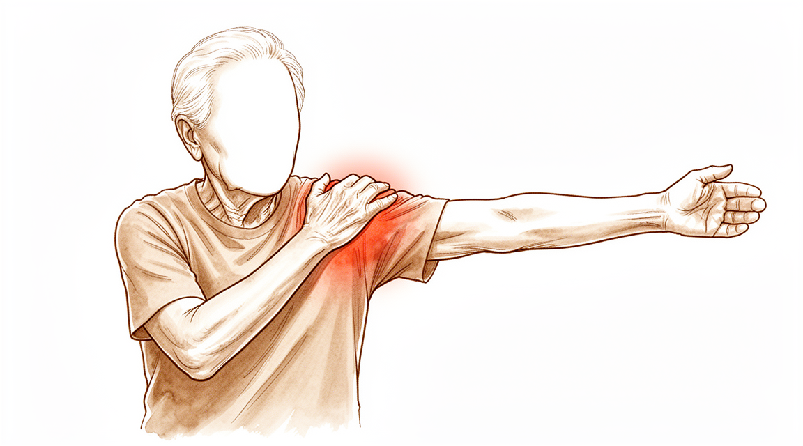
Những triệu chứng mà bạn đang gặp phải
Bệnh khớp do rách chóp xoay là tình trạng viêm khớp ở vai phát triển sau khi bị rách chóp xoay lâu ngày. Chóp xoay là nhóm gân giữ phần chỏm (đầu tròn) của khớp vai nằm ở chính giữa khi bạn nâng cánh tay. Khi các gân này không còn nữa, khớp bị mòn theo cách mà nó không bao giờ được thiết kế để chịu, và sự mài mòn đó chính là những gì bạn đang cảm thấy.
Cơn đau thường nằm sâu bên trong chính khớp vai, tại chỗ chỏm tiếp xúc với ổ khớp. Cơn đau cũng thường xuất hiện ở mặt ngoài của cánh tay. Nâng tay lên cao quá đầu, với tay lên kệ, hoặc xách túi đồ bằng tay bên đó đều làm cơn đau nặng hơn. Nhiều người thấy cơn đau bùng lên vào ban đêm, và nằm đè lên vai đó có thể khiến bạn thức giấc. Cơn đau thường dịu đi một chút khi bạn đã thức dậy và cử động, rồi lại tăng lên sau một ngày bận rộn.
Vì chóp xoay không còn làm được nhiệm vụ của nó, cách cánh tay bạn cử động cũng thay đổi. Bạn có thể nhận thấy tình trạng yếu chứ không chỉ đau: việc nâng cánh tay lên ngang vai hoặc cao hơn trở nên khó khăn, ngay cả khi có lấy đà vung tay lên. Với tay ra sau để cài dây chuyền, phơi quần áo, hoặc kéo áo len chui qua đầu có thể trở nên vụng về hoặc không thể làm được. Một số người xuất hiện một khối u tròn, nhẵn gần đỉnh vai, nơi chỏm đã bị trượt lên trên.
Qua nhiều tuần và nhiều tháng, những vấn đề này thường tăng dần từ từ chứ không xuất hiện cùng một lúc. Những việc đơn giản như với tay lấy dây an toàn, rót nước từ ấm đun, hoặc mặc áo khoác có thể trở thành phần khó khăn nhất trong ngày.
Một số dấu hiệu cần được chăm sóc khẩn cấp. Nếu vai của bạn trở nên nóng, đỏ, sưng và đau, đặc biệt khi kèm theo sốt, hãy đến khoa cấp cứu ngay trong ngày. Nếu cánh tay của bạn trở nên nhợt nhạt, lạnh, trắng bệch hoặc tím tái, hoặc bạn đột ngột mất cảm giác hay mất khả năng cử động ở cánh tay, cũng hãy làm như vậy. Nếu các triệu chứng của bạn không thuyên giảm, ngày càng nặng hơn qua nhiều tuần, hoặc khiến bạn thức giấc vào ban đêm, hãy đến gặp bác sĩ đa khoa hoặc đề nghị chúng tôi khám chuyên khoa.
Chuyện gì đang xảy ra thực sự
Một khớp vai khỏe mạnh hoạt động giống như một quả bóng nằm trong một ổ khớp nông. Các gân chóp xoay bao quanh chỏm và giữ nó ở chính giữa khi bạn nâng tay. Hãy hình dung chúng như những sợi dây néo giữ cho cột lều đứng vững. Khi những sợi dây đó không còn nữa, cột lều sẽ bị nghiêng.
Ở vai của bạn, cơ delta khỏe ở mặt ngoài vai vẫn kéo khi bạn nâng cánh tay. Khi không còn chóp xoay để giữ chỏm ở dưới và ở chính giữa, lực kéo đó kéo chỏm lên trên. Khi đó chỏm cọ xát vào phần xương phía trên nó, tức là mỏm cùng vai, gờ xương nằm ở đỉnh vai của bạn. Hai bề mặt này vốn không bao giờ được thiết kế để chạm vào nhau, vì vậy chúng làm mòn lẫn nhau. Sự mài mòn đó chính là viêm khớp, và đó là lý do cơn đau của bạn nằm sâu trong khớp và lý do chỏm đã bị trượt lên tạo thành khối u tròn mà bạn có thể đã sờ thấy.
Sự mài mòn này cũng giải thích tình trạng yếu tay. Việc nâng cánh tay phụ thuộc vào việc chỏm được giữ ở chính giữa, và chỏm khớp của bạn không còn như vậy nữa. Vì thế bạn có dùng sức nhưng cánh tay không nâng lên được như bình thường.
Có một cách để khắc phục điều này. Phẫu thuật thay khớp vai ngược chiều hoán đổi vị trí của chỏm và ổ khớp: chỏm được đặt ở phía ổ khớp và ổ khớp được đặt ở phía cánh tay. Sự thay đổi đó hạ thấp tâm của khớp và cho cơ delta một cánh tay đòn dài hơn để hoạt động, giống như cầm tay nắm cửa ở vị trí xa bản lề hơn. Khi đó cơ có thể tự nâng cánh tay mà không cần đến các gân chóp xoay đã mất. Khớp mới cũng được thiết kế để chỏm không thể trượt ra ngoài, giúp khớp luôn vững khi bạn cử động.
Vì cơ delta đảm nhận công việc này, sau phẫu thuật vai sẽ cử động theo cách khác. Xương bả vai của bạn tham gia vào chuyển động nhiều hơn so với trước đây. Hầu hết mọi người thích nghi với điều này mà không hề nhận ra, và đây là một phần trong cách khớp mới khôi phục khả năng cử động.
Những biện pháp chúng tôi có thể áp dụng
Bác sĩ Kieran Hirpara, bác sĩ phẫu thuật chi trên tại Bệnh viện Mater Private Rockhampton, sẽ bắt đầu bằng các phương pháp ít xâm lấn nhất phù hợp với tình trạng của bạn. Thông thường, bệnh nhân được bác sĩ đa khoa giới thiệu đến phòng khám chúng tôi; nếu nhà vật lý trị liệu khuyên bạn đến gặp chúng tôi, bạn vẫn cần có giấy giới thiệu từ bác sĩ đa khoa để được hưởng mức hoàn trả từ Medicare. Việc đánh giá tại phòng khám, bao gồm hỏi bệnh sử, thăm khám và chụp hình ảnh khi cần, sẽ giúp xác định chẩn đoán.
Bước đầu tiên thường là điều trị không phẫu thuật. Thay đổi cách bạn sử dụng vai sẽ có ích: tránh với tay lên cao quá đầu, nâng đồ bằng tay còn lại, và sắp xếp công việc để bạn không phải mang vác vật nặng ở bên đó. Vật lý trị liệu nhằm giữ cho vai cử động được, tăng cường sức mạnh cho những cơ vẫn còn hoạt động, và làm dịu cơn đau. Chúng tôi thường cho phương pháp này thử nghiệm đầy đủ trong vài tháng trước khi nghĩ đến biện pháp khác.
Thuốc giảm đau có thể hỗ trợ song song với các biện pháp này. Các loại thuốc giảm đau đơn giản và thuốc chống viêm, tức là những loại thuốc giúp giảm sưng và kích ứng, là lựa chọn ban đầu thông thường. Bác sĩ đa khoa có thể tư vấn loại nào an toàn cho bạn.
Nếu những biện pháp này không mang lại đủ cải thiện, phẫu thuật có thể được cân nhắc. Đối với bệnh khớp do rách chóp xoay, phẫu thuật là thay khớp vai ngược chiều, hoán đổi vị trí của chỏm và ổ khớp để cơ delta có thể nâng cánh tay mà không cần đến các gân chóp xoay đã mất. Chúng tôi sẽ cùng bạn trao đổi về những gì ca phẫu thuật bao gồm, những gì nó có thể và không thể làm được cho bạn, và liệu nó có phù hợp với mục tiêu của bạn hay không. Đây là quyết định bạn đưa ra cùng với chúng tôi, dựa trên cơn đau, khả năng cử động của bạn và những điều quan trọng với bạn trong cuộc sống hằng ngày.
Những điều có thể mong đợi
Bệnh khớp do rách chóp xoay thường tiến triển chậm trong nhiều tháng, nhiều năm chứ không xuất hiện đột ngột. Nếu không điều trị, tình trạng đau và yếu thường tiếp tục gây cản trở. Sự mài mòn trong khớp không tự hồi phục, vì vậy hầu hết mọi người thấy vai ngày càng hạn chế họ nhiều hơn theo thời gian chứ không ít đi.
Với sự chăm sóc phù hợp, triển vọng sẽ khác. Điều trị không phẫu thuật như vật lý trị liệu và thuốc giảm đau đơn giản có thể làm dịu tình trạng trong một thời gian, và nhiều người xoay xở tốt với các biện pháp này trong nhiều tháng hoặc lâu hơn. Nếu cần phẫu thuật, thay khớp vai ngược chiều đã có thành tích tốt đối với tình trạng này. Phẫu thuật giúp giảm đau và khôi phục khả năng cử động một cách đáng tin cậy, vì cơ delta đảm nhận công việc mà các gân đã mất trước đây vẫn làm. Hầu hết những người vốn năng động trước phẫu thuật đều có thể quay lại các hoạt động của mình sau đó, và việc trở lại chơi thể thao là thường gặp.
Hồi phục sau phẫu thuật cần thời gian và nỗ lực. Phục hồi chức năng chủ động, sớm dưới sự hướng dẫn của nhà vật lý trị liệu là an toàn và hiệu quả, và có thể mang lại lợi ích sớm hơn so với cách tiếp cận chậm hơn, thận trọng hơn. Vật lý trị liệu cũng quan trọng sau những tuần đầu: nó quyết định bạn lấy lại được bao nhiêu khả năng cử động và bạn xoay xở với các công việc hằng ngày dễ dàng đến mức nào trong nhiều tháng, nhiều năm sau đó. Một số người thấy chương trình tập tại nhà tự thực hiện cũng hiệu quả như các buổi tập chính thức có giám sát, vì vậy có nhiều cách để làm tốt việc này.
Hãy thực tế về những gì phẫu thuật có thể và không thể làm được. Một khớp vai được thay do bệnh khớp do rách chóp xoay bắt đầu từ tình trạng xấu hơn so với khớp được thay do viêm khớp đơn thuần, vì vậy kết quả thường kém hơn một chút so với những trường hợp đơn giản hơn đó. Biến chứng có thể xảy ra với bất kỳ ca thay khớp vai nào. Các biến chứng thường gặp nhất là mất vững khớp, tức là khớp cử động theo cách không nên có, và nhiễm trùng. Một số biến chứng có thể được điều trị thành công nếu xảy ra, và phẫu thuật làm lại, tức là thay lại một phần hoặc toàn bộ khớp nhân tạo, vẫn mang lại cải thiện thực sự về đau, khả năng cử động và chức năng, dù kết quả nhìn chung không tốt bằng sau lần phẫu thuật đầu tiên.
Điểm xuất phát của chính bạn rất quan trọng. Tình trạng vai của bạn trước phẫu thuật là một trong những yếu tố dự báo mạnh nhất về tình trạng vai sau phẫu thuật, và chúng tôi dựa vào đó để cùng bạn lên kế hoạch hồi phục.
Khi nào nên gặp bác sĩ
Bệnh khớp do rách chóp xoay thường tiến triển chậm, vì vậy hầu hết mọi người đến khám khi vai bắt đầu hạn chế cuộc sống hằng ngày chứ không phải như một trường hợp cấp cứu. Hãy đề nghị được khám chuyên khoa nếu cơn đau không thuyên giảm khi nghỉ ngơi và dùng thuốc giảm đau đơn giản, nếu cơn đau ngày càng nặng hơn qua nhiều tuần, hoặc nếu nó khiến bạn thức giấc vào ban đêm. Điều tương tự cũng áp dụng nếu tình trạng yếu tay hoặc khối u tròn ở đỉnh vai đang khiến bạn không thể làm việc hay sử dụng cánh tay.
Một số dấu hiệu cần được chăm sóc ngay trong ngày. Hãy đến khoa cấp cứu nếu vai của bạn trở nên nóng, đỏ, sưng và đau, đặc biệt khi kèm theo sốt, hoặc nếu cánh tay của bạn trở nên nhợt nhạt, lạnh, trắng bệch hoặc tím tái. Đột ngột mất cảm giác hoặc mất khả năng cử động ở cánh tay cũng cần được đánh giá cấp cứu.
Nếu bạn đã được phẫu thuật thay khớp vai và nhận thấy cơn đau mới xuất hiện ở phần gốc của mỏm cùng vai, tình trạng yếu mới xuất hiện, hoặc cơn đau đột ngột tăng lên hay mất chức năng, hãy báo cho chúng tôi ngay. Nếu bạn không thể liên hệ với phòng khám, hãy đến khoa cấp cứu gần nhất.
Phân tích chi tiết hơn
Advanced reading: the deeper science (optional)
Phần này đi sâu hơn mức cần thiết để bạn có thể tự đưa ra quyết định điều trị. Bệnh khớp do rách chóp xoay và các vết rách lớn không thể khâu phục hồi rất đáng để đọc thêm, bởi chính số lượng các phương pháp phẫu thuật được mô tả cho những trường hợp này là thông tin nói lên nhiều điều nhất: khi có nhiều phương pháp cùng cạnh tranh, không phương pháp nào tốt hơn rõ rệt.
Mọi phương pháp đều có tác dụng phần nào, nhưng không có phương pháp nào vượt trội rõ rệt
Trên 2.000 bệnh nhân, hiệu quả điều trị có ý nghĩa lâm sàng được ghi nhận ở cả mười một phương thức điều trị khác nhau được nghiên cứu đối với các vết rách chóp xoay vùng sau trên không thể khâu phục hồi; tuy nhiên sự khác biệt về đặc điểm bệnh nhân, các can thiệp đi kèm, cách báo cáo kết quả và thời gian theo dõi đã khiến việc so sánh giữa các phương pháp trở nên khó khăn [1]. Một nghiên cứu khác trên 3.363 bệnh nhân cho thấy cả sáu lựa chọn điều trị không thay khớp đều giúp cải thiện đáng kể phạm vi vận động và các chỉ số đánh giá do bệnh nhân tự báo cáo sau một năm hoặc lâu hơn; tỷ lệ phải phẫu thuật lại cũng ở mức thấp [2].
Mười một phương pháp điều trị, sáu lựa chọn khác nhau; tất cả đều mang lại cải thiện, nhưng không phương pháp nào chứng tỏ được tính ưu việt. Xu hướng này thường cho thấy hai điều: tiến triển tự nhiên của bệnh cũng có thể dẫn đến sự cải thiện nào đó, và các nghiên cứu có quá nhiều yếu tố không đồng nhất khiến việc phân biệt hiệu quả giữa các phương pháp trở nên khó khăn.
Cải thiện ban đầu, suy giảm về sau
Có một phát hiện cần được nhấn mạnh vì dễ bị bỏ qua trong các báo cáo ngắn hạn. Trên 2.790 bệnh nhân trải qua phẫu thuật tái tạo bao khớp trên, khâu phục hồi một phần, đặt mảnh ghép xen giữa và các thủ thuật liên quan, các chỉ số đánh giá khớp vai đều cải thiện đáng kể ngay từ đầu ở tất cả các kỹ thuật, dù tỷ lệ rách lại cao; đồng thời các chỉ số này có thể suy giảm ở giai đoạn theo dõi trung hạn và dài hạn [3].
Vì vậy, cùng một ca phẫu thuật có thể cho kết quả khả quan sau một năm nhưng lại kém hiệu quả sau năm năm. Khi đọc về kết quả tốt của một kỹ thuật nào đó, khoảng thời gian theo dõi cũng quan trọng không kém gì con số kết quả.
Một phân tích năm 2026 trên 4.963 bệnh nhân đã cố gắng giải quyết vấn đề này bằng cách xếp hạng các phương pháp điều trị dựa trên tỷ lệ thất bại thay vì chỉ dựa vào các chỉ số kết quả; dù không xác định được phương pháp điều trị nào là tốt nhất, nghiên cứu này vẫn đưa ra thứ tự tin cậy của các phương pháp điều trị [4]. Việc xếp hạng dựa trên tỷ lệ thất bại có lẽ là cách đánh giá trung thực hơn khi các chỉ số ban đầu của các phương pháp điều trị gần như tương đương nhau.
Tại sao khớp bị thoái hóa sau khi cơ chóp xoay bị tổn thương
Cơ chế này giải thích tại sao đây là một tình trạng bệnh lý riêng biệt chứ không đơn thuần chỉ là một vết rách lớn ở cơ chóp xoay. Cơ chóp xoay có nhiệm vụ giữ đầu xương cánh tay nằm đúng vị trí trong ổ khớp trong lúc cơ delta nâng cánh tay. Khi không còn chóp xoay, lực kéo của cơ delta sẽ đẩy đầu xương cánh tay lên phía trên, áp vào mặt dưới của mỏm cùng vai.
Điều này dẫn đến một dạng tổn thương đặc trưng: trên phim X-quang, đầu xương cánh tay bị dịch lên trên; mỏm cùng vai và đầu xương cánh tay cọ xát vào nhau ở những vùng vốn không bao giờ tiếp xúc; các bề mặt khớp bị thoái hóa thứ phát. Đây là dạng viêm khớp do yếu tố cơ học gây ra chứ không phải do bệnh lý khớp nguyên phát; vì vậy việc điều trị viêm khớp mà không khắc phục các yếu tố cơ học là không hiệu quả.
Đây cũng là lý do tại sao phẫu thuật thay khớp vai ngược lại có thể giải quyết vấn đề: phương pháp này giúp cơ delta đảm nhiệm chức năng nâng đỡ mà không cần đến sự hỗ trợ của cơ chóp xoay. Khi bệnh khớp đã hình thành, phẫu thuật này được trình bày trong một trang riêng.
Cách đọc các số liệu kết quả và đặt kỳ vọng phù hợp
Một lưu ý quan trọng khi diễn giải kết quả: các kết quả sau phẫu thuật thay khớp ngược chiều trong trường hợp này thấp hơn so với các chỉ định khác ở 6.698 bệnh nhân [5]. Phẫu thuật này vẫn mang lại hiệu quả, nhưng không nên kỳ vọng rằng khớp vai được thay do bệnh khớp do rách chóp xoay sẽ có kết quả tương đương với khớp vai được thay do viêm khớp thông thường khi chóp xoay còn nguyên vẹn; tình trạng ban đầu của bệnh nhân ở nhóm đầu đã tồi tệ hơn.
Tài liệu tham khảo
[1] Kooistra B, Gurnani N, Weening A, van den Bekerom M, van Deurzen D. Mức độ bằng chứng còn hạn chế đối với mọi phương pháp điều trị vết rách cơ chóp xoay vùng sau trên không thể vá lại. Knee Surg Sports Traumatol Arthrosc. 2019;27(12):4038-48. https://doi.org/10.1007/s00167-019-05710-0
[2] Hughes JD, Davis B, Whicker E, Sprowls GR, Barrera L, Baradaran A, và cộng sự. Các phương pháp điều trị không cần thay khớp cho các vết rách cơ chóp xoay lớn và không thể vá lại đã giúp cải thiện kết quả theo đánh giá của bệnh nhân. Knee Surg Sports Traumatol Arthrosc. 2022;31(5):1883-902. https://doi.org/10.1007/s00167-022-07099-9
[3] Davies A, Singh P, Reilly P, Sabharwal S, Malhas A. Tái tạo màng bao khớp vùng trên, vá một phần cơ chóp xoay, đặt mảnh ghép, bóng đệm dưới mỏm cùng vai hoặc tạo hình củ lớn xương cánh tay: một phân tích có hệ thống. J Orthop Surg Res. 2022;17(1). https://doi.org/10.1186/s13018-022-03411-y
[4] Cooke SP, Koh JL, Amirouche F. Phân tích tỷ lệ thất bại của các phương pháp điều trị đối với các vết rách cơ chóp xoay lớn đến rất lớn không thể vá lại. J Shoulder Elbow Arthroplasty. 2026;10(1-2):100019. https://doi.org/10.1016/j.jsea.2026.100019
[5] Yazdanpanah S, Soth BT, Eskew JR, Dancy M, Fu MC, Taylor SA, và cộng sự. Kết quả lâm sàng và chức năng giảm sút sau khi thực hiện phẫu thuật thay khớp vai ngược để điều trị viêm khớp do vết rách cơ chóp xoay: một phân tích có hệ thống. JSES Rev Rep Tech. 2026;6(2):100691. https://doi.org/10.1016/j.xrrt.2026.100691
Evidence & references
This is the clinical evidence summary written for health professionals. It is technical, and it lists the research this page was built from. You do not need to read it to understand your treatment or to make a decision about it.
Overview
- Favorable clinical outcomes can be achieved after hemiarthroplasty for glenohumeral arthritis complicated by massive rotator cuff tears [1].
- Shoulder arthroplasty restores shoulder function well in both osteoarthritis and rotator cuff tear arthropathy [2].
- Reverse total shoulder arthroplasty provides comparable clinical outcomes across indications of irreparable rotator cuff tear, rotator cuff tear arthropathy, and primary osteoarthritis at 2 years [3].
- Patients with rotator cuff arthropathy associated with a significant loss of deltoid function may be better managed with glenohumeral arthrodesis [4, 5].
- The short-term results of subacromial balloon spacers for management of massive rotator cuff tears demonstrate clinically relevant improvements in shoulder range of motion and substantial improvements in patient-reported outcome measures [6].
- Patients with irreparable massive rotator cuff tears without the presence of osteoarthritis have a high likelihood of achieving a painless shoulder and functional improvements after reverse shoulder arthroplasty [7].
- In patients with rotator cuff-intact glenohumeral osteoarthritis, anatomic total shoulder arthroplasty and reverse total shoulder arthroplasty demonstrated similar short-term to midterm clinical outcomes after propensity score matching, with no significant differences observed across age strata [8].
- Bipolar arthroplasty is indicated for patients with rotator cuff arthropathy [11].
- The functional results of large head or any type of hemiarthroplasty are unpredictable in rotator cuff tear arthropathy, but pain relief, the primary goal of surgery, is a predictable result of surgery [12].
- In selected and willing patients, deltoid release can be an efficacious alternative to shoulder prosthesis or to other technically complicated procedures [14].
- The most common postoperative complication in humeral head replacement was rotator cuff tearing, found in 23 (18.1%) of 127 shoulders [16, 25].
- Differences in postoperative patient-reported outcomes and improvement from baseline demonstrate a trend toward lower outcomes in patients with prior rotator cuff repair, but these differences may be below the minimal clinically important difference [17].
- Reverse shoulder arthroplasty provides optimal outcomes with low complication rates across a short term of follow-up for glenohumeral osteoarthritis with an intact rotator cuff [20].
- Irrespective of tissue source, superior capsular reconstruction serves as a reasonable joint-preserving option for massive, irreparable rotator cuff tears, with favorable short- to midterm improvements in patient-reported outcomes and range of motion [26].
- Under optimal circumstances, simultaneous shoulder arthroplasty is feasible [38].
- Tenodesis at the time of primary rotator cuff repair may be associated with a reduction in the utilization of ipsilateral shoulder revision surgery rates [41].
- Nonprosthetic glenoid arthroplasty is being expanded to individuals who do not want the risk of a glenoid prosthesis regardless of age for the treatment of glenohumeral arthritis [55].
- All six nonarthroplasty treatment options for irreparable rotator cuff tears resulted in statistically significant improvements in range of motion and patient-reported outcomes at 1 year follow-up or more, with low rates of revision and conversion to arthroplasty [56].
- Hemiarthroplasty does not provide for a successful outcome in all patients with rotator cuff arthropathy [60].
Anatomy & Pathophysiology
Bony Anatomy
- The glenoid is a convex structure of shallow depth shaped like an inverted pear [74].
- The glenoid articular surface radius of curvature is 2 to 3 mm larger than that of the humeral head [86].
- The average neck-shaft angle of the proximal humerus is 45 degrees (±5 degrees), with a range of 30 to 50 degrees [86].
- Arthritic shoulders have a flatter neck-shaft angle close to 50 degrees [86].
- The superior margin of the humeral head articular surface is normally superior to the top of the greater tuberosity by 8 to 10 mm [86].
- The distance from the lateral base of the coracoid process to the lateral margin of the greater tuberosity is called the lateral humeral offset [86].
- A significant decrease in lateral humeral offset reduces the lever arms for the deltoid and supraspinatus muscles, weakening abduction and impairing function [86].
- A significant increase in lateral humeral offset causes excessive tension on the soft tissues, resulting in loss of motion and likely accelerating polyethylene wear [86].
- Humeral articular malposition of more than 4 mm leads to increased subacromial contact [86].
- An offset of 8 mm in any direction significantly decreases passive range of motion [86].
- Proximal humeral retroversion is highly variable, ranging from 0 to 55 degrees depending on the method used for measurement [86].
- The glenoid averages 5° of retroversion in relation to the axis of the scapular body [77].
- The humeral head averages 19° of retroversion and 41° of inclination (neck-shaft angle) [77].
- The articular head of the humerus is spherical and has a diameter of 37 to 57 mm [74].
- The most superior portion of the articular surface of the humeral head averages 8 mm above the greater tuberosity [74].
- The humeral version averages 29.8 degrees (range, 10 to 55 degrees) [74].
- The head is inclined approximately 130 degrees with respect to the humeral shaft [74].
- The neck-shaft angle measures an average of 135 degrees, and the humeral head is retroverted an average of 30 degrees [75].
- The glenoid cavity is a shallow socket, approximately one third the size of the humeral head [75].
- The linear correlation between glenoid inclination and acromial angle suggests the presence of a balance between the glenoid inclination and the acromial coverage in a healthy shoulder [39].
- The critical shoulder angle should be considered as a “combined shoulder angle” with balanced contributions of glenoid inclination and acromial angle in shoulder arthritis progression [120].
Soft Tissue Anatomy
- The rotator cuff consists of four muscles: the subscapularis, supraspinatus, infraspinatus, and teres minor [75].
- The teres major is not a rotator cuff muscle [75].
- The cuff muscles serve as depressors of the humeral head to allow the deltoid to efficiently abduct the humerus [75].
- The infraspinatus and teres minor are external rotators, while the subscapularis is an internal rotator of the humerus [75].
- The greater tuberosity provides attachment for the supraspinatus, infraspinatus, and teres minor muscles [75].
- The lesser tuberosity contains the attachment of the subscapularis muscle [75].
- The rotator cuff is a sheet of conjoined tendons closely applied over the shoulder capsule and inserting mainly into the greater tuberosity of the humerus, with the subscapularis inserted into the lesser tuberosity [82].
- The coracoacromial arch is formed by the acromion process posterosuperiorly, the coracoid process anteriorly, and the coracoacromial ligament joining them [82].
- The subacromial bursa separates the tendons from the coracoacromial arch and allows them to glide [82].
- The rotator interval is defined medially by the base of the coracoid, superiorly by the supraspinatus tendon, and inferiorly by the subscapularis tendon [77].
- The rotator interval contains the coracohumeral ligament, the superior glenohumeral ligament, and the intra-articular portion of the long head of the biceps tendon [77].
- Laxity of the rotator interval results in inferior laxity (the sulcus sign), and contracture of the interval is seen with adhesive capsulitis [77].
- The coracohumeral ligament restricts external rotation in adduction and is a static restraint to inferior and posterior translation in adduction and external rotation [77].
- The superior glenohumeral ligament is a primary static restraint against anterior translation with the arm at the side [77].
- With the coracohumeral ligament, the superior glenohumeral ligament forms a pulley that provides restraint against medial subluxation of the long head of the biceps tendon [77].
- The middle glenohumeral ligament is a primary static restraint against anterior translation with the arm in external rotation and 45° of abduction [77].
- The anterior band of the inferior glenohumeral ligament is a primary static restraint against anterior-inferior dislocation of the glenohumeral joint in 90° of abduction and external rotation [77].
- The posterior band of the inferior glenohumeral ligament is a primary static restraint against posterior-inferior translation in internal rotation and adduction [77].
- The glenoid labrum provides concavity and up to 50% of marginal glenoid socket depth [77].
- The fibrocartilaginous glenoid labrum deepens the socket by 50% around the humeral head and increases stability [87].
- The glenoid articular surface and the labrum combine to create a socket that is approximately 9 mm deep in the superoinferior direction and 5 mm deep in the anteroposterior direction [87].
- Adding the glenoid labrum increases the glenoid surface to 75% of the humeral head vertically and 57% horizontally [87].
- The tendinous insertions of the rotator cuff muscles, the articular capsule, the coracohumeral ligament, and the glenohumeral ligament complex blend into a confluent sheet before insertion into the humeral tuberosities [87].
- The tendons of the infraspinatus and supraspinatus muscles join approximately 15 mm proximal to their insertion and cannot be readily separated by blunt dissection [87].
- The infraspinatus and teres minor fuse near their musculotendinous junctions [87].
- The supraspinatus and subscapularis tendons join as a sheath that surrounds the biceps tendon at the entrance of the bicipital groove [87].
- The roof of the biceps sheath consists of a portion of the supraspinatus tendon, and a sheet of the subscapularis tendon forms the floor [87].
- The coracoacromial ligament is a thick band of fibrous tissue extending from the coracoid process along the surface of the capsule to the tuberosities between the supraspinatus and subscapularis tendons [87].
- The coracoacromial ligament contributes to anterosuperior stability in rotator cuff deficiency and should be preserved with irreparable cuff tears to prevent anterosuperior escape [88].
- The subscapular bursa lies between the subscapularis tendon and the neck of the scapula and communicates with the joint cavity between the superior and middle glenohumeral ligaments [78].
- The subscapular bursa often houses loose bodies in the shoulder and is a region in which synovitis of the shoulder may be most intense [78].
- The humeroscapular motion interface lies between the inner structures of the proximal humerus, rotator cuff, coracohumeral ligament, and biceps tendon sheath and the superficial layer of the acromion, deltoid, coracoacromial ligament, coracoid process, and the conjoined tendon [80].
- Smooth, unrestricted motion at the humeroscapular motion interface is vital to shoulder mobility [80].
- The axillary nerve has an intimate relationship within the humeroscapular motion interface [80].
- The anterior and middle deltoid muscle receives sole innervation from the anterior branch of the axillary nerve [80].
- The posterior deltoid muscle innervation varies, with supply only from the anterior branch in 2.3% of cases, from the posterior branch in 8.5%, and from both branches in 89.1% [80].
- The proximal humerus receives its blood supply from the anterior and posterior humeral circumflex branches from the third division of the axillary artery [74].
- The anterior humeral circumflex artery provides vascular inflow to the humeral head by way of its terminal anterolateral branch known as the artery of Laing (also known as the arcuate artery) [74].
- Injury to the arcuate artery may result in osteonecrosis of the humeral head [74].
- Additional extraosseous collateral branches can permit humeral head perfusion despite complete ligation of the arcuate artery [74].
- The anterolateral ascending branch of the anterior humeral circumflex artery provides the primary blood supply to the humeral head [77].
- The terminal intraosseous portion of the artery enters at the proximal aspect of the intertubercular groove as the arcuate artery [77].
- Quantitative assessment has shown that 64% of the humeral head blood supply arises from the posterior humeral circumflex artery [81].
- The scapula is suspended by muscles alone and reflects the adaptive development of the shoulder [84].
- Broadening of the infraspinatus fossa has resulted in a change in the vector of muscle pull from the axillary border of the scapula to the glenoid fossa [84].
- This adaptation allows the infraspinatus and teres minor muscles to be more effective in their roles as depressors and external rotators of the humeral head [84].
- The acromion has enlarged over time, reflecting the increasing role of the deltoid muscle in shoulder function [84].
- The broader attachment of the deltoid on the acromion and its more distal insertion on the humerus have increased its mechanical advantage in shoulder motion [84].
- With the shoulder in 90 degrees of abduction, the coracoid extension over the glenohumeral joint can mechanically limit anterior translation of the humerus relative to the glenoid [84].
Pathophysiology
- Cuff tear arthropathy (CTA) is the final stage of the shoulder impingement syndrome spectrum [102].
- CTA affects patients with long-term insufficient massive rotator cuff tears, superior migration of the humeral head toward the acromion, subchondral osteoporosis, humeral head collapse, and painful debilitating shoulder arthritis [102].
- CTA was initially known as Milwaukee shoulder syndrome due to the rapidly progressive destruction of cartilage and bone, noninflammatory joint effusion containing calcium hydroxyapatite crystals, synovial hyperplasia, and multiple loose bodies [102].
- CTA affects women (3:1 female to male ratio), over 70 years old, more commonly on the dominant shoulder [102].
- Risk factors for CTA include chronic rotator cuff tears, hemorrhagic shoulder (oral anticoagulants and hematologic diseases), rheumatic disease, and crystal-induced arthropathy [102].
- Neer suggested mechanical, nutritional, and crystal-induced arthropathy pathways for CTA, but no definitive pathogenesis has been identified [102].
- Mechanical factors in CTA include insufficient cuff, superior migration of the humeral head, instability, eccentric wear of the glenoid, humeral head deformity, and decreased shoulder function [102].
- Nutritional factors in CTA include hypomobility-induced cartilage atrophy, poor nutrition (decrease in glycosaminoglycans), dehydration, and subchondral osteoporosis [102].
- Crystalline-induced arthropathy in CTA involves synovial-based matrix proteins degradation destroying rotator cuff tendons and cartilage, with end-stage calcium-phosphate crystal deposition [102].
- A massive cuff tear induces both nutritional and mechanical factors that provide a logical explanation for the development of humeral head collapse [15].
- Hydroxyapatite is strongly related to the pathogenesis of Milwaukee shoulder syndrome, which is nearly identical to cuff tear arthropathy [15].
- CPPD crystal deposition can hasten the development of cuff tear arthropathy [15].
- Cuff tear arthropathy is defined as a combination of massive cuff tear, elevation and collapse of the humeral head, and damage to the glenohumeral joint [15].
- The dysfunction of the rotator cuff results in loss of the concavity-compression mechanism, instability, and a predictable wear pattern with superior humeral migration and ultimate acetabularization of the acromion [70].
- The reverse total shoulder design counteracts a rotator cuff-deficient system by changing the center of rotation of the native glenohumeral joint to one that is more distal and medial [70].
- Distalization of the humerus lengthens the lever arm of the deltoid and increases the resting tension of the muscle, thus increasing the compression between the glenosphere and humeral prosthesis [70].
- Medialization minimizes the shear forces experienced at the bone-base plate interface [70].
- The compressive, stabilizing force generated by deltoid pressing along the lateral aspect of the proximal humerus is referred to as deltoid wrapping [70].
- Impingement-rotator cuff tears are a spectrum of disease that start with tendinitis (20 to 35 years old), progress to tendinosis (35 to 45 years old), rotator cuff tears (>45 years old), and, if not treated, evolve to cuff arthropathy (>65 years old) [118].
- Intrinsic degeneration in rotator cuff tears involves age-related (>60 years old) changes in collagen, proteoglycan, water content, and vascularity (tendinosis), usually involving the supraspinatus and infraspinatus starting on the articular side [118].
- Extrinsic rotator cuff tears result from chronic impingement on the coracoacromial arch, usually starting on the bursal side of the tendon, and may be associated with a hook-shaped acromion [118].
- Acute traumatic rotator cuff tears occur after a fall and/or dislocation of the shoulder in patients under 40 years old [118].
- The hypovascular critical zone is located on the articular side of the rotator cuff close to the insertion on the greater tuberosity [118].
- Microangiographic studies showed an area of hypovascularity near Codman’s “critical zone” just proximal to the supraspinatus insertion into the greater tuberosity [101].
- This hypoperfusion is believed to initiate degenerative changes, which subsequently lead to calcification or susceptibility to tearing [101].
- Other histologic studies showed no evidence of inadequate vascularization, and the supraspinatus, including the critical zone, was found to be well supplied with an anastomosis of vessels [101].
- One histologic study demonstrated neovascularization and neoinnervation in calcific tendonitis, with an associated substantial inflammatory response as the cause of pain [101].
- Calcific tendinitis follows a definite progression in most patients, with resolution seen in almost all of them [101].
- The precalcification stage of calcific tendinitis involves fibrocartilaginous metaplasia at the site of predilection for calcification, possibly a site with diminished blood supply [101].
- During the calcification stage, calcium is deposited into matrix vesicles, which are excreted by the cells and coalesce into larger calcium deposits [101].
- The resorption phase of calcific tendinitis involves an inflammatory response and is exquisitely painful [101].
- The precise pathogenesis of calcific tendinitis remains unclear, but an active, cell-mediated process is widely accepted [116].
- The precalcific stage of calcific tendinitis consists of predominantly fibrocartilaginous metaplasia presumably within less vascular areas of the tendon [116].
- In the formative phase of the calcific stage, matrix vesicles unite to form calcium hydroxyapatite deposits that are separated by fibrocollagenous tissue [116].
- Without a clear trigger, the resorption phase involves an inflammatory response [116].
- Pain in calcific tendinitis is variable during the proliferative phase and is correlated with the macrophage activity during the resorptive phase [116].
- Rotator cuff pathology spans a spectrum of severity that includes rotator cuff tendinopathy, partial-thickness tears, full-thickness tears, and rotator cuff arthropathy [93].
- The requisites for normal cuff function include healthy, strong cuff muscles, normal capsular laxity, intact cuff tendons, a smooth contour of the underside of the coracoacromial arch, a thin, lubricating bursa, a smooth upper surface of the cuff and tuberosities, and concent
Classification
- The hypothesis proposed by Neer concerning the pathomechanics of cuff tear arthropathy is that a massive cuff tear induces both nutritional and mechanical factors that provide a logical explanation for the development of humeral head collapse [15].
- McCarty described a shoulder condition called the Milwaukee shoulder syndrome, which was nearly identical to cuff tear arthropathy, and emphasized that hydroxyapatite was strongly related to the pathogenesis of this shoulder condition [15].
- CPPD crystal deposition has been suggested to hasten the development of cuff tear arthropathy [15].
- Primary osteoarthritis is confirmed where glenohumeral joint narrowing is observed on radiographs, together with sclerotic osteophytes on the humeral head, and acromiohumeral distance >6 mm [33].
- Secondary osteoarthritis is confirmed where glenohumeral joint narrowing is observed on radiographs, together with proximal humeral migration, as a consequence of large or massive rotator cuff tears observed on MRI or CTA [33].
- Secondary osteoarthritis includes cuff tear arthropathy (Hamada stage 5), as well as early stage of OA combined with mRCT (Hamada stages 1 and 2), and shoulders with no humeral necrosis [33].
- Fatty infiltration of the supraspinatus, infraspinatus, and subscapularis is graded using the classification of Goutallier et al [33].
- Fatty infiltration of the rotator cuff muscles is dichotomized as either functional (Goutallier classification 0, 1, or 2) or nonfunctional (Goutallier classification 3 or 4) [33].
- The Walch classification is used to assess glenoid morphology in the transverse plane [33].
- The Hamada classification is used to confirm secondary osteoarthritis due to rotator cuff tears [33].
Clinical Presentation
- Hydroxyapatite is strongly related to the pathogenesis of Milwaukee shoulder syndrome, which was described as nearly identical to cuff tear arthropathy [15].
- CPPD crystal deposition may hasten the development of cuff tear arthropathy [15].
- Overhead function is experienced only in cases when the rotating cuff was intact or properly reconstructed [13].
- Rotator cuff deficiency and instability are important factors for a poor result in shoulder arthroplasty for non-tumorous glenohumeral desintegration [128].
- The glenoid should not be resurfaced in the setting of a deficient rotator cuff, significant bone loss, or a young active patient because of the increased failure rates in these populations [19].
- Favorable clinical outcome can be achieved after hemiarthroplasty for glenohumeral arthritis complicated by massive rotator cuff tears [1].
- The functional results of large head or any type of hemiarthroplasty are unpredictable in rotator cuff tear arthropathy, but pain relief is a predictable result of surgery [12].
- Significant theoretical advantages exist for the BiPolar shoulder design which are particularly important in cases of cuff tear arthropathy [42, 62].
- Results with the Neer Shoulder prosthesis are adversely influenced by cuff tear arthritis, with a Constant Score of 45 per cent [18].
- In cadaveric studies, subacromial balloon spacers resist superior humeral head migration and reduce subacromial pressure [22].
- Arthroscopic debridement is one of the useful methods for osteoarthritis with massive rotator cuff tears, but its utility is limited [40].
- Patients with irreparable massive rotator cuff tears without presence of osteoarthritis have a high likelihood of achieving a painless shoulder and functional improvements after reverse shoulder arthroplasty [7].
- Reverse total shoulder arthroplasty can provide reliable improvement in clinical outcomes regardless of preoperative diagnosis, with few differences across diagnostic groups regarding preoperative to postoperative improvement [133].
- Within a group of patients with primary glenohumeral degenerative joint disease, shoulder arthroplasty was effective within a relatively short time frame in improving the patients' assessment of both shoulder function and overall health status [27].
- Shoulder function and outcome scores showed no significant deterioration between 5 and 20 years of follow-up after reverse total shoulder arthroplasty for rotator cuff dysfunction [35].
- Patients undergoing primary reverse shoulder arthroplasty demonstrated clinically significant improvements in both range of motion and clinical outcome scores [63].
- The most common diagnosis in a systematic review of patient satisfaction after reverse total shoulder arthroplasty was cuff tear arthropathy (25.8%) [30].
- Cuff tear arthropathy was the most common indication for reverse shoulder arthroplasty in a systematic review of outcomes following failed rotator cuff repair [31].
- Reverse total shoulder arthroplasty has continued to increase in clinical utility and popularity as an effective treatment for cuff tear arthropathy [121].
Investigations
Plain Radiography
- The purpose of shoulder imaging is to help establish the diagnosis, determine the severity of the pathoanatomy, assist in surgical planning, and enable the surgeon to illustrate the condition of the shoulder to the patient [49].
- Standardized plain films are almost always sufficient to garner the information needed for shoulder arthroplasty planning [49].
- The first key radiographic view is the anteroposterior (AP) view in the plane of the scapula, taken so that the x-ray beam passes through the glenohumeral joint [49].
- The AP view in the plane of the scapula shows the superoinferior position of the humeral head relative to the glenoid, the presence of osteophytes on the humeral head and glenoid, narrowing of the joint space, and the degree of medial displacement of the humerus in relation to the lateral acromial line [49].
- The AP view also demonstrates the quality of the humeral and glenoid bone, the presence of loose bodies, and whether there is humeral head collapse or deformity [49].
- The second key radiographic view is the axillary view taken with the arm in the functional position of elevation in the plane of the scapula [49].
- The axillary view is oriented so that both the spinoglenoid notch and the scapular neck are visible [49].
- The axillary view shows a different perspective of the humeral anatomy, the amount of glenoid bone, the shape of the glenoid, its version in relation to the plane of the scapula, and the relationship of the humeral head to the glenoid fossa [49].
- The axillary view is referred to as the "truth view" because it demonstrates the glenohumeral relationships in the functional position of elevation [49].
- CT scans have the disadvantage of being taken with the arm in the adducted position, whereas the axillary truth view is taken in elevation [49].
- When taken properly, standardized anteroposterior and axillary views indicate the thickness of the cartilage space between the humerus and the glenoid, relative positions of the humeral head and the glenoid, presence of osteophytes, degree of osteopenia, and extent of bony deformity and erosion [49].
- Joint space narrowing is most evident on the axillary truth view as opposed to images made with the arm at the side [49].
- The axillary truth view can show posterior subluxation or "functional decentering" that is not evident in images taken with the arm at the side [49].
- The degree of posterior subluxation can be measured as the position of the center of the humeral head in relation to the plane of the scapula, the position of the center of the humeral head in relation to the glenoid face, or the point of contact of the humeral articular surface on the glenoid articular surface [49].
- The point of contact of the humeral articular surface on the glenoid articular surface reflects the degree of centering of the net humeral joint reaction force on the glenoid [49].
- Malcentering of the joint reaction force leads to posterior instability, posterior glenoid wear, and "rocking horse" loosening of prosthetic glenoid components [49].
- At least two X-ray views should be obtained: an anteroposterior in the plane of the glenoid and an axillary projection with the arm in abduction to show the relationship of the humeral head to the glenoid [91].
- The standard shoulder series should include orthogonal views of the shoulder, including a true AP view in the scapular plane, an AP view, an axillary view, and a scapular Y view [104].
- The true AP view in the scapular plane visualizes the anterior greater tuberosity in profile and can reveal proximal humeral migration when the arm is held in neutral rotation with the shoulder in slight abduction [104].
- The AP view with the arm held in internal rotation visualizes the posterior aspect of the greater tuberosity and the lesser tuberosity in profile [104].
- The axillary view enables determination of the humeral head position in the glenoid fossa and may detect occult, locked posterior shoulder dislocation [104].
- The axillary view is helpful in evaluation of glenoid morphology in glenohumeral osteoarthritis and provides good visualization of the coracoid process, acromion, and distal clavicle [104].
- The scapular Y view provides visualization of the coracoacromial arch and can reveal coracoacromial spurs associated with rotator cuff pathology [104].
- The scapular Y view is a reliable alternative for evaluation of glenohumeral subluxation and dislocation and can show scapular body abnormalities and acromial shape [104].
- The acromiohumeral distance is normally 7 to 14 mm [104].
- The width of the glenohumeral joint space should be symmetric superiorly and inferiorly [104].
- The coracoclavicular distance is normally 1.1 to 1.3 cm [104].
- Neer classified acromial morphology as type I (flat), type II (curved), and type III (hooked) [104].
- Type III acromial morphology has been shown to have a correlation with the presence of rotator cuff disease, although no direct causal relationship has been demonstrated [104].
- The glenohumeral offset ratio in normal shoulders can be reliably calculated from a single radiograph [168].
- Plain radiographs are appropriate for patients presenting with shoulder pain with any history of trauma, dislocation, night pain, or chronic shoulder pain [104].
- Arthritis, calcific tendinitis, and osteolysis of the distal clavicle can be observed on plain radiograph [100].
Computed Tomography
- CT imaging is frequently used to evaluate fractures of the shoulder, to assess for bony lesions in recurrent instability cases, or for preoperative templating for shoulder arthritis [100].
- CT with three-dimensional reconstructions is the advanced imaging study of choice for determining the extent of glenoid bone loss in the setting of shoulder instability [104].
- CT is helpful for planning fracture surgery and shoulder joint replacement [91].
- Although CT scans may offer a few degrees of increased precision in the measurement of glenoid version, this precision does not necessarily improve the quality of the surgery or the clinical outcome [49].
- Three-dimensional reconstructions can reveal fine details of the shoulder anatomy, but this additional information rarely changes the planning or conduct of the arthroplasty [49].
Magnetic Resonance Imaging
- MRI is useful to identify osteonecrosis of the humeral head, or a bone tumour [91].
- MRI can identify labral tears and rotator cuff tears, although the accuracy for these is enhanced by combining the scan with arthrography [91].
- MRI is the modality of choice for evaluating the rotator cuff, biceps, and subacromial/subdeltoid bursa [100].
- T1-weighted MRI can reveal Hill-Sachs lesions and is often used with magnetic resonance arthrograms to provide a more detailed picture of the joint surfaces [100].
- T2-weighted MRI provides better visualization of full thickness rotator cuff tears [100].
- Magnetic resonance accuracy in identifying labral and rotator cuff tears in the literature ranges from 70% to 100% [98].
- The acquired multi-planar imaging of MRI allows for the detailed evaluation of the glenoid, labrum, joint capsule, and rotator cuff in different planes [98].
- MR arthrography (MRA) refers to MRI of a joint that has been injected with an intra-articular contrast agent such as diluted gadolinium or saline solution [98].
- MRA increases the sensitivity for detecting tears and other lesions by distending the joint capsule and outlining the cartilage, ligaments, and labrum with contrast [98].
- MRA has proven utility by increasing both sensitivity and specificity in detecting injuries to the capsulolabral–ligamentous complex as compared to traditional MRI [98].
- In a meta-analysis of 6 studies including 4,667 shoulders, MRA had greater diagnostic test accuracy for the detection of glenoid labral lesions than MRI, with MRA sensitivity of 88% and specificity of 93% versus MRI sensitivity of 76% and specificity of 87% [98].
- Abduction and external rotation (ABER) of the arm is an alternative position utilized to increase the sensitivity and specificity for detecting anteroinferior labroligamentous injury [98].
- Limited range of motion or pain may prohibit patients from performing the ABER provocative maneuver [98].
- MRAs can demonstrate a patulous capsule on the coronal, sagittal, and axial imaging in patients with multidirectional instability [98].
- MRAs can be helpful in evaluating lesions of the rotator interval and other associated findings that may affect the eventual surgical plan [98].
- The presence of glenoid dysplasia, increased capsular cross-sectional area, and increased glenoid retroversion have been found to be associated with increased posterior labral tears and symptomatic instability [98].
- Glenoid retroversion was significantly increased in patients with symptomatic posterior labral tears, but there was no significant association between instability and increased humeral head subluxation [98].
- The diagnosis of multidirectional instability is a clinical one, and the need for expensive and/or invasive imaging should be weighed against the information that will be gained from these studies [98].
- MR arthrography is considered the benchmark for evaluation of labral tears and is rarely indicated for evaluation of rotator cuff pathology [100].
- When MRI or MR arthrography is contraindicated, such as in patients with a pacemaker or vascular clips, CT arthrography is indicated [100].
Ultrasonography
- Ultrasonography is a simple and accurate test for identifying rotator cuff tears and calcific tendinitis [91].
- Ultrasonography can be useful in guiding injections or barbotage, such as aspirating calcific deposits in the rotator cuff [91].
- Ultrasonography is a low-cost alternative to MRI and arthrography for evaluating both skeletal and soft-tissue structures of the shoulder [100].
- Ultrasonography can provide immediate, real-time visualization of the rotator cuff, biceps tendon, and calcific deposits [100].
- Ultrasonography can be used to measure the subacromial space and detect atrophy of rotator cuff muscles [100].
- As a result of providing images in real-time, ultrasonography can evaluate impingement in various positions and motions [100].
- Ultrasonography is highly operator dependent and is not as useful for evaluating labral tears or rotator cuff tears that are very small or larger than 3 cm [100].
- The most commonly performed joint examination using ultrasonography is the shoulder examination [89].
- Accuracy of rotator cuff ultrasonography depends on the skill of the scanner operator and an awareness of pitfalls that are encountered [89].
- Ten common pitfalls of rotator cuff ultrasonography have been identified to reduce overdiagnosis or underdiagnosis of rotator cuff pathology [89].
Arthroscopy
- Arthroscopy is useful for diagnosing and treating subacromial impingement, intra-articular lesions, detachment of the glenoid labrum and rotator cuff tears [91].
Treatment
Non-Operative Management
- Nonoperative modalities for shoulder arthritis include activity modification, NSAIDs, physical therapy focusing on capsular stretching, and corticosteroid injections [107].
- Any surgical treatment for glenohumeral arthritis should be preceded by an adequate trial of conservative management that includes activity modification, physical therapy, antiinflammatory medication, and corticosteroid injections [92].
- Injectable viscosupplementation is an additional nonoperative treatment option for glenohumeral arthritis, although there is a paucity of evidence that supports its use in the shoulder and it is not currently approved by the U.S. Food and Drug Administration for injection in joints other than the knee [92].
- The minimal duration of non-operative treatment prior to surgery for irreparable posterosuperior rotator cuff tears varied from 0 months to 6 months across studies, with 28 studies not reporting on prior non-operative treatment [137].
Arthroscopic and Joint-Preserving Procedures
- Arthroscopic debridement for glenohumeral arthritis lacks high-quality evidence to support its routine use [92].
- Arthroscopic debridement with a combination of subacromial decompression, tuberoplasty, subacromial bursectomy, and biceps tenotomy produces good functional outcomes and improvement in pain at mid to long term follow up for the low-demand population greater than 65 years of age looking for pain relief over substantial increase in function [162].
- Comprehensive arthroscopic management consists of glenohumeral débridement, capsular release, and removal of humeral osteophytes [107].
- The optimal treatment of glenohumeral arthritis in young patients is not firmly established [44].
- Arthroscopic repairs of chronic, massive rotator cuff tears, whether complete or partial, are associated with significant improvements in pain, function and objective outcome scores [36].
- Superior capsule reconstruction serves as a reasonable joint-preserving option for massive, irreparable rotator cuff tears, with favorable short- to midterm improvements in patient-reported outcomes and range of motion irrespective of tissue source [26].
- Superior capsule reconstruction is a useful treatment modality for patients with irreparable rotator cuff tears, associated with significantly improved functional outcome scores and preserved or increased mean acromiohumeral distance [160].
- Shoulder scores may decline at mid- to long-term follow-up for large and massive irreparable rotator cuff tears treated with superior capsule reconstruction, partial cuff repair, graft interposition, arthroscopic debridement, or balloon spacers [21].
- Subacromial balloon spacer implantation for patients with massive irreparable rotator cuff tears may achieve satisfactory outcomes between 3 months and 3 years of follow-ups [127].
- Patients undergoing subacromial spacer implantation for the treatment of massive irreparable rotator cuff tears have satisfactory outcomes at the 2- to 3-year follow-up with a low rate of complications [58].
- Placement of the subacromial balloon spacer is a minimally invasive, technically simple procedure with favorable patient-reported outcomes at limited short-term follow-up [161].
- In selected and willing patients, deltoid release can be an efficacious alternative to shoulder prosthesis or to other technically complicated procedures for symptomatic cuff tear arthropathy [14].
Hemiarthroplasty
- Hemiarthroplasty is an option for young and active patients with severe glenohumeral arthritis, but glenoid erosion and need for early revision have been challenges in this patient population [117].
- Hemiarthroplasty is associated with a high rate of glenoid erosion and revision, and its use should be limited to a young patient with severe glenohumeral arthritis and a high level of heavy activity [117].
- The authors are optimistic for the role of shoulder hemiarthroplasty with nonprosthetic glenoid arthroplasty in the treatment of glenohumeral arthritis and have begun to expand indications to individuals who do not want the risk of a glenoid prosthesis regardless of age [55].
- Evaluation of patients suggests that bipolar arthroplasty is indicated for patients with rotator cuff arthropathy [11].
Anatomic Total Shoulder Arthroplasty
- Anatomic total shoulder arthroplasty is the benchmark for surgical treatment of primary glenohumeral arthritis with an intact rotator cuff and ample glenoid bone stock [117].
- Both keeled and pegged glenoid components yield similar pain relief, functional gains, and shoulder motion across most patient-reported outcome measures in total shoulder arthroplasty for primary osteoarthritis [37].
- Primary total shoulder arthroplasty performed through the rotator interval allows access to the glenohumeral joint without sacrificing the integrity of the subscapularis complex, maintains soft tissue balancing, allows optimal placement of the glenoid component, and allows patients to be started on a nonrestrictive physical therapy regimen [69].
Reverse Total Shoulder Arthroplasty
- Reverse total shoulder arthroplasty provides comparable clinical outcomes across indications of irreparable rotator cuff tear, rotator cuff tear arthropathy, and primary osteoarthritis at 2 years, supporting its broad applicability [3].
- Reverse total shoulder arthroplasty can address multiple issues including poor rotator cuff function, instability, and poor glenoid bone stock and is the treatment of choice in severe cuff tear arthropathy, and revision arthroplasty in elderly patients [117].
- The complication rate of reverse total shoulder arthroplasty is higher than anatomic total shoulder arthroplasty and patients commonly achieve less internal rotation postoperatively [117].
- Differences in postoperative patient-reported outcomes and improvement from baseline demonstrate a trend toward lower outcomes in patients with prior rotator cuff repair undergoing reverse shoulder arthroplasty, but these differences may be below the minimal clinically important difference [17].
- The presence of os acromiale does not appear to have a negative impact on the clinical outcomes after surgery and reverse total shoulder arthroplasty remains a safe and effective treatment option [59].
- Pre-operative glenoid bone mineral density varies significantly by indication for reverse total shoulder arthroplasty [139].
- Reverse shoulder arthroplasty provides the least benefit in forward flexion among multiple surgical treatments for massive irreparable rotator cuff tears in patients younger than 70 years of age [65].
- The postoperative rehabilitation protocol for reverse total shoulder arthroplasty generally occurs in 4 phases: 0 to 2 weeks (shoulder immobilizer with abduction pillow, non-weight-bearing), 2 to 6 weeks (active and passive ROM exercise), 6 to 16 weeks (weight-bearing as tolerated, strengthening exercises), and 16 to 24 weeks (gradual return to full activities) [66].
- In the overall study population of a systematic review on patient satisfaction after reverse total shoulder arthroplasty, the most common diagnosis was cuff tear arthropathy at 25.8%, followed by glenohumeral osteoarthritis at 20.6%, and rotator cuff tear at 19.4% [30].
- Within the subset of patients stratified by diagnosis in a systematic review on patient satisfaction after reverse total shoulder arthroplasty, the most common diagnoses were glenohumeral osteoarthritis at 34.5%, cuff tear arthropathy at 33.4%, and massive rotator cuff tear at 32.1% [30].
Arthrodesis
- Shoulder arthrodesis remains an alternative for failed prosthetic reconstructions, combined rotator cuff and deltoid deficiency, paralytic disorders, brachial plexus palsies, infection, and intractable instability in a patient who is not a candidate for reverse total shoulder arthroplasty [107].
- The position of fusion for shoulder arthrodesis is 30 degrees of abduction, 30 degrees of forward flexion, and 30 degrees of internal rotation [107].
Contraindications and Special Considerations
- Contraindications to shoulder arthroplasty include nonfunctioning deltoid and rotator cuff deficiency, intractable instability (though reverse arthroplasty may still be indicated), active infection, Charcot arthropathy, and poor patient compliance [107].
- Nonarthroplasty surgical interventions for shoulder arthritis are generally reserved for relatively young patients [107].
Complications
Postoperative Complications and Revision Rates
- In a literature review of 782 reverse total shoulder arthroplasties (RTSAs), the rate of postoperative complications was 20% [159].
- In a series of 825 RTSAs performed between 1996 and 2013, there were 84 reinterventions, including 60 revision surgeries [159].
- The complication rate for revision RTSA was 33.3%, which was almost 3-fold higher than the 13.4% rate for primary RTSA [159].
- In a cohort of 127 shoulders treated for osteonecrosis of the humeral head, the most common postoperative complication was rotator cuff tearing, occurring in 23 (18.1%) of shoulders [16, 25].
- In a study of total shoulder arthroplasty with an uncemented glenoid component, radiolucencies were noted around the glenoid component and/or screws in 45% of shoulders [64].
- Glenoid loosening is the most common long-term complication of total shoulder replacement [10].
- In a systematic review and meta-analysis of patients over 70 without a full-thickness rotator cuff tear, higher revision rates were identified following anatomic total shoulder arthroplasty (aTSA) compared to reverse total shoulder arthroplasty (rTSA) [130].
- In patients with rotator cuff-intact glenohumeral osteoarthritis, aTSA and rTSA demonstrated similar short-term to midterm clinical outcomes with no significant differences observed across age strata [8].
- In a systematic review of 2879 shoulders, differences in postoperative patient-reported outcomes and improvement from baseline demonstrated a trend toward lower outcomes in patients with prior rotator cuff repair, but these differences may be below the minimal clinically important difference [17].
- In a systematic review of 2149 patients undergoing reverse shoulder arthroplasty following failed rotator cuff repair, 760 patients had a history of prior rotator cuff repair and 1389 did not [31].
Risk Factors for Fracture and Complications
- Risk factors for acromial and scapular fractures following reverse shoulder arthroplasty include osteoporosis, inflammatory arthritis, female gender, and previous rotator cuff repair [23].
- The most commonly cited risk factors for acromial stress fractures following rTSA include osteoporosis, rheumatoid arthritis, female sex, and rotator cuff arthropathy [54].
Functional Limitations and Contraindications
- Overhead function was experienced only in cases when the rotating cuff was intact or properly reconstructed [13].
- In patients younger than 50 years, clinical outcomes after total shoulder arthroplasty tended to decline, ultimately with a large number of unsatisfactory results [110].
- In patients older than 80 years, total shoulder arthroplasty is associated with an increased risk for perioperative medical complications [110].
Disease-Specific Complications and Outcomes
- In patients with cuff tear arthritis or post-traumatic arthritis, the use of the Neer Shoulder prosthesis is more difficult and adversely influences results, with a Constant Score of 45% in both categories [18].
- In a rare case of cuff tear arthropathy associated with chondrocalcinosis, microscopic examination of the subscapularis tendon stump revealed calcium deposition closely approximating those of CPPD crystal on X-ray diffraction analysis [15].
Recovery
Arthroplasty Outcomes
- Patients with irreparable massive rotator cuff tears without osteoarthritis have a high likelihood of achieving a painless shoulder and functional improvements after reverse shoulder arthroplasty [7].
- In patients with rotator cuff-intact glenohumeral osteoarthritis, anatomic total shoulder arthroplasty and reverse total shoulder arthroplasty demonstrated similar short-term to midterm clinical outcomes with no significant differences observed across age strata [8].
- Shoulder function and outcome scores showed no significant deterioration between 5 and 20 years of follow-up for reverse total shoulder arthroplasty for rotator cuff dysfunction [35].
- All 68 shoulders achieved satisfactory long term results in total shoulder replacement for the treatment of primary glenohumeral osteoarthritis [61].
- Results with the Neer Shoulder prosthesis are influenced by etiology, with excellent and good results predictable with osteoarthritis (Constant score: 75 per cent) and rheumatoid arthritis (Constant score: 59 per cent), while cuff tear arthritis and post-traumatic arthritis make the procedure more difficult and adversely influence the results (Constant Score: 45 per cent in both categories) [18].
- Patients with prior rotator cuff repair undergoing reverse shoulder arthroplasty have worse postoperative functional scores and pain scores than those without prior repair [68].
- Patients in the proximal humerus fracture cohort were less likely to report persistent shoulder pain at all evaluated time points compared to the osteoarthritis cohort, suggesting that symptom relief following treatment of traumatic pathology may differ fundamentally from that of chronic degenerative disease [173].
Complications and Risks
- Glenoid loosening remains the most common long-term complication of total shoulder replacement [10].
- The most common postoperative complication was rotator cuff tearing, found in 23 (18.1%) of 127 shoulders in a study of osteonecrosis of the humeral head replacement [16].
- Further long-term studies are needed to assess durability for stemless versus stemmed reverse total shoulder arthroplasty as primary treatment in the elderly [140].
Joint-Preserving and Alternative Procedures
- Irrespective of tissue source, superior capsule reconstruction serves as a reasonable joint-preserving option for massive, irreparable rotator cuff tears, with favorable short- to midterm improvements in patient-reported outcomes and range of motion [26].
- Shoulder scores may decline at mid- to long-term follow-up for superior capsule reconstruction, partial cuff repair, graft interposition, arthroscopic debridement or balloon spacers for large and massive irreparable rotator cuff tears [21].
- Early results for meniscal allograft interposition arthroplasty for the arthritic shoulder appear promising, and the procedure does not preclude conversion to a total shoulder replacement or arthrodesis should this become necessary in the future [174].
Technical and Surgical Considerations
- Radiographs at 6 month follow-up demonstrate a space between the humeral head and the glenoid in all cases for surgical treatment of glenohumeral arthritis in the young patient [28].
- All glenoid components remain well fixed, with no loss of position noted, in patient-specific instrument-assisted structural glenoid bone grafting in reverse shoulder arthroplasty [67].
Key Evidence
- [L4] Favorable clinical outcome can be achieved after hemiarthroplasty for glenohumeral arthritis complicated by massive rotator cuff tears. [1] (10.1067/mse.2000.105138)
- [L3] Shoulder arthroplasty restores shoulder function well in both osteoarthritis (OA) and rotator cuff tear arthropathy (CTA). [2] (10.1016/j.otsr.2024.103852)
- [L3] rTSA provides comparable clinical outcomes across indications of irreparable rotator cuff tear, rotator cuff tear arthropathy, and primary osteoarthritis at 2 years, supporting its broad applicability. [3] (10.1016/j.jsea.2026.100080)
- [L4] Patients with rotator cuff arthropathy associated with a significant loss of deltoid function may be better managed with glenohumeral arthrodesis. [4] (10.1016/s1058-2746(95)80214-2)
- [L4] Patients with rotator cuff arthropathy associated with a significant loss of deltoid function may be better managed with glenohumeral arthrodesis. [5] (10.1016/s1058-2746(96)80227-8)
- [L4] The short-term results of subacromial balloon spacers for management of massive rotator cuff tears demonstrate clinically relevant improvements in shoulder range of motion and substantial improvements in patient-reported outcome measures. [6] (10.1016/j.arthro.2023.05.028)
- [L1] Patients with irreparable massive rotator cuff tears without presence of osteoarthritis have a high likelihood of achieving a painless shoulder and functional improvements after reverse shoulder arthroplasty. [7] (10.1016/j.jse.2017.03.039)
- [L3] In patients with rotator cuff-intact glenohumeral osteoarthritis, aTSA and rTSA demonstrated similar short-term to midterm clinical outcomes after PSM, with no significant differences observed across age strata. [8] (10.1016/j.jsea.2026.100050)
- [Paper] Glenoid loosening remains the most common long-term complication of total shoulder replacement. [10] (10.1097/00132589-200303000-00002)
- [L4] Evaluation of these patients suggests that Bipolar arthroplasty is indicated for patients with rotator cuff arthropathy. [11] (10.1016/s1058-2746(97)90083-5)
- [L4] The functional results of large head or any type of hemiarthroplasty are unpredictable in rotator cuff tear arthropathy, but pain relief, the primary goal of surgery, is a predictable result of surgery. [12] (10.1016/s1058-2746(95)80213-4)
- [L4] Overhead function was experienced only in cases when the rotating cuff was intact or properly reconstructed. [13] (10.1016/s1058-2746(95)80151-0)
- [L4] The author believes that in selected and willing patients, deltoid release can be an efficacious alternative to shoulder prosthesis or to other technically complicated procedures. [14] (10.1097/01.bte.0000159728.28049.43)
- [L5] [15] (10.1016/s1058-2746(98)90111-2)
- [L4] The most common postoperative complication was rotator cuff tearing, found in 23 (18.1%) of 127 shoulders. [16] (10.1067/mse.2000.105126)
- [L4] Differences in postoperative patient-reported outcomes and improvement from baseline demonstrate a trend toward lower outcomes in patients with prior rotator cuff repair, but these differences may be below the minimal clinically important difference. [17] (10.1177/17585732241268712)
- [L4] Results with the Neer Shoulder prosthesis are influenced by the etiology: excellent and good results are predictable with osteoarthritis (Constant score: 75 per cent) and rheumatoid arthritis (Constant score: 59 per cent) while cuff tear arthritis and post-traumatic arthritis makes the procedure more difficult and adversely influence the results (Constant Score: 45 per cent in both categories). [18] (10.1016/s1058-2746(96)80401-0)
- [L5] The glenoid should not be resurfaced in the setting of a deficient rotator cuff, significant bone loss, or a young active patient because of the increased failure rates in these populations. [19] (10.1097/bte.0b013e3181e0b319)
- [L4] Reverse shoulder arthroplasty provides optimal outcomes with low complication rates across a short term of follow-up for glenohumeral osteoarthritis with an intact rotator cuff. [20] (10.1016/j.jse.2021.06.010)
- [L1] Shoulder scores may decline at mid- to long-term follow-up. [21] (10.1186/s13018-022-03411-y)
- [L1] In cadaveric studies, subacromial balloon spacers resist superior humeral head migration and reduce subacromial pressure. [22] (10.1016/j.asmr.2020.06.011)
- [L1] Other risk factors identified included osteoporosis, inflammatory arthritis, female gender, and previous rotator cuff repair. [23] (10.1016/j.xrrt.2025.08.015)
- [L4] The most common postoperative complication was rotator cuff tearing, found in 23 (18.1%) of 127 shoulders. [25] (10.1016/s1058-2746(00)90052-1)
- [L1] Irrespective of tissue source, SCR serves as a reasonable joint-preserving option for massive, irreparable rotator cuff tears, with favorable short- to midterm improvements in patient-reported outcomes and range of motion. [26] (10.1016/j.asmr.2020.09.002)
- [L4] The data presented indicate that within a group of patients with primary glenohumeral degenerative joint disease, shoulder arthroplasty was effective within a relatively short time frame in improving the patients' assessment of both shoulder function and overall health status. [27] (10.1016/s1058-2746(95)80203-7)
- [L4] Radiographs at 6 month follow-up demonstrate a space between the humeral head and the glenoid in all cases. [28] (10.1097/01.bte.0000135965.23606.f0)
- [L4] [30] (10.1016/j.jse.2024.03.036)
- [L1] [31] (10.1177/17585732231194785)
- [L3] [33] (10.1016/j.jse.2023.07.027)
- [L1] Shoulder function and outcome scores also showed no significant deterioration between 5 and 20 years of follow-up. [35] (10.1016/j.jse.2018.10.005)
- [L2] Arthroscopic repairs of chronic, massive RCTs, whether complete or partial, are associated with significant improvements in pain, function and objective outcome scores. [36] (10.1007/s00167-020-06190-3)
- [L2] Both designs yield similar pain relief, functional gains, and shoulder motion across most patient-reported outcome measures. [37] (10.5397/cise.2025.01480)
- [L5] Under optimal circumstances simultaneous shoulder arthroplasty is feasible. [38] (10.1016/s1058-2746(96)80400-9)
- [L4] However, the linear correlation between GI and AA suggests the presence of a balance between the glenoid inclination and the acromial coverage in a healthy shoulder. [39] (10.1016/j.jseint.2024.08.157)
- [L4] Arthroscopic debridement is one of the useful methods for osteoarthritis with massive rotator cuff tears, but limited. [40] (10.1016/s1058-2746(96)80524-6)
- [L3] This suggests that tenodesis at the time of primary rotator cuff repair may be associated with a reduction in the utilization of ipsilateral shoulder revision surgery rates. [41] (10.5435/jaaosglobal-d-24-00046)
- [L4] Significant theoretical advantages exist for the BiPolar shoulder design which are particularly important in cases of cuff tear arthropathy. [42] (10.1016/s1058-2746(96)80462-9)
- [L5] The optimal treatment of glenohumeral arthritis in young patients is not firmly established. [44] (10.1097/bte.0b013e31825ce947)
- [L4] The most commonly cited risk factors for ASFs following rTSA include osteoporosis, rheumatoid arthritis, female sex, and rotator cuff arthropathy. [54] (10.1016/j.jse.2025.02.032)
- [Paper] The authors are optimistic for its role in the treatment of glenohumeral arthritis and have begun to expand indications to individuals who do not want the risk of a glenoid prosthesis regardless of age. [55] (10.1097/bte.0b013e3181976bb9)
- [L4] All six nonarthroplasty treatment options for irreparable rotator cuff tears resulted in statistically significant improvements in range of motion and patient-reported outcomes at 1 year follow-up or more, with low rates of revision and conversion to arthroplasty. [56] (10.1007/s00167-022-07099-9)
- [L1] Patients undergoing subacromial spacer implantation for the treatment of massive irreparable rotator cuff tears have satisfactory outcomes at the 2- to 3-year follow-up with a low rate of complications. [58] (10.1016/j.arthro.2018.08.006)
- [L4] The presence of os acromiale does not appear to have a negative impact on the clinical outcomes after surgery and rTSA remains a safe and effective treatment option. [59] (10.1016/j.xrrt.2025.01.002)
- [L4] Hemiarthroplasty does not provide for a successful outcome in all patients with rotator cuff arthropathy. [60] (10.1016/s1058-2746(96)80079-6)
- [L4] All 68 shoulders achieved satisfactory long term results. [61] (10.1016/s1058-2746(96)80399-5)
- [L4] Significant theoretical advantages exist for the BiPolar shoulder design which are particularly important in cases of cuff tear arthropathy. [62] (10.1016/s1058-2746(95)80108-1)
- [L1] Additionally, patients demonstrated clinically significant improvements in both range of motion and clinical outcome scores. [63] (10.1016/j.jse.2022.06.005)
- [L4] Radiolucencies were noted around the glenoid component and/or screws in 45% of shoulders. [64] (10.1097/00132589-200412000-00002)
- [L3] Reverse shoulder arthroplasty provides the least benefit in forward flexion. [65] (10.1177/03635465231204623)
- [L3] [66] (10.5435/jaaosglobal-d-22-00264)
- [L4] All glenoid components remain well fixed, with no loss of position noted. [67] (10.1097/bte.0000000000000123)
- [L1] Patients with prior rotator cuff repair undergoing reverse shoulder arthroplasty have worse postoperative functional scores and pain scores than those without prior repair. [68] (10.1016/j.xrrt.2023.01.006)
- [L5] This novel approach is advantageous because it allows access to the glenohumeral joint without sacrificing the integrity of the subscapularis complex, maintains soft tissue balancing, allows optimal placement of the glenoid component, and patients can be started on a nonrestrictive physical therapy regimen. [69] (10.1097/bte.0b013e3181b170ed)
- [L4] [70] (10.2106/jbjs.rvw.23.00238)
- [L4] Thus, CSA should indeed be considered as a “combined shoulder angle.” [120] (10.1016/j.xrrt.2026.100812)
- [L1] [121] (10.1016/j.jse.2021.07.014)
- [L1] Subacromial balloon spacer implantation for patients with massive irreparable rotator cuff tears may achieve satisfactory outcomes between 3 months and 3 years of follow-ups. [127] (10.1007/s00167-019-05834-3)
- [L4] Rotator cuff deficiency and instability are important factors for a poor result. [128] (10.1016/s1058-2746(96)80409-5)
- [L1] Higher revision rates were identified following aTSA in our study population, although admittedly this is within retrospective studies. aTSA displayed equal functional results and postoperative complications compared to rTSA in patients over 70 without a full-thickness rotator cuff tear. [130] (10.1177/24715492231206685)
- [L4] Reverse total shoulder arthroplasty can provide reliable improvement in clinical outcomes regardless of preoperative diagnosis, with few differences across diagnostic groups regarding preoperative to postoperative improvement. [133] (10.1016/j.jse.2020.10.003)
- [L4] [137] (10.1007/s00167-019-05710-0)
- [L4] Pre-operative glenoid bone mineral density (BMD) varies significantly by indication for reverse total shoulder arthroplasty. [139] (10.1016/j.jseint.2026.101720)
- [L1] Further long-term studies are needed to assess durability. [140] (10.1177/17585732251388447)
- [L4] [159] (10.1016/j.otsr.2015.06.031)
- [L1] This review demonstrates that SCR is a useful treatment modality for patients with irreparable rotator cuff tears, associated with significantly improved functional outcome scores and preserved or increased mean AHD. [160] (10.1016/j.otsr.2019.07.022)
- [L4] Placement of the subacromial balloon spacer is a minimally invasive, technically simple procedure with favorable patient-reported outcomes at limited short-term follow-up. [161] (10.1177/2325967119875717)
- [L1] Arthroscopic debridement with a combination of subacromial decompression, tuberoplasty, subacromial bursectomy, and biceps tenotomy produces good functional outcomes and improvement in pain at mid to long term follow up for the low-demand population greater than 65 years of age looking for pain relief over substantial increase in function. [162] (10.1016/j.xrrt.2021.08.012)
- [L4] The evidence indicates that a fairly constant glenohumeral offset ratio in normal shoulders can be reliably calculated from a single radiograph. [168] (10.1016/s1058-2746(09)80050-5)
- [L3] Patients in the proximal humerus fracture (PHF) cohort were less likely to report persistent shoulder pain at all evaluated time points compared to the osteoarthritis (OA) cohort, suggesting that symptom relief following treatment of traumatic pathology may differ fundamentally from that of chronic degenerative disease. [173] (10.1016/j.jsea.2026.100012)
- [L5] Early results appear promising, and the procedure does not preclude conversion to a total shoulder replacement or arthrodesis should this become necessary in the future. [174] (10.1097/00132589-200112000-00004)
References
[1] Hemiarthroplasty for cuff tear arthropathy. Journal of Shoulder and Elbow Surgery. 2000. DOI: 10.1067/mse.2000.105138
[2] Cost-effectiveness of shoulder arthroplasty for osteoarthritis and rotator cuff tear arthropathy. An economic analysis using real-world data. Orthopaedics & Traumatology: Surgery & Research. 2024. DOI: 10.1016/j.otsr.2024.103852
[3] Comparing the use of reverse total shoulder arthroplasty for glenohumeral osteoarthritis, irreparable rotator cuff tears, and rotator cuff tears with arthritis: a 2-year clinical outcome analysis using the Enovis AltiVate reverse. Journal of Shoulder and Elbow Arthroplasty. 2026. DOI: 10.1016/j.jsea.2026.100080
[4] Hemiarthroplasty of the shoulder for rotator cuff arthropathy. Journal of Shoulder and Elbow Surgery. 1995. DOI: 10.1016/s1058-2746(95)80214-2
[5] Hemiarthroplasty of the shoulder for rotator cuff arthropathy. Journal of Shoulder and Elbow Surgery. 1996. DOI: 10.1016/s1058-2746(96)80227-8
[6] Subacromial Balloon Spacer Implantation Is a Promising Alternative for Patients With Massive Irreparable Rotator Cuff Tears: A Systematic Review. Arthroscopy. 2023. DOI: 10.1016/j.arthro.2023.05.028
[7] Reverse shoulder arthroplasty for irreparable massive rotator cuff tears: a systematic review with meta-analysis and meta-regression. Journal of Shoulder and Elbow Surgery. 2017. DOI: 10.1016/j.jse.2017.03.039
[8] Comparative outcomes of anatomic and reverse total shoulder arthroplasty for rotator cuff-intact glenohumeral osteoarthritis: a propensity score–matched, age-stratified exploratory analysis. Journal of Shoulder and Elbow Arthroplasty. 2026. DOI: 10.1016/j.jsea.2026.100050
[10] Management of Glenoid Bone Deficiency During Shoulder Replacement. Techniques in Shoulder and Elbow Surgery. 2003. DOI: 10.1097/00132589-200303000-00002
[11] Bipolar shoulder arthroplasty for rotator cuff arthropathy. Journal of Shoulder and Elbow Surgery. 1997. DOI: 10.1016/s1058-2746(97)90083-5
[12] Hemiarthroplasty in rotactoc cuff tear arthropathy. Journal of Shoulder and Elbow Surgery. 1995. DOI: 10.1016/s1058-2746(95)80213-4
[13] Technical difficulties in implanting tep of glenohumeral joints. Journal of Shoulder and Elbow Surgery. 1995. DOI: 10.1016/s1058-2746(95)80151-0
[14] Distal Release of the Deltoid for the Treatment of Symptomatic Cuff Tear Arthropathy: A Preliminary Report. Techniques in Shoulder & Elbow Surgery. 2005. DOI: 10.1097/01.bte.0000159728.28049.43
[15] 12 A rare case of cuff tear arthropathy associated with chondrocalcinosis. Journal of Shoulder and Elbow Surgery. 1998. DOI: 10.1016/s1058-2746(98)90111-2
[16] Osteonecrosis of the humeral head: Results of replacement. Journal of Shoulder and Elbow Surgery. 2000. DOI: 10.1067/mse.2000.105126
[17] Is the impact of previous rotator cuff repair on the outcome of reverse shoulder arthroplasty clinically relevant? A systematic review of 2879 shoulders. Shoulder & Elbow. 2024. DOI: 10.1177/17585732241268712
[18] Clinical and radiological results with the neer shoulder prosthesis. Journal of Shoulder and Elbow Surgery. 1996. DOI: 10.1016/s1058-2746(96)80401-0
[19] Glenoid Exposure in Shoulder Arthroplasty. Techniques in Shoulder & Elbow Surgery. 2010. DOI: 10.1097/bte.0b013e3181e0b319
[20] Glenohumeral osteoarthritis with intact rotator cuff treated with reverse shoulder arthroplasty: a systematic review. Journal of Shoulder and Elbow Surgery. 2021. DOI: 10.1016/j.jse.2021.06.010
[21] Superior capsule reconstruction, partial cuff repair, graft interposition, arthroscopic debridement or balloon spacers for large and massive irreparable rotator cuff tears: a systematic review and meta-analysis. Journal of Orthopaedic Surgery and Research. 2022. DOI: 10.1186/s13018-022-03411-y
[22] Implantable Subacromial Balloon Spacers in Patients With Massive Irreparable Rotator Cuff Tears: A Systematic Review of Clinical, Biomechanical, and Financial Implications. Arthroscopy, Sports Medicine, and Rehabilitation. 2020. DOI: 10.1016/j.asmr.2020.06.011
[23] Risk factors for acromial and scapular fractures following reverse shoulder arthroplasty: a meta-analysis of over 100,000 shoulders. JSES Reviews, Reports, and Techniques. 2026. DOI: 10.1016/j.xrrt.2025.08.015
[25] Osteonecrosis of the humeral head: results of replacement. Journal of Shoulder and Elbow Surgery. 2000. DOI: 10.1016/s1058-2746(00)90052-1
[26] Clinical Outcomes of Superior Capsular Reconstruction for Massive, Irreparable Rotator Cuff Tears: A Systematic Review Comparing Acellular Dermal Allograft and Autograft Fascia Lata. Arthroscopy, Sports Medicine, and Rehabilitation. 2020. DOI: 10.1016/j.asmr.2020.09.002
[27] Effectiveness of shoulder arthroplasty in improving function & General health status. Journal of Shoulder and Elbow Surgery. 1995. DOI: 10.1016/s1058-2746(95)80203-7
[28] Surgical Treatment for Glenohumeral Arthritis in the Young Patient. Techniques in Shoulder & Elbow Surgery. 2004. DOI: 10.1097/01.bte.0000135965.23606.f0
[30] Defining patient satisfaction after reverse total shoulder arthroplasty: a systematic review. Journal of Shoulder and Elbow Surgery. 2024. DOI: 10.1016/j.jse.2024.03.036
[31] Reverse shoulder arthroplasty following failed rotator cuff repair: A systematic review and meta-analysis. Shoulder & Elbow. 2023. DOI: 10.1177/17585732231194785
[33] Reverse shoulder arthroplasty for primary glenohumeral osteoarthritis: significantly different characteristics and outcomes in shoulders with intact vs. torn rotator cuff. Journal of Shoulder and Elbow Surgery. 2024. DOI: 10.1016/j.jse.2023.07.027
[35] Long-term results of reverse total shoulder arthroplasty for rotator cuff dysfunction: a systematic review of longitudinal outcomes. Journal of Shoulder and Elbow Surgery. 2019. DOI: 10.1016/j.jse.2018.10.005
[36] Primary arthroscopic repair of massive rotator cuff tears results in significant improvements with low rate of re‐tear. Knee Surgery, Sports Traumatology, Arthroscopy. 2020. DOI: 10.1007/s00167-020-06190-3
[37] Keeled versus pegged glenoid components in total shoulder arthroplasty for primary osteoarthritis: a meta-analysis. Clinics in Shoulder and Elbow. 2026. DOI: 10.5397/cise.2025.01480
[38] Simultaneous bilateral shoulder arthroplasty, a case report. Journal of Shoulder and Elbow Surgery. 1996. DOI: 10.1016/s1058-2746(96)80400-9
[39] Critical Shoulder Angle: Impact Of Glenoid Inclination And Acromial Angle On Shoulder Arthritis?. JSES International. 2024. DOI: 10.1016/j.jseint.2024.08.157
[40] Study for osteoarthritis of glenohumeral joint, in association with massive rotator cuff tears. Journal of Shoulder and Elbow Surgery. 1996. DOI: 10.1016/s1058-2746(96)80524-6
[41] The Effect of Concomitant Biceps Tenodesis on Revision Surgery Rates After Primary Rotator Cuff Repair. JAAOS: Global Research and Reviews. 2024. DOI: 10.5435/jaaosglobal-d-24-00046
[42] Bipolar shoulder arthroplasty for rotator cuff arthropathy — A preliminary report. Journal of Shoulder and Elbow Surgery. 1996. DOI: 10.1016/s1058-2746(96)80462-9
[44] Comprehensive Arthroscopic Management (CAM) Procedure for Shoulder Osteoarthritis. Techniques in Shoulder & Elbow Surgery. 2012. DOI: 10.1097/bte.0b013e31825ce947
[49] Rockwood And Matsen S The Shoulder. Arthroscopic Management of Prearthritic and Arthritic Conditions of the Shoulder and the Postarthroplasty Shoulder > Radiographic Evaluation.
[54] Inconsistent reporting of risk factors for acromial stress fractures following reverse total shoulder arthroplasty: a systematic review. Journal of Shoulder and Elbow Surgery. 2025. DOI: 10.1016/j.jse.2025.02.032
[55] Shoulder Hemiarthroplasty With Nonprosthetic Glenoid Arthroplasty. Techniques in Shoulder & Elbow Surgery. 2009. DOI: 10.1097/bte.0b013e3181976bb9
[56] Nonarthroplasty options for massive, irreparable rotator cuff tears have improvement in range of motion and patient‐reported outcomes at short‐term follow‐up: a systematic review. Knee Surgery, Sports Traumatology, Arthroscopy. 2022. DOI: 10.1007/s00167-022-07099-9
[58] Subacromial Spacer Implantation for the Treatment of Massive Irreparable Rotator Cuff Tears: A Systematic Review. Arthroscopy. 2018. DOI: 10.1016/j.arthro.2018.08.006
[59] Clinical implications of reverse total shoulder arthroplasty with an os acromiale: a systematic review. JSES Reviews, Reports, and Techniques. 2025. DOI: 10.1016/j.xrrt.2025.01.002
[60] Hemiarthroplasty of the shoulder for rotator cuff arthropathy. Journal of Shoulder and Elbow Surgery. 1996. DOI: 10.1016/s1058-2746(96)80079-6
[61] Total shoulder replacement for the treatment of primary glenohumeral osteoarthritis. Journal of Shoulder and Elbow Surgery. 1996. DOI: 10.1016/s1058-2746(96)80399-5
[62] Bi-polar shoulder arthroplasty for rotator cuff arthropathy a preliminary report. Journal of Shoulder and Elbow Surgery. 1995. DOI: 10.1016/s1058-2746(95)80108-1
[63] Outcomes and complications of primary reverse shoulder arthroplasty with minimum of 2 years’ follow-up: a systematic review and meta-analysis. Journal of Shoulder and Elbow Surgery. 2022. DOI: 10.1016/j.jse.2022.06.005
[64] Total Shoulder Arthroplasty With an Uncemented Glenoid Component. Techniques in Shoulder and Elbow Surgery. 2004. DOI: 10.1097/00132589-200412000-00002
[65] Comparison of Multiple Surgical Treatments for Massive Irreparable Rotator Cuff Tears in Patients Younger Than 70 Years of Age: A Systematic Review and Network Meta-analysis. The American Journal of Sports Medicine. 2024. DOI: 10.1177/03635465231204623
[66] Reverse Total Shoulder Arthroplasty for Younger Patients: A Comparable Analysis of Patients Older and Younger Than 65 Years. JAAOS: Global Research and Reviews. 2023. DOI: 10.5435/jaaosglobal-d-22-00264
[67] Patient-specific Instrument-assisted Structural Glenoid Bone Grafting in Reverse Shoulder Arthroplasty. Techniques in Shoulder & Elbow Surgery. 2017. DOI: 10.1097/bte.0000000000000123
[68] Outcomes of reverse shoulder arthroplasty in patients with previous rotator cuff repair: a systematic review and meta-analysis. JSES Reviews, Reports, and Techniques. 2023. DOI: 10.1016/j.xrrt.2023.01.006
[69] Primary Total Shoulder Arthroplasty Performed Through the Rotator Interval. Techniques in Shoulder & Elbow Surgery. 2009. DOI: 10.1097/bte.0b013e3181b170ed
[70] The Influence of Component Design and Positioning on Soft-Tissue Tensioning and Complications in Reverse Total Shoulder Arthroplasty. JBJS Reviews. 2024. DOI: 10.2106/jbjs.rvw.23.00238
[74] Rockwood And Matsen S The Shoulder. Shoulder and Elbow Specialty Clinic Workers’ Survey > ANATOMY.
[75] A Lange Medical Book Current Diagnosis Treatment In Orthopedics Fifth Edition. 2Musculoskeletal Trauma Surgery > SHOULDER AND ARM INJURIES.
[77] Aaos Comprehensive Orthopaedic Review 3. Anatomy of the Shoulder, Arm, and Elbow > I. Shoulder.
[78] Rockwood And Matsen S The Shoulder. Developmental Anatomy of the Shoulder and Anatomy of the Glenohumeral Joint > Bursae.
[80] Rockwood And Matsen S The Shoulder. Developmental Anatomy of the Shoulder and Anatomy of the Glenohumeral Joint > EDITOR COMMENTARY.
[81] Rockwood And Matsen S The Shoulder. Fractures, Dislocations, and Acquired Problems of the Shoulder in Children > FRACTURES OF THE PROXIMAL HUMERUS.
[82] Apley And Solomon S Concise System Of Orthopaedics And Trauma. DISORDERS OF THE ROTATOR CUFF.
[84] Rockwood And Matsen S The Shoulder. Developmental Anatomy of the Shoulder and Anatomy of the Glenohumeral Joint > Development of Individual Regions.
[86] Campbell S Operative Orthopaedics 4 Volume Set. RECONSTRUCTIVE PROCEDURES OF THE SHOULDER AND ELBOW IN ADULTS > ANATOMY AND BIOMECHANICS.
[87] Campbell S Operative Orthopaedics 4 Volume Set. ANTERIOR CRUCIATE LIGAMENT RECONSTRUCTION WITH BONE-PATELLAR TENDON-BONE GRAFT > SHOULDER INJURIES > ANATOMY AND BIOMECHANICS.
[88] Miller S Review Of Orthopaedics. Genetics of musculoskeletal conditions and abnormalities are summarized in Table 1.27 > UPPER EXTREMITY > SHOULDER.
[89] Orthopaedic Knowledge Update Sports Medicine 6. Diagnostic Ultrasonography and Ultrasonography-Guided Procedures > Annotated References.
[91] Apley And Solomon S Concise System Of Orthopaedics And Trauma. INVESTIGATION.
[92] Rockwood And Matsen S The Shoulder. Arthroscopic Management of Prearthritic and Arthritic Conditions of the Shoulder and the Postarthroplasty Shoulder > INDICATIONS FOR ARTHROSCOPIC TREATMENT.
[93] Rockwood And Matsen S The Shoulder. Fractures, Dislocations, and Acquired Problems of the Shoulder in Children > Tendon Healing.
[98] Rockwood And Green S Fractures In Adults. 29: Principles of Nonunion and Bone Defect Treatment > Magnetic Resonance Imaging and Arthrography.
[100] Orthopaedic Knowledge Update 13 Ebook Without Multimedia. Shoulder Anatomy and Biomechanics, Clinical Evaluation, Imaging > Clinical Evaluation > Imaging.
[101] Campbell S Operative Orthopaedics 4 Volume Set. ANTERIOR CRUCIATE LIGAMENT RECONSTRUCTION WITH BONE-PATELLAR TENDON-BONE GRAFT > CALCIFIC TENDINITIS.
[102] Aaos Comprehensive Orthopaedic Review 3. Rotator Cuff Tears and Cuff Tear Arthropathy > II. Cuff Tear Arthropathy.
[104] Aaos Comprehensive Orthopaedic Review 3. Imaging of the Shoulder and Elbow > I. Shoulder.
[107] Miller S Review Of Orthopaedics. SECTION 16 PATELLAR TRACKING IN TOTAL KNEE ARTHROPLASTY > TREATMENT.
[110] Campbell S Operative Orthopaedics 4 Volume Set. RECONSTRUCTIVE PROCEDURES OF THE SHOULDER AND ELBOW IN ADULTS > OSTEOARTHRITIS.
[116] Orthopaedic Knowledge Update 13 Ebook Without Multimedia. Shoulder Instability, Rotator Cuff Disorders, Muscular Ruptures, Adhesive Capsulitis, Calcific Tendinitis > Calcific Tendinitis > Pathophysiology.
[117] Orthopaedic Knowledge Update 13 Ebook Without Multimedia. Shoulder Arthritis and Arthroplasty > Summary.
[118] Aaos Comprehensive Orthopaedic Review 3. Rotator Cuff Tears and Cuff Tear Arthropathy > I. Rotator Cuff Tears.
[120] Critical shoulder angle: a “combined shoulder angle”—balanced contributions of glenoid inclination and acromial angle in shoulder arthritis progression. JSES Reviews, Reports, and Techniques. 2026. DOI: 10.1016/j.xrrt.2026.100812
[121] Clinical outcomes of reverse total shoulder arthroplasty for elective indications versus acute 3- and 4-part proximal humeral fractures: a systematic review and meta-analysis. Journal of Shoulder and Elbow Surgery. 2022. DOI: 10.1016/j.jse.2021.07.014
[127] Subacromial balloon spacer implantation for patients with massive irreparable rotator cuff tears achieves satisfactory clinical outcomes in the short and middle of follow‐up period: a meta‐analysis. Knee Surgery, Sports Traumatology, Arthroscopy. 2020. DOI: 10.1007/s00167-019-05834-3
[128] Results of shoulder arthroplasty in non-tumorous glenohumeral desintegration. Journal of Shoulder and Elbow Surgery. 1996. DOI: 10.1016/s1058-2746(96)80409-5
[130] Anatomic Total Shoulder Arthroplasty Versus Reverse Total Shoulder Arthroplasty in Patients Aged Over 70 Without a Full-Thickness Rotator Cuff Tear: A Systematic Review and Meta-Analysis. Journal of Shoulder and Elbow Arthroplasty. 2023. DOI: 10.1177/24715492231206685
[133] Does preoperative diagnosis impact patient outcomes following reverse total shoulder arthroplasty? A systematic review. Journal of Shoulder and Elbow Surgery. 2021. DOI: 10.1016/j.jse.2020.10.003
[137] Low level of evidence for all treatment modalities for irreparable posterosuperior rotator cuff tears. Knee Surgery, Sports Traumatology, Arthroscopy. 2019. DOI: 10.1007/s00167-019-05710-0
[139] Pre-operative glenoid bone density varies by indication for reverse shoulder arthroplasty: a comparison of glenoid bone density in proximal humerus fractures, glenohumeral osteoarthritis, and rotator cuff arthropathy. JSES International. 2026. DOI: 10.1016/j.jseint.2026.101720
[140] Stemless vs. stemmed reverse total shoulder arthroplasty as primary treatment in the elderly: A meta-analysis. Shoulder & Elbow. 2025. DOI: 10.1177/17585732251388447
[159] Complications and revision of reverse total shoulder arthroplasty. Orthopaedics & Traumatology: Surgery & Research. 2016. DOI: 10.1016/j.otsr.2015.06.031
[160] The role of Superior Capsule Reconstruction in the irreparable rotator cuff tear — A systematic review. Orthopaedics & Traumatology: Surgery & Research. 2019. DOI: 10.1016/j.otsr.2019.07.022
[161] Outcomes of Subacromial Balloon Spacer Implantation for Massive and Irreparable Rotator Cuff Tears: A Systematic Review. Orthopaedic Journal of Sports Medicine. 2019. DOI: 10.1177/2325967119875717
[162] Arthroscopic debridement for management of massive, irreparable rotator cuff tears: a systematic review of outcomes. JSES Reviews, Reports, and Techniques. 2022. DOI: 10.1016/j.xrrt.2021.08.012
[168] The glenohumeral offset ratio: A radiographic study. Journal of Shoulder and Elbow Surgery. 1993. DOI: 10.1016/s1058-2746(09)80050-5
[173] Complication rates following total shoulder arthroplasty for osteoarthritis versus proximal humerus fracture: a propensity-matched cohort comparison of 9,190 patients. Journal of Shoulder and Elbow Arthroplasty. 2026. DOI: 10.1016/j.jsea.2026.100012
[174] Meniscal Allograft Interposition Arthroplasty for the Arthritic Shoulder: Description of a New Surgical Technique. Techniques in Shoulder & Elbow Surgery. 2001. DOI: 10.1097/00132589-200112000-00004