Patients › Hand
Nang nhầy
Mucous cysts – common bumps near finger joints, often linked to arthritis, and treatment options.
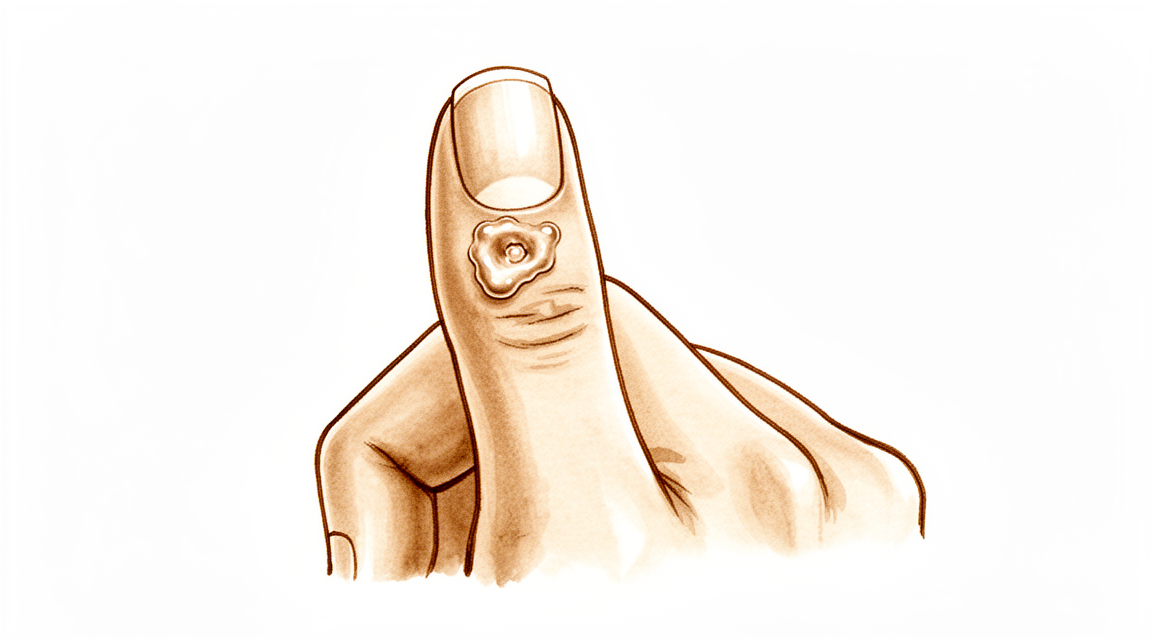
Những triệu chứng bạn đang gặp phải
Nang nhầy là một khối nhỏ, cứng xuất hiện ở mặt sau ngón tay, thường ngay sau gốc móng. Bên trong nang chứa chất dịch đặc, trong suốt. Bản thân khối này thường không gây đau, nhưng nó có thể đè lên các cấu trúc lân cận và gây khó chịu.
Bạn có thể nhận thấy những thay đổi ở chính móng tay. Nang nằm sát nền móng, nên khi to lên nó có thể đè lên móng. Điều này thường tạo ra một rãnh hoặc gờ chạy dọc theo chiều dài móng. Lớp da phủ trên nang cũng có thể trở nên mỏng và bóng. Trong một số trường hợp, da bị loét và nang rỉ dịch ra ngoài qua một đường thông nhỏ.
Khối này thường nằm ngay tại chỗ ngón tay gập ở khớp cuối cùng trước móng. Điều đó khiến một số việc hàng ngày trở nên vướng víu. Thò tay vào một chiếc túi chật, nắm nắp lọ hoặc nhặt những đồng xu nhỏ có thể gây khó chịu. Gõ phím hoặc viết có thể đè lên khối này và làm nó đau.
Nang có liên quan đến tình trạng viêm khớp do thoái hóa ở chính khớp đó. Một mấu xương nhỏ, gọi là gai xương (osteophyte), kích thích khớp và tạo ra dịch làm đầy nang. Vì vậy, khớp có thể đau nhức sau khi bạn sử dụng bàn tay nhiều, và khi đó khối này có thể đau hơn khi chạm vào.
Nếu lớp da phủ trên nang bị nứt ra và dịch rỉ ra ngoài, hãy giữ vùng đó sạch sẽ và nhanh chóng đi khám. Một đường thông từ da vào khớp ngón tay có thể để vi khuẩn xâm nhập, và khớp bị nhiễm trùng cần được điều trị ngay. Nếu ngón tay trở nên nóng, đỏ, sưng và đau, đặc biệt là kèm theo sốt, hãy đến phòng cấp cứu ngay trong ngày. Không cần giấy giới thiệu của bác sĩ đa khoa.
Không được tự chọc thủng hoặc nặn nang. Việc này có thể khiến vi khuẩn xâm nhập vào khớp.
Nếu khối này đang to lên, tình trạng biến dạng móng làm bạn phiền lòng, hoặc các triệu chứng không thuyên giảm sau nhiều tuần, hãy đến gặp bác sĩ đa khoa hoặc đề nghị được bác sĩ chuyên khoa khám.
Điều gì đang thực sự xảy ra
Khớp cuối cùng của ngón tay hoạt động giống như một bản lề nhỏ. Giống như mọi khớp khác, nó được lót bởi một lớp bao mềm tạo ra một ít dịch giúp các bề mặt khớp chuyển động trơn tru. Khi có nang nhầy, khớp đó đã bị viêm khớp do thoái hóa. Màng khớp bị kích ứng và bắt đầu tạo ra nhiều dịch hơn mức khớp cần.
Đồng thời, viêm khớp thường làm hình thành một gai xương nhỏ ở mép khớp. Gai xương đó đè lên và làm mòn thủng bao khớp từ bên trong. Sau đó dịch rỉ ra qua chỗ bị mòn và tích tụ dưới da ở mặt sau ngón tay. Khối mà bạn có thể nhìn thấy và sờ thấy chính là lượng dịch bị giữ lại đó, nằm lệch về một bên của khớp, ngay sau gốc móng.
Bản thân nang là một túi có thành mỏng chứ không phải là một khối mô tăng sinh. Thành nang được tạo nên từ các sợi mô nâng đỡ, và nang không có lớp lót thực sự của riêng mình. Vì thành nang mỏng và lớp da phủ bên trên thường còn mỏng hơn, chỉ một va chạm nhỏ cũng có thể làm vỡ nang. Khi đó, chất dịch trong, giống như thạch, bên trong sẽ rỉ ra ngoài.
Điều này giải thích các triệu chứng mà bạn vừa đọc. Khối này nằm sát nền móng, nên áp lực từ nang có thể tạo rãnh trên móng khi móng mọc. Khớp đau nhức vì tình trạng viêm khớp cũng có ở đó, và nang là một dấu hiệu của nó. Gai xương ở mép khớp là thứ khiến dịch tiếp tục chảy ra, vì vậy chừng nào gai xương còn đó, nang có xu hướng đầy dịch trở lại ngay cả sau khi đã rỉ dịch hoặc được rút dịch.
Đó cũng là lý do việc điều trị tập trung vào khớp chứ không chỉ vào khối nang. Loại bỏ gai xương sẽ chặn chỗ rò rỉ ngay tại nguồn, và nang sẽ xẹp đi khi không còn được bổ sung dịch nữa.
Những biện pháp chúng tôi có thể áp dụng
Bác sĩ Kieran Hirpara, bác sĩ phẫu thuật chi trên tại Bệnh viện tư nhân Mater Rockhampton, sẽ bắt đầu với các phương pháp ít xâm lấn nhất phù hợp với tình trạng của bạn. Thông thường, bệnh nhân được bác sĩ đa khoa giới thiệu đến phòng khám chúng tôi; nếu nhà vật lý trị liệu khuyên bạn nên đến gặp chúng tôi, bạn vẫn cần có giấy giới thiệu từ bác sĩ đa khoa để được hưởng mức hoàn trả từ chương trình Medicare. Trong buổi khám tại phòng khám, chúng tôi sẽ hỏi bệnh sử, khám ngón tay và chỉ định chẩn đoán hình ảnh nếu cần thiết. Hầu hết các nang nhầy có liên quan đến viêm khớp do thoái hóa ở khớp, vì vậy chúng tôi thường thử các biện pháp điều trị không phẫu thuật trước và cân nhắc phẫu thuật khi các biện pháp đó không mang lại đủ cải thiện.
Bước đầu tiên thường chỉ đơn giản là theo dõi và chờ đợi. Một số nang vẫn nhỏ và ít gây phiền toái. Bảo vệ ngón tay sẽ có ích: tránh va chạm lặp đi lặp lại vào khối nang, và cẩn thận với những việc đè lên nó. Nếu khớp đau nhức sau khi sử dụng nhiều, việc giảm bớt hoạt động đó có thể giúp tình trạng dịu đi. Trị liệu bàn tay có thể giúp giảm tình trạng cứng khớp và giúp ngón tay tiếp tục cử động thoải mái. Hãy áp dụng các biện pháp đơn giản này một cách nghiêm túc trong vài tuần trước khi quyết định điều gì tiếp theo.
Việc giảm đau khá đơn giản. Thuốc giảm đau thông thường mua tại nhà thuốc, dùng theo hướng dẫn, có thể làm dịu khớp đau nhức. Thuốc chống viêm có thể làm giảm tình trạng viêm khớp gây ra nang. Các loại thuốc này điều trị cảm giác khó chịu chứ không điều trị chính khối nang.
Một lựa chọn mà chúng tôi không áp dụng là rút dịch nang bằng kim. Đây là một thủ thuật tiện lợi có thể làm tại phòng khám, nhưng sau đó nang tái phát trong khoảng một nửa số trường hợp. Ngoài ra còn có nguy cơ đưa vi khuẩn vào khớp, đó cũng là lý do chúng tôi khuyên bạn không nên tự chọc thủng hoặc nặn nang tại nhà.
Phẫu thuật được cân nhắc khi nang liên tục đầy dịch trở lại, tình trạng biến dạng móng làm bạn phiền lòng, hoặc lớp da phủ trên nang đã trở nên mỏng hoặc đang bị loét. Ca phẫu thuật điều trị khớp chứ không chỉ điều trị khối nang. Gai xương ở mép khớp được loại bỏ, giúp chặn chỗ rò rỉ dịch ngay tại nguồn, và nang sẽ xẹp đi khi không còn được bổ sung dịch nữa. Trong một số trường hợp, nang và một vạt da nhỏ tại chỗ cũng được cắt bỏ, cách này phù hợp với những nang đã làm mỏng lớp da phía trên. Loại bỏ gai xương cùng với cuống nang mang lại khả năng tái phát thấp nhất. Chúng tôi sẽ trao đổi về phương pháp phù hợp với ngón tay của bạn và cùng bạn quyết định.
Những điều có thể xảy ra
Nếu không được điều trị, nang nhầy có xu hướng liên tục tái phát thay vì biến mất. Chừng nào gai xương ở mép khớp còn đó, nó vẫn tiếp tục bổ sung dịch cho nang. Một số nang vẫn nhỏ và ít gây phiền toái trong thời gian dài. Những nang khác to lên từ từ, làm rãnh trên móng sâu thêm, hoặc làm mỏng lớp da phủ trên khối nang cho đến khi rỉ dịch.
Nếu không điều trị, các nguy cơ chính là những điều bạn đã đọc: móng vẫn có gờ chừng nào nang còn đè lên nó, và da bị loét tạo thành một đường thông vào khớp. Đường thông đó cần được xem xét nghiêm túc. Nếu ngón tay trở nên nóng, đỏ, sưng và đau, đặc biệt là kèm theo sốt, hãy đến phòng cấp cứu ngay trong ngày. Không cần giấy giới thiệu của bác sĩ đa khoa.
Với phương pháp điều trị nhắm vào khớp, triển vọng khá rõ ràng. Loại bỏ gai xương sẽ chặn chỗ rò rỉ dịch ngay tại nguồn, và nang sẽ xẹp đi khi không còn được bổ sung dịch nữa. Khi gai xương được loại bỏ, nang rất hiếm khi tái phát. Ở những trường hợp chính nang cũng được cắt bỏ, tỷ lệ tái phát vẫn thấp: khoảng 2 trên 100 nang được điều trị bằng phẫu thuật bị tái phát, và khoảng 1,4 trên 100 khi sử dụng một vạt da tại chỗ để khâu kín vùng đó. Những người được điều trị theo cách này nhìn chung hài lòng với vẻ ngoài của vết sẹo và sẵn lòng thực hiện lại.
Quá trình hồi phục không phải là việc quá nặng nề, nhưng cũng không phải là không đáng kể. Ngón tay thường bị cứng và đau khi chạm vào trong vài tuần, và các việc hàng ngày như cầm nắm hoặc kẹp đồ vật giữa các ngón tay cần một thời gian mới cảm thấy bình thường trở lại. Trị liệu bàn tay sau phẫu thuật được thực hiện với bà Ruby Doolan tại trung tâm Extend Rehabilitation. Ruby là chuyên viên trị liệu bàn tay: bà sẽ hướng dẫn các bài tập của bạn và làm bất kỳ loại nẹp nào bạn cần. Hầu hết mọi người sử dụng bàn tay bình thường trở lại trong vòng vài tuần đến vài tháng.
Rãnh trên móng thường cũng cải thiện khi áp lực lên nền móng không còn. Hãy kiên nhẫn với phần này: phần móng bị biến dạng phải mọc hết ra ngoài, việc này mất vài tháng.
Nếu các triệu chứng không thuyên giảm sau nhiều tuần, hoặc đang nặng hơn, hãy đến gặp bác sĩ đa khoa hoặc đề nghị được bác sĩ chuyên khoa khám.
Khi nào nên đi khám bác sĩ
Hầu hết các nang nhầy không phải là trường hợp khẩn cấp, và nhiều nang dịu đi với cách chăm sóc đơn giản. Tuy nhiên, có một vài dấu hiệu cần được xử lý.
Hãy đến phòng cấp cứu ngay trong ngày nếu ngón tay trở nên nóng, đỏ, sưng và đau, đặc biệt là kèm theo sốt. Đó là dấu hiệu nhiễm trùng trong khớp, cần được điều trị ngay. Điều tương tự cũng áp dụng nếu lớp da phủ trên nang đã bị nứt ra và dịch đang rỉ ra ngoài, vì lỗ hở đó có thể để vi khuẩn xâm nhập.
Hãy đến gặp bác sĩ đa khoa hoặc đề nghị được bác sĩ chuyên khoa khám nếu khối nang tiếp tục to lên, rãnh trên móng làm bạn phiền lòng, hoặc lớp da phủ trên nang đã trở nên mỏng. Đây là những dấu hiệu cho thấy nên trao đổi về việc điều trị. Nếu các triệu chứng không thuyên giảm sau nhiều tuần, hoặc đang nặng hơn, bác sĩ đa khoa có thể sắp xếp giấy giới thiệu.
Phân tích sâu hơn
Advanced reading: the deeper science (optional)
Phần này đi sâu hơn mức cần thiết để bạn tự đưa ra quyết định điều trị. Các nang nhầy là chủ đề đáng để tìm hiểu thêm, vì có một nhận định phẫu thuật quan trọng và được chứng minh rõ ràng: nang nhầy không phải là nguyên nhân gây bệnh; đồng thời phương pháp phẫu thuật hiệu quả nhất cũng không nhất thiết phải là phương pháp loại bỏ nang đó.
Gai xương mới là nguyên nhân, chứ không phải nang
Nang nhầy hình thành do khớp ngón tay bị viêm khớp. Gai xương, hay còn gọi là osteophyte, làm kích ứng và làm rách màng bao khớp; dịch khớp sau đó tràn ra qua chỗ rách và tích tụ dưới da. Nang chỉ là kết quả cuối cùng có thể nhìn thấy được của quá trình này, chứ không phải nguyên nhân gây ra nó.
Hiểu biết này có ảnh hưởng trực tiếp đến phương pháp phẫu thuật: trong một loạt các ca bệnh, việc cắt bỏ gai xương mà không cắt bỏ nang đã giúp các triệu chứng biến mất hoàn toàn ở hầu hết các trường hợp, và được xem là phương pháp điều trị hiệu quả với mức độ xâm lấn thấp hơn [1].
Việc loại bỏ gai xương giúp ngăn chặn sự rò rỉ dịch khớp. Khi không còn dịch được bơm vào nữa, nang cũng dần biến mất. Đây cũng là nguyên lý tương tự trong trường hợp nang gan ở cổ tay: phần “cuống” nang mới là yếu tố quyết định khả năng tái phát; điều này cũng giải thích tại sao việc chỉ chọc hút hoặc rút dịch nang nhầy thường không mang lại hiệu quả.
Tại những vị trí mà u nang được cắt bỏ, kết quả điều trị cũng khá tốt
Phương pháp thay thế là cắt bỏ u nang cùng với vạt da cục bộ để khâu kín vùng khuyết hổng. Phương pháp này cũng đáng tin cậy: trên 69 bệnh nhân, việc cắt bỏ u nang kết hợp với vạt da di chuyển cục bộ cho thấy tỷ lệ tái phát là 1,4%; bệnh nhân đều hài lòng về vết sẹo và sẵn lòng thực hiện lại thủ thuật này [2].
Cả hai phương pháp đều hiệu quả và đều tác động đến khớp bị tổn thương. Sự khác biệt thực tế nằm ở mức độ da bị ảnh hưởng: u nang tồn tại lâu ngày sẽ làm mỏng lớp da phía trên, đôi khi dẫn đến tình trạng chảy dịch; trong trường hợp đó, lớp da mỏng này cần được cắt bỏ và thay thế, bất kể việc xử lý xương ra sao.
Tại sao móng bị biến dạng và liệu có hồi phục được không?
Việc xuất hiện rãnh hoặc gờ dọc theo chiều dài móng là hiện tượng khá phổ biến; điều này khiến nhiều người lo lắng hơn cả việc xuất hiện khối u. Nguyên nhân là do yếu tố cơ học: nang nằm ngay phía trên mô mầm – tức là vùng của nền móng có chức năng tạo ra móng – và gây áp lực lên vùng này, khiến móng phát triển với những khiếm khuyết.
Điều đáng mừng là áp lực này chỉ gây biến dạng chứ không làm hư hại móng. Sau khi nang được giảm áp, móng thường sẽ mọc trở lại bình thường; tuy nhiên phải mất vài tháng thì phần móng bị biến dạng mới mọc hết ra ngoài. Do đó, tình trạng biến dạng móng là lý do để điều trị nang, chứ không phải là hậu quả vĩnh viễn của nó.
Lý do cần thận trọng khi chọc hút nang này
Nang nhầy có đường thông trực tiếp với khớp. Việc chọc hút nang này, dù là cố ý hay do da bị rách trên một nang lớn, sẽ tạo ra lối vào từ môi trường bên ngoài vào khớp ngón tay; viêm khớp nhiễm trùng ở các khớp nhỏ là vấn đề nghiêm trọng hơn nhiều so với chính nang này.
Đây là lý do thực tiễn giải thích tại sao không nên tự chọc hút nang tại nhà khi thấy đó chỉ là một mụn nước chứa dịch; đồng thời cũng là lý do vì sao những nang tự vỡ cần được điều trị ngay lập tức thay vì chỉ theo dõi.
Tài liệu tham khảo
[1] Lee H, Kim P, Jeon I, Kyung H, Ra I, Kim T. Cắt bỏ gai xương mà không cắt bỏ nang dịch trong điều trị nang dịch ngón tay. Tạp chí Phẫu thuật Bàn tay châu Âu. 2013;39(3):258-61. https://doi.org/10.1177/1753193413478549
[2] Johnson SM, Treon K, Thomas S, Cox QGN. Phương pháp phẫu thuật đáng tin cậy để điều trị các nang dịch ở ngón tay. Tạp chí Phẫu thuật Bàn tay châu Âu. 2013;39(8):856-60. https://doi.org/10.1177/1753193413508540
Evidence & references
This is the clinical evidence summary written for health professionals. It is technical, and it lists the research this page was built from. You do not need to read it to understand your treatment or to make a decision about it.
Overview
- The scientific data regarding mucous cysts consist almost entirely of retrospective studies, and much of what is done or recommended is based on expert opinion [3].
- Surgical treatment of mucous cysts is not a totally benign procedure, and complications do occur contrary to literature reporting glowing results [5].
- In-office excision is a safe and effective option for treating digital mucous cysts [6].
- Complications are rare and occur only in cysts associated with fistula, justifying their early surgical treatment [7].
- Surgery provided definitive treatment with no major long-term problems [10].
- Surgical excision was the primary treatment, with no reported recurrences [11].
- Incomplete excisions will invariably result in recurrence [12].
- Surgical excision with a local advancement skin flap is a reliable treatment for digital mucous cysts, demonstrating a low recurrence rate of 1.4% and high patient satisfaction regarding the scar and willingness to undergo the procedure again [13].
- The 2% recurrence rate (2 of 113) obtained in this series illustrates the advantage of this procedure [16].
- Osteophyte excision without cyst excision may be a good treatment choice for mucous cyst of the finger, providing a less invasive method with complete resolution in most cases [20].
- The final recurrence rate of less than 1.5% may be acceptable and comparable with other techniques [21].
- Excision with local flap cover offers benefit in terms of a reduced recurrence rate and is particularly valuable where the cyst has caused marked thinning of the skin [30].
- Excision with local flap cover would seem to offer benefit in terms of a reduced recurrence rate, and is particularly valuable where the cyst has caused marked thinning of the skin [34].
- Excision of the cyst and complete removal of the marginal osteophyte eradicates mucous cysts with extremely rare recurrence [1].
- A total dorsal capsulectomy alone was a simple treatment for mucous cysts and did not lead to any recurrence [8].
Anatomy & Pathophysiology
Clinical Presentation and Location
- Mucous cysts are small, firm, cystic masses that appear just distal to the distal interphalangeal joint [23].
- The lesion is always located to one side of the midline [23].
- Mucous cysts are rarely greater than fifteen millimeters in diameter [23].
- The skin over the mucous cyst is thinned out and occasionally may be ulcerated [23].
- Pressure on the mucous cyst does not usually result in a decrease in its size [23].
- Longitudinal grooving of the nail may be noted, occasionally occurring prior to the appearance of the cyst [23].
- Mucous cysts are usually small and located to one side as they emerge from the joint beside the extensor mechanism [28, 29].
- Occasionally, mucous cysts may spread across the whole of the dorsum of the digit between the distal interphalangeal joint and nail fold [28, 29].
- The overlying skin of a mucous cyst is frequently thin, and minimal trauma may result in rupture [28, 29].
Histology and Ultrastructure
- The histological appearance of a mucous cyst is essentially identical to that of ganglions, synovial cysts, and similar lesions appearing on other areas of the extremities [23].
- The mucous cyst is surrounded by an undemarcated fibrous capsule [23].
- The basic structure of the mucous cyst is myxomatous with interspersed fibroblasts [23].
- Areas of myxomatous degeneration tend to coalesce to form a multiloculated cyst [23].
- An epithelial lining has not been reported for mucous cysts [23].
- Digital mucous cysts and wrist ganglia show no anatomic difference at the light and scanning electron microscopic levels [19].
- The wall of a digital mucous cyst consists of multidirectional strata of collagen fibers with rare fibroblast-like cells on the inner surface [19].
- There are no signs of myxomatous degeneration in the wall of a digital mucous cyst [19].
- The surface of the digital mucous cyst wall is generally flat, with some areas showing localized elevations [19].
- Multifunctional mesenchymal cells are located beneath the lining of the ganglion and digital mucous cyst [19].
- These multifunctional mesenchymal cells contain abundant rough endoplasmic reticulum, smooth-walled vacuoles, and Golgi complexes [19].
- The ganglion fluid may be produced by these multifunctional cells and reaches the ganglion cavity by flowing through the porous collagen matrix of the ganglion wall [19].
- A digital mucous cyst has a large globular dome-shaped interconnecting cavity having a wall of collagen fibers covered by a fenestrated pseudomembrane [19].
- A porous network of collagen fibers is present in the wall of the digital mucous cyst on cross section [19].
Etiology and Pathogenesis
- Degenerative arthritis of the distal interphalangeal joint is a frequent associated finding with mucous cysts [23].
- An incidence of 78 percent for degenerative arthritis of the distal interphalangeal joint has been reported in association with mucous cysts [23].
- The etiology indicated by evidence is that the lesion arises from the joint capsule [23].
- Mucous cysts are often associated with degenerative changes at the distal interphalangeal joints [28, 29].
- Theories on the pathogenesis of mucous cysts include myxoid degeneration, repeated local trauma, vascular insufficiency, and synovial herniation [28, 29].
- Studies suggest that mucous cysts arise due to excessive hyaluronic acid production from a degenerate distal interphalangeal joint [28, 29].
Classification
- The lesion is a small, firm, cystic mass which appears just distal to the distal interphalangeal joint [23].
- The lesion is always located to one side of the mid-line [23].
- The lesion is rarely greater than fifteen millimeters in diameter [23].
- The skin over the lesion is thinned out and occasionally may be ulcerated [23].
- Pressure on the lesion does not usually result in a decrease in its size [23].
- Degenerative arthritis of the distal interphalangeal joint is a frequent associated finding [23].
- An incidence of 78 per cent has been reported for degenerative arthritis of the distal interphalangeal joint associated with mucous cysts [23].
- The histological appearance is essentially identical to that of ganglions, synovial cysts, and similar lesions appearing on other areas of the extremities [23].
- The lesion is surrounded by an undemarcated fibrous capsule [23].
- The basic structure is myxomatous with interspersed fibroblasts [23].
- An epithelial lining has not been reported [23].
- In spite of their different clinical settings, digital mucous cysts and wrist ganglia show no anatomic difference at the light and scanning electron microscopic levels [19].
- The wall of the digital mucous cyst consisted of multidirectional strata of collagen fibers with rare fibroblast-like cells on the inner surface [19].
- There were no signs of myxomatous degeneration in the wall of the digital mucous cyst [19].
- The surface of the digital mucous cyst was generally flat, with some areas showing localized elevations [19].
- Multifunctional mesenchymal cells beneath the lining of the ganglion are postulated to produce collagen fibers, elastic fibers, and the interfibrillary mucopolysaccharide matrix [19].
- The porous nature of the ganglion wall was confirmed in the ultrastructural study of digital mucous cysts [19].
- A previous ultrastructural study of digital mucous cysts demonstrated a large globular dome-shaped interconnecting cavity having a wall of collagen fibers covered by a fenestrated pseudomembrane [19].
- The current study confirmed the anatomic organization of a large globular dome-shaped interconnecting cavity having a wall of collagen fibers covered by a fenestrated pseudomembrane [19].
- A porous network of collagen fibers in the wall was revealed on cross section [19].
Clinical Presentation
- The mucous cyst presents as a small, firm, cystic mass located just distal to the distal interphalangeal joint [23].
- The lesion is easily distinguished from a Heberden's node by its placement distal to the distal interphalangeal joint [23].
- The histological appearance of the mucous cyst is essentially identical to that of ganglions, synovial cysts, and similar lesions appearing on other areas of the extremities [23].
- The basic structure of the lesion is myxomatous with interspersed fibroblasts [23].
- An epithelial lining has not been reported for the mucous cyst [23].
- The wall of the digital mucous cyst consists of a porous network of collagen fibers [19].
- The fluid within the digital mucous cyst is produced by underlying mesenchymal cells [19].
Investigations
- Ultrasound is a powerful modality for evaluation of pathologic conditions in the hand and wrist, providing a cost-effective and expedient alternative and/or adjunct to MRI [33].
- Ultrasound is best used when there is a specific clinical question regarding a well-localized abnormality [33].
- Ultra-sonography provides a new ability to study the complex anatomical structures of the hand, with the technique being quick and simple to perform [38].
- A careful physical examination is essential to direct care and future testing if indicated [35].
- Diagnostic tests such as imaging and serum laboratory studies are useful in this determination but can be expensive, time consuming, and often nonspecific [35].
Treatment
Operative Techniques
- Total dorsal capsulectomy alone is a simple treatment for mucous cysts that did not lead to any recurrence [8].
- Osteophyte-sparing treatment of mucous cysts has a final recurrence rate of less than 1.5% [21].
- Surgical excision was the primary treatment in a series with no reported recurrences [11].
- Surgery provided definitive treatment for mucous cysts with no major long-term problems [10].
- The 2% recurrence rate (2 of 113) obtained in a series of 191 excised cysts illustrates the advantage of the procedure [4, 16].
- The authors of a study on osteophyte excision without cyst excision state that their principle is not to do more damage to the friable skin because the skin is not the culprit of the cyst [15].
Outcomes and Complications
- Complications of surgical treatments include infection, recurrence, nail deformity, swelling, stiffness and pain [28].
- This report provides additional basis for prophylactic extirpation of mucous cyst of the finger [2].
- This case emphasizes the utility of a pathohistological analysis in cases where doubts arise about the initial diagnosis of a benign tumorous lesion [14].
Non-Operative and Adjunctive Considerations
- Aspiration with injection of steroid is a convenient technique that can be performed in an outpatient setting but is followed by a recurrence rate of 50% [28].
- Surgical treatment has a higher cure rate than aspiration with injection of steroid [28].
- Radical treatment with complete excision of the stalk and underlying osteophytes seems to reduce recurrence rates [28].
Complications
- Complications are rare and occur only in cysts associated with fistula [7].
- Aspiration with injection of steroid is followed by a recurrence rate of 50% [28].
- The 2% recurrence rate (2 of 113) was obtained in a series of surgically treated mucous cysts [16].
- Surgical excision with a local advancement skin flap demonstrated a low recurrence rate of 1.4% [13].
- A total dorsal capsulectomy alone did not lead to any recurrence [8].
- Eccrine porocarcinomas have a substantial risk of metastasis, high risk of local recurrence, and are potentially fatal [24].
Recovery
- Surgical excision with a local advancement skin flap demonstrated a low recurrence rate of 1.4% and high patient satisfaction regarding the scar and willingness to undergo the procedure again [13].
Key Evidence
- [L4] Excision of the cyst and complete removal of the marginal osteophyte eradicates mucous cysts with extremely rare recurrence. [1] (10.2106/00004623-197355030-00013)
- [L4] This report provides additional basis for prophylactic extirpation of mucous cyst of the finger. [2] (10.1016/s0363-5023(84)80152-5)
- [L4] The scientific data regarding mucous cysts consist almost entirely of retrospective studies, and much of what is done or recommended is based on expert opinion. [3] (10.1016/j.jhsa.2010.01.029)
- [L5] Of the 191 mucous cysts excised, 113 had at least 6 months of follow-up time. [4] (10.1016/j.jhsa.2010.07.028)
- [L4] Surgical treatment of mucous cysts is not a totally benign procedure, and complications do occur contrary to literature reporting glowing results. [5] (10.1016/s0266-7681(97)80067-8)
- [Paper] In-office excision is a safe and effective option for treating digital mucous cysts. [6] (10.1177/15589447251350168)
- [L4] Complications are rare and occur only in cysts associated with fistula, justifying their early surgical treatment. [7] (10.1016/j.main.2015.06.001)
- [L4] A total dorsal capsulectomy alone was a simple treatment for mucous cysts and did not lead to any recurrence. [8] (10.1016/j.jhsa.2014.03.004)
- [L4] Surgery provided definitive treatment with no major long-term problems. [10] (10.1053/jhsu.2003.50088)
- [L5] Incomplete excisions will invariably result in recurrence. [12] (10.1016/0363-5023(93)90302-j)
- [L4] Surgical excision with a local advancement skin flap is a reliable treatment for digital mucous cysts, demonstrating a low recurrence rate of 1.4% and high patient satisfaction regarding the scar and willingness to undergo the procedure again. [13] (10.1177/1753193413508540)
- [L5] This case emphasizes the utility of a pathohistological analysis in cases where doubts arise about the initial diagnosis of a benign tumorous lesion. [14] (10.1007/s00402-008-0794-4)
- [L5] The authors state that their principle is not to do more damage to the friable skin because the skin is not the culprit of the cyst. [15] (10.1177/1753193414546990)
- [L4] The 2% recurrence rate (2 of 113) obtained in this series illustrates the advantage of this procedure. [16] (10.1016/0363-5023(94)90071-x)
- [L5] [19] (10.1016/s0363-5023(88)80143-6)
- [L4] Osteophyte excision without cyst excision may be a good treatment choice for mucous cyst of the finger, providing a less invasive method with complete resolution in most cases. [20] (10.1177/1753193413478549)
- [L4] The final recurrence rate of less than 1.5% may be acceptable and comparable with other techniques. [21] (10.5435/jaaosglobal-d-21-00164)
- [L4] [23] (10.2106/00004623-197254070-00008)
- [L4] Prompt recognition and appropriate treatment are critical because eccrine porocarcinomas have a substantial risk of metastasis, high risk of local recurrence, and are potentially fatal. [24] (10.1016/j.jhsa.2016.07.112)
- [L4] [28] (10.1177/1753193408103498)
- [L4] [29] (10.1177/1753193409103498)
- [L4] Excision with local flap cover offers benefit in terms of a reduced recurrence rate and is particularly valuable where the cyst has caused marked thinning of the skin. [30] (10.1016/0266-7681(90)90064-b)
- [L5] Ultrasound is a powerful modality for evaluation of pathologic conditions in the hand and wrist, providing a cost-effective and expedient alternative and/or adjunct to MRI, best used when there is a specific clinical question regarding a well-localized abnormality. [33] (10.1016/j.jhsa.2009.02.010)
- [L4] Excision with local flap cover would seem to offer benefit in terms of a reduced recurrence rate, and is particularly valuable where the cyst has caused marked thinning of the skin. [34] (10.1016/0266-7681_90_90064-b)
- [L4] Ultra-sonography provides a new ability to study the complex anatomical structures of the hand, with the technique being quick and simple to perform. [38] (10.1016/0266-7681(90)90133-o)
References
[1] Marginal Osteophyte Excision in Treatment of Mucous Cysts. The Journal of Bone & Joint Surgery. 1973. DOI: 10.2106/00004623-197355030-00013
[2] Infected mucous cyst of the finger. The Journal of Hand Surgery. 1984. DOI: 10.1016/s0363-5023(84)80152-5
[3] Mucous Cysts. The Journal of Hand Surgery. 2010. DOI: 10.1016/j.jhsa.2010.01.029
[4] Mucous Cysts. The Journal of Hand Surgery. 2010. DOI: 10.1016/j.jhsa.2010.07.028
[5] Complications Following Mucous Cyst Excision. Journal of Hand Surgery. 1997. DOI: 10.1016/s0266-7681(97)80067-8
[6] Safety and Cost-Effectiveness of In-Office Digital Mucous Cyst Excisions. HAND. 2025. DOI: 10.1177/15589447251350168
[7] Surgical treatment of mucous cysts by subcutaneous excision and osteophyte resection: Results in 68 cases at a mean 6.63 years’ follow-up. Chirurgie de la Main. 2015. DOI: 10.1016/j.main.2015.06.001
[8] Total Dorsal Capsulectomy for the Treatment of Mucous Cysts. The Journal of Hand Surgery. 2014. DOI: 10.1016/j.jhsa.2014.03.004
[10] Treatment of mucous cysts of the fingers: Review of 134 cases with minimum 2-year follow-up evaluation. The Journal of Hand Surgery. 2003. DOI: 10.1053/jhsu.2003.50088
[11] 10.1177-15589447261433068. n.d..
[12] Juvenile hyaline fibromatosis of the hand in an adult. The Journal of Hand Surgery. 1993. DOI: 10.1016/0363-5023(93)90302-j
[13] A reliable surgical treatment for digital mucous cysts. Journal of Hand Surgery (European Volume). 2013. DOI: 10.1177/1753193413508540
[14] Malignant Natural-Killer cell neoplasm presenting as a mucous cyst on the distal interphalangeal joint of the finger. Archives of Orthopaedic and Trauma Surgery. 2008. DOI: 10.1007/s00402-008-0794-4
[15] Re: Lee, H.-J., Kim, P.-T., Jeon, I.-H., Kyung, H.-S., Ra, I.-H. and Kim, T.-K. Osteophyte excision without cyst excision for a mucous cyst of the finger. J Hand Surg Eur. 2014, 39: 258–61. Journal of Hand Surgery (European Volume). 2014. DOI: 10.1177/1753193414546990
[16] Outcome of surgically treated mucous cysts of the hand. The Journal of Hand Surgery. 1994. DOI: 10.1016/0363-5023(94)90071-x
[19] A surface ultrastructure study of ganglia and digital mucous cysts. The Journal of Hand Surgery. 1988. DOI: 10.1016/s0363-5023(88)80143-6
[20] Osteophyte excision without cyst excision for a mucous cyst of the finger. Journal of Hand Surgery (European Volume). 2013. DOI: 10.1177/1753193413478549
[21] Osteophyte-Sparing Treatment of Mucous Cysts: Case Analysis and Surgical Technique. JAAOS: Global Research and Reviews. 2021. DOI: 10.5435/jaaosglobal-d-21-00164
[23] Etiology and Treatment of the So-Called Mucous Cyst of the Finger. The Journal of Bone & Joint Surgery. 1972. DOI: 10.2106/00004623-197254070-00008
[24] Eccrine Porocarcinoma Presenting as a Hand Cyst. The Journal of Hand Surgery. 2016. DOI: 10.1016/j.jhsa.2016.07.112
[28] Use of Wolfe Graft for the Treatment of Mucous Cysts. Journal of Hand Surgery (European Volume). 2009. DOI: 10.1177/1753193408103498
[29] Use of Wolfe Graft for the Treatment of Mucous Cysts. Journal of Hand Surgery (European Volume). 2009. DOI: 10.1177/1753193409103498
[30] Mucous cyst of the distal interphalangeal joint: Treatment by simple excision or excision and rotation flap. The Journal of Hand Surgery: Journal of the British Society for Surgery of the Hand. 1990. DOI: 10.1016/0266-7681(90)90064-b
[33] Ultrasound of the Hand and Wrist. The Journal of Hand Surgery. 2009. DOI: 10.1016/j.jhsa.2009.02.010
[34] Mucous Cyst of the Distal Interphalangeal Joint: Treatment by Simple Excision or Excision and Rotation Flap. Journal of Hand Surgery. 1990. DOI: 10.1016/0266-7681_90_90064-b
[35] Orthopaedic Knowledge Update 13 Ebook Without Multimedia. Anatomy, Evaluation, Clinical Examination, and Imaging > Evaluation and Clinical Examination: Current Concepts.
[38] Diagnostic medical ultrasound in the management of hand injuries. The Journal of Hand Surgery: Journal of the British Society for Surgery of the Hand. 1990. DOI: 10.1016/0266-7681(90)90133-o