Patients › Wrist
Nội soi khớp cổ tay
Diagnostic and therapeutic wrist arthroscopy — what it is, when it's used, and recovery.
Lý do phẫu thuật này được đề xuất
Bác sĩ Kieran Hirpara, bác sĩ phẫu thuật chi trên tại Bệnh viện tư nhân Mater Rockhampton, thường bắt đầu bằng các phương pháp ít xâm lấn nhất phù hợp với tình trạng của bệnh nhân. Thông thường, bệnh nhân được bác sĩ đa khoa giới thiệu đến phòng khám chúng tôi; nếu nhà vật lý trị liệu khuyên bạn nên đến gặp chúng tôi, bạn vẫn cần có giấy giới thiệu từ bác sĩ đa khoa để được hưởng mức hoàn trả chi phí từ Medicare. Trong buổi hẹn khám, chúng tôi sẽ hỏi kỹ về tiền sử bệnh, khám cổ tay và chỉ định chụp chiếu khi cần thiết để tìm ra điều gì đang gây đau cho bạn.
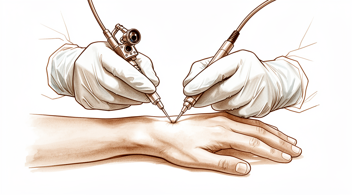
Nội soi khớp cổ tay là một ca phẫu thuật sử dụng một camera nhỏ đưa vào bên trong khớp cổ tay để quan sát kỹ các bề mặt khớp và các mô mềm. Chúng tôi thường đề nghị phẫu thuật này khi cơn đau cổ tay đã kéo dài hơn 3 tháng, không thuyên giảm với các biện pháp điều trị không phẫu thuật như thay đổi hoạt động sinh hoạt, trị liệu tay, đeo nẹp hoặc tiêm thuốc, và khi các phim chụp và việc thăm khám chưa đưa ra được câu trả lời đủ rõ ràng. Phẫu thuật này cũng có thể được khuyến nghị sau khi bị gãy xương hoặc chấn thương dây chằng, để kiểm tra các bề mặt khớp và các mô mềm, đồng thời điều trị một số vấn đề ngay trong cùng một lần mổ. Ca phẫu thuật cũng có thể giúp cải thiện tình trạng cứng khớp, một số loại viêm khớp và các vấn đề dây chằng kéo dài. Những người được phẫu thuật vì đau cổ tay dai dẳng có cải thiện trung bình khoảng 50% sau một năm, dù hầu hết vẫn còn đau và hạn chế chức năng ở một mức độ nào đó. Mục tiêu chính là giảm đau, cử động tốt hơn và cổ tay vững hơn, được quyết định cùng với bạn.
Trước khi phẫu thuật
Bác sĩ phẫu thuật sẽ đưa ra những hướng dẫn rõ ràng cho bạn trước ngày phẫu thuật, và dưới đây chúng tôi trình bày những điểm thực tế. Bạn cần ngừng ăn và uống từ bảy giờ trước khi phẫu thuật. Chúng tôi yêu cầu bảy giờ thay vì sáu giờ như thông thường để ca phẫu thuật của bạn có thể được đẩy lên sớm hơn nếu lịch mổ diễn ra nhanh hơn dự kiến. Hãy mang theo danh sách các loại thuốc bạn đang dùng, và bác sĩ phẫu thuật sẽ cho bạn biết nếu có loại nào cần tạm ngưng trước phẫu thuật. Hãy sắp xếp người đưa bạn về nhà sau phẫu thuật, và mặc quần áo rộng rãi, thoải mái. Các hình ảnh chụp chiếu như X-quang, MRI hoặc siêu âm giúp lên kế hoạch phẫu thuật, và việc này thường được sắp xếp trong buổi hẹn khám của bạn. Nếu bạn mắc các bệnh lý khác, có thể bạn sẽ cần làm xét nghiệm máu hoặc được bác sĩ gây mê thăm khám.
Vào ngày phẫu thuật
Vào ngày phẫu thuật, bạn sẽ đến khu vực tiếp nhận bệnh nhân phẫu thuật của bệnh viện. Tại đây, nhân viên sẽ làm thủ tục nhập viện cho bạn và chuẩn bị cho bạn trước khi vào phòng mổ. Sau đó, bạn sẽ gặp bác sĩ gây mê – bác sĩ phụ trách việc gây mê và giảm đau cho bạn. Ca phẫu thuật này được thực hiện dưới gây mê toàn thân. Bạn sẽ hoàn toàn ngủ say trong suốt quá trình phẫu thuật. Một số bệnh nhân có thể được tiêm thuốc gây tê vùng để giảm đau sau mổ; quyết định này do bác sĩ gây mê đưa ra vào ngày phẫu thuật, dựa trên tình trạng cụ thể của từng người.
Sau đó, bạn sẽ được đưa vào phòng mổ để tiến hành ca phẫu thuật. Khi mổ xong, bạn sẽ tỉnh dậy tại khu vực hồi sức. Tại đó, các điều dưỡng sẽ theo dõi bạn cho đến khi thuốc gây mê hết tác dụng. Khi sức khỏe ổn định, bạn sẽ được chuyển sang phòng bệnh hoặc xuất viện về nhà, tùy thuộc vào loại phẫu thuật và tiến triển hồi phục của bạn.
Quy trình phẫu thuật
Nội soi khớp cổ tay là một ca phẫu thuật lỗ khóa (xâm lấn tối thiểu). Bác sĩ phẫu thuật tạo vài vết rạch nhỏ quanh vùng cổ tay, và một camera nhỏ được đưa qua một trong các vết rạch này để có thể quan sát bên trong khớp trên màn hình. Trong lúc phẫu thuật, cánh tay của bạn được giữ cố định và các ngón tay được treo nhẹ nhàng trong các dụng cụ giữ ngón tay mềm, với một lực kéo ổn định lên cổ tay giúp mở rộng khớp và tạo khoảng trống để thao tác. Dung dịch nước muối chảy qua khớp để giữ tầm nhìn rõ ràng.
Khi camera đã vào bên trong, bác sĩ phẫu thuật có thể quan sát kỹ các bề mặt khớp và các mô mềm, bao gồm lớp sụn bao phủ các xương và các dây chằng giữ các xương nhỏ của cổ tay với nhau. Những gì diễn ra tiếp theo phụ thuộc vào những gì được phát hiện. Các mảnh sụn hoặc xương lỏng lẻo có thể được lấy ra. Lớp mô lót khớp bị viêm có thể được cắt bỏ. U nang hoặc dải mô dày có thể được xử lý. Các dây chằng bị rách, bao gồm cả sụn tam giác ở phía ngón út của cổ tay, có thể được cắt gọt hoặc khâu sửa. Nếu xương bị gãy, các mảnh xương có thể được nắn thẳng hàng chính xác dưới sự quan sát của camera và được cố định bằng vít hoặc đinh trong khi chúng liền lại. Nếu cổ tay đã bị cứng sau chấn thương hoặc sau một ca phẫu thuật trước đó, mô bị co rút có thể được giải phóng để cổ tay cử động tự do hơn.
Khi kết thúc, các vết rạch nhỏ được khâu lại. Băng gạc được đặt lên vùng cổ tay, và bàn tay của bạn thường được nâng đỡ bằng băng quấn hoặc nẹp. Băng gạc sẽ được giữ nguyên trong khoảng 10 ngày, và chúng tôi sẽ hướng dẫn bạn cách chăm sóc vết mổ trong thời gian này.
Sau khi phẫu thuật
Bạn sẽ tỉnh dậy tại khu vực hồi sức, nơi các điều dưỡng sẽ theo dõi bạn cho đến khi thuốc gây mê hết tác dụng. Thông thường đây là ca phẫu thuật ngoại trú nên bạn có thể về nhà ngay trong ngày; tuy nhiên đôi khi bệnh nhân cũng phải ở lại qua đêm. Cổ tay của bạn sẽ được băng lại, và bàn tay có thể được nâng đỡ bằng băng quấn hoặc nẹp. Chúng tôi sẽ giữ băng trên cổ tay khoảng 10 ngày; vui lòng không tự ý tháo băng ra trước thời hạn đó trừ khi có chỉ định của chúng tôi. Chúng tôi sẽ thay hoặc gỡ băng khi bạn quay lại tái khám. Thuốc giảm đau được lên kế hoạch cho bạn trước khi bạn về nhà; hãy dùng thuốc theo chỉ dẫn. Nếu bạn được gây tê vùng, tình trạng tê và yếu trong 24 giờ đầu là điều bình thường. Khi thuốc tê hết tác dụng, vào khoảng 24 giờ, hãy gọi cho phòng khám nếu các ngón tay vẫn còn tê hoặc bạn không thể cử động chúng. Hãy nhờ người ở lại với bạn trong 24 giờ đầu. Hãy gọi cho phòng khám ngay trong ngày nếu bạn bị sốt, vùng đỏ lan rộng quanh vết mổ, có dịch hoặc mủ rỉ ra từ vết mổ, hoặc cơn đau ngày càng tăng dù đã dùng thuốc giảm đau. Hãy đến khoa cấp cứu nếu các ngón tay hoặc bàn tay trở nên nhợt nhạt, lạnh, trắng bệch, xanh tím hoặc sẫm màu, vì đây có thể là vấn đề về tuần hoàn máu. Hãy kê bàn tay cao khi nghỉ ngơi để giúp giảm sưng. Hầu hết mọi người có thể đi lại trong nhà bình thường ngay từ ngày phẫu thuật; bạn không cần phải nằm nghỉ trên giường.
Quá trình hồi phục
Trong vài ngày đầu, cổ tay của bạn sẽ đau nhức và sưng, và các ngón tay có thể cảm thấy cứng. Kê bàn tay cao khi nghỉ ngơi giúp giảm sưng. Hãy dùng thuốc giảm đau theo chỉ dẫn, và cảm giác khó chịu sẽ giảm dần đều đặn theo từng ngày.
Hầu hết mọi người có thể đi lại trong nhà bình thường ngay từ ngày phẫu thuật. Lúc đầu, bạn sẽ cần người khác giúp mang vác vật nặng và lái xe. Khi nẹp (nếu có) đã được tháo ra và bạn có thể cầm vô lăng một cách thoải mái, bạn có thể lái xe trở lại; trang của chúng tôi về việc lái xe sau phẫu thuật chi trên giải thích điều này chi tiết hơn. Băng gạc sẽ được giữ nguyên trong khoảng 10 ngày, và chúng tôi sẽ kiểm tra vết mổ khi bạn quay lại tái khám.
Trị liệu tay đóng vai trò quan trọng trong việc giúp cổ tay cử động trở lại. Bạn sẽ phục hồi chức năng với Ruby Doolan tại Extend Rehabilitation. Ruby là chuyên gia trị liệu tay: cô ấy sẽ hướng dẫn bạn các bài tập và làm nẹp cho bạn nếu cần trong suốt quá trình. Lúc đầu, trọng tâm là cử động nhẹ nhàng và kiểm soát tình trạng sưng. Khi cổ tay ổn định hơn, các bài tập sẽ tăng dần sang cầm nắm, nâng đồ vật và sử dụng bàn tay cho các công việc hằng ngày. Quá trình hồi phục diễn ra theo từng bước, và khả năng phối hợp cử động giữa cổ tay và các ngón tay tiếp tục cải thiện theo từng tuần.
Thời gian bạn có thể quay lại làm việc, chơi thể thao hoặc tập gym phụ thuộc vào những gì được phát hiện và sửa chữa bên trong cổ tay, cũng như vào công việc và sở thích của bạn. Lộ trình của bạn có thể khác với người khác; chúng tôi và chuyên viên trị liệu sẽ hướng dẫn bạn ở từng giai đoạn.
Những biến chứng có thể xảy ra
Hầu hết bệnh nhân đều hồi phục tốt, nhưng đôi khi vẫn có thể gặp phải các vấn đề. Bác sĩ phẫu thuật và đội ngũ y tế sẽ theo dõi sát sao để phát hiện sớm bất kỳ dấu hiệu bất thường nào.
Nhiễm trùng ít gặp sau phẫu thuật này, nhưng khi xảy ra thì cần được quan tâm. Hãy gọi cho phòng khám ngay trong ngày nếu bạn bị sốt, vùng đỏ lan rộng quanh vết mổ, hoặc có dịch hoặc mủ rỉ ra từ vết mổ. Cũng hãy gọi cho phòng khám ngay trong ngày nếu cơn đau ngày càng tăng dù đã dùng thuốc giảm đau. Trong trường hợp nhiễm trùng nặng nằm sâu trong cổ tay, đôi khi cần phải phẫu thuật thêm, và điều này rất hiếm gặp.
Các dụng cụ giữ ngón tay trong lúc phẫu thuật có thể khiến các khớp ngón tay bị đau nhức sau đó, hoặc gây bầm mô mềm hay các dây thần kinh nhỏ ở một ngón tay. Trong hầu hết các trường hợp, tình trạng này tự hết. Hãy nói với bác sĩ trong lần tái khám tiếp theo nếu một ngón tay vẫn còn đau hoặc có cảm giác lạ.
Dịch dùng để làm đầy khớp trong lúc phẫu thuật đôi khi có thể rò rỉ vào các mô của cẳng tay. Hãy chú ý nếu cẳng tay sưng lên nhanh chóng, kèm theo cảm giác căng cứng và cơn đau ngày càng tăng. Hãy gọi ngay cho phòng khám nếu bạn nhận thấy điều này.
Các dây thần kinh gần cổ tay có thể bị kích thích hoặc tổn thương trong lúc phẫu thuật. Bạn có thể thấy tê, ngứa ran hoặc một vùng da có cảm giác khác lạ sau khi thuốc gây tê vùng hết tác dụng, vào khoảng 24 giờ. Tình trạng tê và yếu trong 24 giờ đầu sau khi gây tê vùng là điều bình thường. Hãy gọi cho phòng khám nếu tình trạng tê kéo dài quá thời gian đó, hoặc nếu bạn không thể cử động các ngón tay hoặc bàn tay sau khi thuốc tê đã hết tác dụng. Nếu không thể liên lạc với phòng khám, hãy đến khoa cấp cứu gần nhất.
Bảng liệt kê các biến chứng ở trang này cung cấp tỷ lệ xảy ra của từng tình trạng nếu bạn muốn biết chi tiết.
Khi nào nên gọi cho chúng tôi
Hầu hết mọi người hồi phục mà không gặp vấn đề gì, nhưng biết những điều cần chú ý sẽ có ích. Hãy gọi cho phòng khám ngay trong ngày nếu bạn bị sốt, vùng đỏ lan rộng quanh vết mổ, có dịch hoặc mủ rỉ ra từ vết mổ, hoặc cơn đau ngày càng tăng dù đã dùng thuốc giảm đau. Hãy gọi cho phòng khám nếu các ngón tay vẫn còn tê hoặc bạn không thể cử động chúng sau khi thuốc gây tê vùng đã hết tác dụng, vào khoảng 24 giờ. Hãy đến khoa cấp cứu nếu bắp chân bị sưng hoặc đau, nếu bạn bị khó thở hoặc đau ngực, hoặc nếu các ngón tay hoặc bàn tay trở nên nhợt nhạt, lạnh, trắng bệch, xanh tím hoặc sẫm màu. Đây có thể là dấu hiệu của cục máu đông hoặc vấn đề về tuần hoàn máu. Nếu không thể liên lạc với phòng khám, hãy đến khoa cấp cứu gần nhất.
Nơi để đọc thêm về tình trạng bệnh
Trang này nói về chính ca phẫu thuật. Còn về tình trạng bệnh mà ca phẫu thuật này điều trị, bao gồm cả những bằng chứng cho thấy khi nào phẫu thuật có ích và khi nào không, sẽ được trình bày chi tiết hơn trên trang Chấn thương dây chằng cổ tay.
Evidence & references
This is the clinical evidence summary written for health professionals. It is technical, and it lists the research this page was built from. You do not need to read it to understand your treatment or to make a decision about it.
Overview
- Wrist arthroscopy is a valuable means of diagnosis and treatment of wrist disorders [2].
- Wrist arthroscopy is a safe procedure with a low rate of major and minor complications [3].
- Wrist arthroscopy provides views of and access to the intraarticular spaces of the wrist that are otherwise difficult to achieve without widely open approaches [4].
- The indications for wrist arthroscopy are growing with the description of volar portals for the radiocarpal and scaphotrapezial trapezoid joint [1].
- Volar portals for wrist arthroscopy have certain advantages over the standard dorsal portals for visualizing dorsal capsular structures as well as the palmar aspects of the carpal ligaments [15].
- The routine use of wrist arthroscopy as a replacement or supplement to arthrography cannot be justified [5].
- Wrist arthroscopy may be recommended in cases of chronic wrist pain where the clinical suspicion is strong and the operating team is well-versed with the technique [6].
- Postoperative infection after wrist arthroscopy is uncommon but clinically relevant, particularly in elderly, male patients with systemic comorbidities or undergoing synovectomy [7].
- Arthroscopic arthrolysis is a suitable and promising surgical option for the treatment of wrist stiffness after trauma or surgery [8].
- In a prospective comparative study, the arthroscopically guided procedure for intra-articular distal radius fracture was superior to the conventional open procedure [12].
- Arthroscopic exploration of the extensor carpi ulnaris and its subsheath using standard wrist arthroscopy is relatively simple to perform without the need of a further portal [11].
- Complete avulsion of the distal posterior interosseous nerve during wrist arthroscopy is a possible cause of persistent pain after arthroscopy [10].
- It is questioned whether distal posterior interosseous nerve avulsion could be responsible for symptomatic improvement in certain patients after arthroscopy due to partial wrist denervation [10].
- A simple, effective, and cost-efficient solution exists to overcome oversized finger traps for wrist arthroscopy distraction [9].
- The concept to use a physical model for wrist arthroscopy training is well perceived and indicates the potential for continued development [14].
Anatomy & Pathophysiology
Bony Anatomy
- The wrist includes the distal radioulnar, radiocarpal, and ulnocarpal joints and the eight carpal bones and their proximal and distal articulations and attached ligaments [38].
- The carpus comprises eight ossicles traditionally separated into two rows: the proximal row (scaphoid, lunate, triquetrum, pisiform) and the distal row (trapezium, trapezoid, capitate, hamate) [39].
- The distal radius articular surface has two concave facets for the scaphoid and lunate, separated by the scapholunate ridge [39].
- The sigmoid notch along the ulnar border of the distal radius is a shallow concavity for the articulating ulnar head at the distal radioulnar joint [39].
- The distal ulna is covered with hyaline cartilage on its dorsal, lateral, palmar, and distal surfaces [39].
- The ulnar styloid projects distally, and at its base, the fovea is the insertion for the triangular fibrocartilaginous complex (TFCC) [39].
- The scaphoid's primary vascular supply is a branch of the radial artery at the dorsal ridge, with smaller vessels entering the palmar tubercle to supply the distal 30% [39].
- The lunate has a dorsal and palmar vascular supply in 80% of wrists, while 20% have only a palmar supply [39].
- The capitate head often relies on a retrograde vascular supply [39].
- The pisiform is a sesamoid bone within the flexor carpi ulnaris tendon and serves as the origin for the abductor digiti minimi [39].
- The distal radial articular surface has a double obliquity of 12–15 degrees in the lateral view and 15–20 degrees in the anteroposterior view [43].
- The carpal articular surface has a smaller diameter of curvature than the radius, with stability in the medial and palmar aspects dependent on ligamentous and capsular resistance [43].
- The scaphoid presents a long axis inclined by 45 degrees to the long axis of the radius [43].
- The triquetrum is not in contact with the ulnar head; a fibro-cartilage disc, the triangular ligament, separates the two bones [43].
Ligaments and Soft Tissue Anatomy
- The triangular fibrocartilage complex (TFCC) consists of the central meniscus homolog, dorsal and volar radioulnar ligaments, the floor of the extensor carpi ulnaris tendon sheath, and volar ulnocarpal ligaments [39].
- The TFCC arises from the radial border of the distal radius and inserts into the base of the ulnar styloid and distal ulna through the ligamentum subcruentum [39].
- The dorsal and volar radioulnar ligaments are the primary stabilizers of the distal radioulnar joint [39].
- Only the peripheral 10% to 40% of the volar, ulnar, and dorsal TFCC has a vascular supply [39].
- The scapholunate interosseous ligament is C-shaped in the sagittal plane, with the dorsal third being the thickest and strongest portion [39].
- The volar portion of the lunotriquetral ligament is the thickest [39].
- The extrinsic wrist ligaments include the dorsal intercarpal ligament and the dorsal radiocarpal ligament [39].
- The dorsal radiocarpal ligament originates at the dorsal lip of the distal radius adjacent to Lister's tubercle and inserts into the lunate and triquetrum [39].
- The dorsal intercarpal ligament arises from the triquetrum and attaches to the scaphoid, trapezoid, and capitate [39].
- The terminal branches of the radial, ulnar, and anterior interosseous arteries provide extraosseous blood supply to the carpus through three dorsal and three palmar transverse arterial arches [41].
- The dorsal intercarpal arch is the largest of the dorsal arches and supplies the distal carpal row and, through anastomoses, the lunate and triquetrum [41].
- The deep palmar arch at the level of the metacarpal bases is consistent and communicates with the dorsal basal metacarpal arch and palmar metacarpal arteries [41].
- The extensor carpi ulnaris tendon rotates around the ulnar head, situated on the ulnar side of the styloid in pronation and on the radial side in supination [44].
- The extensor carpi radialis brevis is the primary wrist extensor with a moment arm of 16.30 mm, compared to 12.50 mm for the extensor carpi radialis longus [44].
Kinematics and Biomechanics
- The wrist functions as a two-joint system linking the hand to the forearm around the highly mobile bones of the proximal carpal row [40].
- With axial loading through the neutral wrist, approximately 80% of forces are transmitted through the distal radius (60% scaphoid facet, 40% lunate facet) and 20% through the distal ulna [39].
- With wrist flexion, 60% of the motion is midcarpal and 40% is radiocarpal [39].
- With wrist extension, 33% of the motion is midcarpal and 66% is radiocarpal [39].
- The proximal carpal row has no muscular or tendinous attachments and is an intercalary segment [39].
- With ulnar deviation, the proximal row extends relative to the forearm/distal row, while with radial deviation, the proximal row flexes [39].
- The dart-thrower's path of radial extension to ulnar flexion defines the transition between flexion and extension of the scaphoid and lunate [40].
- Dart-thrower's motion occurs almost exclusively through the midcarpal joint [40].
Pathophysiology of Wrist Disorders
- Scapholunate advanced collapse (SLAC) wrist pathophysiology involves scapholunate interosseous ligament injury leading to palmar flexion of the scaphoid and extension of the lunate (DISI) [36].
- In SLAC wrist, the radioscaphoid joint becomes incongruous, leading to altered contact forces and arthrosis at the radioscaphoid and capitolunate joints [36].
- The radiolunate joint is typically spared in SLAC wrist due to its spheroid shape [36].
- Ulnocarpal impingement is a degenerative condition resulting from a discrepancy in the relative length of the distal articular surfaces of the radius and ulna (positive ulnar variance) [36].
- Posttraumatic causes of ulnocarpal impingement include distal radius fracture with shortening, Galeazzi or Essex-Lopresti fracture, and childhood epiphyseal plate injuries [36].
- Congenital causes of ulnocarpal impingement include dyschondroplasia (Madelung deformity) and naturally occurring positive ulnar variance [36].
- In rheumatoid arthritis, the classic pattern of wrist deformity involves destabilization of the carpus caused by attenuation of extrinsic wrist ligaments, resulting in ulnar-palmar translocation and wrist supination [49].
- Three main pathophysiological factors drive rheumatoid wrist deformation: cartilage destruction, synovial expansion, and ligamentous laxity [49].
- In juvenile arthritis, uncontrolled joint synovitis stretches essential ligaments and erodes cartilage and bone, resulting in a progressive shift of the carpus ulnarward and volarward [50].
- The dorsal wrist ganglion is the prototype of hand and wrist ganglions, accounting for 60% to 70% of all hand and wrist ganglions [23].
- The main cyst of a dorsal wrist ganglion is usually located directly over the scapholunate ligament [23].
- A small, mucin-filled duct invariably pierces the transverse fibers of the scapholunate ligament, connecting the underlying scapholunate joint with the main ganglion cyst [23].
TFCC Tear Classification
- The Palmer classification categorizes triangular fibrocartilage complex (TFCC) tears into traumatic (class 1) or degenerative (class 2) [24].
- Subtypes within the Palmer classification are based on the specific location within the TFCC [24].
- The class and location of a TFCC tear have important implications for treatment [24].
- Class 1A traumatic TFCC injuries are characterized by central perforation or tear [24].
- Class 1B traumatic TFCC injuries are characterized by ulnar avulsion with or without ulnar styloid fracture [24].
- Class 1C traumatic TFCC injuries are characterized by distal avulsion involving the origins of the ulnolunate and ulnotriquetral ligaments [24].
- Class 1D traumatic TFCC injuries are characterized by radial avulsion involving the dorsal and/or volar radioulnar ligaments [24].
- Class 2A degenerative TFCC tears are characterized by TFCC wear or thinning [24].
- Class 2B degenerative TFCC tears are characterized by Class 2A findings plus lunate and/or ulnar chondromalacia [24].
- Class 2C degenerative TFCC tears are characterized by TFCC perforation plus lunate and/or ulnar chondromalacia [24].
- Class 2D degenerative TFCC tears are characterized by Class 2C findings plus lunotriquetral ligament disruption [24].
- Class 2E degenerative TFCC tears are characterized by Class 2D findings plus ulnocarpal and distal radioulnar joint arthritis [24].
- Arthroscopy is the gold standard for the detection of TFCC tears [24].
- The arthroscopic trampoline test is performed to assess TFCC resiliency by balloting the central portion with a small probe [24].
- The arthroscopic hook test can be used to demonstrate peripheral detachment of the TFCC [24].
- The arthroscopic suction test can show laxity of the TFCC when peripherally scarred in or foveal detachment when the distal radioulnar joint is clinically unstable [24].
Clinical Presentation
Diagnostic Indications
- Indications for diagnostic wrist arthroscopy include chronic wrist pain of uncertain etiology with more than 3 months interval, unresponsive to conservative treatment [21, 37].
- Indications for diagnostic wrist arthroscopy include assessment of acute ligamentous injuries, including scapholunate, lunotriquetral, and triangular fibrocartilage complex (TFCC) injuries [21].
- Indications for diagnostic wrist arthroscopy include evaluation of carpal instability [21].
- Indications for diagnostic wrist arthroscopy include assessment of chondral lesions [21].
- Indications for diagnostic wrist arthroscopy include evaluation of associated soft tissue injury in fracture conditions, including distal radius, scaphoid, ulnar styloid, and other carpal bone fractures [21].
- Indications for diagnostic wrist arthroscopy include assessment of scaphoid healing in delayed union and nonunion [21].
- Indications for diagnostic wrist arthroscopy include staging of posttraumatic arthritis, including scapholunate advanced collapse (SLAC), scaphoid nonunion advanced collapse (SNAC), and distal radius fractures [21].
- Indications for diagnostic wrist arthroscopy include evaluation of monoarticular arthritis and synovial biopsy [21].
- Indications for diagnostic wrist arthroscopy include evaluation of Kienböck disease [21, 37].
- Patients without positive provocative sign on examination seldom yield positive findings at wrist arthroscopy [37].
- Arthroscopic findings need to correlate with clinical examination [37].
Clinical Examination Principles
- The natural inclination to study radiographs or special imaging studies and reports prior to a thorough history and physical examination should be avoided, as this introduces cognitive bias which can affect thinking and decision making [34].
- Physical examination always needs to be preceded by a thorough investigation of the patient’s medical history, with special emphasis on the mechanism of injury and acuity [34].
- The patient should be asked details about the location, duration, and characteristics of any pain, including aggravating and relieving factors and previous treatments [34].
- With chronic problems, it is important to inquire about the patient’s jobs and hobbies, and whether there has been exposure to repetitive stress, vibrating tools, or potentially dangerous instruments [34].
- Elucidating a history of ligamentous laxity or multiple joint instabilities, especially in younger patients presenting with chronic wrist pain, is important [34].
- Such an evaluation should also include an assessment of the patient's stress coping skills [34].
- Swelling is generally moderate in wrist dislocations, and bone displacements may be evident only if the patient is seen immediately after experiencing trauma [34].
- If there has been a delay since the accident, swelling may have increased substantially, making visualization of the displaced bones more difficult [34].
- Skin abrasions, contusions, or ecchymosed areas may be helpful in determining the mechanism of injury and the potential areas of damage [34].
- Range of motion is usually limited by pain in acute injuries, whereas it may be reduced or normal in more chronic cases [34].
- In chronic cases, passive assessment of mobility is valuable not only in determining the presence of abnormal motion or crepitus but also in reproducing the patient’s pain [34].
- The physician should record the motion in the contralateral, uninjured wrist for comparative purposes [34].
- Palpation for areas of maximal tenderness is one of the most useful tools in the diagnosis of wrist pathology, especially in patients with chronic dysfunctions [34].
- In acute dislocations, tenderness is seldom elicited at specific points but rather in a diffuse manner due to extensive soft tissue damage [34].
- Palpation should always be performed in a methodical way, starting from the basal joint of the thumb and proceeding across the proximal carpal row from the scaphoid to the triquetrum and continuing from the hamate back across the distal row and CMC joints [34].
- A careful assessment of neural and vascular status is imperative, with particular attention being paid to the median and ulnar nerves [34].
- The median and ulnar nerves may be injured by direct contusion at the moment of impact, by compression from displaced bones or by swelling within the carpal canal [34].
- A thorough set of provocative maneuvers should be performed to rule out alternative or even concurrent diagnoses [34].
- The examination should begin in a nontender area and proceed rotationally around the carpus, ending at the most symptomatic area [34].
- Bilateral grip and pinch strength are useful to uncover underlying pathology in chronic cases [34].
- Strength may be diminished due to muscle atrophy, pain inhibition, or learned behaviors [34].
- Rapid alternating grip assessment may be helpful in determining voluntary effort [34].
- A local injection of anesthetic to a painful joint or selected tendon sheath may help normalize the dynamometer readings and narrow the diagnostic spectrum [34].
- Sensory testing should always accompany an examination of suspected nerve compression, using threshold or density testing [34].
Specific Provocative Maneuvers
- The ligamentous habitus of a given individual must be assessed using information from the normal wrist because of the wide variation in mobility and laxity [54].
- The Watson test, or scaphoid shift test, is designed to show scaphoid instability [54].
- In the Watson test, the examiner puts one hand on the radial border of the distal forearm with the thumb on the palmar aspect of the scaphoid while moving the patient’s hand to bring about ulnar then radial deviation of the wrist [54].
- The Watson test causes a dorsal subluxation of the scaphoid, accompanied by a painful click [54].
- A positive Watson shift test results when the scaphoid subluxates dorsally out of the scaphoid fossa and relocates when pressure is released [36].
- The ballotment tests, or shear tests, consist of demonstrating abnormal movements between adjacent bones by exerting pressure in opposite directions [54].
- The scapholunate ballotment test involves stressing of the scapholunate joint by volar pressure on the tubercle of the scaphoid and dorsal pressure on the lunate [54].
- If there is any ligament damage, pressure on these two bones causes a painful shearing of the scapholunate joint [54].
- Instability of the lunotriquetral joint can be demonstrated by Reagan’s test, which involves dorsal pressure on the lunate and volar pressure on the triquetrum [54].
- The midcarpal joint "pivot shift" test consists of supinating and volar subluxing the distal row of the carpus [54].
- The pivot shift test is performed by placing the patient elbow upon a firm surface, holding the elbow at 90 degrees, putting the hand into a fully supine position and holding the distal forearm firmly [54].
- The hand is moved into full radial deviation and then the ulnar side of the carpus is forced into further supination and a volar subluxed position [54].
- The wrist must not be flexed during the pivot shift test [54].
- The hand, still with the displacing force applied, is gently moved from radial to full ulnar deviation [54].
- The normal wrist will notch into a less supinated position as the head of the capitate engages the lunate, because of the restraint of the anterior capsule and triquetrolunate interosseous ligaments [54].
- Rupture, attenuation or excess laxity allow the capitate to drift out of the lunate [54].
- Triquetral hamate instability is demonstrated with the wrist straight with ulnar deviation [54].
- After a range of about 20 degrees, triquetral hamate instability produces a firm block [54].
- If one forces a sharp click accompanied by discrete posterior movement of the wrist, the proximal row of the carpus has moved from the VISI position to that of DISI, thus allowing a complete ulnar deviation of the wrist joint [54].
- The radio-carpal and midcarpal joint anteroposterior drawer test involves one hand holding the patient’s hand by the metacarpals to apply axial traction while the other hand stabilizes the patient’s forearm [54].
- While holding this traction, anteroposterior force is applied and a drawer is elicited at the radio-carpal, then the midcarpal joint [54].
- The ability to push the midcarpal joint volarward is present to a varying degree in most normal asymptomatic patients [54].
- A marked drawer test is a sign of congenital laxity [54].
- An exaggeration of the normal ulna head prominence is seen in dorsal subluxation or articular effusion [54].
- The prominence of the ulna head may be temporarily reduced by direct pressure over the ulna head [54].
- In the rheumatoid wrist, the ulna head prominence is further exaggerated by a supination deformity of the carpus [54].
- If the hand is held in full ulnar deviation and the ulna head is held forward by the examiner’s thumb, significant pain may be precipitated by this movement alone, suggesting DRUJ pathology [54].
- Pain precipitated by pronosupination while the ulna head is pressed volarward and the pisiform pressed dorsally is usually indicative of some form of ulnar impingement or abutment syndrome [54].
- The diagnosis of DRUJ arthrosis is confirmed by improvement in rotation and grip strength with injection of a local anesthetic into the DRUJ [36].
- Clinical findings for DRUJ arthrosis include pain that increases with proximal rotation of the forearm and compression of the ulna against the radius [36].
- Symptoms of ulnocarpal impingement include pain on the dorsal side of the DRUJ and an intermittent clicking sensation [36].
- Pain in ulnocarpal impingement is exacerbated by forearm rotation and ulnar deviation [36].
- Pain in ulnocarpal impingement occurs with axial loading of the ulnar side of the wrist [36].
- Pain in ulnocarpal impingement occurs with dorsal and palmar displacement of the distal ulna, with the wrist in ulnar deviation (positive ballottement test) [36].
- Symptoms of SLAC wrist include reduced grip and pinch strength [36].
- Symptoms of SLAC wrist include stiffness with extension and radial deviation [36].
- Symptoms of SLAC wrist include localized tenderness at the radioscaphoid articulation [36].
- Symptoms of SLAC wrist include decreased wrist motion on extension and radial deviation [36].
Imaging and Preoperative Evaluation
- After the history and physical examination, radiographic evaluation is helpful in determining the diagnosis, prognosis, and management of wrist problems [32].
- MRI should be added for evaluation of the triangular fibrocartilage; the distal radioulnar joint (DRUJ); and vascularity of the various carpal bones, extrinsic ligaments, joint surfaces, and surrounding soft tissues to confirm clinical suspicion and correlate with physical examination findings [32].
- A high rate of false-positive findings on MR images of normal subjects has been reported [32].
- A dedicated wrist coil provides enhanced resolution of wrist structures [32].
- Routine radiographic series for the wrist consist of four views: posteroanterior, lateral, oblique, and ulnar-deviated posteroanterior scaphoid view [32].
- Spot views of the carpal bones for detail (carpal tunnel view) are a useful radiographic technique [32].
- Fluoroscopic spot views of the wrist are a useful radiographic technique [32].
- A series of views for instability includes anteroposterior clenched fist, posteroanterior in neutral, radial, and ulnar deviation, lateral in neutral and full flexion and extension, semipronated oblique 30 degrees from the posteroanterior, and semisupinated oblique 30 degrees from the lateral [32].
- Diagnostic ultrasound is a useful radiographic technique for evaluating a painful wrist [32].
- Cine or video fluoroscopy is a useful radiographic technique for evaluating a painful wrist [32].
- Bone scanning is a useful radiographic technique for evaluating a painful wrist [32].
- Arthrography of the wrist (triple injection when indicated) is a useful radiographic technique [32].
- CT is a useful radiographic technique for evaluating a painful wrist [32].
- MRI is a useful radiographic technique for evaluating a painful wrist [32].
- The authors do not feel that the routine use of wrist arthroscopy as a replacement or supplement to arthrography can be justified [5].
- Perform repeated clinical examinations for chronic wrist pain as part of preoperative evaluation [37].
Investigations
Diagnostic Role and Indications
- Arthroscopic assessment of intercarpal ligament injuries and instability is considered by many the “gold standard” for evaluation of these conditions [31].
- Arthroscopy is the gold standard for detection of triangular fibrocartilage complex (TFCC) tears [24].
- Arthroscopy has been found to be more accurate than arthrography in identifying the location and size of triangular fibrocartilage and interosseous ligament injuries [31].
- Arthroscopy is more accurate than triple-injection cinearthrography in detecting tears of the dorsal sensory branch of the ulnar nerve during arthroscopic repair of the triangular fibrocartilage [31].
- Indications for wrist arthroscopy include the evaluation of ligamentous injuries, examination of joint articular surfaces, removal of loose bodies, biopsy of synovium, irrigation and debridement of joints, and confirmation and supplementation of wrist arthrography [31].
Arthroscopic Tests
Imaging Modalities
- MRI should be added for evaluation of the triangular fibrocartilage, the distal radioulnar joint, and vascularity of the various carpal bones, extrinsic ligaments, joint surfaces, and surrounding soft tissues to confirm clinical suspicion and correlate with physical examination findings [32].
- With proper technique, injuries to the triangular fibrocartilage complex can be demonstrated with MRI [46].
- Perforations in the triangular fibrocartilage complex appear as linear defects or gaps filled with hyperintense fluid on coronal gradient-echo or T2-weighted pulse sequences [46].
- The addition of arthrographic contrast improves the visualization of carpal ligaments on MR images [46].
- MRI is useful in detecting additional marrow abnormalities in osteonecrosis, as seen in the lunate in Kienböck disease or in the scaphoid after fracture [46].
- Asymmetry of marrow signal in proximal and distal fragments of a fractured scaphoid is suggestive of proximal pole ischemia [46].
- MRI has a limited role in the evaluation of carpal tunnel syndrome, which remains a clinical diagnosis [46].
- Axial imaging with T2 weighting can clearly display masses within the confines of the carpal tunnel, as well as edema and swelling of the median nerve [46].
- MRI provides earlier detection of synovitis and erosive bone changes associated with rheumatoid arthritis than do radiographs [46].
- MRI has an expanding role in the evaluation of inflammatory arthritis [46].
- MRI has gained a greater role in the evaluation of acute wrist trauma, where bone marrow edema may reveal fractures of the carpal bones or distal radius that are radiographically occult [46].
- MRI is controversial for TFCC pathology, but newer innovations suggest value in detection and localization of TFCC pathology [24].
Radiographic Techniques
- Spot views of the carpal bones are used for detail, including the carpal tunnel view [32].
- Arthrography of the wrist, including triple injection when indicated, is a useful radiographic technique [32].
Preoperative Assessment
- The natural inclination to study radiographs or special imaging studies and reports prior to a thorough history and physical examination should be avoided, as this introduces cognitive bias [34].
Treatment
Indications and Diagnostic Role
Operative Technique and Setup
- The patient is positioned supine on the operating table with a traction device applied to distract the wrist joint [21].
- Traction force of 10 to 12 lb is applied through plastic finger traps over the index and middle fingers, or more preferably, the middle three fingers [21].
- Overdistraction or use of wire finger traps may cause postoperative finger joint pain or localized contusion to soft tissue or digital nerves [21].
- Nylon finger traps are more comfortable and atraumatic to the patient, especially in awake cases [21].
- For arthroscopy over the scaphotrapeziotrapezoid joint, an additional trap and traction can be put on the thumb [21].
- When an overhead traction boom is employed, countertraction is provided by securing the arm to the hand table, and the operated limb is draped free up to the elbow level [21].
- When a dedicated wrist traction device is being used, the limb is draped up to the axilla level and the lower arm is wrapped to the basal plate of the device close to the elbow level [21].
- A traction device should be sterilizable and allow flexible positioning of the wrist intraoperatively in varying degrees of extension, flexion, and radial and ulnar deviation [21].
- Tourniquet use is optional and is often unnecessary, especially for diagnostic and uncomplicated therapeutic procedures performed under local anesthesia without sedation [21].
- Joint visibility is maintained by saline inflow, as the small volume of the wrist makes fluid distention rather impractical compared to the knee or shoulder joint [21].
- The main maneuver in creating the working space is controlled traction, while saline maintains a clear view by removing intraarticular debris through the outflow portal [21].
- The hydrostatic pressure generated by saline irrigation serves a hemostatic role when arthroscopy is performed without a tourniquet [21].
- Continuous irrigation is achieved with a 3 L bag of normal saline suspended 1.5 m above the patient and instilled under gravity [21].
- Caution should be used to avoid extravasation of fluid that may lead to compartment syndrome [21].
Therapeutic Procedures
- In the treatment of central triangular fibrocartilage complex tears, partial excision using the holmium:Yttrium-aluminum-garnet laser has been performed over a five-year experience [13].
- Arthroscopically guided procedure for intra-articular distal radius fracture was found to be superior to the conventional open procedure based on a prospective comparative study [12].
- Ablative procedures in wrist arthroscopy include TFCC debridement, debridement of ligament tears, synovectomy, wrist ganglionectomy, removal of loose body, capsulotomy/capsulectomy, lavage, and arthrolysis [21].
- Ablative bony procedures in wrist arthroscopy include scaphoidectomy, radial styloidectomy, wafer procedure, proximal row carpectomy, and proximal hamate excision [21].
- Ablative cartilage procedures in wrist arthroscopy include debridement of chondral and osteochondral lesions [21].
- Reparative soft tissue procedures in wrist arthroscopy include peripheral TFCC tear repair, TFCC foveal avulsion repair, scapholunate ligament injury repair, and lunotriquetral ligament injury repair [21].
- Reparative bony tissue procedures in wrist arthroscopy include arthroscopic-assisted reduction and internal fixation (ARIF) for distal radius fracture and scaphoid fracture [21].
- Reparative cartilage procedures in wrist arthroscopy include drill/abrasion chondroplasty [21].
- Reconstructive soft tissue procedures in wrist arthroscopy include arthroscopic TFCC reconstruction with tendon graft and arthroscopic-assisted scapholunate ligament reconstruction with tendon graft [21].
- Reconstructive bony procedures in wrist arthroscopy include arthroscopic bone grafting for scaphoid nonunion, limited carpal fusion, intraosseous bone cyst, and intraosseous ganglion [21].
- Reconstructive cartilage tissue procedures in wrist arthroscopy include osteochondral grafting [21].
Complications and Safety
- DPIN avulsion could be responsible for symptomatic improvement in certain patients after arthroscopy due to partial wrist denervation [10].
Complications
- Overdistraction during wrist arthroscopy distraction can cause postoperative finger joint pain [21].
- Use of wire finger traps for wrist arthroscopy distraction may cause localized contusion to soft tissue or digital nerves [21].
- Nylon finger traps are more comfortable and atraumatic to the patient than other traction methods, especially in awake cases [21].
- Extravasation of fluid during wrist arthroscopy may lead to compartment syndrome [21].
- Postoperative infection after wrist arthroscopy is uncommon but clinically relevant [7].
- Postoperative infection risk after wrist arthroscopy is particularly elevated in elderly, male patients with systemic comorbidities or those undergoing synovectomy [7].
- Complete avulsion of the distal posterior interosseous nerve can occur during wrist arthroscopy [10].
- Distal posterior interosseous nerve avulsion is a possible cause of persistent pain after wrist arthroscopy [10].
- Inadvertent damage to the dorsal branch of the ulnar nerve can be avoided by placing the 6R portal in the proximal fifth (19%) of a line drawn from the ulnar styloid to the fourth dorsal web space [22].
Recovery
- Postoperative infection risk is particularly relevant in elderly, male patients with systemic comorbidities or undergoing synovectomy [7].
Key Evidence
- [Paper] The indications for wrist arthroscopy are growing with the description of volar portals for the radiocarpal and scaphotrapezial trapezoid joint. [1] (10.1097/00130911-200603000-00007)
- [L4] From these data it appears that wrist arthroscopy is a valuable means of diagnosis and treatment of wrist disorders. [2] (10.1016/s0266-7681(96)80100-8)
- [L4] Wrist arthroscopy is a safe procedure with a low rate of major and minor complications. [3] (10.1016/j.jhsa.2003.12.020)
- [L5] Wrist arthroscopy can be a useful tool in one’s armamentarium in the diagnosis and treatment of wrist pathology, providing views of and access to the intraarticular spaces of the wrist that are otherwise difficult to achieve without widely open approaches. [4] (10.1016/j.eats.2024.103223)
- [L4] The authors do not feel that the routine use of wrist arthroscopy as a replacement or supplement to arthrography can be justified. [5] (10.1016/0266-7681(94)90256-9)
- [L4] It may be recommended in cases of chronic wrist pain where the clinical suspicion is strong and the operating team is well-versed with the technique. [6] (10.1177/1558944716660555ig)
- [L3] Postoperative infection after wrist arthroscopy is uncommon but clinically relevant, particularly in elderly, male patients with systemic comorbidities or undergoing synovectomy. [7] (10.1016/j.otsr.2026.104771)
- [L4] Arthroscopic arthrolysis is a suitable and promising surgical option for the treatment of wrist stiffness after trauma or surgery. [8] (10.1016/s0363-5023(03)80309-x)
- [L5] We present a simple, effective, and cost-efficient solution to overcome oversized finger traps for wrist arthroscopy distraction. [9] (10.1016/j.eats.2025.103662)
- [L5] An alternate question is whether DPIN avulsion could be responsible for symptomatic improvement in certain patients after arthroscopy due to partial wrist denervation. [10] (10.1053/jhsu.1999.0240)
- [L5] They found this arthroscopic exploration relatively simple to perform without the need of a further portal. [11] (10.1177/1753193420948404)
- [L3] On the basis of our prospective comparative study, we found that the arthroscopically guided procedure was superior to the conventional open procedure. [12] (10.1016/j.jhsb.2006.03.177)
- [L4] [13] (10.1053/jhsu.2001.20157)
- [Paper] The concept to use a physical model for wrist arthroscopy training is well perceived, and indicates the potential for continued development. [14] (10.1016/j.main.2015.10.087)
- [L5] Volar portals for wrist arthroscopy have certain advantages over the standard dorsal portals for visualizing dorsal capsular structures as well as the palmar aspects of the carpal ligaments. [15] (10.1097/00130911-200412000-00006)
- [L5] Inadvertent damage to the dorsal branch of the ulnar nerve can be avoided if care is taken to place the 6R portal in the ‘‘safe area’’ found within the proximal fifth (19%) of a line drawn from the ulnar styloid to the fourth dorsal web space. [22] (10.1016/j.jhsb.2005.10.005)
References
[1] Uses and Abuses of Wrist Arthroscopy. Techniques in Hand & Upper Extremity Surgery. 2006. DOI: 10.1097/00130911-200603000-00007
[2] The Value of Wrist Arthroscopy. Journal of Hand Surgery. 1996. DOI: 10.1016/s0266-7681(96)80100-8
[3] Complications of wrist arthroscopy. The Journal of Hand Surgery. 2004. DOI: 10.1016/j.jhsa.2003.12.020
[4] Wrist Arthroscopy: Positioning, Portal Placement, and Diagnostic Evaluation. Arthroscopy Techniques. 2024. DOI: 10.1016/j.eats.2024.103223
[5] Wrist Arthroscopy. Journal of Hand Surgery. 1994. DOI: 10.1016/0266-7681(94)90256-9
[6] Wrist Arthroscopy as a Diagnostic Tool in Chronic Wrist Pain. HAND. 2016. DOI: 10.1177/1558944716660555ig
[7] Incidence and risk factors of postoperative infection after wrist arthroscopy: an 11-year nationwide population-based cohort study in South Korea. Orthopaedics & Traumatology: Surgery & Research. 2026. DOI: 10.1016/j.otsr.2026.104771
[8] Arthroscopic wrist arthrolysis. The Journal of Hand Surgery. 2003. DOI: 10.1016/s0363-5023(03)80309-x
[9] Tip to Overcome Oversized Finger Traps in Wrist Arthroscopy Distraction. Arthroscopy Techniques. 2025. DOI: 10.1016/j.eats.2025.103662
[10] Complete avulsion of the distal posterior interosseous nerve during wrist arthroscopy: A possible cause of persistent pain after arthroscopy. The Journal of Hand Surgery. 1999. DOI: 10.1053/jhsu.1999.0240
[11] Visualization of the extensor carpi ulnaris and its subsheath using standard wrist arthroscopy. Journal of Hand Surgery (European Volume). 2020. DOI: 10.1177/1753193420948404
[12] 2.3 Wrist arthroscopy in intra-articular distal radius fracture. The Journal of Hand Surgery: Journal of the British Society for Surgery of the Hand. 2006. DOI: 10.1016/j.jhsb.2006.03.177
[13] The holmium:Yttrium-aluminum-garnet laser in wrist arthroscopy: A five-year experience in the treatment of central triangular fibrocartilage complex tears by partial excision. The Journal of Hand Surgery. 2001. DOI: 10.1053/jhsu.2001.20157
[14] Development of a wrist arthroscopy simulator. Chirurgie de la Main. 2015. DOI: 10.1016/j.main.2015.10.087
[15] Clinical Applications of Volar Portals in Wrist Arthroscopy. Techniques in Hand & Upper Extremity Surgery. 2004. DOI: 10.1097/00130911-200412000-00006
[21] Green S Operative Hand Surgery. AUTHOR'S PREFERRED METHOD OF TREATMENT: ARTHROSCOPIC PARTIAL WRIST FUSION > SURGICAL TECHNIQUE FOR DIAGNOSTIC ARTHROSCOPY > Setup.
[22] The Anatomy of the Dorsal Cutaneous Branch of the Ulnar Nerve – a Safe Zone for Positioning of the 6r Portal in Wrist Arthroscopy. Journal of Hand Surgery. 2006. DOI: 10.1016/j.jhsb.2005.10.005
[23] Green S Operative Hand Surgery. BOX 59.1 Ganglions of the Hand and Wrist > Operative Treatment > Dorsal Wrist Ganglion.
[24] Miller S Review Of Orthopaedics. DISTAL RADIOULNAR JOINT, TRIANGULAR FIBROCARTILAGE COMPLEX, AND WRIST ARTHROSCOPY > 2. TFCC tears.
[31] Campbell S Operative Orthopaedics 4 Volume Set. NERVE INJURIES AT THE LEVEL OF THE HAND AND WRIST > ARTHROSCOPY OF THE WRIST.
[32] Campbell S Operative Orthopaedics 4 Volume Set. NERVE INJURIES AT THE LEVEL OF THE HAND AND WRIST > RADIOGRAPHIC TECHNIQUES.
[34] Green S Operative Hand Surgery. Diagnosis and Treatment > Assessment of the Symptomatic Wrist.
[36] Aaos Comprehensive Orthopaedic Review 3. Arthritides of the Hand and Wrist* > IV. Posttraumatic Arthritis.
[37] Green S Operative Hand Surgery. AUTHOR'S PREFERRED METHOD OF TREATMENT: ARTHROSCOPIC PARTIAL WRIST FUSION > Diagnostic Wrist Arthroscopy.
[38] Campbell S Operative Orthopaedics 4 Volume Set. NERVE INJURIES AT THE LEVEL OF THE HAND AND WRIST > ANATOMY.
[39] Aaos Comprehensive Orthopaedic Review 3. Anatomy of the Hand and Wrist > VII. The Wrist.
[40] Green S Operative Hand Surgery. WRIST BIOMECHANICS > Carpal Kinematics.
[41] Campbell S Operative Orthopaedics 4 Volume Set. NERVE INJURIES AT THE LEVEL OF THE HAND AND WRIST > CIRCULATION.
[43] Exam Of The Hand Wrist 2Ed. 1.2 SKELETON OF THE WRIST.
[44] Exam Of The Hand Wrist 2Ed. Extension of the wrist > Muscles of wrist extension.
[46] Campbell S Operative Orthopaedics 4 Volume Set. WRIST AND ELBOW.
[49] Green S Operative Hand Surgery. WRIST INVOLVEMENT IN RA.
[50] Tachdjian S Pediatric Orthopaedics From The Texas Scottish Rite Hospital For Children E Book. Wrist Involvement.
[54] Exam Of The Hand Wrist 2Ed. Examination.