Patients › Wrist
Viêm màng bao gân De Quervain
Patient-facing topic on De Quervain's tenosynovitis (first dorsal compartment) — diagnosis, conservative management, and indications for de Quervain's release.
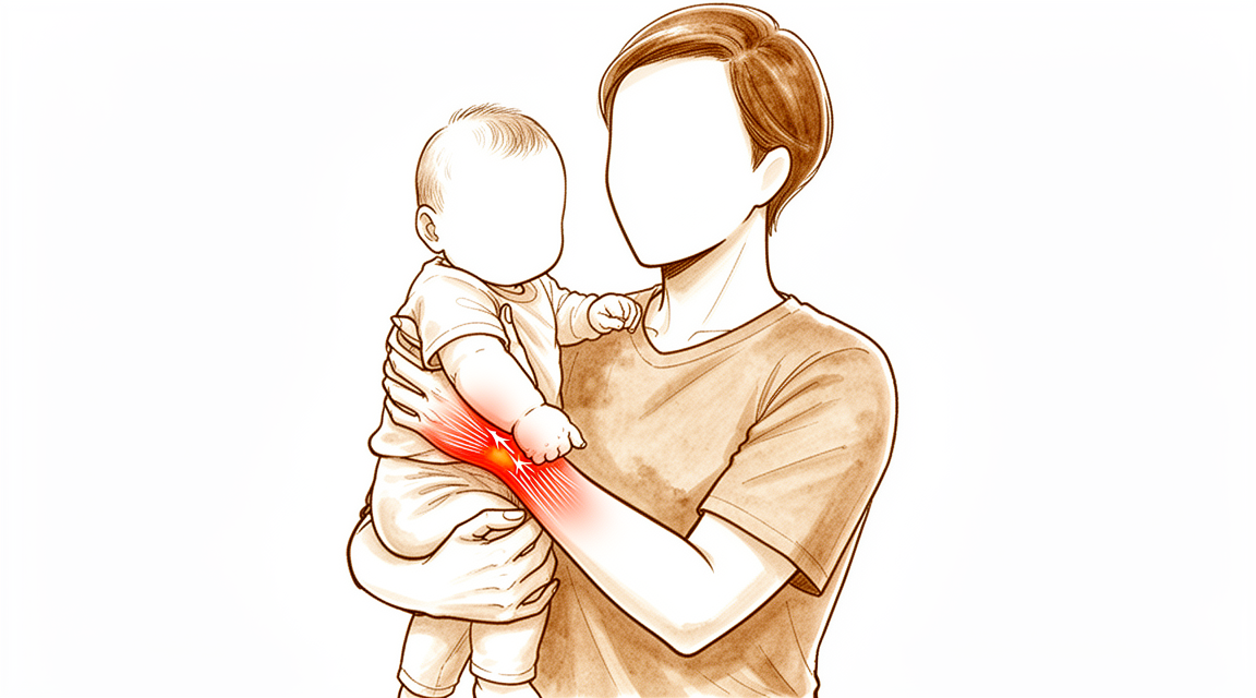
Những triệu chứng bạn đang gặp phải
Viêm màng bao gân De Quervain là một vấn đề ở hai gân tại phía ngón tay cái của cổ tay. Đường hầm mà các gân này đi qua bị sưng và chật lại, nên các gân trượt qua đó gây đau. Bạn cảm thấy đau nhức và đau khi ấn ở phía ngón tay cái của cổ tay, gần chỗ xương nhô lên ở gốc ngón tay cái. Vùng đó thường cũng bị sưng.
Một số động tác làm tăng cơn đau. Gập cổ tay về phía ngón út, nắm chặt tay trong lúc gập cổ tay về phía ngón tay cái, hoặc kẹp ngón tay cái và ngón trỏ lại với nhau đều có thể gây đau. Những việc hằng ngày cần đến các động tác này trở nên khó khăn: nhấc ấm nước, xoay tay nắm cửa, bế một đứa trẻ nhỏ, vắt khăn hoặc dùng kéo. Một số người nhận thấy cảm giác lạo xạo dưới da khi ấn vào chỗ đau.
Hầu hết mọi người thấy cơn đau tăng lên khi sử dụng tay và dịu đi khi nghỉ ngơi. Cơn đau có thể nặng hơn sau một ngày sử dụng tay nhiều. Một số người nhận thấy đau ngay khi vừa thức dậy vào buổi sáng hoặc trong đêm.
Một số ít người (khoảng 1 trên 100) thấy ngón tay cái bị vướng hoặc kẹt lại, ngoài việc bị đau. Bạn nên nói ra điều này, vì nó có thể làm thay đổi việc phương pháp điều trị nào sẽ hiệu quả.
Khi nào cần tìm sự trợ giúp
Hãy đến gặp bác sĩ đa khoa hoặc đề nghị được bác sĩ chuyên khoa khám nếu cơn đau không thuyên giảm, ngày càng nặng hơn qua nhiều tuần, khiến bạn thức giấc vào ban đêm, hoặc khiến bạn không thể làm việc hay sử dụng bàn tay.
Hãy đến khoa cấp cứu ngay trong ngày nếu bàn tay hoặc cánh tay của bạn trở nên nóng, đỏ, sưng và đau, đặc biệt khi kèm theo sốt. Đây có thể là tình trạng nhiễm trùng cần được chăm sóc ngay trong ngày, và bạn không cần giấy giới thiệu của bác sĩ đa khoa. Cũng hãy đi khám ngay trong ngày nếu các ngón tay hoặc bàn tay của bạn chuyển sang nhợt nhạt, lạnh, trắng hoặc tím tái, hoặc nếu bạn đột ngột mất cảm giác hay mất khả năng cử động sau một chấn thương.
Nếu bạn không liên lạc được với phòng khám, chẳng hạn như ngoài giờ làm việc hoặc vào cuối tuần, hãy đến khoa cấp cứu gần nhất.
Điều gì đang thực sự xảy ra
Có hai gân giúp đưa ngón tay cái ra xa bàn tay và duỗi thẳng nó. Chúng chạy song song với nhau qua một đường hầm hẹp ở phía ngón tay cái của cổ tay. Đường hầm có một phần mái chắc chắn giữ các gân áp sát vào xương, giống như một chiếc khoen dẫn giữ hai sợi dây cố định tại chỗ.
Vấn đề là các gân và đường hầm của chúng bị viêm. Lớp lót bị sưng chiếm chỗ bên trong một không gian vốn đã chật, nên các gân bị cọ xát khi trượt. Sự ma sát đó gây sưng nhiều hơn và đau nhiều hơn, và bản thân đường hầm có thể hẹp lại theo thời gian. Nguồn cấp máu qua lối đi hẹp này kém, khiến tình trạng kích ứng khó tự lắng dịu.
Một số người bẩm sinh có thêm một vách ngăn bên trong đường hầm, chia nó thành hai lối đi riêng biệt. Điều này quan trọng vì cả hai gân đều cần không gian để trượt. Nếu một gân nằm trong lối đi riêng bị ngăn kín, lối đi đó có thể vẫn còn chật ngay cả khi đường hầm chính đã được điều trị.
Điều này giải thích những gì bạn đang cảm thấy. Chỗ đau ở phía ngón tay cái của cổ tay chính là đường hầm. Cơn đau khi nắm, kẹp và nâng đồ vật là do các gân bị kéo qua khoảng không gian bị hẹp. Sức kẹp và sức của ngón tay cái có thể giảm vì các gân không thể trượt tự do. Cảm giác lạo xạo mà một số người nhận thấy là do bề mặt gân bị sần sùi chuyển động dưới da.
Khi ngón tay cái bị vướng hoặc kẹt lại kèm theo đau, tình trạng hẹp đã trở nên đủ nặng để giữ kẹt gân trong chốc lát. Đây là dạng tiến triển hơn của cùng một vấn đề, và nên được báo sớm vì nó làm thay đổi việc phương pháp điều trị nào sẽ hiệu quả.
Cơn đau ở vùng này cũng có thể đến từ các cấu trúc lân cận đường hầm thay vì chính đường hầm: viêm khớp ở gốc ngón tay cái, các gân bị kích ứng ở cao hơn trên cẳng tay, hoặc một dây thần kinh nông chạy ngang qua phần này của cổ tay. Những tình trạng này cần các phương pháp điều trị khác, đó là lý do vì sao việc xác định chính xác nguồn gốc của cơn đau là quan trọng.
Những biện pháp chúng tôi có thể áp dụng
Bác sĩ Kieran Hirpara, bác sĩ phẫu thuật chi trên tại Bệnh viện tư nhân Mater Rockhampton, sẽ bắt đầu với các phương pháp ít xâm lấn nhất phù hợp với tình trạng của bạn. Chúng tôi thường bắt đầu bằng việc thay đổi cách bạn sử dụng bàn tay, cùng với trị liệu tay để làm dịu tình trạng kích ứng và giảm tải cho các gân. Một chiếc nẹp giữ yên ngón tay cái và cổ tay có thể giúp các gân được nghỉ ngơi và giảm ma sát đang gây đau. Hãy kiên trì áp dụng các bước này trong một thời gian hợp lý trước khi chuyển sang bước tiếp theo, và cho chúng tôi biết nếu tình trạng không cải thiện.
Phương pháp điều trị có bằng chứng mạnh nhất cho tình trạng này là tiêm corticosteroid. Corticosteroid là thuốc chống viêm giúp làm dịu tình trạng sưng bên trong đường hầm. Chỉ một lần tiêm đã giúp giảm triệu chứng ở 82% người bệnh, và hơn một nửa trong số đó duy trì tình trạng không còn triệu chứng ít nhất 12 tháng. Sử dụng siêu âm để hướng dẫn tiêm có thể cải thiện kết quả, và cũng giúp chúng tôi xác nhận chính xác vấn đề đang được điều trị. Việc kết hợp tiêm thuốc với nẹp giữ yên cổ tay mang lại hiệu quả tốt hơn so với chỉ tiêm đơn thuần. Nếu triệu chứng quay trở lại, tiêm lần thứ hai vẫn là một lựa chọn hợp lý chứ không phải dấu hiệu cho thấy việc tiêm đã thất bại. Nếu bạn mắc bệnh tiểu đường, khả năng một lần tiêm mang lại hiệu quả sẽ thấp hơn so với những người khác, nhưng mỗi lần tiêm tiếp theo vẫn hiệu quả như lần tiêm trước.
Hầu hết mọi người sẽ không cần phẫu thuật. Chúng tôi cân nhắc phẫu thuật khi điều trị không phẫu thuật chưa mang lại đủ cải thiện, hoặc khi ngón tay cái bị vướng và kẹt lại kèm theo đau, vì tình trạng này thường không tự thuyên giảm. Ca phẫu thuật giải phóng phần mái bị chật của đường hầm để các gân có thể trượt tự do trở lại. Đây là một ca phẫu thuật nhỏ và có trang thông tin riêng mô tả chi tiết.
Những điều có thể xảy ra
Ở hầu hết mọi người, tình trạng này không tự biến mất nhanh chóng. Hầu hết những người mắc bệnh vẫn còn triệu chứng sau một năm nếu không làm gì, và cơn đau thường tăng lên khi sử dụng tay chứ không giảm dần đều. Một số người (khoảng một trên ba) cuối cùng sẽ cần phẫu thuật trong vòng hai năm, và hầu hết những người cần phẫu thuật đi đến quyết định đó trong năm đầu tiên.
Triển vọng khi được điều trị là khả quan, và cần được trình bày một cách trung thực. Một lần tiêm corticosteroid giúp hết hoàn toàn triệu chứng ở 70% người bệnh, và thêm 10% được chữa khỏi nhờ lần tiêm thứ hai. Tính chung, việc tiêm thành công trong 73,4% trường hợp với tối đa hai lần tiêm. Khi cần phẫu thuật, phẫu thuật mang lại hiệu quả tốt: 88% người bệnh hoàn toàn hài lòng sau phẫu thuật, và ca phẫu thuật chữa khỏi bệnh ở 91% trường hợp.
Cảm giác trong quá trình hồi phục phụ thuộc vào hướng điều trị bạn chọn. Sau khi tiêm, cơn đau nhức thường dịu đi trong vài ngày đến vài tuần khi tình trạng sưng bên trong đường hầm lắng xuống. Sau phẫu thuật, các gân trượt tự do trở lại, và sự cải thiện thường đến sớm hơn với phương pháp nội soi (lỗ khóa) so với phương pháp mổ mở. Việc trị liệu tay sau phẫu thuật được thực hiện với Ruby Doolan tại Extend Rehabilitation. Ruby là chuyên gia trị liệu tay: cô ấy sẽ điều phối quá trình trị liệu của bạn và chế tạo bất kỳ loại nẹp nào bạn cần.
Có một số điều bạn nên biết trước. Nếu bạn mắc bệnh tiểu đường hoặc hội chứng chuyển hóa (một nhóm các vấn đề sức khỏe bao gồm huyết áp cao và tích mỡ vùng bụng), khả năng tiêm thuốc thành công ở bạn sẽ thấp hơn so với những người khác. Cách bạn nhìn nhận về tình trạng này cũng quan trọng: những người luôn nghĩ đến điều tồi tệ nhất thường cho biết họ đau nhiều hơn và chức năng kém hơn, vì vậy sẽ có ích nếu bạn có những kỳ vọng chính xác thay vì lo sợ về ý nghĩa của tình trạng này.
Nếu các triệu chứng của bạn không thuyên giảm, ngày càng nặng hơn qua nhiều tuần, khiến bạn thức giấc vào ban đêm, hoặc khiến bạn không thể làm việc hay sử dụng bàn tay, hãy đến gặp bác sĩ đa khoa hoặc đề nghị được bác sĩ chuyên khoa khám.
Khi nào nên đi khám
Hầu hết các dấu hiệu cảnh báo của tình trạng này đã được đề cập trong phần đầu tiên ở trên: cơn đau không thuyên giảm, ngày càng nặng hơn qua nhiều tuần, khiến bạn thức giấc vào ban đêm, hoặc khiến bạn không thể làm việc hay sử dụng bàn tay. Nếu bạn có bất kỳ dấu hiệu nào trong số đó, hãy đến gặp bác sĩ đa khoa hoặc đề nghị được bác sĩ chuyên khoa khám.
Có một dấu hiệu cần được nhắc lại ở đây. Nếu ngón tay cái của bạn bị vướng hoặc kẹt lại kèm theo đau, hãy báo cho người thăm khám cho bạn. Dạng bệnh này thường không thuyên giảm chỉ với nghỉ ngơi hoặc đeo nẹp, và nó làm thay đổi việc phương pháp điều trị nào sẽ hiệu quả, vì vậy nên báo sớm thay vì chờ xem nó có tự hết hay không.
Lời khuyên tương tự cũng áp dụng nếu cơn đau nằm hơi lệch khỏi vị trí thường gặp, chẳng hạn như cao hơn trên cẳng tay hoặc ở gốc ngón tay cái. Một số tình trạng khác có thể có biểu hiện giống bệnh này, và chúng đáp ứng với các phương pháp điều trị khác. Việc thăm khám cẩn thận sẽ xác định được bạn mắc tình trạng nào.
Hãy đến khoa cấp cứu ngay trong ngày nếu bàn tay hoặc cánh tay của bạn trở nên nóng, đỏ, sưng và đau, đặc biệt khi kèm theo sốt, hoặc nếu các ngón tay chuyển sang nhợt nhạt, lạnh, trắng hoặc tím tái.
Phân tích chi tiết hơn
Advanced reading: the deeper science (optional)
Phần này đi sâu hơn so với những gì bạn cần biết để đưa ra quyết định điều trị. Viêm màng bao gân De Quervain đáng để đọc thêm vì có một biến thể giải phẫu nhỏ gây ra phần lớn các trường hợp không hài lòng sau ca phẫu thuật vốn vẫn được coi là hiệu quả; đồng thời, phương pháp điều trị không phẫu thuật tốt nhất là sự kết hợp của hai phương pháp chứ không phải chỉ chọn một trong số chúng.
Sự kết hợp giữa các phương pháp điều trị mang lại hiệu quả vượt trội so với từng phương pháp riêng lẻ
Khoang cổ tay lưng bàn tay chứa hai gân nằm trong một đường hầm ở phía ngón cái. Mục tiêu điều trị là giảm viêm trong đường hầm này và giảm áp lực tác động lên chúng.
Một phân tích mạng lưới trên 823 bệnh nhân cho thấy việc tiêm corticosteroid kết hợp với thời gian cố định khớp ngắn vẫn là phương pháp điều trị chính và hiệu quả nhất; liệu pháp sóng xung kích ngoài cơ thể là lựa chọn thứ hai [1]. Khi xem xét riêng từng thành phần điều trị, việc kết hợp sử dụng nẹp chỉnh hình và tiêm corticosteroid mang lại hiệu quả cao hơn so với việc áp dụng từng phương pháp một [2].
Đây là hướng dẫn cụ thể hơn so với lời khuyên “hãy dùng nẹp trước, sau đó tiêm thuốc nếu không hiệu quả”. Các bằng chứng khoa học ủng hộ việc áp dụng cả hai phương pháp cùng lúc, trong đó bệnh nhân cần đeo nẹp trong một khoảng thời gian ngắn sau khi tiêm.
Việc tiêm lần thứ hai vẫn là một lựa chọn đáng cân nhắc
Khi các triệu chứng tái phát, người ta thường cho rằng các mũi tiêm đã không hiệu quả và cần chuyển sang phẫu thuật. Tuy nhiên, dữ liệu thu thập từ một nhóm bệnh nhân lớn lại cho thấy điều ngược lại: mặc dù tỷ lệ thành công giảm đi sau nhiều lần tiêm, việc tiêm lại vẫn có tỷ lệ thành công cao và là một phương án điều trị khả thi [3].
Việc hiệu quả điều trị giảm dần theo số lần tiêm là điều dễ hiểu. Tuy nhiên, tỷ lệ thành công thấp hơn không có nghĩa là phương pháp này vô ích; việc tiêm lần thứ hai vẫn là một bước đi hợp lý chứ không phải là cách để trì hoãn điều trị.
Biến thể giải thích nguyên nhân gây thất vọng sau phẫu thuật
Việc giải phóng áp lực trong màng bao cổng cổ tay thứ nhất mang lại hiệu quả; còn những trường hợp không đạt kết quả thì thường có nguyên nhân cụ thể. Sự không hài lòng của bệnh nhân có thể xuất phát từ việc giải phóng không triệt để, sự trật khớp của gân, tổn thương dây thần kinh, hoặc đơn giản là thời gian hồi phục kéo dài; đồng thời việc không phát hiện và không giải phóng được màng bao riêng của gân cơ duỗi ngón cái ngắn cũng là một nguyên nhân gây thất vọng rõ rệt [4].
Điều này cần được phân tích kỹ lưỡng vì đây là thông tin hữu ích nhất trong mục này. Ở một tỷ lệ đáng kể bệnh nhân, màng bao này không phải là một ống duy nhất mà gồm hai ống riêng biệt; gân cơ duỗi ngón cái ngắn chạy trong màng bao riêng của nó, song song với các gân khác. Nếu chỉ mở phần màng bao chính mà dừng lại, màng bao thứ hai vẫn còn nguyên, khiến gân bên trong vẫn bị chèn ép. Kết quả là vết mổ đã hoàn tất như mô tả, nhưng các triệu chứng vẫn tiếp diễn.
Đây cũng là lý do vì sao hai nguyên nhân còn lại cũng rất quan trọng: nếu mở màng bao quá rộng, các gân có thể trật khỏi rãnh chứa gân khi cổ tay cử động, gây ra tiếng “lách cách” đau đớn; đồng thời nhánh nông của dây thần kinh quay lại đi ngay phía trên vùng phẫu thuật. Vì vậy, màng bao cần được mở hoàn toàn nhưng không quá mức – đặc biệt khi có dây thần kinh nằm gần đó – và đó là lý do vì sao một ca phẫu thuật tưởng chừng đơn giản vẫn đòi hỏi sự cẩn trọng tương đương với các ca phẫu thuật phức tạp hơn.
Không phải mọi cơn đau ở vùng cổ tay đều là bệnh de Quervain
Các nguyên nhân gây đau ở vùng này rất đa dạng và cần được phân biệt rõ, vì phương pháp điều trị cũng khác nhau: viêm khớp ở gốc ngón tay cái, hội chứng chéo nằm cách đó vài centimet dọc theo cẳng tay, và tình trạng kích thích dây thần kinh quay đều có thể gây đau ở những vùng tương tự nhau. Triệu chứng đặc trưng để xác định nguyên nhân là tình trạng đau nhạy cảm cục bộ ngay tại vùng bao gân; cơn đau sẽ tăng lên khi cổ tay bị lệch sang phía trụ trong trong khi ngón tay cái được gập vào lòng bàn tay. Đây là dấu hiệu chỉ ra nguyên nhân nằm ở bao gân chứ không phải các cấu trúc lân cận.
Tài liệu tham khảo
[1] Chong HH, Pradhan A, Dhingra M, Liong W, Hau MY, Shah R. Những tiến bộ trong việc điều trị viêm màng bao gân De Quervain: phân tích mạng lưới toàn diện các thử nghiệm lâm sàng ngẫu nhiên có đối chứng. J Hand Surg Am. 2024;49(6):557-69. https://doi.org/10.1016/j.jhsa.2024.03.003
[2] Cavaleri R, Schabrun SM, Te M, Chipchase LS. Liệu pháp trị liệu tay so với tiêm corticosteroid trong điều trị bệnh De Quervain: tổng quan có hệ thống và phân tích tổng hợp. J Hand Ther. 2016;29(1):3-11. https://doi.org/10.1016/j.jht.2015.10.004
[3] Hassan K, Sohn A, Shi L, Lee M, Wolf JM. Viêm màng bao gân De Quervain: đánh giá về dịch tễ học và hiệu quả của việc tiêm nhiều lần dựa trên cơ sở dữ liệu quốc gia. J Hand Surg Am. 2022;47(3):284.e1-284.e6. https://doi.org/10.1016/j.jhsa.2021.04.018
[4] Rogozinski B, Lourie GM. Mức độ không hài lòng sau phẫu thuật giải phóng bao gân vùng mu bàn tay thứ nhất đối với bệnh viêm gân De Quervain. J Hand Surg Am. 2016;41(1):117-9. https://doi.org/10.1016/j.jhsa.2015.09.003
Evidence & references
This is the clinical evidence summary written for health professionals. It is technical, and it lists the research this page was built from. You do not need to read it to understand your treatment or to make a decision about it.
Overview
Anatomy and Diagnosis
- Anatomical variations in the first extensor compartment are clinically relevant to the pathophysiology and treatment of De Quervain's tenosynovitis [1].
- Wrist radiography does not influence the management of patients presenting with de Quervain tendinopathy [6].
- The tethered thumb sign is a clinical maneuver that supports the diagnosis of de Quervain tenosynovitis and assists in determining an effective treatment algorithm [12].
- Ultrasound can be used as a diagnostic tool in the management of de Quervain’s disease [18].
Non-Operative Treatment
- Corticosteroid injection is the only available nonsurgical treatment that can potentially modify the course of de Quervain's tenosynovitis and is the preferred initial treatment [2].
- Corticosteroid injection with a short duration of immobilization remains the primary and effective treatment for de Quervain tenosynovitis [28].
- Corticosteroid injections lead to treatment success 73.4% of the time within 2 injections [22].
- Although the success rate for the treatment of De Quervain's tenosynovitis decreases with multiple injections, repeat injections have a high rate of success and are a viable clinical option [46].
- Corticosteroid injection for De Quervain tenosynovitis is not as effective in patients with metabolic syndrome compared with age- and sex-matched controls in terms of functional outcomes and treatment failure [5].
- Patients with diabetes mellitus have a decreased probability of success following a single corticosteroid injection for de Quervain tenosynovitis in comparison to nondiabetic patients [40].
- The effectiveness of each additional corticosteroid injection does not appear to diminish in patients with diabetes mellitus [40].
- Ultrasound-guided triamcinolone injection should be considered to improve the treatment outcome for de Quervain's disease [18].
Operative Treatment
- Surgical release of De Quervain's tenosynovitis remains the gold standard treatment [10].
- Surgical intervention is effective as definitive therapy for de Quervain's tenosynovitis, with a cure rate of 91% and 88% of patients indicating full satisfaction [11].
- The release of the first extensor compartment for refractory de Quervain's disease results in good clinical outcomes with minimal morbidity [24].
- Longitudinal incision offers the advantage of easy identification of the compartment, more complete releases of tendon sheath and peritendinous adhesions, and less risk of palmar subluxation of tendons [10].
- The longitudinal incision for release of the first extensor compartment is a safe and effective technique that provides good exposure, protects vital structures, prevents possible tendon subluxation in the postoperative period, and is cosmetically acceptable [17].
- Endoscopic release for de Quervain's tenosynovitis seems to provide earlier improvement after surgery, with fewer superficial radial nerve complications and greater scar satisfaction, when compared with open release [33].
- One portal endoscopic assisted release of the extensor compartment is an effective and safe minimal invasive procedure with similar complication rates reported previously in open and endoscopic procedures [31].
- Endoscopic release of the extensor compartment is an effective and safe procedure in patients with de Quervain's disease who are unresponsive to nonoperative treatments, despite a little increase in operation time [41].
- Pulley reconstruction should be considered part of the surgical treatment for de Quervain tenosynovitis and gives satisfactory medium-term results [4].
Prognosis and Patient Factors
- Patients presenting with de Quervain's tenosynovitis may favor initial nonsurgical management [7].
- Patients who scored lower than 40 for physical function or higher than 60 for pain interference had significantly increased odds of eventually undergoing surgical release for de Quervain tenosynovitis [34].
- The failure of an operation in De Quervain tenosynovitis is due to anatomical variations, the involvement of the radial superficial nerve, and the ignoring of another syndrome in the proximity [13].
- Addressing misconceptions about de Quervain's tenosynovitis regarding consequences for patients and symptom duration allows patients to make informed decisions about treatment [9].
Anatomy & Pathophysiology
Anatomical Variations
- The first dorsal compartment of the wrist exhibits immense anatomical variation, including commonly found supernumerary tendons or tendon slips [107].
- Variation in tendon insertion is present in the population of the first dorsal compartment [107].
- Septa or aberrant compartments are present in the first dorsal compartment [107].
- Bilateral variations are present in the population regarding the anatomy of the first dorsal compartment [107].
- The superficial branch of the radial nerve is expected to be encountered in more than 50% of patients undergoing surgery for De Quervain's tenosynovitis [94].
Pathophysiology
- De Quervain's syndrome is defined as a condition where the abductor pollicis longus and extensor pollicis brevis tendons in the first dorsal compartment become inflamed through friction within a tight osteoligamentous tunnel [3].
- Compromised blood flow and nutrition in the first dorsal compartment lead to the development of adhesions and tendon stenosis [3].
- De Quervain's syndrome may involve myxoid degeneration, defined as a degenerative process where connective tissues are replaced by a gelatinous or mucoid substance [3].
- The pathophysiology of De Quervain's disease does not involve inflammation, with histopathological examination showing predominant degenerative changes such as myxoid degeneration and fibrocartilagenous metaplasia [38].
- De Quervain's tenosynovitis is caused by impaired gliding of the abductor pollicis longus and extensor pollicis brevis tendons [38].
- Impaired gliding in De Quervain's tenosynovitis is most probably caused by thickening of the extensor retinaculum of the wrist [38].
- De Quervain's syndrome may not be an isolated pathology and may be associated with trauma, extensor carpi ulnaris involvement, ligament instability, or repetitive strain [3].
- Operative treatment for distal radius fractures confers a slightly greater risk of developing De Quervain tenosynovitis within 1 year of injury or surgery [105].
Clinical Presentation & Assessment
- De Quervain's tenosynovitis is characterized by pain, tenderness, and swelling over the thumb side of the wrist at the radial styloid process [38].
- The condition is especially associated with sideward movements of the wrist and often leads to impairment of thumb function [38].
- The tethered thumb sign is a clinical maneuver that can support the diagnosis of De Quervain tenosynovitis and assist in determining an effective treatment algorithm [12].
- Subjects with stenosing tenosynovitis demonstrate a significant decrease in maximum velocity in slow fist tasks [81].
- In vivo extensor pollicis brevis tendon excursion measures have been quantified, and wrist position has been found to have an influence on excursion [73].
- The proportion of wrists with a positive extensor pollicis brevis entrapment test was significantly higher among those with two compartments (18 of 22) than among those with one compartment (0 of 4) [97].
Etiology & Risk Factors
- Women are more affected by De Quervain tenosynovitis than men, potentially due to manual work such as housekeeping activities and child rearing influencing first compartment dynamics [72].
- Training intensity is a major factor in determining De Quervain disease in volleyball players, with patients in the higher intensity group showing a mean training time quotient of 155 compared to 74 in the lower intensity group [72].
- The close association of work and repetitive trauma with De Quervain’s disease has been observed, although the exact relationship is controversial [21].
- Recent studies have disputed the view that repetitive work is causative of De Quervain’s disease, with one review of 100 patients finding no increased incidence in the dominant hand [21].
- A study of workers in a car plant reported 544 cases of tenosynovitis but only 2 cases of De Quervain’s disease [21].
- Repetitive work is less contentious as a factor in aggravating symptoms, as tendon movement within a stenosed sheath during activity logically causes pain [21].
- The process of tendon movement within a stenosed sheath may cause tendon damage through wear and tear, potentially leading to tendon rupture [21].
Complications & Associated Pathology
- Rupture of the abductor pollicis longus tendon in association with De Quervain’s disease has never been reported in the literature prior to the 2006 case report [21].
- Volar subluxation of the tendons of the first dorsal compartment can occur after surgery for De Quervain's stenosing tenosynovitis [48].
- Volar tendon subluxation after De Quervain's release can be treated with a distally based flap of the brachioradialis tendon to prevent tendon prolapse [48].
- Symptomatic palmar tendon subluxation after surgical release for De Quervain's disease can be avoided by leaving a palmarly based flap of extensor retinaculum [15].
- Dorsal tendon dislocation after De Quervain's release can occur if the patient overstresses the wrist through forceful extension [80].
- Repeat provocative testing for volar tendon subluxation following De Quervain’s release revealed volar tendon translation with wrist flexion, with no pain reported [85].
- Osteoid osteoma of the radial styloid can mimic stenosing tenosynovitis, with correct diagnosis often requiring histological examination when clinical presentation and X-rays are not typical [45].
Classification
Anatomical Variations and Subcompartmentalization
- The prevalence of a septated first dorsal compartment is considerably higher than previously reported, most notably in patients with De Quervain tenosynovitis [52].
- In a prospective study of 87 wrists, 22 of 30 patients (73%) who failed non-operative treatment and underwent operative release were found to have a separate compartment for the extensor pollicis brevis [55].
- The prevalence of a separate compartment for the extensor pollicis brevis in patients failing non-operative treatment is significantly higher than that in the general population, as shown in anatomical studies of cadavers [55].
- The persistence of an intracompartmental septum between the abductor pollicis longus and extensor pollicis brevis tendons has been described in a mean 43% of wrists in cadaveric studies, with a wide variation ranging from 20 to 75% [39].
- When the extensor pollicis brevis tendon resides in a subcompartment, it is likely to do so particularly in patients with de Quervain's disease [53].
- Ultrasound is a useful imaging technique for diagnosing de Quervain's disease and provides important information about anatomic variations within the first extensor compartment [50].
- The presence of an intracompartmental septum detected by ultrasound is associated with the failure of ultrasound-guided steroid injection in de Quervain’s syndrome [39].
Diagnostic Mimics and Associated Pathologies
- Extensor pollicis longus tenosynovitis caused by the extensor pollicis longus's unusual course through the first extensor compartment is a very rare condition that mimics de Quervain’s disease [30].
- An osteoid osteoma of the radial styloid can mimic stenosing tenosynovitis, with correct diagnosis often requiring histological examination when clinical presentation and X-rays are not typical [45].
- A hemangioma of the extensor pollicis brevis in the first dorsal compartment is an unusual cause of bilateral de Quervain's disease [14].
- De Quervain’s syndrome may not be an isolated pathology, with potential associations to trauma, extensor carpi ulnaris involvement, ligament instability, and repetitive strain [3].
- Failure of surgical treatment in De Quervain tenosynovitis can be attributed to anatomical variations, the involvement of the radial superficial nerve, and the ignorance of another syndrome in the proximity [13].
- The complexities of de Quervain's tendinitis and the diagnosis and treatment of associated diseases and complications are described to help prevent surgical failures and prolonged disabilities in working women [8].
Clinical Presentation and Physical Examination
- Triggering or locking in extension is an uncommon symptom in de Quervain’s stenosing tenosynovitis, with a prevalence of 1.3% based on a review of 827 consecutive patients [20].
- A new test for evaluating the first extensor compartment is a useful clinical tool in the diagnosis of De Quervain's tenosynovitis [36].
- Ultrasound can be used as a diagnostic tool in the management of de Quervain’s disease, in addition to improving treatment outcomes [18].
Epidemiology and Risk Factors
- In a young, active population, the unadjusted incidence rate of de Quervain’s tenosynovitis was 0.94 per 1000 person-years [54].
- The adjusted incidence rate ratio for females compared to male patients was 4.45 (95% CI 4.28, 4.62) when controlling for race, age, service, and rank [54].
- The highest incidence rate of de Quervain’s tenosynovitis was seen in the ≥40-year-old group, with an incidence rate of 1.37 per 1000 person-years [54].
- The adjusted incidence rate ratio for the ≥40-year-old group compared to the <20-year-old group was 3.65 (95% CI 3.26, 4.09) [54].
- Non-white race is a risk factor for de Quervain’s tenosynovitis, with an adjusted incidence rate ratio of 1.31 (95% CI 1.21, 1.42) for black race and 1.17 (95% CI 1.05, 1.25) for others compared to white race [54].
- De Quervain’s syndrome affects around 1.3% of women and 0.5% of men in a population of adults of working age [39].
Pathophysiology
- De Quervain’s syndrome is defined as a condition where the abductor pollicis longus and extensor pollicis brevis tendons in the first dorsal compartment become inflamed through friction, leading to compromised blood flow, nutrition, adhesions, and tendon stenosis [3].
- De Quervain’s syndrome may represent a myxoid degeneration, defined as a degenerative process in which connective tissues are replaced by a gelatinous or mucoid substance [3].
- The role of repetitive work in the causation of de Quervain’s disease is controversial, with some studies disputing it as a primary cause while acknowledging it may aggravate symptoms [21].
- Rupture of the abductor pollicis longus tendon in association with De Quervain’s disease has never been reported in the literature prior to the 2006 report [21].
Clinical Presentation
Symptoms and Signs
- Patients typically complain of soreness and tenderness on the radial side of the distal radius that is exacerbated by ulnar deviation of the thumb [59].
- Symptoms are also exacerbated by a strong grasp combined with flexion and radial deviation of the wrist or by a firm pinching together of the index finger and thumb [59].
- Physical examination reveals tenderness and swelling directly over the first dorsal compartment [59].
Diagnostic Testing
- De Quervain disease is diagnosed clinically and no imaging studies are required [64].
- Wrist radiography does not influence management of patients presenting with de Quervain tendinopathy [6].
- In a study of 200 patients, no radiographic findings altered the course of treatment in patients with isolated de Quervain's tenosynovitis [62].
- Wrist imaging is required only in the presence of associated processes that may need to be evaluated, such as previous distal radius or scaphoid fracture, arthritis of the thumb, and instability of the wrist [64].
- Ultrasound can be useful as a diagnostic tool in the management of de Quervain’s disease [18].
- Ultrasound is a worthwhile preoperative investigation in case of de Quervain's disease [51].
- The wrist hyperflexion and abduction of the thumb (WHAT) test is a more specific and sensitive test to diagnose de Quervain tenosynovitis than the Eichhoff’s Test [59].
- Finkelstein's test is superior to Eichhoff's test in the investigation of de Quervain's disease [63].
- A new clinical maneuver, the tethered thumb sign, can support the diagnosis of de Quervain tenosynovitis and assist in determining an effective treatment algorithm [12].
Differential Diagnosis and Mimics
- Extensor pollicis longus tenosynovitis caused by the EPL's unusual course through the first extensor compartment is a very rare condition that mimics de Quervain’s disease [30].
- Congenital synostosis between the scaphoid and the trapezium can cause tenosynovitis simulating de Quervain's disease [43].
- The failure of surgical treatment in De Quervain tenosynovitis can be due to anatomical variations, the involvement of the radial superficial nerve, and the ignoring of another syndrome in the proximity [13].
- In cases with symptoms of de Quervain's syndrome where the constriction involves only the extensor pollicis brevis in a separate compartment, exploration of both compartments is advised [27].
Patient Factors and Perception
- Providers should remain cognizant that patients presenting with de Quervain's tenosynovitis may favor initial nonsurgical management [7].
- Most informed patients initially elect symptomatic treatment for de Quervain's disease, and most experience symptom resolution within one year [32].
- More negative perceptions of the consequences of de Quervain's tenosynovitis and worse pain catastrophizing are associated with worse pain and reduced function at baseline in patients awaiting surgical decompression [25].
- Addressing misconceptions about de Quervain's tenosynovitis in terms of the consequences for patients and how long their symptoms will last should allow patients to make informed decisions about the treatment that best matches their values [9].
Investigations
Clinical Examination
- Finkelstein's test is described as a descriptive error that can produce a false positive [16].
- A staged description of the Finkelstein test has been proposed to address diagnostic accuracy issues [16].
- A new clinical test for evaluating the first extensor compartment is a useful tool in the diagnosis of De Quervain's tenosynovitis [36].
- In cases where symptoms suggest de Quervain's syndrome but constriction involves only the extensor pollicis brevis in a separate compartment, exploration of both compartments is advised [27].
Imaging
- Deferring routine wrist radiography does not affect the management of de Quervain tendinopathy patients [6].
- Ultrasound is a useful imaging technique for diagnosing de Quervain's disease and provides information about anatomic variations within the first extensor compartment [50].
- Ultrasound is a worthwhile preoperative investigation in cases of de Quervain's disease [51].
- Radial styloid abnormalities do not affect the outcome of management for de Quervain's disease [19].
- An osteoid osteoma of the radial styloid can mimic stenosing tenosynovitis, and correct diagnosis may require histological examination when clinical presentation and X-rays are atypical [45].
- Congenital synostosis between the scaphoid and the trapezium can cause tenosynovitis that simulates de Quervain's disease [43].
- Extensor pollicis longus tenosynovitis caused by an unusual course through the first extensor compartment is a rare condition that mimics de Quervain’s disease [30].
Anatomical Variations
- Anatomical variations in the first extensor compartment are clinically relevant in the pathophysiology and treatment of De Quervain's tenosynovitis [1].
- The presence of an intracompartmental septum does not significantly affect clinical outcomes or complications following endoscopic release for de Quervain's syndrome [42].
- When the extensor pollicis brevis tendon is associated with thumb interphalangeal joint extension, it is likely to reside in a subcompartment of the first dorsal compartment, particularly in patients with de Quervain's disease [53].
- De Quervain's disease may be secondary to extensor pollicis brevis entrapment [29].
Patient Factors and Perception
- Addressing misconceptions about the consequences of de Quervain's tenosynovitis and symptom duration allows patients to make informed decisions about treatment [9].
- Growth hormone abuse is associated with a more recalcitrant form of de Quervain tenosynovitis that does not respond well to nonsurgical treatment, increasing the likelihood of surgical decompression [79].
Treatment
Non-Operative Management
- One or two local injections of 1 ml triamcinolone acetonide 10 mg/ml provided by general practitioners leads to short-term improvement in participants with de Quervain's tenosynovitis compared to placebo [26].
- Corticosteroid injections are a useful treatment for de Quervain's tenosynovitis, leading to treatment success 73.4% of the time within 2 injections [22].
- A single cortisone injection was effective in alleviating symptoms of de Quervain tendinopathy in 82% of patients, with over half remaining symptom-free for at least 12 months [67].
- The combined technique of corticosteroid injection and thumb spica casting was better than injection alone in the treatment of de Quervain tenosynovitis in terms of treatment success and functional outcomes [68].
- Patients with diabetes mellitus have a decreased probability of success following a single corticosteroid injection for de Quervain tenosynovitis in comparison to nondiabetic patients, but the effectiveness of each additional injection does not appear to diminish [40].
- Ultrasound should be considered to improve the treatment outcome of corticosteroid injections and can also be useful as a diagnostic tool in the management of de Quervain’s disease [18].
- Pain and clinical outcomes significantly improved after corticosteroid injection in de Quervain's disease [49].
- Preliminary results indicate that betamethasone is a superior treatment for De Quervain's tenosynovitis compared to ketorolac injection [70].
- Splinting has not been shown to provide long-lasting relief beyond allowing the joints to rest in an immobilized position [65].
- Iontophoresis with dexamethasone may improve functional outcomes, while therapeutic pulsed ultrasound may be effective in decreasing pain in patients with de Quervain's tenosynovitis [69].
Operative Management
- The release of the first extensor compartment for refractory de Quervain's disease resulted in good clinical outcomes with minimal morbidity [24].
- An oblique incision coursing along the extensor brevis tendon is preferred for surgical release, though transverse, oblique, or longitudinal incisions are also satisfactory [35].
- The longitudinal incision for release of the first extensor compartment offers the advantage of easy identification of the compartment, more complete releases of tendon sheath and peritendinous adhesions, and less risk of palmar subluxation of tendons [10].
- The first dorsal compartment should be opened on its dorsoulnar side during surgical release [35].
- A volarly based retinacular flap must remain over the released tendons to prevent volar tendon subluxation [35].
- Symptomatic palmar tendon subluxation after surgical release can be avoided by leaving a palmarly based flap of extensor retinaculum to prevent palmar subluxation of the tendons with wrist flexion [15].
- Volar subluxation of the tendons of the first dorsal compartment after surgery can be treated with a distally based flap of the brachioradialis tendon [48].
- One portal endoscopic assisted release of the extensor compartment is an effective and safe minimal invasive procedure with similar complication rates reported previously in open and endoscopic procedures in patients with de Quervain's disease who are unresponsive to non-operative treatments [31].
- Endoscopic release of the extensor compartment seems to be an effective and safe procedure in patients with de Quervain's disease who are unresponsive to nonoperative treatments, despite a little increase in operation time [41].
- Pulley reconstruction should be considered part of the surgical treatment for de Quervain tenosynovitis as it gives satisfactory medium-term results [4].
- The failure of an operation in De Quervain tenosynovitis is due to anatomical variations, the involvement of the radial superficial nerve, and the ignorance of another syndrome in the proximity [13].
- Anatomical variations are clinically relevant in the pathophysiology and treatment of De Quervain's tenosynovitis [1].
- Anatomic variations are frequently encountered in recalcitrant cases of de Quervain tenosynovitis, including multiple slips of APL and EPB in its own separate compartment [83].
- First dorsal compartment release during volar approach for distal radius fracture fixation reduces symptoms in patients with pre-existing De Quervain disease compared with no release [75].
Complications
Surgical Complications
- Surgical failure in De Quervain tenosynovitis is attributed to anatomical variations, involvement of the radial superficial nerve, and the failure to diagnose another syndrome in the proximity [13].
- Symptomatic palmar tendon subluxation is a complication of surgical release that can be avoided by leaving a palmarly based flap of extensor retinaculum [15].
- The longitudinal incision for release of the first extensor compartment prevents possible tendon subluxation in the postoperative period [17].
- Endoscopic release for de Quervain's tenosynovitis is associated with fewer superficial radial nerve complications compared with open release [33].
- The longitudinal incision offers a less risk of palmar subluxation of tendons compared to other approaches [10].
Tendon and Structural Complications
- Rupture of the abductor pollicis longus tendon in association with De Quervain’s disease has been reported, with pathogenesis potentially linked to ongoing tendon damage from wear and tear within the stenosed sheath [21].
- Longitudinal split tears of the extensor pollicis brevis tendon have been reported in cases of de Quervain's tenosynovitis [16].
Diagnostic and Associated Pathology
- De Quervain's syndrome may be associated with trauma, extensor carpi ulnaris involvement, ligament instability, or repetitive strain, indicating it may not be an isolated pathology [3].
- Extensor pollicis longus tenosynovitis caused by the EPL's unusual course through the first extensor compartment is a rare condition that mimics de Quervain’s disease [30].
- In cases where constriction involves only the extensor pollicis brevis in a separate compartment, exploration of both compartments is advised to avoid diagnostic error [27].
- The complexities of de Quervain's tendinitis and associated diseases and complications are described to help prevent surgical failures and prolonged disabilities in working women [8].
Recovery
Non-Operative Management
- A single injection of steroid gives complete and lasting relief in 70% of patients with de Quervain's disease, and a further 10% will be cured by a second injection [91].
Operative Management
- Only 34.9% of patients with new stenosing tenosynovitis required surgery within a 2-year follow-up period, with most progressing to surgery within 1 year of presentation [93].
Surgical Technique and Complications
- Pulley reconstruction as part of the surgical treatment for de Quervain tenosynovitis gives satisfactory medium-term results and should be considered part of the surgical treatment [4].
Patient Factors and Prognosis
- More negative perceptions of the consequences of de Quervain's tenosynovitis and worse pain catastrophizing are associated with worse pain and reduced function at baseline in patients awaiting surgical decompression of de Quervain's tenosynovitis [25].
Key Evidence
- [L4] These variations are clinically relevant in the pathophysiology and treatment of De Quervain's tenosynovitis. [1] (10.1016/j.bjps.2016.08.020)
- [L4] According to the limited evidence available, injection of corticosteroids is the only available nonsurgical treatment that can potentially modify the course of de Quervain's tenosynovitis and is therefore the preferred initial treatment. [2] (10.1016/j.jhsa.2008.12.030)
- [L4] [3] (10.1177/1758998315599796)
- [L4] The authors believe the technique gives satisfactory medium-term results and should be considered part of the surgical treatment for de Quervain tenosynovitis. [4] (10.1055/s-0035-1556862)
- [L3] Although considerable improvements in symptom severity and hand function will likely occur in patients with metabolic syndrome, corticosteroid injection for De Quervain tenosynovitis is not as effective in these patients compared with age- and sex-matched controls in terms of functional outcomes and treatment failure. [5] (10.1177/1753193417694112)
- [L3] Wrist radiography does not influence management of patients presenting with de Quervain tendinopathy. [6] (10.1055/s-0037-1606124)
- [L4] Providers should remain cognizant that patients presenting with de Quervain's tenosynovitis may favor initial nonsurgical management. [7] (10.1016/j.jhsg.2024.01.009)
- [L4] The complexities of de Quervain's tendinitis and the diagnosis and treatment of associated diseases and complications are described to help prevent surgical failures and prolonged disabilities in these groups of working women. [8] (10.1016/s0363-5023(87)80204-6)
- [L3] Addressing misconceptions about de Quervain's tenosynovitis in terms of the consequences for patients and how long their symptoms will last should allow patients to make informed decisions about the treatment that best matches their values. [9] (10.1097/corr.0000000000001577)
- [L4] Surgical release of De Quervain's tenosynovitis remains the gold standard treatment, and longitudinal incision offers advantage of easy identification of compartment, more complete releases of tendon sheath and peritendinous adhesions and less risk of palmar subluxation of tendons. [10] (10.1007/s12306-018-0585-1)
- [L4] Surgical intervention is effective as definitive therapy for de Quervain's tenosynovitis, with a cure rate of 91% and 88% of patients indicating full satisfaction. [11] (10.1053/jhsu.1999.1071)
- [L4] We can conclude that the failure of an operation in the De Quervain tenosynovitis is due to anatomical variations, the involvement of the radial superficial nerve, and the ignoration of another syndrome in the proximity. [13] (10.1177/1558944716660555gr)
- [L5] [14] (10.1016/s0363-5023(96)80112-2)
- [L5] This complication can be avoided by leaving a palmarly based flap of extensor retinaculum to prevent palmar subluxation of the tendons with wrist flexion. [15] (10.1016/s0363-5023(84)80017-9)
- [L4] [16] (10.1016/j.jhsa.2014.09.024)
- [L4] In our experience, the longitudinal incision for release of the first extensor compartment for de Quervain tenosynovitis is a safe and effective technique that provides good exposure, protects vital structures, prevents possible tendon subluxation in the postoperative period, and is cosmetically acceptable. [17] (10.1097/01.bth.0000181105.24124.c0)
- [L4] Therefore, ultrasound should not only be considered to improve the treatment outcome, but can also be useful as a diagnostic tool in the management of de Quervain’s disease. [18] (10.1016/j.main.2013.09.002)
- [L4] Though considered as a manifestation of de Quervain's disease by some authors, styloid abnormalities do not affect the outcome of management as proved in this study. [19] (10.1007/s11552-010-9258-8)
- [L4] Triggering or locking in extension is an uncommon symptom in de Quervain’s stenosing tenosynovitis, with a prevalence of 1.3% based on a review of 827 consecutive patients. [20] (10.1053/jhsu.1999.1311)
- [L4] [21] (10.1016/j.jhsb.2005.09.020)
- [L3] This study indicates that corticosteroid injections are a useful treatment for de Quervain's tenosynovitis, leading to treatment success 73.4% of the time within 2 injections. [22] (10.1177/1558944716681976)
- [L4] The release of the first extensor compartment for refractory de Quervain's disease resulted in good clinical outcomes with minimal morbidity. [24] (10.4055/cios.2014.6.4.405)
- [L3] More negative perceptions of the consequences of de Quervain's tenosynovitis and worse pain catastrophizing are associated with worse pain and reduced function at baseline in patients awaiting surgical decompression of de Quervain's tenosynovitis. [25] (10.1097/corr.0000000000000992)
- [L1] One or two local injections of 1 ml triamcinolonacetonide 10 mg/ml provided by general practitioners leads to improvement in the short term in participants with de Quervain's tenosynovitis when compared to placebo. [26] (10.1186/1471-2474-10-131)
- [L4] In cases with symptoms of de Quervain's syndrome where the constriction involves only the extensor pollicis brevis in a separate compartment, exploration of both compartments is advised. [27] (10.2106/00004623-194931040-00019)
- [L1] Corticosteroid injection with a short duration of immobilization remains the primary and effective treatment for de Quervain tenosynovitis. [28] (10.1016/j.jhsa.2024.03.003)
- [L4] We conclude that de Quervain's disease is secondary to EPB entrapment. [29] (10.1016/s0363-5023(98)80160-3)
- [L5] Extensor pollicis longus tenosynovitis caused by the EPL's unusual course through the first extensor compartment is a very rare condition that mimics de Quervain’s disease. [30] (10.1016/j.jhsa.2003.11.012)
- [L4] One portal endoscopic assisted release of the extensor compartment is an effective and safe minimal invasive procedure with similar complication rates reported previously in open and endoscopic procedures in patients with de Quervain's disease who are unresponsive to non-operative treatments. [31] (10.1016/j.aott.2018.10.004)
- [L4] Most informed patients initially elect symptomatic treatment for de Quervain's disease, and most experience symptom resolution within one year. [32] (10.1007/s12593-009-0018-3)
- [L1] Endoscopic release for de Quervain's tenosynovitis seems to provide earlier improvement after surgery, with fewer superficial radial nerve complications and greater scar satisfaction, when compared with open release. [33] (10.1302/0301-620x.95b7.31486)
- [L4] Patients who scored lower than 40 for physical function or higher than 60 for pain interference had significantly increased odds of eventually undergoing surgical release for de Quervain tenosynovitis. [34] (10.1016/j.jhsa.2023.07.005)
- [L3] The new test is a useful clinical tool in the diagnosis of De Quervain's tenosynovitis. [36] (10.1016/j.main.2011.10.017)
- [L1] [38] (10.1002/14651858.cd005616.pub2)
- [L3] [39] (10.1177/1753193415611414)
- [L4] Patients with diabetes mellitus have a decreased probability of success following a single corticosteroid injection for de Quervain tenosynovitis in comparison to nondiabetic patients, but the effectiveness of each additional injection does not appear to diminish. [40] (10.1016/j.jhsa.2022.02.018)
- [L4] Endoscopic release of the extensor compartment seems to be an effective and safe procedure in patients with de Quervain's disease who are unresponsive to nonoperative treatments, despite a little increase in operation time. [41] (10.1016/j.bjps.2011.05.015)
- [L4] The presence of a septum does not significantly affect clinical outcomes or complications following endoscopic release for de Quervain's syndrome. [42] (10.1177/17531934231214137)
- [L5] We describe a case of nonsyndromic congenital fusion of the scaphoid and the trapezium as a cause of tendinitis simulating de Quervain's stenosing tenosynovitis, an association not previously reported. [43] (10.1016/s1297-3203(00)73478-0)
- [L5] As the clinical presentation and X-rays of the wrist were not typical of an osteoid osteoma, only the histological examination led to the correct diagnosis. [45] (10.1016/0266-7681(92)90100-g)
- [L2] Although the success rate for the treatment of De Quervain's tenosynovitis decreases with multiple injections, repeat injections have a high rate of success and are a viable clinical option. [46] (10.1016/j.jhsa.2021.04.018)
- [L4] [48] (10.1016/s0363-5023(10)80007-3)
- [L1] Pain and clinical outcomes significantly improved after corticosteroid injection in de Quervain's disease. [49] (10.1016/j.otsr.2019.11.015)
- [L5] Ultrasound is a useful imaging technique for diagnosing de Quervain's disease and provides important information about anatomic variations within the first extensor compartment. [50] (10.1136/bcr-2021-242173)
- [L4] Ultrasound is a worthwhile preoperative investigation in case of de Quervain's disease. [51] (10.1007/s12593-009-0001-z)
- [L3] The prevalence of a septated first dorsal compartment is considerably higher than previously reported, most notably in patients with De Quervain tenosynovitis. [52] (10.1177/1558944718810864)
- [L4] When it does, particularly in patients with de Quervain's disease, it is likely to reside in a subcompartment of the first dorsal compartment. [53] (10.1016/j.jhsa.2008.12.015)
- [L2] [54] (10.1016/j.jhsa.2008.08.020)
- [L2] [59] (10.1177/1753193412475043)
- [L4] [62] (10.1055/s-0040-1716522)
- [L3] [63] (10.1055/s-0038-1626690)
- [L5] [64] (10.5435/00124635-200712000-00009)
- [L4] [65] (10.1007/s11552-014-9649-3)
- [L4] A single cortisone injection was effective in alleviating symptoms of de Quervain tendinopathy in 82% of patients, with over half remaining symptom-free for at least 12 months. [67] (10.1016/j.jhsa.2014.12.027)
- [L2] The combined technique of corticosteroid injection and thumb spica casting was better than injection alone in the treatment of de Quervain tenosynovitis in terms of treatment success and functional outcomes. [68] (10.1016/j.jhsa.2013.10.013)
- [L4] This study demonstrated that iontophoresis with dexamethasone may improve functional outcomes, while therapeutic pulsed ultrasound may be effective in decreasing pain in patients with de Quervain's tenosynovitis. [69] (10.1016/j.jht.2014.08.032)
- [L1] Preliminary results indicate that betamethasone is a superior treatment for De Quervain's tenosynovitis. [70] (10.1016/j.jhsa.2017.06.086)
- [L4] [72] (10.1177/0363546504268134)
- [L4] In vivo EPB tendon excursion measures have been quantified, and wrist position has been found to have an influence on excursion. [73] (10.1016/j.jht.2017.12.004)
- [L1] The current results demonstrated a significantly greater reduction in de Quervain disease symptoms in the release group compared with the no release group during the short-term follow-up. [75] (10.1016/j.jhsg.2024.03.009)
- [L3] Our results suggest that growth hormone abuse is associated with a more recalcitrant form of de Quervain tenosynovitis that does not respond well to nonsurgical treatment, thus leading to increased likelihood of surgical decompression. [79] (10.1177/0363546509337993)
- [L5] The patient overstressed his wrist through forceful extension during over-zealous weightlifting. [80] (10.1177/1753193419871862)
- [L3] Those subjects demonstrate a significant decrease in maximum velocity in slow fist tasks, highlighting the need for comprehensive assessment to ascertain the full extent of functional limitations that can occur in the setting of hand pathology. [81] (10.1177/1558944717729218)
- [L4] Repeat provocative testing revealed volar tendon translation with wrist flexion, with no pain reported. [85] (10.1097/bth.0000000000000054)
- [L4] A single injection of steroid gives complete and lasting relief in 70% of patients with de Quervain's disease, and a further 10% will be cured by a second injection. [91] (10.1016/s0363-5023(09)91110-8)
- [L2] Only 34.9% of patients with new stenosing tenosynovitis required surgery within a 2-year follow-up period, with most progressing to surgery within 1 year of presentation. [93] (10.1016/j.jhsa.2017.06.088)
- [L2] The anatomical findings are consistent with previous studies, with an expectation to encounter the superficial branch of the radial nerve in more than 50% of patients. [94] (10.1055/s-0039-1688700)
- [L3] The proportion of wrists with a positive EPB entrapment test was significantly higher among those with 2 compartments (18 of 22) than among those with 1 compartment (0 of 4). [97] (10.1053/jhsu.2002.35309)
- [Paper] Operative treatment for distal radius fractures confers a slightly greater risk of developing DeQuervain tenosynovitis within 1 year of injury or surgery. [105] (10.1177/15589447251366456)
- [Paper] There is immense anatomical variation present in the first dorsal compartment of the wrist, supernumerary tendons/tendon slips are commonly found, there is variation of insertion present in the population, septum/aberrant compartment is also present, and bilateral variations are present in the population. [107] (10.1007/s12593-012-0073-z)
References
[1] Surgical anatomy of the first extensor compartment: A systematic review and comparison of normal cadavers vs. De Quervain syndrome patients. Journal of Plastic, Reconstructive & Aesthetic Surgery. 2017. DOI: 10.1016/j.bjps.2016.08.020
[2] Nonsurgical Treatment for de Quervain's Tenosynovitis. The Journal of Hand Surgery. 2009. DOI: 10.1016/j.jhsa.2008.12.030
[3] De Quervain’s syndrome: It may not be an isolated pathology. Hand Therapy. 2015. DOI: 10.1177/1758998315599796
[4] Pulley Reconstruction As Part of the Surgical Treatment for de Quervain Disease: Surgical Technique with Medium-Term Results. Journal of Wrist Surgery. 2015. DOI: 10.1055/s-0035-1556862
[5] RETRACTED: Effects of metabolic syndrome on the functional outcomes of corticosteroid injection for De Quervain tenosynovitis. Journal of Hand Surgery (European Volume). 2017. DOI: 10.1177/1753193417694112
[6] Deferring Routine Wrist Radiography Does Not Affect Management of de Quervain Tendinopathy Patients. Journal of Wrist Surgery. 2017. DOI: 10.1055/s-0037-1606124
[7] De Quervain’s Tenosynovitis: As Seen from the Perspective of the Patient. Journal of Hand Surgery Global Online. 2024. DOI: 10.1016/j.jhsg.2024.01.009
[8] de Quervain's release in working women: A report of failures, complications, and associated diagnoses. The Journal of Hand Surgery. 1987. DOI: 10.1016/s0363-5023(87)80204-6
[9] Are Patient Expectations and Illness Perception Associated with Patient-reported Outcomes from Surgical Decompression in de Quervain’s Tenosynovitis?. Clinical Orthopaedics & Related Research. 2020. DOI: 10.1097/corr.0000000000001577
[10] Functional outcome of De Quervain’s tenosynovitis with longitudinal incision in surgically treated patients. MUSCULOSKELETAL SURGERY. 2019. DOI: 10.1007/s12306-018-0585-1
[11] Patient satisfaction and outcomes of surgery for de Quervain's tenosynovitis. The Journal of Hand Surgery. 1999. DOI: 10.1053/jhsu.1999.1071
[12] Tethered_Thumb_Sign_A_Unique_Observation_in_the_Physical_Examination_of_de_Querv_S0363502313005340. n.d..
[13] Failure of Surgical Treatment in the De Quervain Tenosynovitis. HAND. 2016. DOI: 10.1177/1558944716660555gr
[14] Hemangioma of the extensor pollicis brevis in the first dorsal compartment: An unusual cause of bilateral de Quervain's disease. The Journal of Hand Surgery. 1996. DOI: 10.1016/s0363-5023(96)80112-2
[15] Symptomatic palmar tendon subluxation after surgical release for de Quervain's disease: A case report. The Journal of Hand Surgery. 1984. DOI: 10.1016/s0363-5023(84)80017-9
[16] Longitudinal Split Tear of the Extensor Pollicis Brevis Tendon: Report of 2 Cases. The Journal of Hand Surgery. 2015. DOI: 10.1016/j.jhsa.2014.09.024
[17] Longitudinal Incision in Surgical Release of De Quervain Disease. Techniques in Hand & Upper Extremity Surgery. 2005. DOI: 10.1097/01.bth.0000181105.24124.c0
[18] The role of ultrasound-guided triamcinolone injection in the treatment of De Quervain's disease: Treatment and a diagnostic tool?. Chirurgie de la Main. 2013. DOI: 10.1016/j.main.2013.09.002
[19] Does Radial Styloid Abnormality in de Quervain's Disease Affect the Outcome of Management?. HAND. 2010. DOI: 10.1007/s11552-010-9258-8
[20] Extensor triggering in de Quervain's stenosing tenosynovitis. The Journal of Hand Surgery. 1999. DOI: 10.1053/jhsu.1999.1311
[21] Abductor Pollicis Longus Tendon Rupture in De Quervain’s Disease. Journal of Hand Surgery. 2006. DOI: 10.1016/j.jhsb.2005.09.020
[22] Effectiveness of Corticosteroid Injections for Treatment of de Quervain’s Tenosynovitis. HAND. 2016. DOI: 10.1177/1558944716681976
[24] Surgical Release of the First Extensor Compartment for Refractory de Quervain's Tenosynovitis: Surgical Findings and Functional Evaluation Using DASH Scores. Clinics in Orthopedic Surgery. 2014. DOI: 10.4055/cios.2014.6.4.405
[25] Which Psychological Variables Are Associated With Pain and Function Before Surgery for de Quervain’s Tenosynovitis? A Cross-sectional Study. Clinical Orthopaedics & Related Research. 2019. DOI: 10.1097/corr.0000000000000992
[26] Randomised controlled trial of local corticosteroid injections for de Quervain's tenosynovitis in general practice. BMC Musculoskeletal Disorders. 2009. DOI: 10.1186/1471-2474-10-131
[27] AN UNUSUAL FORM OF DE QUERVAINʼS SYNDROME. The Journal of Bone & Joint Surgery. 1949. DOI: 10.2106/00004623-194931040-00019
[28] Advancements in de Quervain Tenosynovitis Management: A Comprehensive Network Meta-Analysis. The Journal of Hand Surgery. 2024. DOI: 10.1016/j.jhsa.2024.03.003
[29] Limited surgical treatment of de quervain's disease: Decompression of only the extensor pollicis brevis subcompartment. The Journal of Hand Surgery. 1998. DOI: 10.1016/s0363-5023(98)80160-3
[30] Extensor pollicis longus tenosynovitis mimicking de quervain’s disease because of its course through the first extensor compartment: a report of 2 cases. The Journal of Hand Surgery. 2004. DOI: 10.1016/j.jhsa.2003.11.012
[31] One portal endoscopic release of the first extensor compartment in de Quervain's disease. Acta Orthopaedica et Traumatologica Turcica. 2020. DOI: 10.1016/j.aott.2018.10.004
[32] Patient-centered care of de Quervain’s disease. Journal of Hand and Microsurgery. 2009. DOI: 10.1007/s12593-009-0018-3
[33] Endoscopicversusopen release in patients with de Quervain’s tenosynovitis. The Bone & Joint Journal. 2013. DOI: 10.1302/0301-620x.95b7.31486
[34] Association of Patient-Reported Outcomes Measurement Information System Measures With Injection and Surgical Treatment Response in Patients With De Quervain Tenosynovitis. The Journal of Hand Surgery. 2023. DOI: 10.1016/j.jhsa.2023.07.005
[35] Campbell S Operative Orthopaedics 4 Volume Set. EXCISION OF NECROTIC MUSCLES COMBINED WITH NEUROLYSIS OF MEDIAN AND ULNAR NERVES FOR SEVERE CONTRACTURE > SURGICAL TREATMENT OF DE QUERVAIN DISEASE.
[36] Évaluation de la première loge des extenseurs à fin diagnostique : un nouveau test dans le diagnostic de la ténosynovite de De Quervain. Chirurgie de la Main. 2011. DOI: 10.1016/j.main.2011.10.017
[38] Corticosteroid injection for de Quervain's tenosynovitis. Cochrane Database of Systematic Reviews. 2009. DOI: 10.1002/14651858.cd005616.pub2
[39] Presence of an intracompartmental septum detected by ultrasound is associated with the failure of ultrasound-guided steroid injection in de Quervain’s syndrome. Journal of Hand Surgery (European Volume). 2015. DOI: 10.1177/1753193415611414
[40] Effectiveness of Corticosteroid Injections in Diabetic Patients With De Quervain Tenosynovitis. The Journal of Hand Surgery. 2022. DOI: 10.1016/j.jhsa.2022.02.018
[41] Does endoscopic release of the first extensor compartment have benefits over open release in de Quervain’s disease?. Journal of Plastic, Reconstructive & Aesthetic Surgery. 2011. DOI: 10.1016/j.bjps.2011.05.015
[42] Prediction of an intracompartmental septum and its effect on outcomes of endoscopic release for de Quervain’s syndrome. Journal of Hand Surgery (European Volume). 2023. DOI: 10.1177/17531934231214137
[43] Congenital synostosis between the scaphoid and the trapezium as a cause of tenosynovitis simulating de Quervain's disease. Chirurgie de la Main. 2000. DOI: 10.1016/s1297-3203(00)73478-0
[45] Osteoid Osteoma of the Radial Styloid Mimicking Stenosing Tenosynovitis. Journal of Hand Surgery. 1992. DOI: 10.1016/0266-7681(92)90100-g
[46] De Quervain Tenosynovitis: An Evaluation of the Epidemiology and Utility of Multiple Injections Using a National Database. The Journal of Hand Surgery. 2022. DOI: 10.1016/j.jhsa.2021.04.018
[48] Tendon subluxation after de Quervain's release: Treatment by brachioradialis tendon flap. The Journal of Hand Surgery. 1991. DOI: 10.1016/s0363-5023(10)80007-3
[49] Prospective randomized comparison of ultrasonography-guided and blind corticosteroid injection for de Quervain's disease. Orthopaedics & Traumatology: Surgery & Research. 2020. DOI: 10.1016/j.otsr.2019.11.015
[50] Type II de Quervain’s disease: depicting subcompartmentalisation with ultrasound. BMJ Case Reports. 2021. DOI: 10.1136/bcr-2021-242173
[51] Preoperative ultrasound in de Quervain’s disease: an investigation worth doing. Journal of Hand and Microsurgery. 2009. DOI: 10.1007/s12593-009-0001-z
[52] Prevalence of a Septated First Dorsal Compartment Among Patients With and Without De Quervain Tenosynovitis: An In Vivo Anatomical Study. HAND. 2018. DOI: 10.1177/1558944718810864
[53] Thumb Interphalangeal Joint Extension By the Extensor Pollicis Brevis: Association With a Subcompartment and de Quervain's Disease. The Journal of Hand Surgery. 2009. DOI: 10.1016/j.jhsa.2008.12.015
[54] Incidence of de Quervain's Tenosynovitis in a Young, Active Population. The Journal of Hand Surgery. 2009. DOI: 10.1016/j.jhsa.2008.08.020
[55] Treatment of de Quervain tenosynovitis. A prospective study of the results of injection of steroids and immobilization in a splint.. The Journal of bone and joint surgery. American volume. 1991.
[59] The wrist hyperflexion and abduction of the thumb (WHAT) test: a more specific and sensitive test to diagnose de Quervain tenosynovitis than the Eichhoff’s Test. Journal of Hand Surgery (European Volume). 2013. DOI: 10.1177/1753193412475043
[62] Are Plain X-Rays Necessary in the Diagnosis of De Quervain's Tenosynovitis?. Journal of Wrist Surgery. 2020. DOI: 10.1055/s-0040-1716522
[63] Finkelstein's Test Is Superior to Eichhoff's Test in the Investigation of de Quervain's Disease. Journal of Hand and Microsurgery. 2018. DOI: 10.1055/s-0038-1626690
[64] de Quervain Tenosynovitis of the Wrist. Journal of the American Academy of Orthopaedic Surgeons. 2007. DOI: 10.5435/00124635-200712000-00009
[65] De Quervain's Tenosynovitis: A Review of the Rehabilitative Options. HAND. 2014. DOI: 10.1007/s11552-014-9649-3
[67] de Quervain Tendinopathy: Survivorship and Prognostic Indicators of Recurrence Following A Single Corticosteroid Injection. The Journal of Hand Surgery. 2015. DOI: 10.1016/j.jhsa.2014.12.027
[68] Corticosteroid Injection With or Without Thumb Spica Cast for de Quervain Tenosynovitis. The Journal of Hand Surgery. 2014. DOI: 10.1016/j.jhsa.2013.10.013
[69] Effective Conservative Treatments for De Quervain's Tenosynovitis: A Retrospective Study. Journal of Hand Therapy. 2016. DOI: 10.1016/j.jht.2014.08.032
[70] Betamethasone Versus Ketorolac Injection for the Treatment of De Quervain's Tenosynovitis: A Double-Blind Randomized Clinical Trial. The Journal of Hand Surgery. 2017. DOI: 10.1016/j.jhsa.2017.06.086
[72] De Quervain Disease in Volleyball Players. The American Journal of Sports Medicine. 2005. DOI: 10.1177/0363546504268134
[73] Ultrasound assessment of extensor pollicis brevis tendon excursion in different wrist positions in healthy people. Journal of Hand Therapy. 2019. DOI: 10.1016/j.jht.2017.12.004
[75] First Dorsal Compartment Release During Volar Approach for Distal Radius Fracture Fixation Reduces Symptoms in Patients With Pre-Existing De Quervain Disease. Journal of Hand Surgery Global Online. 2024. DOI: 10.1016/j.jhsg.2024.03.009
[79] Abuse of Growth Hormone Increases the Risk of Persistent de Quervain Tenosynovitis. The American Journal of Sports Medicine. 2009. DOI: 10.1177/0363546509337993
[80] Dorsal tendon dislocation after de Quervain's release and its surgical management: a case report. Journal of Hand Surgery (European Volume). 2019. DOI: 10.1177/1753193419871862
[81] Dynamic Functional Assessment of Hand Motion Using an Animation Glove: The Effect of Stenosing Tenosynovitis. HAND. 2017. DOI: 10.1177/1558944717729218
[83] Miller S Review Of Orthopaedics. 2. Flexor tendon injury > 5. de Quervain tenosynovitis (Fig. 7.35).
[85] Radial Ridge Excision for Symptomatic Volar Tendon Subluxation Following de Quervain’s Release. Techniques in Hand & Upper Extremity Surgery. 2014. DOI: 10.1097/bth.0000000000000054
[91] De Quervain's disease: Surgical or nonsurgical treatment. The Journal of Hand Surgery. 1990. DOI: 10.1016/s0363-5023(09)91110-8
[93] A Prospective Study of Risk Modeling for Stenosing Tenosynovitis. The Journal of Hand Surgery. 2017. DOI: 10.1016/j.jhsa.2017.06.088
[94] A Prospective Evaluation of the Anatomy of the First Dorsal Compartment in Patients Requiring Surgery for De Quervain's Tenosynovitis. Journal of Wrist Surgery. 2019. DOI: 10.1055/s-0039-1688700
[97] The extensor pollicis brevis entrapment test in the treatment of de Quervain's disease. The Journal of Hand Surgery. 2002. DOI: 10.1053/jhsu.2002.35309
[105] Incidence of DeQuervain Tenosynovitis After Distal Radius Fractures. HAND. 2025. DOI: 10.1177/15589447251366456
[107] A Cadaveric Study of the First Dorsal Compartment of the Wrist and Its Content Tendons: Anatomical Variations in the Indian Population. Journal of Hand and Microsurgery. 2012. DOI: 10.1007/s12593-012-0073-z