Patients › Elbow
Giải phóng khuỷu tay bị cứng (gỡ dính khớp)
Lý do phẫu thuật này được đề xuất
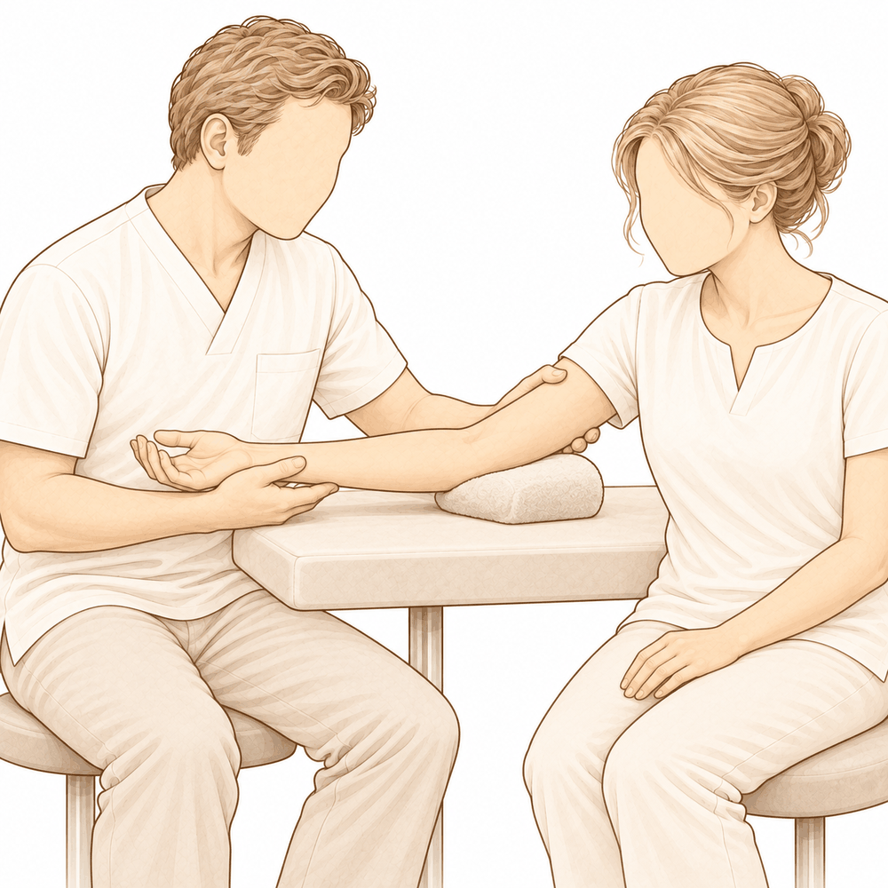
Bác sĩ Kieran Hirpara, bác sĩ phẫu thuật chi trên tại Bệnh viện tư nhân Mater Rockhampton, sẽ bắt đầu bằng các phương pháp ít xâm lấn nhất phù hợp với tình trạng của bạn. Giải phóng khuỷu tay bị cứng, còn gọi là gỡ dính khớp, là ca phẫu thuật giải phóng các mô bị co rút và phần xương bên trong khớp để khuỷu tay có thể gập và duỗi được nhiều hơn.
Chúng tôi thường thử điều trị không phẫu thuật trước. Điều đó bao gồm trị liệu tay, nẹp cố định, và đôi khi tiêm thuốc, trong khoảng 6 đến 12 tuần. Hầu hết các trường hợp cứng khuỷu tay đều cải thiện theo thời gian và khi được sử dụng. Phẫu thuật được tính đến khi liệu trình đó đã kết thúc mà khuỷu tay của bạn vẫn không cử động đủ cho công việc hoặc sinh hoạt hằng ngày.
Bạn có thể phù hợp với phẫu thuật nếu bề mặt khớp vẫn còn trơn láng, tình trạng cứng là do mô bị co rút ở bên ngoài khớp chứ không phải do tổn thương bên trong khớp, và bạn sẵn sàng tham gia trị liệu sau đó. Ca phẫu thuật nhằm mang lại sự cải thiện lâu dài về khả năng cử động, giảm đau và giúp bạn sử dụng cánh tay tốt hơn. Chúng tôi sẽ trao đổi với bạn về những gì phẫu thuật có thể và không thể làm được cho bạn, và cùng nhau quyết định liệu đó có phải là bước đi đúng đắn hay không.
Trước khi phẫu thuật
Bác sĩ phẫu thuật sẽ lên kế hoạch cho ca mổ dựa trên phim X-quang khuỷu tay của bạn, và đôi khi chụp CT, một loại chụp chi tiết cho thấy rõ xương. Những hình ảnh này cho thấy vị trí của phần xương thừa hoặc các mảnh rời nằm bên trong khớp.
Trước phẫu thuật, bạn sẽ nhận được hướng dẫn rõ ràng về việc nhịn ăn. Không ăn hoặc uống trong bảy giờ trước ca mổ. Khoảng thời gian dự phòng này cho phép ca mổ của bạn được đưa lên sớm hơn nếu lịch mổ diễn ra sớm hơn dự kiến. Hãy cho chúng tôi biết về tất cả các loại thuốc bạn đang dùng, kể cả thuốc chống viêm dạng viên, vì một số loại có thể cần tạm ngưng. Hãy mang theo danh sách ghi rõ các loại thuốc của bạn. Hãy sắp xếp người đưa bạn về nhà, vì bạn sẽ không thể lái xe. Mặc quần áo rộng rãi, thoải mái, có tay áo dễ luồn qua cánh tay. Nếu bạn mắc các bệnh lý khác, bạn có thể cần xét nghiệm máu hoặc được bác sĩ gây mê – người thực hiện gây mê cho bạn – khám trước. Hầu hết mọi người không cần cả hai điều này.
Vào ngày phẫu thuật
Bạn sẽ đến đơn vị tiếp nhận bệnh nhân phẫu thuật của bệnh viện, nơi nhân viên làm thủ tục nhập viện và chuẩn bị cho bạn trước ca mổ. Bạn sẽ gặp bác sĩ gây mê tại đó. Ca phẫu thuật này được thực hiện dưới gây mê toàn thân. Đôi khi thuốc gây tê vùng được dùng thêm để giảm đau sau mổ; bác sĩ gây mê sẽ trao đổi với bạn về điều này vào ngày phẫu thuật.
Sau đó, bạn được đưa vào phòng mổ để thực hiện ca phẫu thuật. Khi ca mổ kết thúc, bạn tỉnh dậy tại khu hồi sức. Các điều dưỡng sẽ theo dõi tình trạng của bạn cho đến khi tác dụng của thuốc mê hết. Khi sức khỏe ổn định, tùy thuộc vào loại phẫu thuật và diễn biến hồi phục của bạn, bạn sẽ được chuyển sang phòng bệnh hoặc về nhà.
Quy trình phẫu thuật
Mục tiêu của ca phẫu thuật rất đơn giản: giải phóng mọi thứ bị co rút bên trong và xung quanh khuỷu tay để khuỷu tay có thể gập và duỗi được nhiều hơn. Bác sĩ phẫu thuật có thể thực hiện điều này qua một vài đường rạch nhỏ nội soi, dùng camera để quan sát bên trong khớp, hoặc qua một đường mổ mở. Lựa chọn phụ thuộc vào nguyên nhân gây cứng khớp và mức độ can thiệp mà khớp cần.
Bên trong khớp, bác sĩ phẫu thuật loại bỏ các gai xương, là những mẩu xương thừa nhỏ cản trở cử động. Các mảnh xương hoặc sụn rời cũng được lấy ra. Bao khớp bị co rút, tức là lớp mô bao quanh khớp, được giải phóng hoặc cắt bỏ bớt. Nếu xương đã hình thành trong mô mềm ở nơi lẽ ra không có xương, một tình trạng gọi là hóa xương dị vị, phần xương đó sẽ được lấy bỏ như một phần của việc giải phóng.
Đôi khi ca phẫu thuật cần được thực hiện từ cả hai bên khuỷu tay. Nếu khả năng cử động bị hạn chế nhiều, bác sĩ phẫu thuật cũng có thể giải phóng dây thần kinh trụ, một trong những dây thần kinh chính đi qua khuỷu tay, để dây thần kinh không bị căng khi khớp cử động được nhiều hơn. Trong một số trường hợp, một khung cố định ngoài có bản lề được lắp sau khi giải phóng. Đây là một thanh nằm bên ngoài cánh tay giúp giữ khớp vững và nhẹ nhàng duy trì cử động trong khi khớp lành lại.
Cuối cùng, các đường mổ được khâu lại bằng chỉ và phủ băng gạc. Bạn sẽ tỉnh dậy với khuỷu tay được đặt trong một nẹp có đệm lót dày, gập ở góc 90 độ. Việc cử động được bắt đầu sớm, đây là một phần có chủ đích của quá trình điều trị: khuỷu tay rất dễ bị cứng lại, vì vậy các bài tập nhẹ nhàng được bắt đầu ngay sau phẫu thuật để giữ được biên độ cử động đã đạt được trong phòng mổ.
Sau khi phẫu thuật
Bạn sẽ tỉnh dậy tại khu hồi sức, sau đó được chuyển sang khu nội trú hoặc phòng chờ trong khi bạn tỉnh táo hoàn toàn. Khuỷu tay của bạn được đặt trong một nẹp có đệm lót dày, gập ở góc 90 độ. Việc giảm đau được điều chỉnh phù hợp với bạn; hãy báo cho điều dưỡng nếu thuốc không có tác dụng. Nên có người ở cùng bạn trong 24 giờ đầu tiên. Nếu bạn được chặn dây thần kinh, cánh tay có thể cảm thấy tê hoặc nặng và khó cử động trong khoảng ngày đầu; điều này là bình thường và sẽ hết khi thuốc chặn thần kinh hết tác dụng, thường trong vòng khoảng 24 giờ. Hãy gọi cho phòng khám nếu bạn vẫn còn tê, hoặc không thể cử động cánh tay, bàn tay hay các ngón tay, sau khi thuốc chặn thần kinh đã hết tác dụng. Chúng tôi để nguyên băng gạc khoảng 10 ngày; xin đừng tháo ra trước thời gian đó trừ khi chúng tôi yêu cầu. Chúng tôi sẽ thay hoặc tháo băng khi gặp bạn. Đội ngũ y tế sẽ cho bạn biết bạn được về nhà trong ngày hay phải ở lại bệnh viện một đêm.
Quá trình hồi phục
Bạn sẽ bị đau và sưng trong những ngày đầu. Cánh tay có thể đau nhức theo nhịp, và khuỷu tay sẽ cảm thấy căng và nặng. Nghỉ ngơi, kê cao cánh tay và dùng thuốc giảm đau sẽ giúp giảm bớt tình trạng này. Nếu bạn được chặn dây thần kinh, cảm giác tê sẽ giảm dần trong khoảng ngày đầu, và cơn đau sẽ rõ hơn khi thuốc hết tác dụng.
Bạn sẽ tỉnh dậy với khuỷu tay được đặt trong một nẹp có đệm lót dày, gập ở góc 90 độ. Chúng tôi sẽ tháo hoặc thay băng trong buổi tái khám, khoảng 10 ngày sau phẫu thuật. Việc cử động được bắt đầu sớm, và điều này là có chủ đích: các bài tập nhẹ nhàng được bắt đầu ngay sau ca mổ để giữ được biên độ cử động đã đạt được trong phòng mổ. Việc trị liệu tay của bạn sẽ do Ruby Doolan tại Extend Rehabilitation thực hiện. Ruby hướng dẫn các bài tập của bạn và làm bất kỳ nẹp nào bạn cần. Cử động chủ động, khi bạn tự mình cử động cánh tay, quan trọng hơn việc được người khác cử động giúp, vì nó giúp ổn định khớp.
Ở nhà, trong giai đoạn đầu bạn sẽ tập những buổi ngắn và thường xuyên thay vì những buổi tập dài. Giữ vết mổ được che kín và khô ráo. Hãy ngủ với cánh tay được kê trên gối nếu thấy thoải mái hơn. Bạn sẽ không được lái xe khi đang đeo đai treo tay hoặc nẹp. Khi bạn đã tháo bỏ chúng, khuỷu tay cảm thấy thoải mái, và bạn có thể cầm vô lăng bằng cả hai tay và phản ứng nhanh, bạn có thể lái xe trở lại. Hãy xem trang của chúng tôi về việc lái xe sau phẫu thuật chi trên để biết thêm chi tiết.
Khi tình trạng sưng giảm và khả năng cử động trở lại, các công việc hằng ngày như ăn uống, mặc quần áo và đánh máy sẽ dần trở lại. Quá trình hồi phục ở mỗi người là khác nhau, và lộ trình của bạn có thể khác. Bác sĩ phẫu thuật và chuyên gia trị liệu sẽ hướng dẫn bạn ở từng bước.
Những biến chứng có thể xảy ra
Hầu hết bệnh nhân đều hồi phục tốt, nhưng đôi khi vẫn có thể gặp phải các vấn đề. Bác sĩ phẫu thuật và đội ngũ y tế sẽ theo dõi sát sao để phát hiện sớm bất kỳ bất thường nào.
Ca phẫu thuật được thực hiện gần các dây thần kinh và mạch máu đi qua khuỷu tay, vì vậy có một nguy cơ nhỏ đối với các cấu trúc này. Nếu bạn nhận thấy tê, châm chích như kim châm, hoặc yếu mới xuất hiện ở bàn tay hay các ngón tay sau khi thuốc chặn thần kinh đã hết tác dụng, hãy gọi cho phòng khám.
Dây thần kinh trụ, một trong những dây thần kinh chính đi qua khuỷu tay, cần được lưu ý đặc biệt. Nếu dây thần kinh này bị kích ứng hoặc bị sẹo bám, bạn có thể cảm thấy ngứa ran hoặc tê ở ngón út và ngón áp út, hoặc yếu khi cầm nắm. Không thể dự đoán ai sẽ gặp vấn đề mới với dây thần kinh trụ sau phẫu thuật, vì vậy hãy báo cho chúng tôi trong buổi tái khám nếu bạn nhận thấy những cảm giác này.
Một số triệu chứng có thể xuất phát từ chấn thương ban đầu chứ không phải từ ca phẫu thuật. Một khuỷu tay bị chấn thương nặng thường bị tụ máu bên trong khớp, và mô xung quanh khớp hình thành sẹo và co rút lại. Các vết gãy cũ có thể liền ở vị trí bị lệch, hoặc hoàn toàn không liền. Xương có thể hình thành ở nơi lẽ ra không có, dây chằng có thể bị giãn hoặc mất cân bằng, và cơ có thể bị sẹo và mất độ giãn bình thường. Bất kỳ điều nào trong số này đều có thể làm thay đổi cách khuỷu tay hoạt động, hạn chế cử động và khiến cơn đau kéo dài. Cơn đau cũng có thể khiến bạn vô tình giữ khớp bất động để tự bảo vệ, khiến tình trạng co rút nặng thêm. Nếu cơn đau ngày càng tăng dù đã dùng thuốc giảm đau, hãy gọi cho phòng khám ngay trong ngày.
Nếu được lắp khung cố định ngoài có bản lề, bạn cần biết rằng điều này đi kèm với mất máu nhiều hơn trong lúc mổ, thời gian mổ dài hơn và thời gian nằm viện lâu hơn. Bác sĩ phẫu thuật chỉ sử dụng khung này khi cần thiết.
Nếu phẫu thuật nội soi được sử dụng, một số triệu chứng ban đầu của bạn có thể vẫn còn nếu mô bị co rút chưa được xử lý hoàn toàn.
Bảng các biến chứng ở trang này liệt kê tỷ lệ xảy ra điển hình nếu bạn muốn biết thông tin cụ thể.
Khi nào nên liên hệ với chúng tôi
Hầu hết các vấn đề được phát hiện sớm, và chúng tôi muốn nghe bạn báo tin hơn là để bạn lo lắng ở nhà. Hãy gọi cho phòng khám nếu bạn thấy tê, châm chích như kim châm, hoặc yếu mới xuất hiện ở bàn tay hay các ngón tay sau khi thuốc chặn thần kinh đã hết tác dụng, thường trong vòng khoảng 24 giờ. Hãy gọi cho chúng tôi ngay trong ngày nếu bạn bị sốt, vùng đỏ lan rộng quanh vết mổ, dịch hoặc mủ rỉ ra từ vết mổ, hoặc cơn đau ngày càng tăng dù đã dùng thuốc giảm đau. Hãy đến khoa cấp cứu nếu bạn bị sưng hoặc đau bắp chân, khó thở hoặc đau ngực, vì đây có thể là dấu hiệu của cục máu đông. Cũng hãy đến khoa cấp cứu nếu các ngón tay hoặc bàn tay của bạn chuyển sang nhợt nhạt, lạnh, trắng, xanh tím hoặc sẫm màu. Nếu bạn không thể liên lạc với chúng tôi ngoài giờ làm việc hoặc vào cuối tuần, hãy đến khoa cấp cứu gần nhất.
Phân tích sâu hơn
Advanced reading: the deeper science (optional)
Phần này đi sâu hơn so với những gì bạn cần biết để đưa ra quyết định điều trị. Việc tìm hiểu thêm về cách giải phóng khớp khuỷu bị cứng là rất đáng giá, bởi các bằng chứng cho thấy một điều khá bất ngờ: phương pháp phẫu thuật mà hầu hết bệnh nhân quan tâm – nội soi hay mổ hở – dường như không quan trọng bằng mức độ can thiệp phẫu thuật được thực hiện; kẻ thù thực sự của ca phẫu thuật lại chính là quá trình gây ra tình trạng cứng khớp ngay từ đầu.
Việc chọn phương pháp mổ hở hay nội soi không phải là vấn đề quan trọng
Một phân tích tổng hợp và đánh giá hệ thống được thực hiện trên 4.311 bệnh nhân bị cứng khớp khuỷu tay sau chấn thương; kết quả cho thấy phương pháp mổ hở và phương pháp nội soi đều cho kết quả tương đương nhau [1]. Kết luận của nghiên cứu là kinh nghiệm của bác sĩ phẫu thuật cùng tình trạng cụ thể của bệnh nhân mới là những yếu tố quan trọng hơn so với chính kỹ thuật phẫu thuật được áp dụng [1].
Đây là một kết luận khá thẳng thắn so với các nghiên cứu hệ thống thông thường; chúng ta nên chấp nhận kết luận này một cách nghiêm túc. Đây là ca phẫu thuật đòi hỏi kỹ năng cao, được thực hiện gần ba dây thần kinh quan trọng, trong một khớp vốn đã bị suy giảm chức năng do tình trạng co rút. Việc bác sĩ phẫu thuật quen thuộc với kỹ thuật này có vai trò quan trọng hơn nhiều so với chính kỹ thuật đó.
Ít phẫu thuật, ít biến chứng
Một phân tích có hệ thống trước đây trên 798 bệnh nhân đã đưa ra kết luận vẫn còn giá trị cho đến nay: số lượng biến chứng dường như tăng lên theo mức độ can thiệp phẫu thuật; do đó nên áp dụng phương pháp điều trị xâm lấn tối thiểu nhất có thể [2]. Các tác giả chỉ đánh giá mức độ khuyến nghị này ở mức vừa phải; các bằng chứng khoa học hiện có chưa đủ mạnh để rút ra kết luận thống kê chắc chắn, nhưng xu hướng này vẫn nhất quán.
Đây chính là mâu thuẫn cốt lõi trong công tác phẫu thuật. Việc thực hiện phẫu thuật triệt để hơn sẽ giúp bệnh nhân có được phạm vi vận động tốt hơn ngay trên bàn mổ; tuy nhiên điều này lại gây ra nhiều tổn thương mô mềm hơn, làm tăng lượng máu chảy vào khớp và kích thích phản ứng viêm – những yếu tố dẫn đến tình trạng cứng khớp sau này.
Cần ngăn ngừa hiện tượng hóa xương dị vị
Hóa xương dị vị – hiện tượng xương hình thành trong các mô mềm vốn phải giữ được tính linh hoạt – là cơ chế khiến khớp khuỷu tay vừa được phẫu thuật trở nên cứng lại. Việc dùng thuốc chống viêm để phòng ngừa là phương pháp tiêu chuẩn, dù bằng chứng khoa học vẫn chưa đầy đủ bằng thực hành lâm sàng.
Một phân tích tổng hợp trên 622 bệnh nhân so sánh hiệu quả của các thuốc chống viêm chọn lọc COX-2 và các thuốc chống viêm không chọn lọc sau phẫu thuật chấn thương khớp khuỷu tay cho thấy cả hai loại thuốc đều giảm một cách hiệu quả nguy cơ hóa xương dị vị. Tuy nhiên, các nghiên cứu so sánh trực tiếp giữa hai nhóm thuốc lại không cho thấy sự khác biệt có ý nghĩa thống kê nào; mức độ tin cậy của các bằng chứng cũng còn thấp [3]. Như vậy: việc phòng ngừa có vẻ đáng thực hiện, việc lựa chọn loại thuốc cụ thể không mang tính quyết định rõ ràng, và độ tin cậy của bằng chứng vẫn còn hạn chế.
Bối cảnh lâm sàng cũng giúp xác định mức độ nguy cơ. Sau phẫu thuật thay khớp khuỷu tay toàn phần, hóa xương dị vị là biến chứng hiếm gặp; phần lớn bệnh nhân mắc phải tình trạng này không có triệu chứng và không cần can thiệp phẫu thuật. Chính vì vậy, các bằng chứng hiện có chưa chứng minh được tính cần thiết của việc phòng ngừa thường quy sau ca phẫu thuật này [4]. Nguy cơ hóa xương dị vị không đồng đều ở mọi ca phẫu thuật khớp khuỷu tay; nó tập trung chủ yếu ở các ca chấn thương và các ca phẫu thuật cần giải phóng khớp mạnh.
Một can thiệp y tế không mang lại kết quả như mong đợi
Axit tranexamic giúp giảm chảy máu trong nhiều ca phẫu thuật chỉnh hình; việc giảm lượng máu chảy ra sau khi tiến hành phẫu thuật khớp khuỷu tay dường như sẽ giúp giảm tình trạng cứng khớp. Một phân tích tổng hợp trên 660 bệnh nhân cho thấy axit tranexamic thực sự có thể làm giảm lượng máu chảy ra trong quá trình phẫu thuật giải phóng khớp khuỷu tay, nhưng lại không ảnh hưởng đến phạm vi vận động cuối cùng hay mức độ đau của bệnh nhân [5].
Đây là một ví dụ điển hình về trường hợp một cơ chế có vẻ hợp lý lại không mang lại kết quả mà mọi người quan tâm; đồng thời cũng là lời nhắc nhở rằng “ít chảy máu” chỉ là một chỉ số thay thế, chứ không phải là kết quả thực sự cần đạt được.
Điều gì thực sự quyết định kết quả điều trị
Không có gì trong các tài liệu y khoa này cho thấy có bất kỳ phương pháp kỹ thuật nào giúp rút ngắn quy trình điều trị. Những yếu tố quan trọng cần lưu ý là: ca phẫu thuật nên được thực hiện ở mức độ tối thiểu cần thiết; việc phòng ngừa hiện tượng hình thành xương dị vị là điều đáng làm; và phạm vi vận động được cải thiện sau phẫu thuật chỉ có thể duy trì được nhờ quá trình phục hồi chức năng sau đó. Khớp khuỷu tay có xu hướng cứng lại một cách dễ dàng; vì vậy những tháng ngày sau khi phẫu thuật cũng quan trọng không kém gì chính ca phẫu thuật đó.
Tài liệu tham khảo
[1] Khorram R, Ghayyad K, Vafadar R, Borazjani R, Nezameslami A, Huffman GR, et al. Surgical treatments of post-traumatic elbow stiffness: a systematic review and meta-analysis. J Shoulder Elbow Surg. 2026;35(1):387-407. https://doi.org/10.1016/j.jse.2025.05.004
[2] Kodde IF, van Rijn J, van den Bekerom MP, Eygendaal D. Surgical treatment of post-traumatic elbow stiffness: a systematic review. J Shoulder Elbow Surg. 2013;22(4):574-80. https://doi.org/10.1016/j.jse.2012.11.010
[3] Ahmad A, Khorram R, Ghayyad K, Amin V, Kachooei AR, Huffman GR, et al. Postoperative nonsteroidal anti-inflammatory drug prophylaxis for elbow heterotopic ossification: a systematic review and meta-analysis comparing COX-2 selective and nonselective inhibitors. JSES Rev Rep Tech. 2026;6(2):100628. https://doi.org/10.1016/j.xrrt.2025.100628
[4] Liu EY, Hildebrand A, Horner NS, Athwal GS, Khan M, Alolabi B. Heterotopic ossification after total elbow arthroplasty: a systematic review. J Shoulder Elbow Surg. 2019;28(3):587-95. https://doi.org/10.1016/j.jse.2018.10.003
[5] Nejat MH, Khayami A, Daliri M, Ebrahimzadeh MH, Sadeghi M, Moradi A. Does tranexamic acid diminish hemorrhage and pain in open elbow arthrolysis? A systematic review and meta-analysis. BMC Musculoskelet Disord. 2023;24(1). https://doi.org/10.1186/s12891-023-06835-7
Evidence & references
This is the clinical evidence summary written for health professionals. It is technical, and it lists the research this page was built from. You do not need to read it to understand your treatment or to make a decision about it.
Overview
- In the setting of failed nonsurgical treatment of the stiff elbow, surgical release can yield sustained improvement of range of motion and patient quality of life [1].
- Treatment choices for elbow stiffness must consider non-surgical management and various surgical options including arthroscopic or open capsular release, arthroplasty, and elbow replacement [2].
- Arthroscopic capsular release of the elbow is effective for restoring a functional arc of motion in the short term in most patients with extrinsic contractures [3].
- Arthroscopic elbow contracture release can improve function and range of motion, though outcomes may vary based on preoperative patient characteristics [4].
- Current literature provides state-of-the-art guidance on the management regarding prevention, evaluation, and treatment of elbow stiffness [5].
- Patients with stiff elbows who underwent arthroscopic arthrolysis achieved satisfactory clinical outcomes very early postoperatively [7].
- Current treatment options for post-traumatic elbow stiffness range from conservative to surgical, with varying rates of success, invasiveness, and complications [8].
- A multinational initiative provides the first comprehensive clinical practice guideline for open arthrolysis in adult posttraumatic elbow stiffness [9].
- A subset of pediatric patients with persistent stiffness following medial epicondyle fractures may benefit from additional interventions, including intensive therapy, transposition of the ulnar nerve, and open capsular release [13].
- In most cases, the current use of arthroscopy by experienced surgeons will produce results superior to those of open release given the proper indications [14].
- The use of a hinged external fixator in open arthrolysis for posttraumatic elbow stiffness may result in short-term improvements in flexion-extension range of motion [16].
- The use of a hinged external fixator in open arthrolysis for posttraumatic elbow stiffness is accompanied by increased blood loss, longer operative time, extended hospitalization, and higher costs [16].
- The dual mediolateral mini-open technique allows for a safe and effective release of stiff elbows through small incisions of 3–5 cm in length [18].
Anatomy & Pathophysiology
Bony Anatomy
- The elbow is a trocho-ginglymoid joint consisting of medial and lateral articulations that provide bony stability [28].
- The medial articulation involves the trochlea and the ulna within the greater sigmoid notch, forming the ulnohumeral hinged portion [28].
- The ulnohumeral articulation features highly congruent anatomy through almost 180° of articular contact, except for a bare area of the greater sigmoid notch devoid of cartilage [28].
- The coronoid process has medial and lateral facets that buttress the trochlea anteriorly [28].
- The sublime tubercle is located just distal and medial to the coronoid and serves as the attachment site for the anterior bundle of the medial ulnar collateral ligament [28].
- The lateral articulation involves the capitellum and radial head, forming the radiocapitellar joint [28].
- The radius is held in close approximation to the ulna at the proximal radioulnar joint by the annular ligament [28].
- The radial head is a concave elliptical structure covered with articular cartilage along the radiocapitellar joint and approximately 270° of the articular margin [28].
- The distal humeral articulation is angled 30° from the longitudinal axis [28].
- The axis of rotation is angulated 5° to 7° in the coronal plane relative to the epicondylar axis, with the medial side more distal than the lateral side [28].
- The ulna medially bends approximately 8° at 8 cm from the tip of the olecranon [28].
- The articulation to the tip of the coronoid is approximately 30° from the long axis of the ulna in the sagittal plane [28].
Ligaments and Soft Tissue
- Elbow stability is determined by primary stabilizers (ulnohumeral articulation, MUCL, LUCL complex) and secondary stabilizers (radiocapitellar articulation, common flexor tendon, common extensor tendon, joint capsule) [25].
- The medial ulnar collateral ligament is the primary valgus stabilizer of the elbow [29].
- The anterior bundle of the medial ulnar collateral ligament is the most important component for stability [29].
- The posterior bundle of the medial ulnar collateral ligament becomes taut at flexion beyond 120 degrees [29].
- The lateral ulnar collateral ligament serves as the posterolateral stabilizer of the elbow [29].
- Osborne’s ligament stabilizes the ulnar nerve in the cubital tunnel [29].
- The ligament of Struthers is a variant anatomy arising from the supracondylar process to attach to the medial epicondyle and is a potential site of median nerve compression [29].
- The joint capsule allows maximum distension at approximately 70 to 80 degrees of flexion [29].
- The anterior capsule attaches at a point approximately 6 mm distal to the tip of the coronoid [29].
- Tensile forces are present at the medial elbow, while compressive forces are present at the lateral elbow [29].
Muscles
- The brachialis is the strongest elbow flexor and attaches to the coronoid 11 mm distal to the tip [29].
- The biceps brachii inserts at the ulnar margin of the radial tuberosity and acts as a powerful supinator of the forearm [29].
- The triceps brachii is the primary elbow extensor and inserts on the olecranon process [29].
- The mobile wad consists of the brachioradialis, extensor carpi radialis longus, and extensor carpi radialis brevis [29].
- The flexor-pronator mass includes the pronator teres, flexor carpi radialis, palmaris longus, flexor carpi ulnaris, and flexor digitorum superficialis [29].
Pathophysiology and Functional Range
- The normal elbow has a range of motion from 0° to 140° from extension to flexion [25].
- The normal elbow has a range of motion of 75° in pronation and 85° in supination [25].
- A functional arc of motion for the elbow is 100° for flexion and extension [25].
- A functional arc of motion for the elbow is 100° for forearm rotation [25].
- Successful intervention for a stiff elbow depends on the quality of the joint and the extent of soft tissue damage [11].
- The functional arc of motion serves as a frame of reference rather than an absolute goal for stiff elbow treatment [11].
- Treatment of the stiff elbow requires a thorough understanding of normal anatomy and etiological factors to develop effective strategies [10].
Classification
- The S.T.I.F. classification system highlights the aetiology of elbow stiffness [51].
- The S.T.I.F. classification system provides a framework to better understand the natural history of elbow stiffness [51].
- The S.T.I.F. classification system provides a framework to direct surgical management of elbow stiffness [51].
- The S.T.I.F. classification system provides a framework to predict clinical outcomes for elbow stiffness [51].
- Influencing factors must be taken into consideration for proper surgical indication and prognosis when using the S.T.I.F. classification system [51].
- The S.T.I.F. classification system is beneficial for research because identifying homogenous patterns of stiffness allows for more appropriate comparison between different treatment options [51].
- Based on the S.T.I.F. classification system, a treatment algorithm can be proposed that includes the severity of the disorders as a guide to surgical treatment [51].
- Based on the S.T.I.F. classification system, a treatment algorithm can be proposed that includes the severity of the disorders as a guide to the preferred operative approach (open or scope) [51].
- The prevalent types of stiffness identified by the S.T.I.F. classification system could be applied in the future to Artificial Intelligence to help in diagnosing the cause of elbow stiffness [51].
Clinical Presentation
- Elbow stiffness is a challenging problem with no ideal management solution [6].
- Elbow contracture is challenging to treat, and therefore prevention is of paramount importance [17].
- Restoration of joint motion in the posttraumatic stiff elbow can be a difficult, time-consuming, and costly challenge [15].
- Successful intervention for stiff elbow depends on the quality of the joint and the extent of soft tissue damage [11].
- The functional arc of motion serves as a frame of reference rather than an absolute goal for stiff elbow intervention [11].
- Conservative treatment is indicated for patients that have elbow stiffness due to heterotopic ossification for less than six months [22].
- Current non-surgical treatment measures for elbow stiffness due to heterotopic ossification include physical therapy and manipulation under anesthesia in order to restore the range of motion of the involved joint [22].
- Non-surgical options for heterotopic ossification have limited effects and can be used mainly in cases that cause a small limitation of range of motion [22].
- Surgical options are necessary if nonoperative management fails to restore the elbow function and range of motion after 6 months in patients with heterotopic ossification [22].
- Surgical excision of ectopic bone and contracture release, if present, are the most common surgical options in elbow heterotopic ossification management [22].
- Arthroscopic elbow contracture release can improve function and range of motion, although outcomes may vary based on preoperative patient characteristics [4].
- Although both open and arthroscopic techniques may produce satisfactory results, the current use of arthroscopy by experienced surgeons will produce results superior to those of open release given the proper indications [14].
- Operative management of pediatric elbow contractures is effective [23].
- Patients undergoing operative management of elbow stiffness secondary to heterotopic ossification maintained substantial reductions in pain, improvement in elbow range of motion, and increased overall elbow function [12].
- Treatment of bony encasement of the ulnar nerve secondary to heterotopic ossification of the elbow leads to superior range of motion, improved or resolved ulnar neuropathy, and good to excellent long-term functional outcomes [24].
- Delayed-onset anterior interosseous nerve palsy can develop one week after open elbow contracture release, likely due to a stretch injury [26].
Investigations
History and Physical Examination
- The history for elbow stiffness evaluation includes the duration of the contracture, initial injury, previous surgical procedures, trials of splinting/therapy/injections, surgical complications, and patient work/life demands [34].
- Physical examination must assess the function of the shoulder, wrist, and hand [34].
- The soft tissue surrounding the elbow should be examined for previous skin incisions, grafts, eschar, or infection [34].
- Active and passive flexion, extension, supination, and pronation should be evaluated using a goniometer for accurate measurement [34].
- The contralateral elbow should be examined for comparison during range of motion assessment [34].
- If the elbow has less than 90° to 100° of flexion, the posterior bundle of the medial collateral ligament is contracted and must be released to restore flexion [34].
- Pain assessment during the mid-arc or at terminal ends of motion helps distinguish intrinsic disease from contracture, as mid-arc pain is more common with intrinsic disease and may not improve with contracture release alone [34].
- The ulnar nerve is of utmost importance in the neurovascular examination due to its anatomic proximity to the elbow [34].
- Electromyography and nerve conduction velocity studies should be performed if there is any question about neurologic dysfunction [34].
- An assessment for ulnar nerve subluxation should be performed, as subluxation is a relative contraindication for arthroscopic procedures due to the risk of iatrogenic nerve injury [34].
- The surgeon must verify if the ulnar nerve has been transposed if there is a history of prior surgical procedures [34].
- Successful intervention for stiff elbow depends on the quality of the joint and the extent of soft tissue damage, with the functional arc of motion serving as a frame of reference rather than an absolute goal [11].
Imaging
- Radiographs should always be obtained for the evaluation of elbow stiffness [34].
- Standard radiographic views include AP, lateral, and oblique views, with serial radiography used as follow-up when heterotopic ossification is present [34].
- Primary bony landmarks assessed on radiographs include the ulnohumeral joint, coronoid process, radial head, capitellum, radiocapitellar joint, olecranon tip, coronoid/olecranon fossae, and trochlear ridge [34].
- CT is helpful for assessing malunion architecture and the location and pattern of osteophytes or loose bodies [34].
- Three-dimensional CT is used to check for heterotopic ossification [34].
- CT is not necessary when the stiffness is entirely soft-tissue related, but is beneficial if any joint incongruity or abnormal bony anatomy is present [34].
- MRI can be used to evaluate ligaments and tendons but is rarely indicated for elbow stiffness [34].
- The normal elbow has a range of motion from 0° to 140° from extension to flexion and 75° and 85° in pronation and supination respectively [25].
- A functional arc in each plane is 100° for flexion and extension and forearm rotation [25].
- Elbow stability is determined by primary stabilizers (ulnohumeral articulation, MUCL, LUCL) and secondary stabilizers (radiocapitellar articulation, common flexor tendon, common extensor tendon, joint capsule) [25].
Treatment
General Principles and Indications
- Elbow stiffness is a challenging problem with no ideal management solution; however, functional improvements can be achieved with both nonsurgical and surgical strategies [6].
Arthroscopic Release
- Arthroscopic elbow contracture release can improve function and range of motion; however, outcomes may vary based on preoperative patient characteristics [4].
- Although both open and arthroscopic techniques may produce satisfactory results, the authors believe that in most cases the current use of arthroscopy by experienced surgeons will produce results superior to those of open release given the proper indications [14].
- Additional peripheral nerve block combined with a postoperative nerve block catheter in arthroscopic arthrolysis in cases of elbow stiffness may be an opportunity to enhance postoperative outcomes by achieving better functional ROM, perhaps through reduced postoperative pain [48].
Open Release and Techniques
- This multinational initiative provides the first comprehensive clinical practice guideline for open arthrolysis in adult posttraumatic elbow stiffness [9].
- The use of a hinged external fixator in open arthrolysis for posttraumatic elbow stiffness may result in short-term improvements in flexion-extension range of motion but is accompanied by increased blood loss, longer operative time, extended hospitalization, and higher costs [16].
- The most common reason for a failed contracture release is a failure on the part of the surgeon to abandon an orthodox method in favor of a less assured technique or to use a single anatomic approach to the joint when the potential gain in motion is not achieved at the time of surgery [40].
- The operative contracture release requires a progressive, adaptive surgical approach, and the surgeon must safely increase the exposure in order to excise the offending fibrosis on nearly all aspects of the joint, anteroposterior and mediolateral [40].
- If an uncomplicated, simple dislocation has caused a 30-degree flexion contracture, arthroscopic capsular excision may be sufficient [40].
- The best care for a given patient with a contracture is the most minimal necessary surgery, including minimal anatomic exposure, and rehabilitation to achieve the desired result [40].
- A patient with a range of 40 to 105 degrees and heterotopic ossification will usually require exposure of the capsule for complete excision from both the medial and lateral approaches but not necessarily hinged fixation [40].
- In a circumstance with more massive heterotopic ossification, hinged external fixation may be necessary to stabilize the elbow postoperatively and permit immediate motion with intermittent passive stretch [40].
- The surgeon should be prepared to extend the exposure or approach the joint from an additional direction if full passive motion is not achieved on the operating table [40].
- In older patients with suspected cartilage damage or avascular bone, total elbow replacement should be available and discussed before surgery [40].
Outcomes and Complications
- Overall, patients maintained substantial reductions in pain, improvement in elbow range of motion, and increased overall elbow function after operative management of elbow stiffness secondary to heterotopic ossification [12].
- The gain in range of motion for open arthrolysis was 51° [19].
- The gain in range of motion for arthroscopic arthrolysis was 40° [19].
- The gain in range of motion for open arthrolysis with external fixation was 88° [19].
- The gain in range of motion for open arthrolysis with distraction arthroplasty was 56° [19].
- The average percentage of complications for open arthrolysis was 23% [19].
- The average percentage of complications for arthroscopic arthrolysis was 5% [19].
- The average percentage of complications for open arthrolysis with external fixation was 73% [19].
- The average percentage of complications for open arthrolysis with distraction arthroplasty was 58% [19].
Complications
- The use of a hinged external fixator in open arthrolysis is accompanied by increased blood loss [16].
- The use of a hinged external fixator in open arthrolysis is accompanied by longer operative time [16].
- The use of a hinged external fixator in open arthrolysis is accompanied by extended hospitalization [16].
- The use of a hinged external fixator in open arthrolysis is accompanied by higher costs [16].
- Current treatment options for post-traumatic elbow stiffness have varying rates of complications [8].
Recovery
- Surgical release of the stiff elbow can yield sustained improvement of range of motion and patient quality of life in the setting of failed nonsurgical treatment [1].
- Arthroscopic capsular release is effective for restoring a functional arc of motion in the short term in most patients with extrinsic contractures [3].
- Functional improvements can be achieved with both nonsurgical and surgical strategies for elbow stiffness [6].
- Treatment for bony encasement of the ulnar nerve secondary to heterotopic ossification of the elbow leads to superior range of motion, improved or resolved ulnar neuropathy, and good to excellent long-term functional outcomes [24].
Key Evidence
- [L5] In the setting of failed nonsurgical treatment of the stiff elbow, surgical release can yield sustained improvement of ROM and patient quality of life. [1] (10.5435/jaaos-d-14-00051)
- [L5] Treatment choices must consider non-surgical management and various surgical options including arthroscopic or open capsular release, arthroplasty, and elbow replacement. [2] (10.1016/j.jisako.2023.10.009)
- [L5] Arthroscopic capsular release of the elbow is effective for restoring a functional arc of motion in the short term in most patients with extrinsic contractures. [3] (10.5435/00124635-201105000-00004)
- [L4] Arthroscopic elbow contracture release can improve function and range of motion; however, outcomes may vary based on preoperative patient characteristics. [4] (10.1016/j.jseint.2026.101621)
- [L4] This paper reviews the current literature and provides state-of-the-art guidance on the management regarding prevention, evaluation, and treatment of elbow stiffness. [5] (10.1530/eor-23-0039)
- [L4] Elbow stiffness is a challenging problem with no ideal management solution; however, functional improvements can be achieved with both nonsurgical and surgical strategies, and recent advancements in biology and pathology may lead to future breakthroughs in prevention and treatment. [6] (10.1016/j.jhsa.2013.06.007)
- [L1] Patients with stiff elbows who underwent arthroscopic arthrolysis achieved satisfactory clinical outcomes very early postoperatively. [7] (10.1016/j.jse.2024.06.009)
- [L5] Current treatment options for post-traumatic elbow stiffness range from conservative to surgical, with varying rates of success, invasiveness, and complications. [8] (10.1177/1758573218793903)
- [L5] This multinational initiative provides the first comprehensive clinical practice guideline for open arthrolysis in adult posttraumatic elbow stiffness. [9] (10.1016/j.jse.2025.07.015)
- [L5] Treatment of the stiff elbow requires a thorough understanding of normal anatomy and etiological factors to develop effective strategies. [10] (10.1016/j.jisako.2023.10.006)
- [L5] Successful intervention for stiff elbow depends on the quality of the joint and the extent of soft tissue damage, with the functional arc of motion serving as a frame of reference rather than an absolute goal. [11] (10.1016/j.jisako.2023.09.002)
- [L4] Overall, patients maintained substantial reductions in pain, improvement in elbow range of motion, and increased overall elbow function. [12] (10.1016/j.jse.2024.11.019)
- [L4] A subset of pediatric patients with persistent stiffness following medial epicondyle fractures may benefit from additional interventions, including intensive therapy, transposition of the ulnar nerve, and open capsular release. [13] (10.1016/j.jhsg.2023.07.002)
- [L5] Although both open and arthroscopic techniques may produce satisfactory results, the authors believe that in most cases the current use of arthroscopy by experienced surgeons will produce results superior to those of open release given the proper indications. [14] (10.1016/j.jse.2010.11.029)
- [L4] Restoration of joint motion in the posttraumatic stiff elbow can be a difficult, time-consuming, and costly challenge. [15] (10.1016/j.jhsa.2007.09.015)
- [L3] The use of a hinged external fixator in open arthrolysis for posttraumatic elbow stiffness may result in short-term improvements in flexion-extension range of motion but is accompanied by increased blood loss, longer operative time, extended hospitalization, and higher costs. [16] (10.1186/s12891-024-08167-6)
- [L5] Elbow contracture is challenging to treat, and therefore prevention is of paramount importance. [17] (10.1016/j.jhsa.2009.02.020)
- [L4] The dual mediolateral mini-open technique allows for a safe and effective release of stiff elbows through small incisions of 3–5 cm in length. [18] (10.1186/s13018-025-06288-9)
- [L4] [19] (10.1016/j.jse.2012.11.010)
- [L4] [22] (10.3390/life13122358)
- [L1] Operative management of pediatric elbow contractures is effective. [23] (10.1016/j.jhsa.2024.01.010)
- [L4] This treatment approach leads to superior range of motion, improved or resolved ulnar neuropathy, and good to excellent long-term functional outcomes. [24] (10.1016/j.jse.2023.12.003)
- [Case_report] The authors describe a case of delayed-onset anterior interosseous nerve palsy developing one week after open elbow contracture release, likely due to a stretch injury. [26] (10.5397/cise.2022.00899)
- [L3] Additional peripheral nerve block combined with a postoperative nerve block catheter in arthroscopic arthrolysis in cases of elbow stiffness may be an opportunity to enhance postoperative outcomes by achieving better functional ROM, perhaps through reduced postoperative pain. [48] (10.1016/j.jseint.2024.10.009)
- [L5] [51] (10.1016/j.jisako.2023.10.011)
References
[1] Open Surgical Release for Contractures of the Elbow. Journal of the American Academy of Orthopaedic Surgeons. 2015. DOI: 10.5435/jaaos-d-14-00051
[2] Elbow stiffness: Arthritis and heterotopic ossification. Journal of ISAKOS. 2024. DOI: 10.1016/j.jisako.2023.10.009
[3] Arthroscopic Management of the Stiff Elbow. American Academy of Orthopaedic Surgeon. 2011. DOI: 10.5435/00124635-201105000-00004
[4] Preoperative risk factors associated with patient outcomes following arthroscopic elbow contracture release. JSES International. 2026. DOI: 10.1016/j.jseint.2026.101621
[5] Management of the stiff elbow: a literature review. EFORT Open Reviews. 2023. DOI: 10.1530/eor-23-0039
[6] Prevention and Treatment of Elbow Stiffness: A 5-Year Update. The Journal of Hand Surgery. 2013. DOI: 10.1016/j.jhsa.2013.06.007
[7] Does tranexamic acid reduce elbow swelling and improve early function following arthroscopic arthrolysis? A double-blind randomized controlled trial. Journal of Shoulder and Elbow Surgery. 2024. DOI: 10.1016/j.jse.2024.06.009
[8] Post-traumatic elbow stiffness: Pathogenesis and current treatments. Shoulder & Elbow. 2018. DOI: 10.1177/1758573218793903
[9] Clinical guideline on the open arthrolysis for post-traumatic elbow stiffness in adult patients. Journal of Shoulder and Elbow Surgery. 2026. DOI: 10.1016/j.jse.2025.07.015
[10] The examination and treatment of soft tissue contracture of the elbow. Journal of ISAKOS. 2024. DOI: 10.1016/j.jisako.2023.10.006
[11] Elbow stiffness: Interview with professor Bernard Morrey. Journal of ISAKOS. 2024. DOI: 10.1016/j.jisako.2023.09.002
[12] Assessing long-term outcomes after operative management of elbow stiffness secondary to heterotopic ossification. Journal of Shoulder and Elbow Surgery. 2025. DOI: 10.1016/j.jse.2024.11.019
[13] Outcomes of Therapy and Ulnar Nerve Transposition for Elbow Stiffness After Pediatric Medial Epicondyle Fractures. Journal of Hand Surgery Global Online. 2023. DOI: 10.1016/j.jhsg.2023.07.002
[14] Arthroscopic management of the post-traumatic stiff elbow. Journal of Shoulder and Elbow Surgery. 2011. DOI: 10.1016/j.jse.2010.11.029
[15] The Posttraumatic Stiff Elbow: A Review of the Literature. The Journal of Hand Surgery. 2007. DOI: 10.1016/j.jhsa.2007.09.015
[16] Comparative study of open elbow arthrolysis with and without hinge external fixation for the treatment of post-traumatic elbow stiffness. BMC Musculoskeletal Disorders. 2024. DOI: 10.1186/s12891-024-08167-6
[17] Prevention and Treatment of Elbow Stiffness. The Journal of Hand Surgery. 2009. DOI: 10.1016/j.jhsa.2009.02.020
[18] Dual mediolateral mini-open technique for the release of elbow contracture. Journal of Orthopaedic Surgery and Research. 2025. DOI: 10.1186/s13018-025-06288-9
[19] Surgical treatment of post-traumatic elbow stiffness: a systematic review. Journal of Shoulder and Elbow Surgery. 2013. DOI: 10.1016/j.jse.2012.11.010
[22] Heterotopic Ossification around the Elbow Revisited. Life. 2023. DOI: 10.3390/life13122358
[23] Clinical Outcomes Following Surgical Management of Post-Traumatic Elbow Contractures in the Pediatric Age Group: A Meta-Analysis and Systematic Review. The Journal of Hand Surgery. 2025. DOI: 10.1016/j.jhsa.2024.01.010
[24] Bony encasement of the ulnar nerve secondary to heterotopic ossification of the elbow: an evaluation of long-term outcomes. Journal of Shoulder and Elbow Surgery. 2024. DOI: 10.1016/j.jse.2023.12.003
[25] Orthopaedic Knowledge Update 13 Ebook Without Multimedia. Anatomy, Biomechanics, Physical Examination, and Imaging of the Elbow > Summary and Conclusions.
[26] Anterior interosseous nerve palsy in the early postoperative period after open capsular release for elbow stiffness: a case report. Clinics in Shoulder and Elbow. 2023. DOI: 10.5397/cise.2022.00899
[28] Orthopaedic Knowledge Update 13 Ebook Without Multimedia. Anatomy, Biomechanics, Physical Examination, and Imaging of the Elbow > Anatomy > Bony Anatomy.
[29] Miller S Review Of Orthopaedics. SECTION 16 PATELLAR TRACKING IN TOTAL KNEE ARTHROPLASTY > TABLE 2.3 Shoulder Spaces.
[34] Aaos Comprehensive Orthopaedic Review 3. Elbow Stiffness* > IV. Evaluation.
[40] Green S Operative Hand Surgery. Classifying the Contracture and Matching the Operative Plan.
[48] A comparative analysis of short-term results in range of motion following arthroscopic arthrolysis with vs. without peripheral nerve block in cases of elbow stiffness. JSES International. 2025. DOI: 10.1016/j.jseint.2024.10.009
[51] Classification of elbow stiffness. Journal of ISAKOS. 2024. DOI: 10.1016/j.jisako.2023.10.011