Patients › Hand
Chứng ngón tay cò súng
Trigger finger causes painful catching or locking of a finger, often treated with splinting or steroid injection.
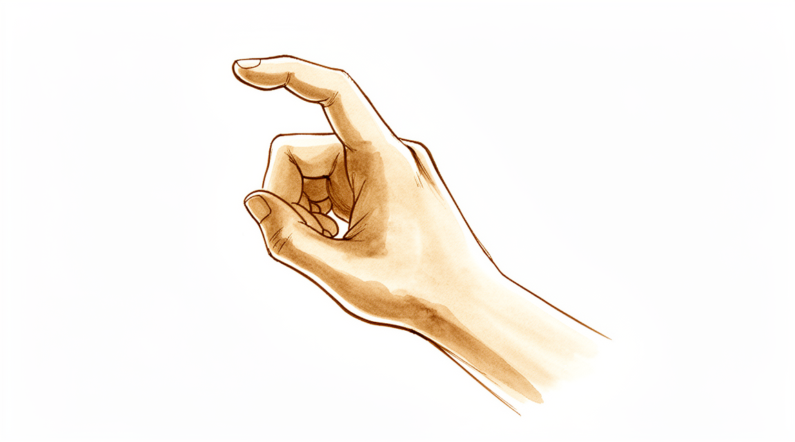
Những triệu chứng bạn đang gặp phải
Chứng ngón tay cò súng ảnh hưởng đến các gân giúp gập ngón tay hoặc ngón cái. Các gân này trượt qua một đường hầm hẹp ở lòng bàn tay gọi là “ròng rọc”. Khi sợi gân dày lên, nó bị mắc kẹt khi di chuyển qua đường hầm đó. Bạn cảm thấy đau và ấn đau ở lòng bàn tay, tại chỗ gốc của ngón tay hoặc ngón cái bị ảnh hưởng. Một số người nhận thấy một cục nhỏ hoặc nốt sần ở lòng bàn tay tại vị trí đó. Cục này là phần bị dày lên của sợi gân hoặc của đường hầm bao quanh nó, và nó di chuyển khi sợi gân di chuyển.
Hiện tượng mắc kẹt thường bắt đầu chỉ như một sự phiền toái nhẹ và tăng dần trong nhiều tuần. Lúc đầu, ngón tay có thể phát ra tiếng “cạch” hoặc bị kẹt khi bạn gập và duỗi, và bạn có thể phải dùng tay kia để duỗi thẳng nó. Khi bệnh nặng hơn, ngón tay có thể bị khóa ở tư thế gập và giữ nguyên như vậy. Nhiều người thấy ngón tay cứng nhất lúc mới thức dậy, rồi cử động dễ dàng hơn khi họ vận động trong ngày. Các động tác nắm và kẹp là những gì làm bệnh nặng thêm, vì vậy những việc như cầm vô lăng, xách túi đồ đi chợ, dùng kéo hoặc cầm chặt nắp lọ có thể trở nên khó chịu. Một số người còn cảm thấy đau âm ỉ ở mặt sau của khớp đốt ngón tay hoặc khớp giữa của chính ngón đó, dù vấn đề thực ra nằm ở lòng bàn tay.
Chứng ngón tay cò súng là bệnh phổ biến. Bệnh ảnh hưởng đến 2% đến 3% dân số, phụ nữ hay mắc hơn nam giới, và thường xuất hiện sau 45 tuổi. Nếu có nhiều hơn một ngón tay bị cò súng, bạn nên nói điều này với bác sĩ phẫu thuật, vì tình trạng này có thể liên quan đến các bệnh khác như bệnh tiểu đường. Chứng ngón tay cò súng cũng thường đi kèm với hội chứng ống cổ tay, gây tê hoặc cảm giác kim châm ở tay; hai bệnh này cùng xuất hiện ở 40% đến 60% số người mắc chứng ngón tay cò súng.
Hãy gặp bác sĩ đa khoa hoặc đề nghị được bác sĩ chuyên khoa khám nếu các triệu chứng không thuyên giảm, nặng dần lên trong nhiều tuần, khiến bạn thức giấc vào ban đêm, hoặc khiến bạn không thể làm việc hay sử dụng bàn tay.
Điều gì đang thực sự xảy ra
Các gân dùng để gập ngón tay giống như những sợi dây chạy qua một loạt vòng dẫn hướng ở lòng bàn tay và ngón tay. Vòng đầu tiên nằm ở lòng bàn tay, tại gốc ngón tay. Nó được gọi là ròng rọc A1. Khi bạn nắm hoặc bóp chặt lặp đi lặp lại, sợi gân có thể bị kích ứng tại chỗ nó trượt qua vòng đó. Vùng bị kích ứng dày lên, khiến sợi dây không còn trượt trơn tru nữa. Thay vào đó, nó bị dồn lại rồi kẹt mạnh qua chiếc vòng đã bị hẹp; đó chính là cảm giác mắc kẹt hoặc bật “cạch” mà bạn cảm nhận được.
Chỗ dày lên trên sợi gân thường là một nốt nhỏ, tức một nút mô bị biến đổi, to hơn một chút so với phần còn lại của sợi dây. Mỗi lần bạn gập ngón tay, nút này phải lách qua bên dưới ròng rọc. Nó trượt qua kèm một tiếng “cạch”, rồi bị mắc kẹt ở phía bên kia khi bạn cố duỗi thẳng. Đó là lý do ngón tay có thể bị khóa ở tư thế gập và cần tay kia giúp gỡ ra, và cũng là lý do vấn đề có cảm giác nặng nhất vào buổi sáng, sau một đêm ngón tay nằm yên ở tư thế gập.
Các bác sĩ mô tả mức độ tiến triển của bệnh theo bốn cấp độ. Cấp độ thứ nhất là đau và ấn đau ở lòng bàn tay, tại chiếc vòng đầu tiên đó. Cấp độ thứ hai có thêm hiện tượng ngón tay bị mắc kẹt. Cấp độ thứ ba là ngón tay bị khóa nhưng bạn vẫn có thể duỗi thẳng bằng cách dùng tay kia đẩy. Cấp độ thứ tư là ngón tay bị kẹt ở tư thế gập và hoàn toàn không thể duỗi thẳng được nữa. Các cấp độ sớm thường thuyên giảm với các phương pháp điều trị đơn giản hơn, trong khi ngón tay đã bị khóa thường cần một thủ thuật nhỏ để mở ròng rọc, giúp sợi gân trượt trơn tru trở lại.
Có một số yếu tố làm tăng khả năng xảy ra tình trạng dày lên này. Việc nắm chặt lặp đi lặp lại góp một phần, vì vậy bệnh thường gặp ở người chèo thuyền và những người khác phải dùng lực nắm nhiều. Bệnh cũng liên quan đến các bệnh như tiểu đường, suy giáp (tuyến giáp hoạt động kém), bệnh gút và viêm khớp dạng thấp, trong đó mô quanh sợi gân bị viêm hoặc biến đổi theo những cách làm hẹp đường hầm. Nếu nhiều ngón tay bị cò súng cùng lúc, bạn nên báo cho bác sĩ biết biểu hiện này, vì nó có thể gợi ý một trong các bệnh nền nói trên.
Những biện pháp chúng tôi có thể áp dụng
Bác sĩ Kieran Hirpara, bác sĩ phẫu thuật chi trên tại Bệnh viện Mater Private Rockhampton, sẽ bắt đầu bằng các phương pháp ít xâm lấn nhất phù hợp với tình trạng của bạn. Thông thường, bệnh nhân được bác sĩ đa khoa giới thiệu đến phòng khám chúng tôi; nếu vật lý trị liệu viên khuyên bạn nên đến gặp chúng tôi, bạn vẫn cần có giấy giới thiệu từ bác sĩ đa khoa để được hưởng mức hoàn trả từ Medicare. Trong buổi hẹn khám, chúng tôi sẽ hỏi bệnh sử, khám tay và chỉ chỉ định chụp hình ảnh khi điều đó làm thay đổi kế hoạch điều trị. Với một vấn đề kéo dài như thế này, chúng tôi thường thử các biện pháp điều trị không phẫu thuật trước và cân nhắc phẫu thuật khi các biện pháp đó chưa mang lại đủ cải thiện.
Bước đầu tiên là giúp bàn tay dịu lại. Việc thay đổi cách cầm nắm và giảm bớt các công việc gây áp lực lên lòng bàn tay, chẳng hạn như mang vác nặng hoặc bóp chặt, sẽ giúp sợi gân bị kích ứng có cơ hội dịu lại. Liệu pháp trị liệu tay nhằm khôi phục khả năng trượt trơn tru của sợi gân qua ròng rọc và giữ cho các khớp ngón tay tiếp tục cử động. Nẹp giữ cố định ngón tay có thể là một phần của quá trình này, và nẹp có hiệu quả nhất khi được đeo đủ lâu: cố định một khớp duy nhất trong 6 đến 10 tuần là khoảng thời gian được bằng chứng ủng hộ, lâu hơn so với dự đoán của hầu hết mọi người. Hãy kiên trì thử các biện pháp này trong vài tuần trước khi đánh giá hiệu quả.
Nếu các biện pháp đơn giản chưa làm tình trạng thuyên giảm, tiêm cortisone vào đường hầm gân là bước tiếp theo thường được áp dụng. Cortisone làm giảm tình trạng sưng và dày lên khiến sợi gân bị mắc kẹt. Những người mắc bệnh tiểu đường thường đáp ứng kém hơn với tiêm cortisone; đây là điều nên biết khi lựa chọn giữa các phương án. Phần lớn mọi người nghĩ rằng mũi tiêm sẽ đau hơn thực tế, và sau đó cho biết nó dễ chịu hơn so với những gì họ đã chuẩn bị tinh thần.
Phẫu thuật được cân nhắc khi ngón tay vẫn bị khóa, hoặc khi tiêm thuốc và đeo nẹp chưa mang lại đủ cải thiện. Ca phẫu thuật giải phóng ròng rọc bị chật ở gốc ngón tay để sợi gân có thể trượt tự do trở lại. Phẫu thuật được thực hiện qua một vết mổ nhỏ ở lòng bàn tay, và đây là một thủ thuật đơn giản, đã được áp dụng rộng rãi từ lâu. Chúng tôi sẽ cùng bạn thảo luận xem phương pháp này có phù hợp với bạn không và cùng quyết định. Sau phẫu thuật, phục hồi chức năng của bạn là trị liệu bàn tay với chuyên viên Ruby Doolan tại Extend Rehabilitation; cô ấy sẽ hướng dẫn bạn các bài tập và làm nẹp nếu bạn cần.
Những điều có thể xảy ra
Một khi ngón tay đã bị mắc kẹt hoặc bị khóa, chứng ngón tay cò súng hiếm khi tự trở lại bình thường. Ở giai đoạn sớm, khi chỉ có đau và ấn đau ở lòng bàn tay, các phương pháp điều trị đơn giản hơn như thay đổi cách cầm nắm, trị liệu tay và đeo nẹp có thể giúp tình trạng dịu lại. Nẹp cần được đeo đủ lâu để có hiệu quả: với một khớp duy nhất, 6 đến 10 tuần là khoảng thời gian được bằng chứng ủng hộ. Nếu các biện pháp này không giúp ích, tiêm cortisone vào đường hầm gân giải quyết được vấn đề ở khoảng hơn một nửa số người, vì vậy tiêm mũi thứ hai hoặc chuyển sang phẫu thuật là bước tiếp theo bình thường chứ không phải dấu hiệu cho thấy điều gì đó đã đi sai hướng.
Khi ngón tay đã đến giai đoạn bị khóa, phẫu thuật giải phóng ròng rọc bị chật là phương pháp điều trị giải quyết được nguyên nhân. Sau đó sợi gân có thể trượt qua đường hầm trở lại, và bạn được khuyến khích sử dụng ngón tay hoặc ngón cái bình thường sau phẫu thuật. Phần lớn mọi người đều vui vì đã điều trị. Tuy vậy, bạn cũng nên biết một cách thẳng thắn những lưu ý nhỏ. Khoảng 1/20 ngón tay gặp một vấn đề nhẹ, ngắn hạn sau đó, thường gặp nhất là đau, cứng hoặc sưng quanh vết mổ. Tình trạng cứng kéo dài xảy ra ở khoảng 8,6% số người, và vết mổ chậm lành ở 1,2%. Các biến chứng nghiêm trọng như tổn thương thần kinh hoặc nhiễm trùng sâu là không phổ biến: tổn thương thần kinh hoặc tê kéo dài xảy ra ở 0,6%, và nhiễm trùng vết mổ ở 2,1%. Khoảng 1/200 ngón tay cần phẫu thuật lần thứ hai, và hiện tượng cò súng tái phát ở 2,2%.
Có một vài yếu tố ảnh hưởng đến tiên lượng của bạn. Nếu khớp giữa của ngón tay đã bị đau trong thời gian dài trước khi điều trị, cơn đau khớp đó có thể không hết hoàn toàn ngay cả sau khi ròng rọc đã được giải phóng. Việc giải phóng nhiều ngón tay cùng lúc thường đồng nghĩa với đau và cứng sau phẫu thuật nhiều hơn so với giải phóng một ngón. Và những người phẫu thuật trong vòng một tháng sau khi tiêm cortisone có nguy cơ nhiễm trùng vết mổ cao hơn sau đó; đây là điều nên cân nhắc khi chọn thời điểm.
Nếu ngón tay của bạn trở nên nóng, đỏ, sưng và đau, đặc biệt là khi kèm theo sốt, hãy đến phòng cấp cứu ngay trong ngày. Nếu các triệu chứng không thuyên giảm, nặng dần lên trong nhiều tuần, khiến bạn thức giấc vào ban đêm, hoặc khiến bạn không thể làm việc hay sử dụng bàn tay, hãy gặp bác sĩ đa khoa hoặc đề nghị được bác sĩ chuyên khoa khám.
Khi nào nên đi khám
Hãy gặp bác sĩ đa khoa hoặc đề nghị được bác sĩ chuyên khoa khám nếu ngón tay hoặc ngón cái của bạn bị kẹt, bị khóa ở tư thế gập, hoặc phải dùng tay kia mới duỗi thẳng được. Điều tương tự cũng áp dụng nếu bạn có một cục ấn đau ở lòng bàn tay di chuyển theo sợi gân, hoặc nếu hiện tượng mắc kẹt khiến bạn không thể cầm nắm, làm việc hoặc ngủ. Nếu có nhiều hơn một ngón tay bị cò súng, cũng hãy cho bác sĩ biết, vì tình trạng này có thể gợi ý một bệnh nền như tiểu đường. Điều này càng quan trọng hơn ở trẻ em: trẻ bị cò súng ở nhiều hơn một ngón tay hoặc ngón cái, hoặc bị cò súng kèm theo cảm giác kim châm hoặc tê, cần được bác sĩ chuyên khoa khám thay vì chỉ theo dõi chờ đợi. Hãy đến phòng cấp cứu ngay trong ngày nếu ngón tay của bạn trở nên nóng, đỏ, sưng và đau, đặc biệt là khi kèm theo sốt, hoặc nếu các ngón tay trở nên nhợt nhạt, lạnh, trắng bệch hoặc xanh tím.
Phân tích chi tiết hơn
Advanced reading: the deeper science (optional)
Phần này đi sâu hơn mức cần thiết để bạn có thể tự đưa ra quyết định điều trị. Chứng ngón tay cò cần được tìm hiểu kỹ hơn vì phương pháp điều trị đầu tay chỉ có hiệu quả khoảng hơn một nửa số ca bệnh; con số này hiếm khi được đề cập. Ngoài ra, ở trẻ em, cùng một biểu hiện lâm sàng lại có ý nghĩa khác hẳn, dẫn đến việc cần tiến hành các xét nghiệm chẩn đoán khác.
Phương pháp tiêm có hiệu quả, và con số trung thực là 57%
Tiêm corticosteroid là biện pháp can thiệp đầu tiên tiêu chuẩn, và thường được mô tả đơn giản là “có hiệu quả”. Con số cụ thể được tổng hợp từ các nghiên cứu hạng I và II cho thấy: tiêm corticosteroid có hiệu quả ở 57% bệnh nhân mắc chứng ngón tay cò. Dù các nghiên cứu này còn nhiều hạn chế, phương pháp này vẫn được khuyến nghị làm điều trị đầu tay nhờ tính đơn giản và khả năng giảm triệu chứng của nó [1].
Con số 57% là điều đáng biết trước khi bạn quyết định tiêm thuốc. Nó giúp chúng ta hiểu rằng việc tiêm lại lần thứ hai hoặc phải phẫu thuật là điều có thể xảy ra với một tỷ lệ đáng kể bệnh nhân, chứ không phải là dấu hiệu cho thấy điều gì đó đã đi sai hướng.
Việc sử dụng nẹp là một phương án thay thế khả thi; thời gian điều trị còn dài hơn nhiều so với dự đoán
Trong những trường hợp tiêm không thích hợp – ví dụ như ở bệnh nhân tiểu đường, nơi việc tiêm có thể làm rối loạn kiểm soát đường huyết – việc điều trị bằng nẹp chỉnh hình có cơ sở bằng chứng khá hợp lý. Các nghiên cứu đều cho thấy kết quả điều trị không phụ thuộc vào khớp nào được cố định, và khuyến nghị là cố định một khớp duy nhất trong khoảng 6 đến 10 tuần [2].
Chính thời gian điều trị này mới là điều mà mọi người thường đánh giá thấp. Việc đeo nẹp chỉ trong hai tuần thì chưa thể coi là một thử nghiệm điều trị bằng nẹp.
Phẫu thuật giải phóng: cuộc tranh luận về kỹ thuật đã ngã ngũ, còn câu hỏi về đường tiếp cận thì chưa
Việc giải phóng ròng rọc A1 có thể thực hiện bằng phương pháp mở, bằng một vết mổ nhỏ dưới quan sát trực tiếp, hoặc qua đường da bằng kim hoặc lưỡi dao. Dựa trên dữ liệu từ 548 bệnh nhân, không có sự khác biệt đáng kể nào giữa phương pháp mở và phương pháp qua da về tỷ lệ phải phẫu thuật lại, biến chứng hay mức độ đau sau phẫu thuật; do đó cả hai phương pháp đều thích hợp [3].
Phân tích quy mô lớn hơn trên 2,114 ngón tay cho thấy phương pháp giải phóng qua da vừa hiệu quả vừa an toàn, đồng thời cung cấp hai thông tin hữu ích: việc sử dụng siêu âm hướng dẫn có thể nâng cao tỷ lệ thành công, còn tỷ lệ thành công thì không bị ảnh hưởng bởi loại dụng cụ sử dụng hay việc có tiêm cortisone kèm theo hay không [4]. Một phân tích năm 2025 trên 685 bệnh nhân cho thấy việc kết hợp giải phóng qua da với tiêm corticosteroid được dung nạp tốt và có tỷ lệ biến chứng thấp [5].
Tóm lại: ròng rọc cần phải được cắt; cách thức thực hiện không quan trọng bằng việc phải cắt đứt hoàn toàn. Loại dụng cụ sử dụng không quyết định kết quả cuối cùng.
Ở trẻ em, cùng một phát hiện lâm sàng lại đòi hỏi cách xử trí khác
Chứng ngón tay cò súng ở trẻ em không đơn thuần là dạng bệnh tương tự ở người lớn mà xuất hiện sớm hơn. Việc có ngón cò ở cả hai bên hoặc ở nhiều ngón, hoặc kèm theo hội chứng ống cổ tay, cần làm tăng nghi ngờ về một bệnh lý nền không điển hình như bệnh mucopolysaccharidosis [6].
Đây là yếu tố có ý nghĩa lâm sàng quan trọng nhất trong mục này. Trẻ em có nhiều ngón tay bị ảnh hưởng, hoặc vừa có triệu chứng ngón tay cò súng vừa có các dấu hiệu thần kinh, cần được kiểm tra để tìm một rối loạn tích tụ chất toàn thân thay vì chỉ tiến hành thủ thuật giải phóng đơn thuần; bởi những biểu hiện ở bàn tay có thể là dấu hiệu đầu tiên của một bệnh lý có ảnh hưởng rộng rãi hơn nhiều so với vùng tay. Trường hợp chỉ có một ngón tay cái bị ảnh hưởng ở trẻ hoàn toàn khỏe mạnh thì lại là tình huống khác và phổ biến hơn nhiều.
Tài liệu tham khảo
[1] Fleisch SB, Spindler KP, Lee DH. Tiêm corticosteroid trong điều trị ngón tay cò: tổng quan có hệ thống cấp độ I và II. J Am Acad Orthop Surg. 2007;15(3):166-71. https://doi.org/10.5435/00124635-200703000-00006
[2] Lunsford D, Valdes K, Hengy S. Các phương pháp điều trị bảo tồn ngón tay cò: tổng quan có hệ thống. J Hand Ther. 2019;32(2):212-21. https://doi.org/10.1016/j.jht.2017.10.016
[3] Casey JC, Daher M, Dworkin M, Cusano J, Garavito J, Gil JA. Phương pháp cố định mở so với qua da trong điều trị ngón tay cò: phân tích tổng hợp về kết quả lâm sàng. J Hand Surg Am. 2024;49(6):570-5. https://doi.org/10.1016/j.jhsa.2024.03.010
[4] Zhao J, Kan S, Zhao L, Wang Z, Long L, Wang J, và cộng sự. Giải phóng ròng rọc vòng thứ nhất qua da cho ngón cò: tổng quan có hệ thống và phân tích tổng hợp các bằng chứng hiện có. J Hand Surg Am. 2014;39(11):2192-202. https://doi.org/10.1016/j.jhsa.2014.07.044
[5] Wen J, Syed B, Khalil R, Shehabat M, Alam M, Sedighi R, và cộng sự. Giải phóng ròng rọc A1 qua da kết hợp tiêm corticosteroid trong điều trị ngón tay cò: tổng quan có hệ thống và phân tích tổng hợp. J Orthop Surg Res. 2025;20(1). https://doi.org/10.1186/s13018-025-05776-2
[6] Wong AL, Wong MJ, Parker R, Wheelock ME. Biểu hiện và nguyên nhân gây ngón tay cò ở trẻ em: tổng quan có hệ thống. J Hand Surg Eur Vol. 2021;47(2):192-6. https://doi.org/10.1177/17531934211035642
Evidence & references
This is the clinical evidence summary written for health professionals. It is technical, and it lists the research this page was built from. You do not need to read it to understand your treatment or to make a decision about it.
Overview
Non-Operative Management
- Female patients presenting with their first trigger finger have the highest rate of long-term treatment success after a single corticosteroid injection [1].
- Factors associated with a worse outcome following a steroid injection were identified and should be considered when choosing the treatment of a trigger finger [4].
- Surgical management may be the next best option in patients with trigger finger who continue to be symptomatic after a single injection [12].
- Management of trigger finger with 2 steroid injections before surgery is the least costly treatment strategy [14].
- Repeated corticosteroid injections for recurrent trigger finger should be considered in patients who prefer nonsurgical treatment, especially in those without factors predictive of failure [33].
- Offering up to three corticosteroid injections before surgical release represents the most cost-effective contemporary strategy for trigger finger management from a current Medicare payer perspective [62].
- A referenced trial found no differences in patient-reported outcomes for pain or function or in reduction of trigger finger severity at 52 weeks among splint alone, steroid alone, and combination treatments [60].
- The authors of the referenced trial recommend splinting alone as the least invasive option for the treatment of adults with trigger finger [60].
Operative Management
- Open trigger finger release is generally a low-risk procedure, although there is potential for complications, some requiring reoperation [2].
- Percutaneous release of the trigger finger should be the treatment of choice for the established trigger finger with symptoms of more than 4 months' duration [3].
- Surgical open release is the “gold standard” of trigger finger treatment because it allows more careful inspection of the surgical area and is highly effective with low complication rates [5].
- Percutaneous trigger finger release can safely be performed on all digits, including the thumb, small fingers, and index fingers [13].
- The study confirms the efficacy and safety of percutaneous trigger finger release but shows no clinical advantage in using pre- or postoperative ultrasonography [15].
- Surgical release can be recommended as an adequate treatment with minimal morbidity for any grade of trigger finger or thumb with failed non-operative treatment and for severe (Grade IV) trigger digits [16].
- Patients who undergo open trigger finger release surgery in the clinic have complication rates similar to reported complication rates of surgery performed in the operating room [17].
- Percutaneous release is permanent, safe, and cost-effective and should be considered as first-line treatment for demonstrable trigger fingers [18].
- The authors recommend open surgery for trigger finger and trigger thumb, citing excellent long-term results with no recurrence and no serious complications such as nerve transection or bowstringing [49].
- Procedure room-based treatment of trigger finger is less costly than release in the operating room [73].
- The percutaneous technique using a L15 blade is recommended for trigger finger release because of lower costs and quicker procedure with equal functional outcome when compared with open surgery [97].
Surgical Technique
- Local anesthetic infiltration in the palm proximal to the incision site is preferred for trigger finger release [20].
- A transverse incision about 2 cm long several millimeters distal to the distal palmar crease is used for middle, ring, and small trigger finger releases [20].
- A transverse incision about 2 cm long several millimeters distal to the proximal palmar crease is used for index trigger finger releases [20].
- Trigger thumb releases can be done through incisions either distal or proximal to the metacarpophalangeal joint flexion crease [20].
- The digital nerves on the thumb are more palmar and closer to the flexor sheath than might be anticipated, with the thumb radial digital nerve being especially vulnerable [20].
- Trigger thumbs require release of only the A1 pulley, whereas trigger digits require division of the A1 and A0, or proximal palmar pulley [20].
- For trigger thumb release, the surgeon should avoid cutting too far distally and disrupting the oblique pulley [20].
- The tendon sheath is incised from proximal to distal, approximately 1 cm, and reassessed for triggering [20].
- Persistent triggering after initial release implies that either the A1 and palmar pulleys are incompletely released or an alternate site of triggering is present [20].
- When the distal A1 pulley edge is released, the divided pulley leaves are parallel rather than ending in a V-shaped pattern [20].
- The compression dressing is removed after 48 hours and sutures are removed at 10 to 14 days postoperatively [20].
- Normal use of the finger or thumb is encouraged after trigger finger release surgery [20].
Patient Preferences and Outcomes
- Patients' preferences for trigger finger treatment often change after consulting with a hand surgeon and during treatment, but these choices do not affect treatment satisfaction [9].
- Comparing patients and hand surgeons, there were some differences in treatment preferences and perceived advantages and disadvantages regarding idiopathic trigger finger [44].
- These differences in treatment preferences between patients and hand surgeons might be addressed by a decision aid [44].
Safety and Complications
- Ultrasound-assisted percutaneous trigger finger release raises questions regarding the safety and efficacy of the procedure, even when adding ultrasound guidance [7].
- Patients with more lifetime trigger fingers and/or prior TFRs for other fingers are more likely to need ulnar superficialis slip resection during trigger finger release [34].
- Patients who are more likely to need ulnar superficialis slip resection may benefit from hand therapy [34].
Anatomy & Pathophysiology
Demographics and Epidemiology
- Trigger finger occurs in 2% to 3% of the general population [24].
- Women are more commonly affected than men [24].
- The condition is most common in women older than 50 years of age [21, 22].
- Middle and ring finger involvement is most common in adults [21, 22].
- The digits are affected in the following order of decreasing prevalence: thumb, ring, long, little, and index [24].
- Trigger finger is more common in patients with systemic diseases such as diabetes mellitus (10% to 20% lifetime incidence), hypothyroidism, sarcoidosis, rheumatoid arthritis, and septic tenosynovitis [24].
- Stenosing tenosynovitis is more common in diabetic patients than in nondiabetic patients [23].
- When multiple digits are involved, the possibility of diabetes should be considered [23].
- Trigger finger is associated with diabetes and inflammatory arthropathy [21, 22].
- A clear difference exists between trigger thumbs and trigger fingers, with thumbs being more frequently affected [6].
Etiology and Pathogenesis
- The precise etiology of trigger finger has not been elucidated [127].
- Repetitive finger movements and local trauma are proposed causes of trigger finger [127].
- Stress and degenerative force account for an increased incidence of trigger finger in the dominant hand [127].
- Trigger finger is possibly associated with repetitive grasping activities [21, 22].
- The causes of trigger finger are multiple and in each individual often multifactorial [127].
- In patients with gout, monosodium urate precipitation elicits a fulminant inflammatory reaction in the tenosynovium [24].
- In patients with calcific tendinitis, calcium salt deposition in the tenosynovium can result in triggering [24].
- In patients with pseudogout, calcium pyrophosphate dihydrate crystal deposition is often localized to the triangular fibrocartilage or within the carpal tunnel [24].
- In patients with amyloidosis, beta-2-microglobulin deposits in thick, plaque-like accumulations along the flexor tendons [24].
- Amyloidosis is most commonly seen in patients with renal failure who are undergoing peritoneal dialysis or hemodialysis [24].
- Trigger thumb in adults is a distinctly separate entity from “congenital” trigger thumb [36].
- Stenosing tenosynovitis leading to inability to extend the flexed digit or flex the extended digit often produces a palpable “triggering” and usually is seen in individuals older than 45 years of age [36].
- When associated with a collagen disease, several fingers may be involved, most often the long and ring fingers [36].
- Trigger finger is an atraumatic medical phenomenon [5].
Histology and Pathology
- Histology of trigger finger shows fibrocartilaginous metaplasia of the pulley and/or FDS tendon [21, 22].
- Pathologic examination of the affected pulleys demonstrates a proliferation of chondrocytes and increased type III collagen [24].
- The flexor digitorum profundus tendon will often demonstrate a pathologic nodule, while the flexor digitorum superficialis is often unaffected [24].
- The lump in the palm may be the thickened area in the first annular pulley or a nodule or fusiform swelling of the flexor tendon just distal to it [36].
- The tendon nodule usually is just proximal to the anulus at the metacarpophalangeal joint level [36].
- In a rheumatoid patient, a nodule distal to the metacarpophalangeal joint level may cause triggering [36].
- In trigger digits, fragmentation of the gliding surface of the A1 pulley is probably a result of anomalous friction forces during flexion-extension movements [136].
- Congenital trigger digit involves narrowing and thickening of the sheath, with occasional formation of a ganglion cyst [123].
- An intratendinous nodule may be present proximal to the first annular pulley in congenital trigger digit, often referred to as Notta’s nodule [123].
- Chronic inflammation is frequent in congenital trigger digit [123].
Clinical Presentation and Classification
- Trigger finger is characterized by pain and tenderness in the palm at the proximal edge of the digital A1 pulley [23].
- Patients frequently note catching or triggering of the affected finger or thumb after forceful flexion [23].
- In more severe cases, the opposite hand must be used to force the finger or thumb passively into extension [23].
- In the most severe cases, the finger becomes locked in a flexed position [23].
- Triggering is often more pronounced in the morning than later in the day [23].
- Pain and tenderness in the distal palm progress to mechanical catching/locking, and may become fixed [21, 22].
- A common complaint is referred pain at the dorsal MCP/PIP area [21, 22].
- Concomitant trigger finger and carpal tunnel syndrome occurs in 40% to 60% of patients [21, 22].
- Physical examination findings may include tenderness to palpation of the flexor tendon at the level of the A1 pulley [24].
- Physical examination findings may include palpable triggering/pain with flexion and extension of the finger [24].
- Physical examination findings may include nodularity of the flexor tendon just proximal to the A1 pulley [24].
- Physical examination findings may include the presence of a volar retinacular ganglion cyst between the A1 and A2 pulleys [24].
- Physical examination findings may include the presence of a fixed flexion deformity of the proximal interphalangeal (PIP) joint [24].
- Green classification Grade I is defined as pain over the A1 pulley [21, 22, 24].
- Green classification Grade II is defined as mechanical catching of the digit without locking [21, 22, 24].
- Green classification Grade III is defined as mechanical locking of the digit which is passively correctable [21, 22, 24].
- Green classification Grade IV is defined as a fixed locked finger [21, 22, 24].
- Patients may experience triggering after operative release because of catching of the tendon on the palmar aponeurosis transverse fibers [36].
- Triggering after operative release due to palmar aponeurosis catching usually resolves with time [36].
- Occasionally, a partially lacerated flexor tendon at the metacarpophalangeal level heals with a nodule sufficiently large to cause triggering [36].
- Local tenderness may be present but is not a prominent complaint in trigger finger [36].
- Pressure accentuates the apparent snapping or triggering of the more distal joints [36].
- Patients frequently state that the problem is in the proximal interphalangeal joint with trigger finger or in the proximal interphalangeal joint with trigger thumb [36].
- Other conditions, such as intraarticular disorders and common extensor tendon subluxation, can cause similar symptoms and must be considered to determine effective treatment for idiopathic trigger finger [36].
- Trigger finger is a very common condition in which a finger gets stuck at the level of the palm [69].
- The finger may click, catch, or may not bend completely to the palm [69].
Anatomical Variations and Associations
- Newer evidence has found a fourth pulley (variable annular pulley) in 75% of patients with trigger thumb, which may contribute to stenosis [21, 22].
- Anatomical studies have identified two annular and one oblique pulley in the thumb [109].
- A clear distinction should always be made between “trigger wrist” and “trigger finger at the wrist” [30].
- Trigger finger at the wrist can be caused by an anomalous flexor digitorum superficialis muscle belly within the carpal tunnel [30].
- Patients with greater volar migration of the flexor tendons after carpal tunnel release are at a higher risk of developing trigger finger [145].
- A relationship between carpal tunnel release and trigger finger is possible, related to anatomy and biomechanics modifications after CTR (e.g. possible volar migration of the flexor tendons) and/or to a coexisting disorder [99].
- The incidence of concurrent Dupuytren’s disease and trigger finger was observed in 16% of trigger finger cases [119].
- When considering middle and ring fingers only, Dupuytren’s disease and trigger finger were present in 25% of cases [119].
- If three additional cases were included where Dupuytren’s disease developed within 1 year after surgery for trigger finger, the incidence would be 28% [119].
- The percentage of patients with combined trigger finger and Dupuytren’s disease strongly increases with age [83].
- 50% of patients in the “80+” age group with trigger finger are suffering from both diseases [83].
- Smith considers trigger finger as an early indication of Dupuytren’s disease [135].
- In the extended finger position, thickening of the pulley wall leads to narrowing of the A1 pulley and synovial congestion [135].
- The additional mechanical irritation of the Dupuytren’s cord above the A1 pulley may provoke an aggravation of the irritation [135].
- In more progressed stages of Dupuytren’s contracture (Stages II or III), the concomitant appearance of trigger finger and Dupuytren’s contracture is rarely seen [135].
- Advanced extension deficit reduces the range of motion of the tendon and thus causes less mechanical irritation at A1 [135].
- The tendon becomes slightly thinner distal to the chiasm of the deep and superficial flexor tendon [135].
Pediatric Anatomy and Pathophysiology
- Congenital trigger digit occurs when the normal gliding movement of the flexor tendon is impeded within the digital flexor sheath [123].
- In contrast to the situation in adults with stenosing tenosynovitis, the congenitally involved finger usually shows a persistent flexion deformity, rather than actual "triggering" [123].
- Congenital trigger digit is a relatively rare condition (2.3%) [123].
- Congenital trigger digit occurs far more commonly in the thumb and is bilateral in about 25% of patients [123].
- The condition occurs sporadically and is not believed to be an inherited trait [123].
- Trigger digits typically occur without other anomalies, but an association with trisomy 13 has been reported [123].
- An association with mucopolysaccharidosis has also been described [123].
- Trigger digits in children are more commonly acquired; 25% are noted at birth [123].
- A prospective study of 5765 newborns did not reveal a single case of congenital trigger thumb [123].
- The condition is frequently not noted until age 1 or 2 years, at which time the child has a relatively fixed flexion posture of the interphalangeal joint of the thumb [123].
- Even with some force, it may be impossible to extend the interphalangeal joint of the thumb fully [123].
- The abnormal clicking or snapping usually is not the presenting complaint as seen in adults [123].
- This condition must be differentiated from the clasped thumb deformity, in which there is primarily metacarpophalangeal flexion [123].
- Fixed contractures are unlikely if the condition resolves or is corrected before the child is 3 years old [123].
- Spontaneous resolution occurs in about 30% of children in whom the condition appears within the first year of life [123].
- Spontaneous resolution occurs in about 12% of children in whom it appears between 6 months and 2 years of age [123].
- Baek et al. noted spontaneous resolution in 63% over a median of 48 months [123].
- Trigger finger is not often associated with a fixed flexion deformity in children [123].
- Trigger finger in children may not respond to a simple A-pulley release [123].
- When surgical intervention for pediatric trigger finger is undertaken, the surgeon should be prepared for a more extensive exploration of the flexor mechanism, which may include excision of one or both slips of the flexor digitorum superficialis tendon and release of the A3 pulley [123].
- In children, trigger finger is distinctly different from trigger thumb, being rarer and not often associated with a fixed flexion contracture [146].
- None of the congenital trigger thumbs resolved spontaneously, whereas two trigger fingers recovered without operation [6].
Classification
- The Green classification system categorizes trigger finger into four grades based on clinical presentation [21].
- Grade I trigger finger is defined as pain and tenderness at the A1 pulley [21].
- Grade II trigger finger is defined as catching of the finger [21].
- Grade III trigger finger is defined as locking of the finger that is passively correctable [21].
- Grade IV trigger finger is defined as a fixed, locked finger [21].
- The Quinnel grading system includes grades I, II, III, and IV for trigger finger [129].
- The modified Green classification includes grades 0, 1, 2, and 3 for triggering [64].
- In a study of 302 fingers, 12% were classified as grade I, 13% as grade II, and 75% as grade III [47].
- There was no statistically significant difference between Green grades I, II, and III regarding the percentage of fingers successfully treated with injection [47].
- In a study of 90 trigger digits, severity was graded according to Green's classification, with Grade I and II grouped as mild triggering and Grade III and IV grouped as severe triggering [63].
- The Quinnel classification is used to assess trigger finger severity in the context of ultrasound-guided acupotomy [134].
- Two distinct clinical types of trigger digits exist: nodular and diffuse [104].
Clinical Presentation
- Trigger finger is defined as stenosing tenosynovitis of the flexor tendons with mechanical impingement of the flexor tendons at the A1 pulley [24].
- Women are more commonly affected by trigger finger than men [24].
- Trigger finger is more common in patients with systemic diseases such as diabetes mellitus, hypothyroidism, sarcoidosis, rheumatoid arthritis, and septic tenosynovitis [24].
- Gout can present with marked pain, erythema, swelling, and warmth that mimics infectious tenosynovitis [24].
- Calcific tendinitis can result in triggering and is affected five times more frequently in males than females [24].
- Pseudogout involves calcium pyrophosphate dihydrate crystal deposition often localized to the triangular fibrocartilage or within the carpal tunnel [24].
- Amyloidosis is characterized by the deposition of beta-2-microglobulin in thick, plaque-like accumulations along the flexor tendons and is most commonly seen in patients with renal failure undergoing dialysis [24].
- Physical examination findings may include palpable triggering or pain with flexion and extension of the finger [24].
- A clinical diagnosis of trigger finger is defined as a history of triggering or locking of a finger with or without pain and tenderness or swelling at the A1 pulley [61].
- Patients may note a lump or knot in the palm, which may be the thickened area in the first annular pulley or a nodule or fusiform swelling of the flexor tendon just distal to it [36].
- The tendon nodule can be palpated by the examiner’s fingertip and moves with the tendon [36].
- Patients may experience triggering after operative release because of catching of the tendon on the palmar aponeurosis transverse fibers, which usually resolves with time [36].
- Occasionally, a partially lacerated flexor tendon at the metacarpophalangeal joint level heals with a nodule sufficiently large to cause triggering [36].
- Local tenderness may be present but is not a prominent complaint [36].
- Pediatric trigger finger is a distinct ailment from adult trigger finger, and a secondary cause must be sought whenever long fingers are affected [55].
- Acute trigger finger caused by partial flexor tendon injury is an uncommon but well-documented presentation [58].
- Ultrasonography can detect various lesions in clinical trigger fingers, and some ultrasonographic findings correlated with clinical findings [59].
Investigations
Clinical Assessment and Classification
- Pain and tenderness at the A1 pulley characterizes Grade I trigger finger [21, 22].
- Mechanical catching of the digit without locking characterizes Grade II trigger finger [21, 22].
- Mechanical locking of the digit which is passively correctable characterizes Grade III trigger finger [21, 22].
- A fixed locked finger characterizes Grade IV trigger finger [21, 22].
- Tenderness to palpation of the flexor tendon at the level of the A1 pulley is a physical examination finding [24].
- Palpable triggering or pain with flexion and extension of the finger is a physical examination finding [24].
- Nodularity of the flexor tendon just proximal to the A1 pulley is a physical examination finding [24].
- The presence of a volar retinacular ganglion cyst between the A1 and A2 pulleys is a physical examination finding [24].
- The presence of a fixed flexion deformity of the proximal interphalangeal (PIP) joint is a physical examination finding [24].
- Observing the triggering of the digit guides the patient's treatment and assists in diagnosing trigger finger [39].
Imaging
- Thickening and hypervascularization of the A1 pulley are the hallmarks of trigger fingers on sonography [130].
- Ultrasound can detect various lesions in clinical trigger fingers, and some ultrasound findings correlated with clinical findings [59].
- Advanced imaging is critical for identifying bony prominences causing locking when common etiologies are absent [120].
Treatment
Non-Operative Management
- Corticosteroid injections are effective in 57% of patients with trigger finger [8].
- A single corticosteroid injection for trigger finger has a 45% long-term success rate [31].
- Steroid injections were an effective first-line intervention for the treatment of trigger finger [43].
- The current evidence supports the use of corticosteroid injection as a first-line treatment for trigger finger [67].
- A prospective, controlled, double-blind trial showed that steroid injection is a satisfactory treatment for trigger finger in 60% of patients [88].
- Thirty-nine percent of second and third corticosteroid injections for trigger finger yield long-term relief [46].
- Trigger finger patients with metabolic syndrome are at risk of poorer functional outcomes and treatment failure after a single corticosteroid injection than age- and sex-matched controls [11].
- Diabetic patients are generally less responsive to corticosteroid injection for trigger finger [21, 22].
- There is no difference between soluble and insoluble corticosteroid preparations for the treatment of trigger finger [21, 22].
- Splinting is an effective short-term conservative treatment for trigger finger, offering symptom relief and functional improvement comparable to corticosteroid injections [45].
- Orthoses are effective for non-surgical management of pediatric and adult trigger finger using various orthotic options [89].
- Initiating conservative treatment with the MCP joint blocking splint has value for patients with trigger finger and positive outcomes in 77% of subjects, whereas use of the DIP joint splint was effective in about half of subjects [87].
- A randomized clinical trial found no differences in patient-reported outcomes for pain or function or in reduction of trigger finger severity at 52 weeks among splint alone, steroid alone, and combination treatments [60].
- The use of an injection without lidocaine is recommended to treat trigger finger [86].
Operative Management
- Open surgical release of the A1 pulley effectively alleviates the subjective and objective manifestations of trigger finger and remains the benchmark procedure [91].
- Surgical release of the A1 pulley provides satisfactory results in >90% of patients [24].
- Approximately 97% of patients have complete resolution after operative treatment for trigger finger [36].
- Open release is an effective treatment for trigger finger with limited need for nonprescription drugs, since almost all patients reported full resolution of triggering within 6 weeks, and analgesic use was minimal [74].
- A study confirms the efficacy and safety of percutaneous trigger finger release but shows no clinical advantage in using pre- or postoperative ultrasonography [15].
- While the clinical significance of findings regarding ultrasound-assisted percutaneous release is unclear, it raises questions regarding the safety and efficacy of percutaneous trigger finger release, even when adding ultrasound guidance [7].
- In patients with rheumatoid arthritis, the preference is to excise a slip of the FDS tendon rather than to release the A1 pulley, because these patients are at risk for ulnar drift at the MCP joint [21, 22].
- The radial digital nerve is at risk of iatrogenic injury during thumb trigger finger release, given its superficial location [21, 22].
- Minor complications of open trigger finger release include wound dehiscence, scar tenderness, and decreased range of motion [21, 22].
- Management of diabetic trigger finger with immediate surgical release in the clinic is the most cost-effective treatment strategy, assuming a corticosteroid injection failure rate of at least 34% [90].
- The incidence and treatment outcome of cases in which trigger finger occurred in conjunction with Dupuytren's disease appeared less predictable than that of ordinary trigger finger [48].
Complications
Open Surgical Release
- Major complications following trigger finger release are unlikely; however, minor complications are prominent [71].
- About 1 in 20 fingers will experience a mild, transient adverse event after surgical release of the A1 pulley for idiopathic trigger finger [72].
- About 1 in 200 fingers have a second surgery after surgical release of the A1 pulley for idiopathic trigger finger [72].
- Incomplete pulley release and damage to the flexor tendons and digital nerves, especially in the index finger and thumb, remain of some concern with limited exposure techniques [36].
- The recurrence rate for surgical treatment of trigger finger was 3%, with only a single patient requiring reoperation in a long-term follow-up study [76].
- No nerve injuries, tendon bowstringing, or ulnar deviation of the digits were observed in a long-term follow-up study of surgical treatment for trigger finger [76].
- There were no wound infections in a long-term follow-up study of surgical treatment for trigger finger [76].
- Open surgery for trigger finger and trigger thumb has been associated with excellent long-term results with no recurrence and no serious complications such as nerve transection or bowstringing [49].
- In patients with rheumatoid arthritis, release of the A1 pulley carries a chance that ulnar drift at the MCP joint can be exacerbated [21].
- Triggering after operative release can occur because of catching of the tendon on the palmar aponeurosis transverse fibers, which usually resolves with time [36].
- Occasionally, a partially lacerated flexor tendon at the A1 pulley level heals with a nodule sufficiently large to cause triggering [36].
- Proximal interphalangeal joint pain in trigger finger patients results from long symptom duration and consequent joint pathology, and is incompletely resolved after A1 pulley release, leading to worse surgical outcomes than expected [25].
- Physicians should consider the duration of preoperative symptoms and preoperative flexion contracture of the PIP joint when deciding timing of surgery for trigger finger patients [28].
- Patients with more lifetime trigger fingers and/or prior trigger finger releases for other fingers are more likely to need ulnar superficialis slip resection [34].
- Overall, 2.9% of patients underwent subsequent treatment in the same digit after initial trigger finger release [112].
- Overall, 37.3% of patients underwent subsequent treatment in another digit after initial trigger finger release [112].
- Smoking and higher comorbidity were independently associated with subsequent treatment in an additional finger after initial trigger finger release [112].
- Patients with a higher BMI were less likely to require subsequent treatment in the digit initially released [112].
- In a cohort of 3,428 patients who underwent trigger finger release, 16% experienced a complication [112].
- In a cohort of 3,428 patients who underwent trigger finger release, 7% experienced a complication requiring either antibiotics, further steroid injection, or pharmacologic treatments [112].
- In a cohort of 3,428 patients who underwent trigger finger release, 2.2% required some form of surgical intervention [112].
- In a study of 795 digits released, the incidence of reoperation was 2.4% [112].
- There were no significant differences in scar quality or improvement in patient-reported disability with transverse or longitudinal incisions for trigger finger release [70].
Percutaneous Release
- Trigger digits were successfully treated in 100% of the cases using the percutaneous technique in a prospective randomized trial [116].
- A greater risk of lesion to the neurovascular sheath exists with the percutaneous release of the thumb and small finger [81].
Corticosteroid Injection
- Corticosteroid injections may elevate serum glucose levels for 5 days or more in patients with diabetes mellitus [36].
- Preoperative hypoglycemia increased infection risk after trigger finger injection and release [19].
- There were no complications from injection and no patient was made worse in a study of 302 fingers treated with steroid injection [47].
Combined Pathologies
- In cases of trigger finger combined with Dupuytren's disease where Dupuytren's tissue was resected, 58% developed a recurrence with induration in the operated area within 1 year [54].
- Patients with trigger finger combined with Dupuytren's disease who underwent resection of Dupuytren's tissue reported problems in flexion of the fingers with permanent tension in the finger as a result of ongoing irritation reactions for months after the operation [54].
Recovery
Non-Operative Recovery
- A single corticosteroid injection for trigger finger has a 45% long-term success rate, with success beyond two years likely predicting lasting symptom relief [31].
- Although symptomatic resolution of trigger finger from a single corticosteroid injection can occur early on, a followup of 4 weeks (28 days) does not allow enough time for symptom resolution [27].
- A dose-response characteristic was demonstrated in the treatment of trigger finger with triamcinolone acetonide [153].
- Patients with marked triggering, symptoms of more than 6 months' duration, and multiple involved digits had a higher rate of failure in both groups [154].
Operative Recovery
- This large retrospective series suggests that about 1 in 20 fingers will experience a mild, transient adverse event after surgical release of the A1 pulley for idiopathic trigger finger and that about 1 in 200 have a second surgery [72].
- Percutaneous A1 pulley release is more effective medium-term therapy for trigger digit than steroid injection, because of lower risk of recurrence [75].
- Simultaneous steroid injection at the time of surgical release provides greater subjective improvement in the early period after percutaneous trigger finger release [148].
- Symptom relief period after open A1 pulley release for trigger fingers was positively correlated with preoperative symptom duration and number of preoperative corticosteroid injections [152].
- Patients with more lifetime trigger fingers and/or prior TFRs for other fingers are more likely to need USSR, and these patients may benefit from hand therapy [34].
Key Evidence
- [L4] Female patients presenting with their first trigger finger have the highest rate of long-term treatment success after a single corticosteroid injection. [1] (10.2106/jbjs.n.00004)
- [L3] Open trigger finger release is generally a low-risk procedure, although there is potential for complications, some requiring reoperation. [2] (10.1007/s11552-014-9716-9)
- [L3] This technique should be the treatment of choice for the established trigger finger with symptoms of more than 4 months' duration. [3] (10.1016/0363-5023(92)90125-9)
- [L3] Factors associated with a worse outcome following a steroid injection were identified and should be considered when choosing the treatment of a trigger finger. [4] (10.1302/0301-620x.104b10.bjj-2022-0058.r3)
- [L5] Surgical open release is the “gold standard” of trigger finger treatment because it allows more careful inspection of the surgical area and is highly effective with low complication rates. [5] (10.1142/s021881041750023x)
- [L4] A clear difference exists between trigger thumbs and trigger fingers, with thumbs being more frequently affected and none resolving spontaneously, whereas two trigger fingers recovered without operation. [6] (10.1016/s0363-5023(96)80213-9)
- [L5] While the clinical significance of these findings is unclear, it raises questions regarding the safety and efficacy of percutaneous trigger finger release, even when adding ultrasound guidance. [7] (10.1007/s11552-008-9137-8)
- [L1] Corticosteroid injections are effective in 57% of patients with trigger finger. [8] (10.5435/00124635-200703000-00006)
- [Paper] Patients' preferences for trigger finger treatment often change after consulting with a hand surgeon and during treatment, but these choices do not affect treatment satisfaction. [9] (10.1007/s12593-015-0203-5)
- [L3] Trigger finger patients with metabolic syndrome are at risk of poorer functional outcomes and treatment failure after a single corticosteroid injection than age- and sex-matched controls. [11] (10.1016/j.jhsa.2016.07.091)
- [L4] These data suggest that surgical management may be the next best option in patients with trigger finger who continue to be symptomatic after a single injection. [12] (10.1016/s0363-5023(05)80194-7)
- [L5] Based on our findings, percutaneous trigger finger release can safely be performed on all digits, including the thumb, small fingers, and index fingers. [13] (10.1007/s11552-007-9069-8)
- [L2] Management of trigger finger with 2 steroid injections before surgery is the least costly treatment strategy. [14] (10.1016/j.jhsa.2009.02.029)
- [L4] The study confirms the efficacy and safety of percutaneous trigger finger release but shows no clinical advantage in using pre- or postoperative ultrasonography. [15] (10.1177/1753193413517992)
- [L4] Surgical release can be recommended as an adequate treatment with minimal morbidity for any grade of trigger finger or thumb with failed non-operative treatment and for severe (Grade IV) trigger digits. [16] (10.1016/j.jhsb.2007.02.016)
- [L4] Patients who undergo open trigger finger release surgery in the clinic have complication rates similar to reported complication rates of surgery performed in the operating room. [17] (10.1016/j.jhsg.2022.01.008)
- [L3] Percutaneous release is permanent, safe, and cost-effective and should be considered as first-line treatment for demonstrable trigger fingers. [18] (10.5435/jaaosglobal-d-25-00445)
- [L4] Proximal interphalangeal joint pain in trigger finger patients results from long symptom duration and consequent joint pathology, and is incompletely resolved after A1 pulley release, leading to worse surgical outcomes than expected. [25] (10.1177/1753193418809771)
- [L3] Although symptomatic resolution of trigger finger from a single corticosteroid injection can occur early on, a followup of 4 weeks (28 days) does not allow enough time for symptom resolution. [27] (10.1142/s2424835518500364)
- [L4] Physicians should consider the duration of preoperative symptoms and preoperative flexion contracture of the PIP joint when deciding timing of surgery for trigger finger patients. [28] (10.1016/j.jhsa.2018.06.023)
- [L5] A clear distinction should always be made between “trigger wrist” and “trigger finger at the wrist”. [30] (10.1016/j.main.2007.08.004)
- [L5] This commentary highlights that a single corticosteroid injection for trigger finger has a 45% long-term success rate, with success beyond two years likely predicting lasting symptom relief, though results vary by sex and number of affected digits. [31] (10.2106/jbjs.n.00832)
- [L2] Repeated corticosteroid injections for recurrent trigger finger should be considered in patients who prefer nonsurgical treatment, especially in those without factors predictive of failure. [33] (10.1016/j.jhsa.2023.12.002)
- [L3] Patients with more lifetime trigger fingers and/or prior TFRs for other fingers are more likely to need USSR, and these patients may benefit from hand therapy. [34] (10.1016/j.jhsa.2024.08.013)
- [L4] Observing the triggering of the digit guides the patient's treatment, and the outlined technique assists in diagnosing and treating one of the most common hand conditions seen in practice. [39] (10.1016/j.jhsa.2011.12.014)
- [L4] Steroid injections were an effective first-line intervention for the treatment of trigger finger. [43] (10.1016/j.jhsa.2014.09.006)
- [L4] Comparing patients and hand surgeons, there were some differences in treatment preferences and perceived advantages and disadvantages regarding idiopathic trigger finger—differences that might be addressed by a decision aid. [44] (10.1016/j.jhsa.2014.08.010)
- [L2] Splinting is an effective short-term conservative treatment for trigger finger, offering symptom relief and functional improvement comparable to corticosteroid injections. [45] (10.1016/j.jhsg.2025.100881)
- [L4] Thirty-nine percent of second and third corticosteroid injections for trigger finger yield long-term relief. [46] (10.1016/j.jhsa.2017.02.001)
- [L4] [47] (10.1016/0363-5023(90)90149-l)
- [Textbook] The incidence and treatment outcome of cases in which trigger finger occurred in conjunction with Dupuytren's disease appeared less predictable than that of ordinary trigger finger. [48] (10.1007/978-3-642-22697-7_31)
- [L4] The authors recommend open surgery for trigger finger and trigger thumb, citing excellent long-term results with no recurrence and no serious complications such as nerve transection or bowstringing. [49] (10.1007/s00402-008-0802-8)
- [L4] Pediatric trigger finger is a distinct ailment from adult trigger finger, and a secondary cause must be sought whenever long fingers are affected. [55] (10.1177/1558944715627634)
- [L4] Acute trigger finger caused by partial flexor tendon injury is an uncommon but well-documented presentation. [58] (10.1177/1558944716681950)
- [L4] US can detect various lesions in clinical trigger fingers, and some US findings correlated with clinical findings. [59] (10.1007/s00296-009-1165-3)
- [L5] The author notes that the referenced trial found no differences in patient-reported outcomes for pain or function or in reduction of trigger finger severity at 52 weeks among splint alone, steroid alone, and combination treatments, leading the trial authors to recommend splinting alone as the least invasive option. [60] (10.1097/corr.0000000000002726)
- [L1] [61] (10.1136/ard.2007.073106)
- [L2] Offering up to three corticosteroid injections before surgical release represents the most cost-effective contemporary strategy for trigger finger management from a current Medicare payer perspective. [62] (10.1016/j.jhsg.2026.101103)
- [L4] [63] (10.4055/cios.2012.4.4.263)
- [L4] [64] (10.1016/j.hansur.2016.02.004)
- [L4] The current evidence supports the use of corticosteroid injection as a first-line treatment for trigger finger. [67] (10.1177/175899830701200304)
- [L1] [69] (10.1177/15589447211058816)
- [L2] There were no significant differences in scar quality or improvement in patient-reported disability with transverse or longitudinal incisions for trigger finger release. [70] (10.1177/1753193419859375)
- [L3] Major complications following trigger finger release are unlikely; however, minor complications are prominent. [71] (10.1177/15589447221081869)
- [L4] This large retrospective series suggests that about 1 in 20 fingers will experience a mild, transient adverse event after surgical release of the A1 pulley for idiopathic trigger finger and that about 1 in 200 have a second surgery. [72] (10.1016/j.jhsa.2012.05.014)
- [L4] The study demonstrates that procedure room-based treatment of trigger finger is less costly than release in the OR. [73] (10.1097/gox.0000000000002509)
- [L2] Open release is an effective treatment for trigger finger with limited need for nonprescription drugs, since almost all patients reported full resolution of triggering within 6 weeks, and analgesic use was minimal. [74] (10.1016/j.jhsa.2025.12.006)
- [L1] Percutaneous A1 pulley release is more effective medium-term therapy for trigger digit than steroid injection, because of lower risk of recurrence. [75] (10.1177/1753193410381824)
- [L4] [76] (10.1016/s0363-5023(05)80195-9)
- [L1] [81] (10.1093/rheumatology/ker315)
- [L1] We recommend the use of an injection without lidocaine to treat trigger finger. [86] (10.1016/j.jhsa.2018.06.090)
- [L1] Initiating conservative treatment with the MCP joint blocking splint has value for patients with trigger finger and positive outcomes in 77% of subjects, whereas use of the DIP joint splint was effective in about half of subjects. [87] (10.1016/j.jhsa.2011.10.038)
- [L1] Our prospective, controlled, double-blind trial showed that steroid injection is a satisfactory treatment for trigger finger in 60% of patients. [88] (10.1016/0266-7681(92)90014-s)
- [L1] Orthoses are effective for non-surgical management of pediatric and adult trigger finger using various orthotic options. [89] (10.1016/j.jht.2023.05.016)
- [L3] Management of diabetic trigger finger with immediate surgical release in the clinic is the most cost-effective treatment strategy, assuming a corticosteroid injection failure rate of at least 34%. [90] (10.1016/j.jhsa.2016.08.007)
- [L5] Open surgical release of the A1 pulley effectively alleviates the subjective and objective manifestations of trigger finger and remains the benchmark procedure. [91] (10.5435/jaaos-d-19-00614)
- [L1] Because of lower costs and quicker procedure with equal functional outcome when compared with open surgery, we recommend the percutaneous technique using a L15 blade for trigger finger release. [97] (10.1097/bth.0b013e31817f289a)
- [L4] However, a relationship is possible, related to anatomy and biomechanics modifications after CTR (e.g. possible volar migration of the flexor tendons) and/or to a coexisting disorder (with the same etiology). [99] (10.1016/j.main.2015.03.003)
- [L3] Two distinct clinical types of trigger digits exist—nodular and diffuse. [104] (10.1016/s0363-5023(89)80024-3)
- [L5] Anatomical studies have identified two annular and one oblique pulley in the thumb. [109] (10.1016/s0363-5023(77)80101-9)
- [L4] [112] (10.1016/j.jhsa.2025.02.009)
- [L1] Trigger digits were successfully treated in 98% of the cases using the open surgical technique and in 100% of the cases using the percutaneous technique. [116] (10.1053/jhsu.2001.24967)
- [L4] This case supports the inclusion of metacarpal head osteochondroma in the differential diagnosis of a 'trigger finger.' Advanced imaging is critical for identifying bony prominences causing locking when common etiologies are absent. [120] (10.1016/j.jhsg.2023.03.010)
- [L5] [127] (10.1007/s12178-007-9012-1)
- [L3] [129] (10.1097/bth.0000000000000231)
- [L4] Thickening and hypervascularization of the A1 pulley are the hallmarks of trigger fingers on sonography. [130] (10.7863/jum.2008.27.10.1407)
- [L1] [134] (10.1186/s13018-023-04127-3)
- [L4] In the trigger digits, this fragmentation of the gliding surface is, probably, a result of anomalous friction forces during flexion-extension movements of the fingers between the pulley and the flexor tendons. [136] (10.1016/j.jhsb.2007.01.013)
- [L3] Patients with greater volar migration of the flexor tendons after CTR are at a higher risk of developing trigger finger. [145] (10.1177/1753193413479506)
- [L4] In children, trigger finger is distinctly different from trigger thumb, being rarer and not often associated with a fixed flexion contracture. [146] (10.1053/jhsu.1999.1156)
- [L1] Simultaneous steroid injection at the time of surgical release provides greater subjective improvement in the early period after percutaneous trigger finger release. [148] (10.1177/1753193418813771)
- [L4] Symptom relief period after open A1 pulley release for trigger fingers was positively correlated with preoperative symptom duration and number of preoperative corticosteroid injections. [152] (10.1016/j.jhsa.2017.06.087)
- [L1] A dose-response characteristic was demonstrated in the treatment of trigger finger with triamcinolone acetonide. [153] (10.1142/s2424835518500157)
- [L4] Patients with marked triggering, symptoms of more than 6 months' duration, and multiple involved digits had a higher rate of failure in both groups. [154] (10.1016/0363-5023(92)90124-8)
References
[1] Long-Term Outcomes Following a Single Corticosteroid Injection for Trigger Finger. Journal of Bone and Joint Surgery. 2014. DOI: 10.2106/jbjs.n.00004
[2] Risk Factors for Complications of Open Trigger Finger Release. HAND. 2014. DOI: 10.1007/s11552-014-9716-9
[3] Percutaneous release of the trigger finger: An office procedure. The Journal of Hand Surgery. 1992. DOI: 10.1016/0363-5023(92)90125-9
[4] Factors associated with conversion to surgical release after a steroid injection in patients with a trigger finger. The Bone & Joint Journal. 2022. DOI: 10.1302/0301-620x.104b10.bjj-2022-0058.r3
[5] Trigger Finger: An Atraumatic Medical Phenomenon. The Journal of Hand Surgery (Asian-Pacific Volume). 2017. DOI: 10.1142/s021881041750023x
[6] Congenital trigger digit. The Journal of Hand Surgery. 1996. DOI: 10.1016/s0363-5023(96)80213-9
[7] Ultrasound-Assisted Percutaneous Trigger Finger Release: Is it Safe?. HAND. 2008. DOI: 10.1007/s11552-008-9137-8
[8] Corticosteroid Injections in the Treatment of Trigger Finger: A Level I and II Systematic Review. Journal of the American Academy of Orthopaedic Surgeons. 2007. DOI: 10.5435/00124635-200703000-00006
[9] Early Patient Satisfaction with Different Treatment Pathways for Trigger Finger and Thumb. Journal of Hand and Microsurgery. 2015. DOI: 10.1007/s12593-015-0203-5
[11] Effect of Metabolic Syndrome on the Outcome of Corticosteroid Injection for Trigger Finger: Matched Case-Control Study. The Journal of Hand Surgery. 2016. DOI: 10.1016/j.jhsa.2016.07.091
[12] Injection versus surgery in the treatment of trigger finger. The Journal of Hand Surgery. 1997. DOI: 10.1016/s0363-5023(05)80194-7
[13] The Safety of Percutaneous Trigger Finger Release. HAND. 2007. DOI: 10.1007/s11552-007-9069-8
[14] Using Evidence to Minimize the Cost of Trigger Finger Care. The Journal of Hand Surgery. 2009. DOI: 10.1016/j.jhsa.2009.02.029
[15] A study of 60 patients with percutaneous trigger finger releases: clinical and ultrasonographic findings. Journal of Hand Surgery (European Volume). 2014. DOI: 10.1177/1753193413517992
[16] Outcome of Open Trigger Digit Release. Journal of Hand Surgery (European Volume). 2007. DOI: 10.1016/j.jhsb.2007.02.016
[17] Office-Based Open Trigger Finger Release Has a Low Complication Rate. Journal of Hand Surgery Global Online. 2022. DOI: 10.1016/j.jhsg.2022.01.008
[18] Percutaneous Release of Trigger Finger: Long-Term Review of Outcomes Using 18-Gauge Needle. JAAOS: Global Research and Reviews. 2026. DOI: 10.5435/jaaosglobal-d-25-00445
[19] Campbell S Operative Orthopaedics 4 Volume Set. EXCISION OF NECROTIC MUSCLES COMBINED WITH NEUROLYSIS OF MEDIAN AND ULNAR NERVES FOR SEVERE CONTRACTURE > REFERENCES > TRIGGER THUMB AND TRIGGER FINGER.
[20] Campbell S Operative Orthopaedics 4 Volume Set. EXCISION OF NECROTIC MUSCLES COMBINED WITH NEUROLYSIS OF MEDIAN AND ULNAR NERVES FOR SEVERE CONTRACTURE > TRIGGER FINGER AND THUMB > SURGICAL RELEASE OF TRIGGER FINGER.
[21] Miller S Review Of Orthopaedics. SECTION 16 PATELLAR TRACKING IN TOTAL KNEE ARTHROPLASTY > 2. Flexor tendon injury > 3. Stenosing tenosynovitis (trigger finger).
[22] Miller S Review Of Orthopaedics. 2. Flexor tendon injury > 3. Stenosing tenosynovitis (trigger finger).
[23] A Lange Medical Book Current Diagnosis Treatment In Orthopedics Fifth Edition. 9Hand Surgery > 2. Flexor Tenosynovitis (Trigger Finger and Trigger Thumb).
[24] Aaos Comprehensive Orthopaedic Review 3. Tendinopathy of the Hand and Wrist* > II. Trigger Finger.
[25] Clinical significance of proximal interphalangeal joint pain in patients with trigger fingers. Journal of Hand Surgery (European Volume). 2018. DOI: 10.1177/1753193418809771
[27] Establishing an Efficient Care Paradigm for Trigger Finger. The Journal of Hand Surgery (Asian-Pacific Volume). 2018. DOI: 10.1142/s2424835518500364
[28] Factors Causing Prolonged Postoperative Symptoms Despite Absence of Complications After A1 Pulley Release for Trigger Finger. The Journal of Hand Surgery. 2019. DOI: 10.1016/j.jhsa.2018.06.023
[30] “Trigger finger at the wrist” due to anomalous flexor digitorum superficialis muscle belly within the carpal tunnel. Chirurgie de la Main. 2007. DOI: 10.1016/j.main.2007.08.004
[31] Doc, Will This Injection Make My Trigger Finger Go Away?. Journal of Bone and Joint Surgery. 2014. DOI: 10.2106/jbjs.n.00832
[33] Factors Influencing the Successful Treatment of Recurrent Trigger Finger With Repeated Corticosteroid Injections: A Prospective Cohort Study. The Journal of Hand Surgery. 2024. DOI: 10.1016/j.jhsa.2023.12.002
[34] Risk Factors for Requiring Ulnar Superficialis Slip Resection During Trigger Finger Release. The Journal of Hand Surgery. 2025. DOI: 10.1016/j.jhsa.2024.08.013
[36] Campbell S Operative Orthopaedics 4 Volume Set. EXCISION OF NECROTIC MUSCLES COMBINED WITH NEUROLYSIS OF MEDIAN AND ULNAR NERVES FOR SEVERE CONTRACTURE > TRIGGER FINGER AND THUMB.
[39] An Observation on Trigger Fingers. The Journal of Hand Surgery. 2012. DOI: 10.1016/j.jhsa.2011.12.014
[43] Long-Term Effectiveness of Corticosteroid Injections for Trigger Finger and Thumb. The Journal of Hand Surgery. 2015. DOI: 10.1016/j.jhsa.2014.09.006
[44] Trigger Finger: Assessment of Surgeon and Patient Preferences and Priorities for Decision Making. The Journal of Hand Surgery. 2014. DOI: 10.1016/j.jhsa.2014.08.010
[45] Efficacy of Splinting in Managing Adult Trigger Finger: A Systematic Review of Short-Term Outcomes. Journal of Hand Surgery Global Online. 2026. DOI: 10.1016/j.jhsg.2025.100881
[46] Long-Term Effectiveness of Repeat Corticosteroid Injections for Trigger Finger. The Journal of Hand Surgery. 2017. DOI: 10.1016/j.jhsa.2017.02.001
[47] Treatment of trigger finger by steroid injection. The Journal of Hand Surgery. 1990. DOI: 10.1016/0363-5023(90)90149-l
[48] 31. The Influence of Dupuytren’s Disease on Trigger Fingers and Vice Versa. Dupuytren’s Disease and Related Hyperproliferative Disorders. 2012. DOI: 10.1007/978-3-642-22697-7_31
[49] Long-term results of surgical release of trigger finger and trigger thumb in adults. Archives of Orthopaedic and Trauma Surgery. 2009. DOI: 10.1007/s00402-008-0802-8
[54] Dupuytren S Disease And Related Hyperproliferative Disorders. 31. The Influence of Dupuytren’s Disease on Trigger Fingers and Vice Versa > 31.5 Discussion > 31.5.3 Surgical Strategies and Recommendations.
[55] Pediatric Trigger Finger due to Osteochondroma. HAND. 2016. DOI: 10.1177/1558944715627634
[58] Closed Partial Flexor Digitorum Profundus Rupture: An Unusual Cause of Pediatric Trigger Finger. HAND. 2016. DOI: 10.1177/1558944716681950
[59] Ultrasonographic assessment of clinically diagnosed trigger fingers. Rheumatology International. 2009. DOI: 10.1007/s00296-009-1165-3
[60] CORR Insights®: Are There Differences in Pain Reduction and Functional Improvement Among Splint Alone, Steroid Alone, and Combination for the Treatment of Adults With Trigger Finger?. Clinical Orthopaedics & Related Research. 2023. DOI: 10.1097/corr.0000000000002726
[61] Corticosteroid injections effective for trigger finger in adults in general practice: a double-blinded randomised placebo controlled trial. Annals of the Rheumatic Diseases. 2008. DOI: 10.1136/ard.2007.073106
[62] Cost-Effectiveness of Contemporary Trigger Finger Treatment Strategies: A Medicare Payer Perspective. Journal of Hand Surgery Global Online. 2027. DOI: 10.1016/j.jhsg.2026.101103
[63] The Efficacy of Steroid Injection in the Treatment of Trigger Finger. Clinics in Orthopedic Surgery. 2012. DOI: 10.4055/cios.2012.4.4.263
[64] Modified percutaneous trigger finger release. Hand Surgery and Rehabilitation. 2016. DOI: 10.1016/j.hansur.2016.02.004
[67] The Use Of Corticosteroid Injections In The Treatment Of Stenosing Tendovaginits (Trigger Finger): Three Comparative Case Studies. The British Journal of Hand Therapy. 2007. DOI: 10.1177/175899830701200304
[69] Patient Comprehension of Trigger Finger Concepts Based on Distribution Format: A Randomized Controlled Trial of Handout Versus Video. HAND. 2021. DOI: 10.1177/15589447211058816
[70] A prospective, randomized clinical trial of transverse versus longitudinal incisions for trigger finger release. Journal of Hand Surgery (European Volume). 2019. DOI: 10.1177/1753193419859375
[71] Complications Following Endoscopic and Open Trigger Finger Release: A Retrospective Comparative Study. HAND. 2022. DOI: 10.1177/15589447221081869
[72] Adverse Events of Open A1 Pulley Release for Idiopathic Trigger Finger. The Journal of Hand Surgery. 2012. DOI: 10.1016/j.jhsa.2012.05.014
[73] A Cost and Efficiency Analysis of the WALANT Technique for the Management of Trigger Finger in a Procedure Room of a Major City Hospital. Plastic and Reconstructive Surgery - Global Open. 2019. DOI: 10.1097/gox.0000000000002509
[74] Patient-Reported Symptom Relief and Use of Analgesics After Open Release of Trigger Finger: A Prospective, Multicenter Cohort Study With 6 Months Follow-Up. The Journal of Hand Surgery. 2026. DOI: 10.1016/j.jhsa.2025.12.006
[75] Percutaneous A1 pulley release vs steroid injection for trigger digit: the results of a prospective, randomized trial. Journal of Hand Surgery (European Volume). 2010. DOI: 10.1177/1753193410381824
[76] The results of surgical treatment of trigger finger. The Journal of Hand Surgery. 1997. DOI: 10.1016/s0363-5023(05)80195-9
[81] Treatment of trigger finger: randomized clinical trial comparing the methods of corticosteroid injection, percutaneous release and open surgery. Rheumatology. 2011. DOI: 10.1093/rheumatology/ker315
[83] Dupuytren S Disease And Related Hyperproliferative Disorders. 31. The Influence of Dupuytren’s Disease on Trigger Fingers and Vice Versa > 31.5 Discussion.
[86] Trigger Finger Corticosteroid Injection With and Without Lidocaine—A Randomized, Double-Blind Controlled Trial. The Journal of Hand Surgery. 2018. DOI: 10.1016/j.jhsa.2018.06.090
[87] Trigger Finger Treatment: A Comparison of 2 Splint Designs. The Journal of Hand Surgery. 2012. DOI: 10.1016/j.jhsa.2011.10.038
[88] Controlled Study of the Use of Local Steroid Injection in the Treatment of Trigger Finger and Thumb. Journal of Hand Surgery. 1992. DOI: 10.1016/0266-7681(92)90014-s
[89] Orthotic intervention options to non-surgically manage adult and pediatric trigger finger: A systematic review. Journal of Hand Therapy. 2023. DOI: 10.1016/j.jht.2023.05.016
[90] Cost of Immediate Surgery Versus Non-operative Treatment for Trigger Finger in Diabetic Patients. The Journal of Hand Surgery. 2016. DOI: 10.1016/j.jhsa.2016.08.007
[91] Current Concepts in the Management of Trigger Finger in Adults. Journal of the American Academy of Orthopaedic Surgeons. 2020. DOI: 10.5435/jaaos-d-19-00614
[97] Open Versus Percutaneous Release of the A1-Pulley for Stenosing Tendovaginitis. Techniques in Hand & Upper Extremity Surgery. 2008. DOI: 10.1097/bth.0b013e31817f289a
[99] Is carpal tunnel release associated with trigger finger?. Chirurgie de la Main. 2015. DOI: 10.1016/j.main.2015.03.003
[104] Nonoperative treatment of trigger fingers and thumbs. The Journal of Hand Surgery. 1989. DOI: 10.1016/s0363-5023(89)80024-3
[109] Anatomy of the flexor tendon sheath and pulleys of the thumb. The Journal of Hand Surgery. 1977. DOI: 10.1016/s0363-5023(77)80101-9
[112] Incidence and Predictors of Subsequent Triggering Requiring Treatment After Trigger Finger Release. The Journal of Hand Surgery. 2026. DOI: 10.1016/j.jhsa.2025.02.009
[116] Prospective randomized trial of open versus percutaneous surgery for trigger digits. The Journal of Hand Surgery. 2001. DOI: 10.1053/jhsu.2001.24967
[119] Dupuytren S Disease And Related Hyperproliferative Disorders. 31. The Influence of Dupuytren’s Disease on Trigger Fingers and Vice Versa > 31.3 Patients Characteristics and Incidence of Concurrent Disease.
[120] Metacarpal Neck Osteochondroma: An Atypical Cause of “Trigger Finger”. Journal of Hand Surgery Global Online. 2023. DOI: 10.1016/j.jhsg.2023.03.010
[123] Campbell S Operative Orthopaedics 4 Volume Set. MULTIPLE Z-PLASTY RELEASE OF A CONGENITAL RING > TRIGGER FINGERS.
[127] Trigger finger: etiology, evaluation, and treatment. Current Reviews in Musculoskeletal Medicine. 2007. DOI: 10.1007/s12178-007-9012-1
[129] Open Surgery for Trigger Finger Required Combined a1-a2 Pulley Release. A Retrospective Study on 1305 Case. Techniques in Hand & Upper Extremity Surgery. 2019. DOI: 10.1097/bth.0000000000000231
[130] Sonographic Appearance of Trigger Fingers. Journal of Ultrasound in Medicine. 2008. DOI: 10.7863/jum.2008.27.10.1407
[134] Ultrasound-guided acupotomy for trigger finger: a systematic review and meta-analysis. Journal of Orthopaedic Surgery and Research. 2023. DOI: 10.1186/s13018-023-04127-3
[135] Dupuytren S Disease And Related Hyperproliferative Disorders. 31. The Influence of Dupuytren’s Disease on Trigger Fingers and Vice Versa > 31.5 Discussion > 31.5.1 Why Is DD Concurring with TF?.
[136] Scanning Electron Microscopic Findings of the Gliding Surface of the A1 Pulley in Trigger Fingers and Thumbs. Journal of Hand Surgery (European Volume). 2007. DOI: 10.1016/j.jhsb.2007.01.013
[145] The relationship of trigger finger and flexor tendon volar migration after carpal tunnel release. Journal of Hand Surgery (European Volume). 2013. DOI: 10.1177/1753193413479506
[146] Trigger finger in children. The Journal of Hand Surgery. 1999. DOI: 10.1053/jhsu.1999.1156
[148] Effects of simultaneous steroid injection after percutaneous trigger finger release: a randomized controlled trial. Journal of Hand Surgery (European Volume). 2018. DOI: 10.1177/1753193418813771
[152] Factors of Delayed Symptom Relief after A1 Pulley Release for Trigger Fingers. The Journal of Hand Surgery. 2017. DOI: 10.1016/j.jhsa.2017.06.087
[153] Different Dosages of Triamcinolone Acetonide Injection for the Treatment of Trigger Finger and Thumb: A Randomized Controlled Trial. The Journal of Hand Surgery (Asian-Pacific Volume). 2018. DOI: 10.1142/s2424835518500157
[154] Trigger fingers and thumb: When to splint, inject, or operate. The Journal of Hand Surgery. 1992. DOI: 10.1016/0363-5023(92)90124-8