Patients › Shoulder
AC Joint Osteoarthritis
AC joint osteoarthritis causes localized shoulder pain with cross-body movements; treatment ranges from activity modification to surgery.
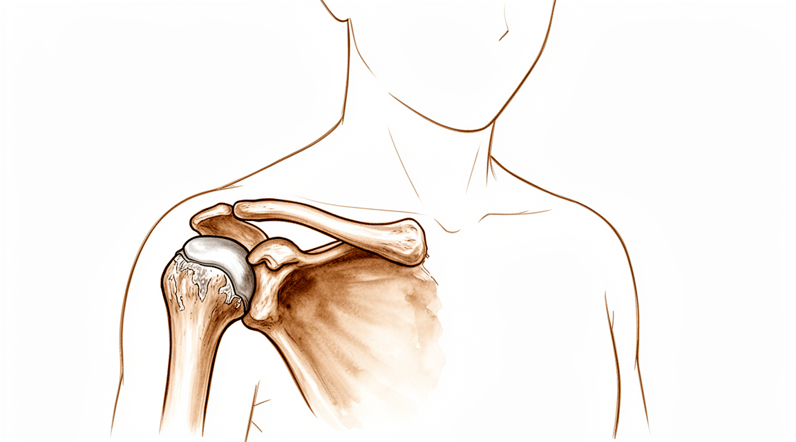
What you're feeling
The pain sits right at the top of your shoulder, at the small joint where your collarbone meets your shoulder blade. It can spread towards the front of your shoulder or up into the muscle that runs from your neck to your shoulder. Lifting anything heavy tends to bring it on. So does lying on that side at night.
Some people notice the pain most after activity, and some feel it when they first wake up. Reaching across your body, such as reaching for a seatbelt or lifting a bag onto the far side of a table, can hurt. Lifting your arm right up overhead can also cause pain at that spot on top of your shoulder.
Here is something worth knowing: arthritis seen on a scan does not always match how much it hurts. Some people have wear-and-tear arthritis at this joint on imaging and feel nothing at all. In fact, most people whose arthritis shows up on a scan but who have no pain stay pain-free over the years that follow. Others have a scan that looks mild but feel a lot of pain. What matters is your symptoms, not just the picture on the scan.
If your shoulder is painful, your surgeon will check for tenderness at that exact spot on top of your shoulder and test how your arm moves. Sometimes a numbing injection into the joint is used to confirm that this joint is the source of your pain. If the pain settles after the injection, that tells us where it is coming from.
What's actually happening
The joint at the top of your shoulder is where your collarbone meets your shoulder blade. It is a small, mobile joint, not a rigid one. It moves a little in every direction as your shoulder blade swings around your chest. Think of it as a small hinge that also slides and turns, letting the two bones stay in step with each other while your arm moves.
Between the two bones sits a soft pad, a bit like a gasket. It cushions the joint and keeps the surfaces gliding smoothly. Over time, this pad wears out. That is where the trouble starts. The joint surfaces begin to grind against each other, and the bone responds by growing small bony spurs at the edges. This is wear-and-tear arthritis.
This kind of arthritis usually shows up from early middle age onwards. It is more common in people who do a lot of repeated overhead work or heavy lifting. An old shoulder separation, even a mild one from years ago, can also lead to painful arthritis at this joint later on.
When the joint wears, it becomes sore exactly where you feel it: that spot on top of your shoulder. Lifting, reaching across your body and lying on that side all press the worn surfaces together, which is why those movements hurt. The bony spurs and the raw joint surfaces are the source of the pain described earlier.
Here is the part that matters most: what the scan shows does not decide what you feel. Many people have arthritis at this joint and never notice it. Most of them stay pain-free over the years that follow. Others have only mild-looking wear but real pain. Your symptoms, not the picture on the scan, are what guide any decision about treatment.
What we can do about it
Dr Kieran Hirpara, an upper-limb surgeon at Mater Private Hospital Rockhampton, starts with the least invasive options that suit your condition. Patients are generally referred to our clinic by their GP; if a physiotherapist has suggested you see us, you will still need a referral from your GP in order to be eligible for the Medicare rebate. At your first visit we take a history, examine your shoulder and arrange imaging if it is needed. Because this is a long-standing wear problem, we usually begin with non-operative care.
The first step is simple: rest from the activities that stir the pain up, and ice when it flares. Anti-inflammatory tablets, which reduce pain and swelling, are often used early on. Physiotherapy works on easing the joint back into comfortable movement and building up the muscles around your shoulder so they take some of the load off the joint. Give this a fair trial before thinking about anything further.
If those measures have not settled things, injections come next. A cortisone injection places anti-inflammatory medicine directly into the joint. It can help confirm where your pain is coming from, and it can also settle the pain itself. In about 47% of people the injection still counts as a success a year later. Not everyone gets lasting relief, but for some it buys a good stretch of comfort.
Surgery enters the picture when pain persists despite all of the above. The operation removes a small sliver of bone from the outer end of your collarbone, which stops the worn, spurred surfaces from grinding against each other. We would talk it through with you only once simpler treatments have had their chance, and the decision is always one we make together.
What to expect
For most people, this is a slow-moving condition rather than a sudden one. The pain tends to come and go with what you do: heavier activity stirs it up, easier weeks calm it down. Some people find it settles with simple measures and stays quiet for long stretches. Others find it grumbles along for months or years, flaring when they overdo it.
If you have no pain, the outlook is straightforward. Most people whose arthritis shows up on a scan but who have no symptoms stay symptom-free over seven years. Wear at this joint that you cannot feel usually stays that way.
If your shoulder is already painful, non-operative care is worth a fair trial. Rest, physiotherapy and anti-inflammatory tablets ease things for many people. A cortisone injection, as covered earlier, still counts as a success a year later for about half the people who have one. When an injection works, it does not seem to matter whether your scan showed a lot of wear or a little: people in both groups report the same level of satisfaction afterwards.
If pain persists despite all of that, surgery to remove a small sliver of bone from the outer end of your collarbone reliably takes the pain away and improves how your shoulder works. That relief holds up over the years that follow, not just for the first few months.
Left alone, painful arthritis at this joint does not usually damage anything else in your shoulder. Wear here that goes untreated does not stop other shoulder repairs from working, and it is not a reason to remove part of the collarbone during other shoulder surgery. The main risk of leaving it is simply that the pain continues.
One honest caveat: surgery is not a blank cheque. Every operation carries its own set of possible complications, and careful technique keeps those risks down rather than removing them altogether. We would talk through the realistic picture with you before any decision, and the choice is always one we make together.
When to see someone
See your GP if the pain at the top of your shoulder keeps coming back, especially with lifting, reaching across your body or lying on that side. Ask for a specialist review if simple measures have not settled it after a fair trial, or if the pain is disturbing your sleep or stopping you working. It is worth getting checked even sooner if the pain is sharp and focused on that one small spot at the top of your shoulder, because tenderness right there is a useful clue about where the problem sits. This condition is not an emergency, and nothing about it needs same-day care. But pain that will not settle deserves a proper look, because the treatments described earlier work best before the problem has worn you down for years.
In more depth
Advanced reading: the deeper science (optional)
This section goes further than you need for your own treatment decisions. Arthritis of the AC joint is worth the extra reading for an uncomfortable reason: it is extremely common on imaging, it is frequently blamed for shoulder pain, and the evidence that treating it helps is weaker than almost anything else on this site.
The evidence does not establish that anything works
A systematic review of the treatment of primary AC joint osteoarthritis pooled 1,902 patients and reached a conclusion that is rarely stated so plainly: the studies varied in indication, intervention and quality, and did not provide evidence that either non-operative or operative interventions are effective [1].
That is not the same as saying nothing helps. It means the trials that would demonstrate it have not been done to a standard that allows the claim. Distal clavicle excision is a long-established operation performed on the reasonable mechanical logic that removing the worn joint surface removes the pain, but "long-established and mechanically sensible" is not evidence, and it is worth knowing the difference when a decision is being made.
Adding it to another operation does not improve that operation
The most direct test comes from patients having something else done at the same time. Among 208 patients with rotator cuff tears, adding distal clavicle resection did not produce better clinical outcome scores or better range of motion [2].
This matters because that is exactly the circumstance in which the joint is most often resected, the surgeon is already inside the shoulder, the AC joint looks degenerate on imaging, and removing a few millimetres of bone is quick. The evidence says the patient does not measurably benefit. If it is proposed as an add-on to your cuff repair, that is a fair thing to ask about.
Technique is not the interesting question
Open versus arthroscopic resection has been compared repeatedly and the answer is consistent: similar functional and clinical outcomes with either approach across 319 patients [3], with an earlier comparison of 429 patients also favouring neither decisively [4].
When two quite different technical approaches produce the same result, the honest inference is that the technique is not what determines the outcome, patient selection is.
Why selection is so difficult here
The AC joint degenerates in almost everyone with age. It is a small, highly loaded joint with a thin disc that wears early, so a report describing AC joint degeneration on your scan is close to an expected finding after middle age rather than a diagnosis.
There is a measurable structural correlate: in symptomatic degenerative AC joints, both the distal clavicle and the acromion are enlarged, whereas in asymptomatic people the relationship between the two is unchanged [5]. That is a useful hint that symptomatic and incidental degeneration differ structurally, but it is a group-level observation, not a test that can be applied to your shoulder.
Practically, this is why a diagnostic injection into the joint carries real weight here. If local anaesthetic placed accurately into the AC joint abolishes the pain, the joint is likely the source. If it does not, degeneration on the scan is probably a bystander, and resecting it is unlikely to help, which is the situation the evidence above is warning about.
References for the advanced reading
- Welch M, Rankin S, How Saw Keng M, Woods D. A systematic review of the treatment of primary acromioclavicular joint osteoarthritis. Shoulder Elbow. 2023;16(2):129-44.
- Wang J, Ma J, Zhu S, Jia H, Ma X. Does distal clavicle resection decrease pain or improve shoulder function in patients with rotator cuff tears? A meta-analysis. Clin Orthop Relat Res. 2018;476(12):2402-14.
- Hohmann E, Tetsworth K, Glatt V. Open versus arthroscopic acromioclavicular joint resection: a systematic review and meta-analysis. Arch Orthop Trauma Surg. 2019;139(5):685-94.
- Pensak M, Grumet RC, Slabaugh MA, Bach BR. Open versus arthroscopic distal clavicle resection. Arthroscopy. 2010;26(5):697-704.
- Bulkmans K, Peeters I, De Wilde L, Van Tongel A. The relationship of the acromion to the distal clavicle in normal and symptomatic degenerative acromioclavicular joints. Arch Orthop Trauma Surg. 2019;140(4):465-72.
Evidence & references
This is the clinical evidence summary written for health professionals. It is technical, and it lists the research this page was built from. You do not need to read it to understand your treatment or to make a decision about it.
Overview
- Asymptomatic acromioclavicular osteoarthritis diagnosed by MRI remained asymptomatic in 90% of cases over a seven-year period [3].
- Symptomatic acromioclavicular joint osteoarthritis occurred in 15.9% of patients following total anatomic shoulder replacement with follow-up of up to 12 years [8].
- Treatment choice for acromioclavicular dislocation should not be influenced by the potential development of acromioclavicular osteoarthritis [1].
- Untreated acromioclavicular joint osteoarthritis, whether symptomatic or not, encountered during arthroscopic rotator cuff repair is associated with a low percentage of failure [4].
- Acromioclavicular joint arthritis is not an indication for routine distal clavicle excision in arthroscopic rotator cuff repair [4].
- Further characterisation of patients in whom mild arthroscopic findings of acromioclavicular joint osteoarthritis are clinically significant and warrant resection is needed [2].
- A distinction between symptomatic and asymptomatic radiographic acromioclavicular osteoarthritis appears unnecessary, as all patients were equally satisfied with the outcome following a preoperative acromioclavicular injection [7].
- Limited distal clavicle excision in patients with acromioclavicular joint osteoarthritis resistant to conservative treatment reduced pain and improved shoulder function at midterm follow-up [10].
- Both open and arthroscopic resection arthroplasty techniques provide predictable pain relief for symptomatic acromioclavicular osteoarthritis [14].
- Open and arthroscopic resection arthroplasty techniques each have a unique set of potential complications that may be minimized with improved understanding of anatomy, biomechanics, and meticulous surgical technique [14].
- Biplanar reconstruction with pectoralis minor tendon and coracoacromial ligament transfer aims to preserve the lateral portion of the clavicle whenever possible [9].
- In biplanar reconstruction for chronic acromioclavicular joint dislocations, resection is limited exclusively to cases of acromioclavicular joint osteoarthritis or irreducible joints [9].
- Clinicians should consider overreduction of the acromioclavicular joint because it may lead to favorable radiological results in acute high-grade dislocations [6].
Anatomy & Pathophysiology
Joint Structure and Biomechanics
- The acromioclavicular (AC) joint is a diarthrodial joint that supports the shoulder girdle through the clavicular “strut” [29].
- The AC joint serves as a primary link between the axial skeleton and the upper extremity [67].
- The AC joint is movable in all planes and is not a rigid structure [67].
- The normal AC joint is capable of translating 4 to 6 mm in the anterior, posterior, and superior planes under 70-N loads [67].
- The AC joint accommodates rotary motion of 5° to 8° during scapulothoracic motion and 40° to 45° with shoulder abduction and elevation [67].
- The AC and coracoclavicular (CC) ligaments are the static stabilizers of the AC joint, whereas the deltoid and trapezius muscles are the dynamic stabilizers [67].
- The horizontal plane stability of the clavicle is provided by the AC ligaments, specifically the posterior and superior portions [15].
- The scapula and clavicle are linked into a single segment referred to as the “claviscapula” [69].
- The AC and CC ligaments play a role in torque transduction and horizontal and vertical stability within the scapulohumeral rhythm [69].
- Kinematic changes resulting from AC joint dislocation can be a potential source of pain and dysfunction in the shoulder [33].
- Scapular and clavicular kinematics are affected in AC separation models [34].
Ligamentous Anatomy and Injury Progression
- Injury to the AC joint results in progressive disruption of the ligamentous support, beginning with the capsular ligaments and progressing to the CC ligaments [16].
- The extent of injury to the AC and CC ligaments, as well as the amount and direction of clavicle displacement, often determines the severity of AC joint separation [41].
- Anatomic techniques that address both coracoclavicular ligaments and the AC capsule are recommended to restore horizontal and vertical stability while allowing physiological rotation [11].
- Although various reconstruction techniques can restore different elements of joint kinematics, none completely restores the shoulder girdle to its preinjured state [31].
Pathoanatomy of Degenerative Conditions
- AC joint osteoarthritis is more common with advanced age following degeneration of the intra-articular disk [15].
- Arthritic deterioration of the AC joint starts in early middle age [15].
- AC joint osteoarthritis is more common in patients engaged in repetitive overhead or lifting activities [15].
- Previous low-grade AC joint separations can result in painful arthritis [15].
- The radiographic severity of AC joint arthritis does not always correlate with patient symptoms [15].
- Asymptomatic AC joint degeneration is frequent and does not always correlate with the presence of symptoms [29].
- Distal clavicle osteolysis involves localized hyperemia of the distal clavicle, resulting in inflammation, bone resorption, microfractures, and secondary arthritis of the AC joint [15].
- Distal clavicle osteolysis is more common in males and seen in younger patients [15].
- Distal clavicle osteolysis is associated with heavy lifting or repetitive motions [15].
Radiographic Anatomy and Normal Values
- The normal coracoclavicular (CC) distance on an AP radiograph should be less than 11 to 13 mm [15].
- The normal coracoclavicular distance, measured between the superior aspect of the coracoid and the inferior clavicle, should be between 11 to 13 mm [16].
- Bone and joint edema on MRI correlate with AC joint pain [15].
- Osteophyte formation, sclerotic reaction, and bone cysts are commonly seen on radiographs of the AC joint in osteoarthritis [15].
Classification
- AC joint osteoarthritis is graded using the Kellgren-Lawrence classification system [61].
- Cuff tear arthropathy is graded using the Hamada classification system [61].
- Radiographic signs of AC joint osteoarthritis include joint space narrowing, osteophytosis, and cyst formation [12].
- Asymptomatic AC joint osteoarthritis is defined by the presence of radiographic signs of osteoarthritis without clinical symptoms such as AC joint tenderness or a positive cross-body adduction test [12].
- Symptomatic AC joint osteoarthritis is characterized by AC joint pain, tenderness to palpation, and temporary pain relief with intra-articular injection [12].
- The Rockwood classification is used to evaluate acute acromioclavicular dislocations, including types III and IV [64].
- The Rockwood classification includes types III through V for severe chronic symptomatic AC joint separations [51].
- The Rockwood classification includes type V for high-grade acromioclavicular dislocations [19].
- The AC-DC measurement and Alexander view are used to define watershed cases of AC joint displacement, specifically types IIIA, IIB, and IV [18].
- Evaluating the integrity of capsuloligamentous structures stabilizing the AC joint provides information on injury severity that may influence treatment decisions [28].
Clinical Presentation
History and Symptoms
- Patients report activity-related pain localized to the AC joint [15].
- Pain may radiate anteriorly or along the trapezius [15].
- Pain is reported with heavy lifting or when sleeping on the affected side [15].
- Asymptomatic AC-OA remained asymptomatic in 90% of patients over a 7-year course [3].
- Symptomatic ACJ OA occurred in 15.9% of patients after total anatomic shoulder replacement with follow-up of up to 12 years [8].
- Radiographic severity of arthritis does not always correlate with patient symptoms [15].
Physical Examination
- Point tenderness is seen at the AC joint [15].
- Horizontal stability should be assessed during physical examination [15].
- Pain at the AC joint with terminal elevation and cross-body motion is often seen [15].
- Selective injection of anesthetic into the AC joint can confirm the diagnosis [15].
- Tenderness to palpation at the AC joint is a risk factor for subsequent distal clavicle resection after rotator cuff repair [26].
- Female sex is a risk factor for subsequent distal clavicle resection after rotator cuff repair [26].
- Surgery on the dominant side is a risk factor for subsequent distal clavicle resection after rotator cuff repair [26].
- Subsequent distal clavicle resection was performed in 40% of cases with a combination of AC joint tenderness, female sex, and surgery on the dominant side [26].
Imaging
- An AP view and/or a Zanca view of the shoulder provides good visualization of the AC joint [15].
- Osteophyte formation, sclerotic reaction, and bone cysts are commonly seen on radiographs [15].
- CT imaging signs of osteoarthritis at the AC joint include joint space narrowing, osteophytosis, and cyst formation [12].
- Radiographic ACJ osteoarthritis is common in patients undergoing reverse shoulder arthroplasty [13].
Investigations
Clinical Evaluation
- Patients with AC joint osteoarthritis report activity-related pain localized to the AC joint, with occasional radiation anteriorly or along the trapezius [15].
- Pain with heavy lifting or when sleeping on the affected side is reported in patients with AC joint osteoarthritis [15].
- Physical examination of symptomatic AC joint osteoarthritis includes point tenderness at the AC joint [15].
- Horizontal stability of the AC joint should be assessed during physical examination [15].
- Pain at the AC joint with terminal elevation and cross-body motion is often seen in patients with AC joint osteoarthritis [15].
- Selective injection of anesthetic into the AC joint can confirm the diagnosis of symptomatic AC joint osteoarthritis [15].
Imaging
- An AP view and/or a Zanca view of the shoulder provides good visualization of the AC joint for evaluating osteoarthritis [15].
- Osteophyte formation, sclerotic reaction, and bone cysts are commonly seen on radiographs of the AC joint in patients with osteoarthritis [15].
- Patients with edema on MRI are more likely to present pain than patients without edema [60].
- Subchondral bone edema on histologic examination is more frequent in patients with pain [60].
- The radiographic severity of arthritis does not always correlate with patient symptoms [15].
- The AC-DC measurement and use of the Alexander view provides a more realistic appreciation of true AC joint displacement, especially in defining watershed cases (ie, IIIA/IIB/IV) [18].
- Normal coracoclavicular distance on an AP radiograph should be less than 11 to 13 mm [15].
- The normal CC distance on an AP radiograph should be between 11 to 13 mm [16].
Diagnostic Injections and Prognostic Indicators
- A distinction between symptomatic and asymptomatic radiographic AC OA is unnecessary, as all patients were equally satisfied with the outcome following preoperative acromioclavicular injection [7].
- Asymptomatic AC-OA remained asymptomatic in 90% of patients over 7 years [3].
- Untreated ACJ osteoarthritis, symptomatic or not, encountered during arthroscopic rotator cuff repair is associated with a low percentage of failure [4].
- Preventive arthroscopic distal clavicle resection is not recommended in patients with radiological but asymptomatic ACJ arthritis [65].
- Further characterisation of patients in whom mild arthroscopic findings of OA of AC joint are clinically significant and warrant resection is needed [2].
Treatment
Non-Operative Management
- Rest, ice, and NSAIDs are used initially for AC joint osteoarthritis [15].
- Corticosteroid injections can be used for diagnostic or therapeutic purposes in AC joint osteoarthritis [15].
- Conservative and surgical treatments are both effective in acromioclavicular joint osteoarthritis management [22].
- AC injections offer a 1-year success rate of 47% in patients with acromioclavicular osteoarthritis [27].
- A distinction between symptomatic and asymptomatic radiographic AC OA is unnecessary, as all patients were equally satisfied with the outcome following preoperative injection [7].
- Additional research is needed to determine the main cause of pain and compare clinical outcomes of intra-articular versus extra-articular injections for isolated acromioclavicular osteoarthritis [24].
- Asymptomatic AC-OA remained asymptomatic in 90% over 7 years [3].
- Non-operative reduction and stabilization of high-grade AC joint separations seems to be a valuable treatment option [46].
- Type V AC dislocations may be given a trial of conservative therapy [19].
Operative Management
- Surgical indications for AC joint osteoarthritis include persistent pain and failure of nonsurgical treatment [15].
- Relative contraindications for surgical treatment of AC joint osteoarthritis include a previous low-grade separation with persistent horizontal plane instability [15].
- Biomechanical evidence suggests a resection of 5 mm is needed to prevent contact between the clavicle and the acromion in the absence of instability during arthroscopic distal clavicle excision [15].
- Care should be taken to preserve the posterior and superior AC ligaments during arthroscopic distal clavicle excision [15].
- Pain relief is reliable in >90% of patients following arthroscopic distal clavicle excision in the absence of instability [15].
- Previous traumatic instability is associated with persistent pain in 30% to 40% of cases following distal clavicle excision [15].
- One systematic review showed slightly better results with arthroscopic excision than with open distal clavicle excision [15].
- Direct comparison studies have shown similar or better results with arthroscopic excision than with open techniques for distal clavicle resection [15].
- Between 5 and 10 mm of the distal clavicle should be resected during an open distal clavicle resection (Mumford procedure) [15].
- Meticulous repair of the deltotrapezial fascia is important during open distal clavicle resection [15].
- Limited distal clavicle excision of patients with AC joint osteoarthritis resistant to conservative treatment reduced pain and improved shoulder function at midterm follow-up [10].
- The biplanar reconstruction procedure aims to preserve the lateral portion of the clavicle whenever possible, limiting resection exclusively to cases of AC joint osteoarthritis or irreducible joints [9].
- Some persistent pain and osteoarthritis progression remain concerns following revision of failed open anterior stabilization of the shoulder [5].
- Clinicians should consider overreduction of the AC joint because it may lead to favorable radiological results in acute high-grade dislocations [6].
Rehabilitation
- Acute rehabilitation (zero to 7 days postoperative) for distal clavicle excision includes a sling, ice, and pendulum exercises [15].
- Subacute rehabilitation (1 to 6 weeks postoperative) for distal clavicle excision involves gradually increasing shoulder ROM, gentle passive stretching and ROM as tolerated, reducing sling use as pain permits, and avoiding heavy lifting or strengthening exercises [15].
- Late recovery (more than 6 weeks postoperative) for distal clavicle excision involves full shoulder ROM and stretching, initiation of rotator cuff, scapular stabilizer, and deltoid strengthening, and heavy weight lifting and return to full activities as tolerated [15].
- Residual pain or soreness can persist for 3 to 4 months after distal clavicle excision and can be aggravated by heavy lifting [15].
- Activity progression after distal clavicle excision should be modified according to symptoms [15].
Complications
Post-operative Osteoarthritis and Joint Degeneration
- Patients with loss of immediate postoperative reduction after AC joint dislocation repair more often developed radiologic and symptomatic AC osteoarthritis than those in whom the AC joint was anatomically restored [17].
- Osteoarthritis is associated with poorer final clinical outcomes following rotator cuff repair [48].
- An unhealed or re-torn rotator cuff increases the risk of osteoarthritis following rotator cuff repair [48].
- Severe acromioclavicular joint osteoarthritis is associated with acromial stress fractures after reverse shoulder arthroplasty [13].
Surgical Technique and Stability Complications
- Clinicians should consider overreduction of the AC joint because it may lead to favorable radiological results [6].
- Both open and arthroscopic resection arthroplasty techniques provide predictable pain relief for symptomatic AC osteoarthritis, though each has a unique set of potential complications that may be minimized with improved understanding of anatomy, biomechanics, and meticulous surgical technique [14].
Natural History and Diagnostic Considerations
- Untreated ACJ osteoarthritis, symptomatic or not, encountered during arthroscopic RCR is associated with a low percentage of failure [4].
- The outcomes of a retrospective study suggest that a distinction between symptomatic and asymptomatic radiographic AC OA is unnecessary, as all patients were equally satisfied with the outcome [7].
Recovery
Non-Operative
- Asymptomatic acromioclavicular osteoarthritis diagnosed by MRI remained asymptomatic in 90% of cases over a 7-year period [3].
- Acromioclavicular injections offer a 1-year success rate of 47% in patients with acromioclavicular osteoarthritis [27].
- Outcomes suggest that a distinction between symptomatic and asymptomatic radiographic acromioclavicular osteoarthritis is unnecessary, as all patients were equally satisfied with the outcome following preoperative injection [7].
- Type V acromioclavicular dislocations may be given a trial of conservative therapy [19].
Operative
- Symptomatic acromioclavicular joint osteoarthritis occurred in 15.9% of patients after total anatomic shoulder replacement with follow-up of up to 12 years [8].
- Osteoarthrotic changes were always associated with fair or poor results in surgical treatment of acromioclavicular dislocation [30].
- Treatment of acromioclavicular dislocation by synthetic ligament reconstruction gave satisfactory results in terms of recovery of strength, but evolution is not risk-free with the onset of significant early osteolysis [70].
- Outcomes after arthroscopic coracoclavicular reconstruction for chronic, type V acromioclavicular dislocations show sustained and statistically significant improvements in functional outcomes as well as high rates of return to sport and maintenance of active-duty military status at long-term follow-up [56].
- Short-term follow-up of patients treated with minimally invasive coracoclavicular ligament augmentation for total acromioclavicular joint dislocation reveals excellent radiologic and clinical results, with no subluxations or dislocations of the acromioclavicular joint noted [68].
General Principles
- Choice of treatment for acromioclavicular dislocation should not be influenced by the potential development of acromioclavicular osteoarthritis [1].
- Anatomic techniques that address both coracoclavicular ligaments and the acromioclavicular capsule are recommended to restore horizontal and vertical stability while allowing physiological rotation [11].
- No long term disability results from the loss of the coraco-acromial ligament from its normal site [50].
Key Evidence
- [L4] Based on the available evidence, treatment choice for AC dislocation should not be influenced by the potential development of AC OA. [1] (10.2106/jbjs.rvw.24.00085)
- [L2] Further characterisation of patients in whom mild arthroscopic findings of OA of AC joint are clinically significant and warrant resection is needed. [2] (10.1007/s00167-014-3114-2)
- [L2] Asymptomatic AC-OA remained asymptomatic in 90% over 7 years. [3] (10.1016/j.jse.2019.04.004)
- [L2] Untreated ACJ osteoarthritis, symptomatic or not, encountered during arthroscopic RCR is associated with a low percentage of failure. [4] (10.1007/s00167-020-06098-y)
- [L4] Some persistent pain and osteoarthritis progression remain concerns. [5] (10.1016/j.arthro.2009.04.073)
- [L3] Clinicians should consider overreduction of the AC joint because it may lead to favorable radiological results. [6] (10.1177/0363546519862850)
- [L4] The outcomes of this study seem to suggest that a distinction between symptomatic and asymptomatic radiographic AC OA is unnecessary, as all patients were equally satisfied with the outcome. [7] (10.5397/cise.2023.00073)
- [L4] Symptomatic ACJ OA occurred in 15.9% of patients after total anatomic shoulder replacement with follow-up of up to 12 years. [8] (10.1177/17585732221114796)
- [L5] The procedure aims to preserve the lateral portion of the clavicle whenever possible, limiting resection exclusively to cases of AC joint osteoarthritis or irreducible joints. [9] (10.1016/j.eats.2024.103104)
- [L4] Limited distal clavicle excision of patients with AC joint osteoarthritis resistant to conservative treatment reduced pain and improved shoulder function at midterm follow-up. [10] (10.1016/j.otsr.2016.01.008)
- [L5] Anatomic techniques that address both coracoclavicular ligaments and the AC capsule are recommended to restore horizontal and vertical stability while allowing physiological rotation. [11] (10.1016/j.arthro.2019.01.038)
- [L3] [12] (10.1007/s00402-019-03258-9)
- [L3] Radiographic ACJ osteoarthritis is common in patients undergoing RSA. [13] (10.1016/j.jseint.2021.11.008)
- [L5] Both open and arthroscopic resection arthroplasty techniques provide predictable pain relief for symptomatic AC osteoarthritis, though each has a unique set of potential complications that may be minimized with improved understanding of anatomy, biomechanics, and meticulous surgical technique. [14] (10.1177/0363546513485359)
- [L4] [17] (10.1016/j.otsr.2017.11.001)
- [L4] The AC-DC measurement and use of the Alexander view provides the clinician with a more realistic appreciation of true AC joint displacement, especially in defining watershed cases (ie, IIIA/IIB/IV) and may better inform the decision-making process regarding management options and recommendations. [18] (10.1016/j.jse.2019.12.014)
- [L4] This suggests that Type V AC dislocations may be given a trial of conservative therapy. [19] (10.1177/2325967115s00017)
- [L4] Conservative and surgical treatments are both effective in acromioclavicular joint osteoarthritis management. [22] (10.1007/s00167-020-06377-8)
- [L4] Additional research is needed to determine the main cause of pain and compare clinical outcomes of intra-articular versus extra-articular injections. [24] (10.5397/cise.2023.00311)
- [L3] Risk factors for subsequent DCE included tenderness to palpation at the AC joint, female sex, and surgery on the dominant side, with subsequent DCE performed in 40% of cases with a combination of these 3 factors. [26] (10.1177/2325967119844295)
- [L4] AC injections offer a 1-year success rate of 47%. [27] (10.5397/cise.2023.00031)
- [L1] Evaluating the integrity of the capsuloligamentous structures stabilizing the AC joint is reproducible and gives additional information on the severity of the injury, which might also influence the treatment decision. [28] (10.1016/j.jse.2020.10.026)
- [L5] [29] (10.5435/00124635-199905000-00004)
- [L4] Osteoarthrotic changes, however, were always associated with fair or poor results. [30] (10.1016/0020-1383(83)90092-x)
- [L5] Although each technique was able to restore different elements of the joint kinematics, none of the strategies completely restored the shoulder girdle to its preinjured state. [31] (10.1177/03635465221095231)
- [L5] The kinematic changes could be a potential source of pain and dysfunction in the shoulder with AC joint dislocation. [33] (10.1177/0363546512458571)
- [L5] Scapular and clavicular kinematics were affected in AC separation models. [34] (10.1016/j.jse.2013.01.004)
- [L4] Non-operative reduction and stabilization of high-grade AC joint separations seems to be a valuable treatment option. [46] (10.1007/s00402-020-03630-0)
- [L4] Osteoarthritis is associated with poorer final clinical outcomes, and an unhealed or re-torn cuff increases the risk of osteoarthritis. [48] (10.1016/j.otsr.2017.03.007)
- [L4] No long term disability results from the loss of the coraco-acromial ligament from its normal site. [50] (10.1016/s0020-1383(80)80045-3)
- [L4] Severe chronic symptomatic AC joint separations (Rockwood types III through V) can be repaired entirely by arthroscopy safely and effectively by transferring the coracoacromial ligament with a bone block in the distal clavicle. [51] (10.1016/j.arthro.2009.08.008)
- [L4] Outcomes after arthroscopic CC reconstruction for chronic, type V AC dislocations in an active-duty military patient population show sustained and statistically significant improvements in functional outcomes as well as high rates of return to sport and maintenance of active-duty military status at long-term follow-up. [56] (10.1016/j.arthro.2025.05.008)
- [L4] Patients with edema on MRI were more likely to present pain than patients without edema, and subchondral bone edema on histologic examination was more frequent in patients with pain. [60] (10.1016/j.jseint.2020.03.007)
- [L3] [61] (10.5397/cise.2023.00465)
- [L4] [64] (10.1016/j.otsr.2010.10.004)
- [L1] Preventive arthroscopic DCR is not recommended in patients with radiological but asymptomatic ACJ arthritis. [65] (10.1177/0363546514547254)
- [L4] [67] (10.1302/2058-5241.3.170027)
- [L4] The short-term follow-up of 15 recently operated patients reveals excellent radiologic and clinical results, with no subluxations or dislocations of the acromioclavicular joint noted. [68] (10.1016/j.arthro.2006.12.015)
- [L5] [69] (10.5435/jaaos-d-24-00360)
- [L4] Treatment of ACD by synthetic ligament reconstruction gave satisfactory results, notably in terms of recovery of strength, but evolution is not risk-free with onset of significant early osteolysis. [70] (10.1016/j.otsr.2010.06.004)
References
[1] Choice of Acromioclavicular Dislocation Treatment Should Not Be Influenced by Risk of Development of Acromioclavicular Osteoarthritis. JBJS Reviews. 2024. DOI: 10.2106/jbjs.rvw.24.00085
[2] Outcome of distal clavicle resection in patients with acromioclavicular joint osteoarthritis and full‐thickness rotator cuff tear. Knee Surgery, Sports Traumatology, Arthroscopy. 2014. DOI: 10.1007/s00167-014-3114-2
[3] Seven-year course of asymptomatic acromioclavicular osteoarthritis diagnosed by MRI. Journal of Shoulder and Elbow Surgery. 2019. DOI: 10.1016/j.jse.2019.04.004
[4] Acromioclavicular joint arthritis is not an indication for routine distal clavicle excision in arthroscopic rotator cuff repair. Knee Surgery, Sports Traumatology, Arthroscopy. 2020. DOI: 10.1007/s00167-020-06098-y
[5] The Role of Arthroscopy in Revision of Failed Open Anterior Stabilization of the Shoulder. Arthroscopy. 2009. DOI: 10.1016/j.arthro.2009.04.073
[6] Factors Predicting the Outcome After Arthroscopically Assisted Stabilization of Acute High-Grade Acromioclavicular Joint Dislocations. The American Journal of Sports Medicine. 2019. DOI: 10.1177/0363546519862850
[7] Diagnostic value of a preoperative acromioclavicular injection for symptomatic acromioclavicular osteoarthritis: a retrospective study of cross-sectional midterm outcomes. Clinics in Shoulder and Elbow. 2024. DOI: 10.5397/cise.2023.00073
[8] The incidence and treatment of symptomatic acromioclavicular joint osteoarthritis following total shoulder arthroplasty. Shoulder & Elbow. 2022. DOI: 10.1177/17585732221114796
[9] Biplanar Reconstruction With Pectoralis Minor Tendon and Coracoacromial Ligament Transfer for Chronic Acromioclavicular Joint Dislocations. Arthroscopy Techniques. 2024. DOI: 10.1016/j.eats.2024.103104
[10] Limited distal clavicle excision of acromioclavicular joint osteoarthritis. Orthopaedics & Traumatology: Surgery & Research. 2016. DOI: 10.1016/j.otsr.2016.01.008
[11] Editorial Commentary: Why We Have To Respect The Anatomy In Acromioclavicular Joint Surgery And Why Clinical Shoulder Scores Might Not Give Us The Information We Need!. Arthroscopy. 2019. DOI: 10.1016/j.arthro.2019.01.038
[12] The relationship of the acromion to the distal clavicle in normal and symptomatic degenerated acromioclavicular joints. Archives of Orthopaedic and Trauma Surgery. 2019. DOI: 10.1007/s00402-019-03258-9
[13] Severe acromioclavicular joint osteoarthritis is associated with acromial stress fractures after reverse shoulder arthroplasty. JSES International. 2022. DOI: 10.1016/j.jseint.2021.11.008
[14] Degenerative Joint Disease of the Acromioclavicular Joint. The American Journal of Sports Medicine. 2013. DOI: 10.1177/0363546513485359
[15] Aaos Comprehensive Orthopaedic Review 3. Disorders of the Acromioclavicular Joint > III. Atraumatic and Degenerative Conditions of the AC Joint.
[16] Aaos Comprehensive Orthopaedic Review 3. Disorders of the Acromioclavicular Joint > II. Traumatic Conditions of the AC Joint.
[17] Arthroscopically assisted reduction of acute acromioclavicular joint dislocation using a single double-button device: Medium-term clinical and radiological outcomes. Orthopaedics & Traumatology: Surgery & Research. 2018. DOI: 10.1016/j.otsr.2017.11.001
[18] Improved identification of unstable acromioclavicular joint injuries in a clinical population using the acromial center line to dorsal clavicle radiographic measurement. Journal of Shoulder and Elbow Surgery. 2020. DOI: 10.1016/j.jse.2019.12.014
[19] Successful Conservative Therapy in Rockwood Type V Acromioclavicular Dislocations. Orthopaedic Journal of Sports Medicine. 2015. DOI: 10.1177/2325967115s00017
[22] No differences between conservative and surgical management of acromioclavicular joint osteoarthritis: a scoping review. Knee Surgery, Sports Traumatology, Arthroscopy. 2021. DOI: 10.1007/s00167-020-06377-8
[24] Isolated acromioclavicular osteoarthritis and steroid injection. Clinics in Shoulder and Elbow. 2023. DOI: 10.5397/cise.2023.00311
[26] Preoperative Factors Associated With Subsequent Distal Clavicle Resection After Rotator Cuff Repair. Orthopaedic Journal of Sports Medicine. 2019. DOI: 10.1177/2325967119844295
[27] Mid- to long-term success rate and functional outcomes of acromioclavicular injections in patients with acromioclavicular osteoarthritis. Clinics in Shoulder and Elbow. 2023. DOI: 10.5397/cise.2023.00031
[28] The ligamentous injury pattern in acute acromioclavicular dislocations and its impact on clinical and radiographic parameters. Journal of Shoulder and Elbow Surgery. 2021. DOI: 10.1016/j.jse.2020.10.026
[29] Painful Conditions of the Acromioclavicular Joint. Journal of the American Academy of Orthopaedic Surgeons. 1999. DOI: 10.5435/00124635-199905000-00004
[30] Surgical treatment of acromioclavicular dislocation: A review of 39 patients. Injury. 1983. DOI: 10.1016/0020-1383(83)90092-x
[31] Differences between Coracoclavicular, Acromioclavicular, or Combined Reconstruction Techniques on the Kinematics of the Shoulder Girdle. The American Journal of Sports Medicine. 2022. DOI: 10.1177/03635465221095231
[33] The Function of the Acromioclavicular and Coracoclavicular Ligaments in Shoulder Motion. The American Journal of Sports Medicine. 2012. DOI: 10.1177/0363546512458571
[34] Acromioclavicular joint ligamentous system contributing to clavicular strut function: a cadaveric study. Journal of Shoulder and Elbow Surgery. 2013. DOI: 10.1016/j.jse.2013.01.004
[41] Orthopaedic Knowledge Update Sports Medicine 6. Disorders of the Acromioclavicular Joint, Sternoclavicular Joint, and Clavicle > AC Joint Injuries.
[46] Can an acute high-grade acromioclavicular joint separation be reduced and stabilized without surgery? A surgeon’s experience. Archives of Orthopaedic and Trauma Surgery. 2020. DOI: 10.1007/s00402-020-03630-0
[48] Osteoarthritis after rotator cuff repair: A 10-year follow-up study. Orthopaedics & Traumatology: Surgery & Research. 2017. DOI: 10.1016/j.otsr.2017.03.007
[50] Disruption of the acromioclavicular joint: surgical anatomy and biological reconstruction. Injury. 1980. DOI: 10.1016/s0020-1383(80)80045-3
[51] All‐Arthroscopic Weaver‐Dunn‐Chuinard Procedure With Double‐Button Fixation for Chronic Acromioclavicular Joint Dislocation. Arthroscopy. 2009. DOI: 10.1016/j.arthro.2009.08.008
[56] Arthroscopic Coracoclavicular Ligament Reconstruction With Double‐Bundle Soft Tissue Allograft for Chronic Type V Acromioclavicular Dislocations Shows Excellent Patient Outcomes and Return to Duty and Sport at Minimum 10‐Year Follow‐Up. Arthroscopy. 2025. DOI: 10.1016/j.arthro.2025.05.008
[60] Histologic and magnetic resonance image evaluation in acromioclavicular joint osteoarthritis. JSES International. 2020. DOI: 10.1016/j.jseint.2020.03.007
[61] Concomitant open distal clavicle excision is associated with greater improvement in range of motion without increased risk of acromial stress fracture after reverse total shoulder arthroplasty: a retrospective cohort study. Clinics in Shoulder and Elbow. 2023. DOI: 10.5397/cise.2023.00465
[64] Endoscopically assisted reconstruction of acute acromioclavicular joint dislocation using a synthetic ligament. Outcomes at 12 months. Orthopaedics & Traumatology: Surgery & Research. 2011. DOI: 10.1016/j.otsr.2010.10.004
[65] Is Arthroscopic Distal Clavicle Resection Necessary for Patients With Radiological Acromioclavicular Joint Arthritis and Rotator Cuff Tears?. The American Journal of Sports Medicine. 2014. DOI: 10.1177/0363546514547254
[67] Acromioclavicular joint injuries: diagnosis, classification and ligamentoplasty procedures. EFORT Open Reviews. 2018. DOI: 10.1302/2058-5241.3.170027
[68] Minimally Invasive Coracoclavicular Ligament Augmentation With a Flip Button/Polydioxanone Repair for Treatment of Total Acromioclavicular Joint Dislocation. Arthroscopy. 2007. DOI: 10.1016/j.arthro.2006.12.015
[69] Effect of Acromioclavicular Joint Injuries on the Acromioclavicular Joint Complex and Scapulohumeral Rhythm: A Functional and Mechanical Perspective. Journal of the American Academy of Orthopaedic Surgeons. 2025. DOI: 10.5435/jaaos-d-24-00360
[70] Acute grade III and IV acromioclavicular dislocations: Outcomes and pitfalls of reconstruction procedures using a synthetic ligament. Orthopaedics & Traumatology: Surgery & Research. 2010. DOI: 10.1016/j.otsr.2010.06.004