Ang iyong nararamdaman
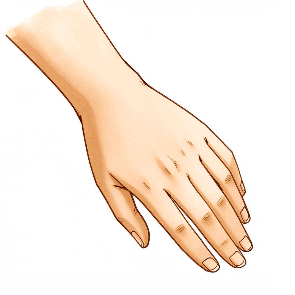
Ang hook of hamate fracture ay nangyayari sa isang iglap. Ang karaniwang sanhi ay isang direktang tama sa panig ng palad ng iyong kamay, malapit sa base ng iyong hinliliit. Karaniwan ang pinsalang ito sa mga manlalaro ng baseball at golf, kung saan ang bat, club o raketa ay dumidiin nang malakas sa lugar na iyon. Maaari rin itong idulot ng isang biglaang puwersa na dumadaan sa mga ligament at tendon ng palad.
Kaagad, nakakaramdam ka ng sakit sa panig ng palad ng iyong kamay, sa base malapit sa iyong hinliliit. Masakit ang lugar kapag hinahawakan. Sa karamihan ng mga bagong bali (acute fracture), palaging naroroon ang pananakit na ito kapag dinidiinan. Masakit ang pagtulak o paghila gamit ang iyong kamay, kaya nagiging mahirap ang paghawak sa doorknob, paghawak ng tasa ng kape o pagpihit ng takip ng garapon. Maaari mong mapansin ang pamamaga. Matalas ang sakit sa simula, pagkatapos ay nagiging malalim na kirot.
Ang mga unang araw at linggo ay maaaring nakakadismaya. Masakit ang iyong kamay kapag ginagamit mo ito, at maaaring makagambala ang kirot sa iyong pagtulog sa simula. Bihira ang pinsalang ito, at madalas itong hindi lumalabas sa karaniwang x-ray ng pulso. Ibig sabihin, maaari itong hindi mapansin sa simula, lalo na kapag mukhang normal ang mga x-ray. Kung tugma ang iyong mga sintomas sa pattern na ito, matutukoy ang bali sa pamamagitan ng isang espesyal na view ng x-ray o ng CT scan. Napakatumpak ng CT scan para sa pinsalang ito.
May ilang babalang senyales na nangangailangan ng agarang pangangalaga. Pumunta sa emergency department sa mismong araw na iyon kung ang iyong kamay o braso ay naging mainit, mapula, namamaga at masakit, lalo na kung may lagnat. Pumunta sa mismong araw na iyon kung may sugat sa balat sa ibabaw ng pinsala, may nakalabas na buto, o may halatang pagkadeporma. Pumunta sa mismong araw na iyon kung ang iyong mga daliri o kamay ay naging maputla, malamig, puti o asul, o kung bigla kang nawalan ng pakiramdam o paggalaw.
Magpatingin sa iyong GP o humingi ng pagsusuri ng isang espesyalista kung ang iyong mga sintomas ay hindi humuhupa, lumalala sa paglipas ng mga linggo, ginigising ka sa gabi, o pumipigil sa iyo na magtrabaho o gamitin ang iyong kamay. Kung hindi mo makontak ang klinika pagkatapos ng oras ng trabaho o sa weekend, pumunta sa pinakamalapit na emergency department.
Ano ang aktwal na nangyayari
Sa kalaliman ng iyong pulso, sa panig ng palad, ay may maliit at kurbadong butong tinatawag na hamate. May maliit itong kawit ng buto na nakaturo pasulong papunta sa iyong palad. Ang kawit na iyon ay isang kapitan para sa ilan sa mga ligament at kalamnan na nagpapagalaw sa iyong hinliliit, at ang mga tendon ng iyong mga daliri ay dumudulas mismo sa tabi nito kapag humahawak ka nang mahigpit.
Kapag bumagsak ka sa iyong kamay o umimbay ng bat, club o raketa, ang kawit na iyon ang sumasalo sa bigat. Ang isang malakas na tama ay maaaring bumitak dito. Ang pag-uulit ng parehong pag-imbay nang libu-libong beses ay maaari ring magpahina rito hanggang sa ito ay mabali, gaya ng pagbigay ng paper clip pagkatapos itong baluktutin nang pabalik-balik. Maliit ang butong ito, at humigit-kumulang 2% lamang ng mga bali sa mga buto ng pulso ang nangyayari dito.
Mahina ang suplay ng dugo sa kawit, at mahalaga iyon. Kailangan ng buto ng dugo upang magdugtong, gaya ng anumang ibang tissue. Gumagalaw din ang kawit kapag humahawak ka nang mahigpit, dahil ang mga tendon na dumidiin dito ay patuloy na nagpapagalaw sa mga baling dulo. Ang bali na patuloy na gumagalaw, na may kaunting daloy ng dugo, ay madalas na nahihirapang humilom. Tinatawag ito ng mga doktor na nonunion, ibig sabihin ay hindi kailanman nagdugtong ang buto.
Kung ang mga baling piraso ay nasa kanilang normal na posisyon, maaari pa ring magdugtong ang buto kung pananatilihing nakapirmi ang kamay sa simula. Kung lumayo sa isa't isa ang mga piraso, o huli nang natuklasan ang bali, nagiging mas maliit ang posibilidad ng simpleng paghilom. Sa ganoong kaso, ang gamutan ay karaniwang alinman sa paghawak sa buto gamit ang isang maliit na screw, o tuluyang pagtanggal sa kawit. Mukhang matindi ang pagtanggal dito, ngunit maayos na nakakaraos ang kamay nang wala ang maliit na kawit na ito, at maraming tao ang nakakabalik sa kanilang sport sa dati nilang antas.
Isa pang bagay na dapat malaman. Ang magaspang at hindi humilom na gilid ng baling kawit ay nasa mismong tabi ng mga tendon ng iyong mga daliri. Sa paglipas ng panahon, maaari nitong mapudpod ang mga ito, parang lubid na kumikiskis sa isang matalim na bato. Kung mapatid ang isang tendon, tinatanggal at pinapakinis ang baling kawit upang muling gumana ang tendon.
Ano ang maaari naming gawin tungkol dito
Si Dr Kieran Hirpara, isang upper-limb surgeon sa Mater Private Hospital Rockhampton, ay iniaangkop ang gamutan sa iyong partikular na pinsala. Ang ilang hook of hamate fracture ay humihilom nang walang operasyon, at ang iba ay nangangailangan ng operasyon sa lalong madaling panahon, kaya mahalaga ang agarang assessment. Ang mga pasyente ay karaniwang nirerefer sa aming klinika ng kanilang GP; kung iminungkahi ng isang physiotherapist na magpatingin ka sa amin, kakailanganin mo pa rin ng referral mula sa iyong GP upang maging eligible para sa Medicare rebate. Sa klinika, kumukuha kami ng history, sinusuri ang iyong kamay, at nag-aayos ng imaging kung saan ito kinakailangan. Dahil madalas na nakatago ang baling ito sa karaniwang x-ray, maaari kaming gumamit ng isang espesyal na view ng x-ray o ng CT scan upang malinaw itong makita.
Kung ang mga baling piraso ay nasa kanilang normal na posisyon at maagang natuklasan ang bali, maaari naming panatilihing nakapirmi ang iyong kamay sa isang splint o cast upang magdugtong ang buto. Binabantayan namin ang paghilom sa pamamagitan ng paulit-ulit na imaging, at ang iyong hand therapist, si Ruby Doolan sa Extend Rehabilitation, ang gumagawa ng anumang splint na kakailanganin mo at gumagabay sa iyong pagbabalik sa paggalaw sa tamang yugto. Pinipili kung minsan ng mga atleta na patuloy na maglaro kahit may bali at harapin ang hindi humilom na piraso sa ibang pagkakataon, at pag-uusapan natin kung ano ang ibig sabihin niyon para sa iyo.
Kung lumayo sa isa't isa ang mga piraso, o hindi stable ang bali, maaari naming irekomenda ang operasyon mula pa sa simula. Ang isang opsyon ay ang pagtanggal sa kawit ng buto, na nagbibigay-daan sa iyo na makabalik sa iyong sport nang medyo mabilis. Ang isa pa ay ang paghawak sa buto gamit ang isang maliit na screw, na maaari naming imungkahi para sa mas batang mga pasyente upang mapanatili ang lakas ng pagkakahawak. Layunin naming protektahan ang kalapit na nerve at suplay ng dugo sa alinman sa mga operasyong ito. Ang pagpili sa pagitan ng mga landas na ito ay tunay na pinagsasaluhan: kung minsan ay maaaring gumana ang splint, ngunit ang sakit o ang huling posisyon ng buto ay maaaring hindi angkop sa iyong kamay o sa iyong sport.
Anumang landas ang piliin mo, magkahawig ang mga unang linggo. Tutulungan ka naming pamahalaan ang sakit, at pinoprotektahan mo ang iyong kamay habang ito ay humihilom. Nagsisimula ang hand therapy kay Ruby sa tamang yugto, upang ligtas na bumalik ang iyong pagkakahawak at paggalaw. Kung ang iyong mga sintomas ay hindi humuhupa, lumalala sa paglipas ng mga linggo, ginigising ka sa gabi, o pumipigil sa iyo na magtrabaho o gamitin ang iyong kamay, magpatingin sa iyong GP o humingi ng pagsusuri ng isang espesyalista.
Ano ang dapat asahan
Nakadepende ang paghilom sa kung nasaan ka na sa kuwento. Kung maagang natuklasan ang iyong bali at nasa normal na posisyon ang mga piraso, ang pagpapanatiling nakapirmi ng iyong kamay sa isang splint o cast ay nagbibigay sa buto ng pagkakataong magdugtong. Kung huli nang natuklasan ang bali, o lumayo ang mga piraso, maaaring hindi kailanman kusang magdugtong ang buto. Sa ganoong kaso, ang operasyon ay karaniwang alinman sa isang maliit na screw upang hawakan ang buto o tuluyang pagtanggal sa kawit.
Kung magpapaopera ka upang tanggalin ang kawit, karamihan sa mga tao ay nakakabalik sa kanilang sport nang medyo mabilis. Para sa mga high-level na amateur athlete, ang operasyon ay nagbibigay-daan sa pagbabalik sa sport sa kanilang antas ng performance bago ang pinsala, na may normal na function ng kamay sa pagsusuri, mas kaunting sakit, at kasiyahan sa resulta. Isang propesyonal na bowler ang may kasiya-siyang function ng kamay 2.5 buwan pagkatapos tanggalin ang kawit. Parehong humahantong ang maaga at huling operasyon sa pagbabalik sa antas ng aktibidad bago ang pinsala. Maaari ring maging maayos ang paghawak sa buto gamit ang isang screw, na may kakaunting problema pagkatapos, at ang pag-aayos (repair) ng baling hindi humilom ay maaaring magpanatili ng lakas ng pagkakahawak at tuluyang mag-alis ng sakit.
Ang paggaling ay unti-unting pagbabalik, hindi parang switch na biglang binubuksan. Kikirot ang iyong kamay sa simula, at humuhupa ang kirot sa loob ng ilang linggo. Bumabalik ang lakas ng pagkakahawak habang muling ginagamit ang kamay, sa gabay ng iyong hand therapist. Nagiging mas madali ang mga pang-araw-araw na gawain tulad ng paghawak ng tasa o pagpihit ng susi habang hindi na naiirita ang mga tendon ng baling piraso.
May mga tapat na panganib na dapat malaman. Mahina ang suplay ng dugo sa kawit, kaya ang mabagal o hindi matagumpay na paghilom ang pangunahing alalahanin. Kung hindi magdugtong ang buto, ang magaspang na gilid ay maaaring kumiskis sa mga tendon ng iyong mga daliri. Ang pagkapatid ng tendon sa hinliliit ay nangyari kahit saan mula 6 na buwan hanggang 25 taon pagkatapos ng orihinal na pinsala, at ang lakas ng pagkakahawak pagkatapos ay umabot sa average na 83% ng kabilang kamay. Kung mapatid nga ang isang tendon, tinatanggal at pinapakinis ang baling kawit upang muling gumana ang tendon. Ang operasyon mismo ay may mababang panganib ng mga minor na komplikasyon. Kung ang iyong mga sintomas ay hindi humuhupa, lumalala sa paglipas ng mga linggo, ginigising ka sa gabi, o pumipigil sa iyo na magtrabaho o gamitin ang iyong kamay, magpatingin sa iyong GP o humingi ng pagsusuri ng isang espesyalista.
Kailan dapat magpatingin
Humingi ng agarang pangangalaga kung may bukas na sugat sa ibabaw ng pinsala, may nakalabas na buto, o may halatang pagkadeporma. Pumunta sa mismong araw na iyon kung ang iyong mga daliri o kamay ay naging maputla, malamig, puti o asul, o kung bigla kang nawalan ng pakiramdam o paggalaw. Ang mga senyales na ito ay nangangailangan ng emergency department, hindi ng waiting room.
Para sa lahat ng iba pa, magsimula sa iyong GP. Humingi ng pagsusuri ng isang espesyalista kung ang sakit ay hindi humuhupa, o kung ang pamamaga, paggalaw o pagkakahawak ay hindi bumubuti linggo-linggo habang humihilom ang buto. Nakatago ang pinsalang ito sa karaniwang x-ray, kaya hindi nito inaalis ang posibilidad ng bali kahit normal ang unang resulta. Natutukoy ito ng CT scan 95% ng pagkakataon, at ang maagang diagnosis ay nagpoprotekta sa iyong pagkakahawak at sa iyong mga tendon sa hinaharap.
Evidence & references
This is the clinical evidence summary written for health professionals. It is technical, and it lists the research this page was built from. You do not need to read it to understand your treatment or to make a decision about it.
Overview
- Hook of hamate fractures are rare injuries [7, 15].
- The most usual mechanism for hook of hamate fractures is a direct blow to the area of the hook [7, 15].
- Fractures of the hook of the hamate have been infrequently reported in the literature [2].
- Hook of hamate fractures with normal standard x-rays may be difficult to diagnose, especially for a nonhand surgeon [1].
- A specific radiographic view is recommended in addition to standard views in all cases of suspected fractures of the hook of the hamate [8].
- The incidence of acute hook of hamate fracture in association with high-energy distal fractures in patients undergoing CT evaluation of the wrist is 2.7% [18].
- Hamate hook fractures can be successfully treated acutely by immediate immobilization [3].
- Non-displaced acute fractures and delayed union of the hamate hook can be treated successfully by dorsal percutaneous cannulated mini-screw fixation with minimal morbidity and complications [5].
- The dorsal percutaneous approach is a reliable method that provides safe fixation of non-displaced fractures of the hook of hamate [23].
- Surgical excision as treatment for hook of hamate fractures is safe and allows a relatively rapid return to play in most cases [4].
- Surgical excision of the hook of the hamate resulted in good postoperative outcomes with a mean follow-up of 6 years in athletes [19].
- Early and late surgical excision of the hook of the hamate leads to excellent results with return to preinjury levels of activity [13].
- Surgical excision of hook of hamate fractures in high-level amateur athletes allows for successful return to sports participation at preinjury performance levels, achievement of normal function as measured by validated objective outcome measures, significant reduction in pain, and high overall patient satisfaction [27].
- Surgical excision is the treatment of choice for fractures of the hook of the hamate in any athlete, as the incidence of nonunion precludes the use of open reduction internal fixation [30].
- The treatment of choice for a symptomatic nonunion, and possibly for an acute fracture, appears to be surgical excision of the hook of the hamate [9].
- Symptomatic partial union of the hook of the hamate fracture in athletes should be managed with excision of the hook of the hamate and repair of the ligament attachments [10].
- The open carpal tunnel approach is a successful technique for open excision of symptomatic ununited hook of hamate fractures due to its familiarity, ease of performance, excellent visualization, and low morbidity [22].
- Both surgical procedures appear to yield acceptable outcomes in the treatment of hook of hamate fractures [11].
- Repair by open reduction and internal fixation for ununited fracture hook of the hamate is feasible and desirable for preservation of grip strength and complete relief of pain [14].
- In younger patients with nonunion of the hook of the hamate, repair of the nonunion rather than simple excision should be aimed for to preserve grip strength [20].
- Hook of hamate regeneration can occur after fracture fragment excision [12].
- The incidence of a hypoplastic hook of the hamate is common in patients with carpal tunnel syndrome [47].
Anatomy & Pathophysiology
Bony Anatomy
- The hamate consists of a body and a hook (hamulus) [59].
- The hook of the hamate serves as an attachment site for the transverse carpal ligament [59].
- The hook of the hamate serves as an origin for the flexor digiti minimi and opponens digiti minimi muscles [59].
- The distal end of the transverse carpal ligament is located 10 mm from the hook of the hamate [36].
- The boundaries of Guyon’s canal include the hook of the hamate radially and the pisiform ulnarly [56].
Vascular Supply
- The extraosseous blood supply to the carpus is provided by terminal branches of the radial, ulnar, and anterior interosseous arteries through dorsal and palmar transverse arterial arches [62].
- The dorsal intercarpal arch is the largest of the dorsal arches and supplies the distal carpal row [62].
- The deep palmar arch at the level of the metacarpal bases is consistent and communicates with the dorsal basal metacarpal arch and palmar metacarpal arteries [62].
- The hamate hook has a poor blood supply, which is a main reason for the increased risk of nonunion [48].
- The hamate hook is vulnerable to osteonecrosis and nonunion due to its vascularity [3].
Ligamentous Anatomy
- The capitohamate ligament is a thick ligament with a cross-section of 5 × 5 mm [57].
- The capitohamate ligament has extensions to the third or fourth metacarpals [57].
- The distal row of carpal bones is rigid with little motion between its bones due to stout intercarpal ligaments [57].
Biomechanics & Kinematics
- The hamate is part of the central column of the wrist, which controls flexion-extension of the wrist [60].
- The hamate is part of the metacolumn in Weber's three-column theory, which includes the triquetrum and hamate [60].
- The lunate, capitate, hamate, trapezium, and trapezoid function collectively as the "stable central column" in the Sandow et al. model [60].
- The hamate hook provides a biomechanical advantage for flexor tendon function [43].
- Cadaveric changes in tendon force after hamate hook excision suggest that power grip may be decreased [43].
- No significant change in the position of deep or superficial digital flexor tendons relative to the hook of hamate was noted with finger extension or flexion [49].
Pathophysiology & Injury Mechanisms
- Hook of hamate fractures are traditionally thought to be caused by direct trauma [69].
- Fracture of the hook of hamate is more likely to result from a fatigue response due to repetitive load applied by adjacent deep flexor tendons [69].
- Direct pressure of tendons on critical local vessels reduces blood flow, leading to mechanical and vascular effects that create pathological osseous change and weakening [69].
- These pathological changes predispose to stress fracture and nonunion in repetitive gripping activities [69].
- High-energy single-impact hamate fractures likely occur in otherwise healthy bone without pre-existing stress response [72].
- Fractures of the hook of the hamate can occur by direct impact applied to the hamate during a fall on the heel of the hand [48].
- Fractures of the hook of the hamate can occur by microtraumatism in sports requiring repeated use of a racquet or a bat [48].
- The growing popularity of golf and other racquet sports has led to an increase in stress fractures of the hamate hook [48].
- Mobility of the fracture site is a main reason for the increased risk of nonunion of the hamate hook [48].
- Delayed diagnosis is a main reason for the increased risk of nonunion of the hamate hook [48].
- Combined fracture of the hook of the hamate and palmar dislocation of the fifth carpometacarpal joint results from a sudden, violent contraction of the flexor carpi ulnaris against a fixed wrist [35].
- Chronic repetitive wear and fraying of flexor tendons against the rough surface of the hook is the proposed etiology for closed rupture of the flexor tendon [37].
- The misleading presentation of a hamate hook stress fracture in a professional bowler is related to a new drilling layout of the bowling ball and technical changes during ball release [29].
- Technical changes in bowling redistribute micro-constraints on the hook of the hamate using an indirect mechanism of injury through ligaments and flexor tendons [29].
Classification
Epidemiology and Mechanism
- Fractures of the hook of the hamate are rare injuries [7].
- Hamate hook fracture represents 2% of carpal bone fractures [48].
- The incidence of acute hook of hamate fracture in association with high-energy distal radius fractures is 2.7% [18].
- Combined intraarticular fracture of the body and hook of hamate is an extremely rare injury pattern [21].
Diagnostic Challenges
- Fractures of the hook of the hamate are not seen on standard two or three view wrist examination [26].
- The similarities between a fracture of the hook of the hamate and other conditions that cause ulnar wrist pain can lead to failure of clinical diagnosis [17].
- Combined intraarticular fracture of the body and hook of hamate requires a high index of suspicion for diagnosis [21].
Imaging and Physical Examination
- The carpal tunnel radiographic view should be considered in addition to routine radiographic views for patients with a possible hook of the hamate fracture [6].
- The hook of hamate pull test is an easy, cheap, sensitive, and specific test that can be readily performed in the clinic to diagnose hook of hamate fractures [25].
- A CT scan has 95% specificity and 95% accuracy in diagnosing hamulus fractures [33].
- Pain is provoked by resistive extension of the ring and middle finger PIP joints in the diagnosis of hamulus fracture or nonunion [33].
Clinical Presentation
Diagnostic Challenges and Rarity
- These fractures are not seen on standard two or three view wrist examination [26].
- Combined intraarticular fracture of the body and hook of hamate is an extremely rare injury pattern requiring a high index of suspicion for diagnosis [21].
- Hook of hamate fractures present with similarities to other conditions that cause ulnar wrist pain [17].
Physical Examination Findings
- Volar tenderness on palpation over the hook of hamate was present in 100% of cases in a study of acute hook of hamate fractures [67].
- The pull test was painful for all patients in a study of acute hook of hamate fractures [67].
- Pain over the ulnar-palmar base of the hand should prompt a presumptive diagnosis of fracture of the hook of the hamate until proven otherwise [31].
Imaging and Diagnostic Modalities
- Lateral trispiral tomography is clearly superior to other diagnostic methods for hamate hook fractures [41].
- An incidence of 2.7% of acute hook of hamate fracture was identified in association with high-energy distal fractures in patients undergoing CT evaluation of the wrist [18].
Mechanism of Injury
- Hamate hook stress fractures can result from an indirect mechanism of injury through the ligaments and flexor tendons due to redistributed micro-constraints [29].
Investigations
Clinical Examination
- Fractures of the hook of the hamate have clinical similarities to other conditions that cause ulnar wrist pain [17].
- In institutions where this protocol is followed, any patient with pain over the ulnar-palmar base of the hand should have a carpal tunnel view obtained with a presumptive diagnosis of hook of hamate fracture until proven otherwise [31].
Radiography
- Hook of hamate fractures may be difficult to diagnose on standard x-rays, particularly for non-hand surgeons [1].
- A specific radiographic view is recommended in addition to standard views for all cases of suspected fractures of the hook of the hamate [8].
- Fractures of the pisiform and hamulus are not seen on standard two or three view wrist examinations [26].
- Three variations of the hook of hamate were identified with radiographic evaluation using the carpal tunnel view [80].
Advanced Imaging
- MRI is the modality of choice for imaging radiographically occult fractures of the hand and wrist [58].
- Bone marrow edema on MRI may reveal fractures of the carpal bones that are radiographically occult [66].
- An incidence of 2.7% of acute hook of hamate fracture was identified in association with high-energy distal radius fractures in patients undergoing CT evaluation of the wrist [18].
Treatment
Non-Operative Management
- Hamate hook fractures diagnosed early may heal with nonoperative management [16].
- Acute hook of hamate fractures in athletes may be treated with casting [34].
- Ultrasound treatment might be useful for nonunion of the hook of the hamate caused by repeated stress [46].
- Low-intensity pulsed ultrasound may be useful in the treatment of ununited fractures of the hook of hamate [50].
Operative Management: Excision
- Surgical excision as treatment for hook of the hamate fractures is safe and allows a relatively rapid return to play [4].
- Surgical excision of the hook of the hamate resulted in good postoperative outcomes with a mean follow-up of 6 years [19].
- Surgical excision remains an effective method of management, with a low risk of minor complications for both acute and chronic hook of hamate fractures in professional baseball players [28].
- MLB players sustaining hook of hamate fractures can reasonably expect to return to their pre-injury performance levels following operative treatment [71].
- The open carpal tunnel approach is a successful technique for open excision of symptomatic ununited hook of hamate fractures because of its familiarity, ease of performance, excellent visualization and low morbidity [22].
- A modified surgical approach through Guyon’s Canal and the proximal ulnar border of the carpal tunnel allows for safe excision of the hook of the hamate [32].
- The modified approach through Guyon’s Canal and the proximal ulnar border of the carpal tunnel allows direct visualization of the vital structures most at risk, including the motor branch of the ulnar nerve, unlike blind or unilateral approaches [32].
- The modified approach through Guyon’s Canal and the proximal ulnar border of the carpal tunnel preserves the nerve supply and blood supply to the hypothenar muscles [32].
- A lateral approach to the hook of hamate for its fracture is believed to replace the conventional volar approach because of its benefits in terms of time, safety, and simplicity [42].
- Functional recovery of the hand was satisfactory 2.5 months after surgical resection of the hamate’s hook in a professional bowler with a stress fracture [29].
Operative Management: Fixation
- The cadaveric study confirms that the dorsal percutaneous approach is a reliable method and provides safe fixation of non-displaced fractures of the hook of hamate [23].
- In younger patients with nonunion of the hook of the hamate, one should aim for the repair of the nonunion rather than simple excision to preserve grip strength [20].
- A simplified dorsal approach to fracture of the hamate hook with percutaneous fixation with screws avoids damage to the blood supply of the hamate, surrounding soft tissues, and the ulnar nerve [45].
- A simplified dorsal approach to fracture of the hamate hook with percutaneous fixation with screws preserves important stabilising ligaments [45].
Outcomes and Biomechanics
- Patient outcomes were good to excellent regardless of the treatment chosen, with minimal pain and good hand functionality [44].
- The hamate hook provides some biomechanical advantage for flexor tendon function [43].
- Cadaveric changes in tendon force after hamate hook excision suggest that power grip may be decreased after hamate hook excision [43].
Diagnostic Challenges
- A specific radiographic view is recommended in addition to standard ones in all cases of suspected fractures of the hook of the hamate [8].
- Fractures of the hook of the hamate share similarities with other conditions that cause ulnar wrist pain, leading to failure of clinical diagnosis [17].
Nonunion and Healing Complications
- Hamate hook fractures that are diagnosed early may heal with nonoperative management [16].
- Symptomatic partial union of the hook of the hamate fracture in athletes should be managed no differently from a symptomatic nonunion [10].
- In younger patients with nonunion of the hook of the hamate, repair of the nonunion is preferred over simple excision to preserve grip strength [20].
- Range of motion and grip strengths were equivalent on the operated and unoperated sides following bone grafting for nonunion [24].
Surgical Complications and Outcomes
- Surgical excision as treatment for hook of the hamate fractures is safe and allows a relatively rapid return to play in most cases [4].
- Dorsal percutaneous cannulated mini-screw fixation for non-displaced acute fractures and delayed union of the hamate hook is associated with minimal morbidity and complications [5].
- Both surgical procedures for hook of hamate fractures appear to yield acceptable outcomes [11].
- The open carpal tunnel approach for excision of symptomatic ununited hook of hamate fractures is associated with low morbidity [22].
- The dorsal percutaneous approach provides safe fixation of non-displaced fractures of the hook of hamate [23].
- Surgical excision for acute and chronic hook of hamate fractures in professional baseball players carries a low risk of minor complications [28].
Associated Injuries and Anatomical Variations
- The incidence of acute hook of hamate fracture in association with high-energy distal radius fractures is 2.7% in patients undergoing CT evaluation of the wrist [18].
- Preoperative evaluation of the morphology of the hooks may help predict adverse events in cases of hypoplastic hooks undergoing endoscopic carpal tunnel release [47].
Recovery
Non-Operative Management
Operative Management
- Dorsal percutaneous cannulated mini-screw fixation treats non-displaced acute fractures and delayed union of the hamate hook with minimal morbidity and complications [5].
- In younger patients with nonunion of the hook of the hamate, repair of the nonunion rather than simple excision is recommended to preserve grip strength [20].
- Range of motion and grip strengths were equivalent on the operated and unoperated sides following bone grafting of a hook of hamate nonunion [24].
Key Evidence
- [Paper] Hook of hamate fractures, with normal standard x-rays, may be difficult to diagnose, especially for a nonhand surgeon. [1] (10.1016/j.hcl.2021.06.013)
- [L4] Fractures of the hook of the hamate have been infrequently reported in the literature. [2] (10.1016/0020-1383(89)90169-1)
- [L5] The data support the recommendation that hamate hook fractures can be successfully treated acutely by immediate immobilization. [3] (10.1016/0363-5023(93)90405-r)
- [L4] In most cases, surgical excision as treatment for hook of the hamate fractures is safe and allows a relatively rapid return to play. [4] (10.1016/j.jhsa.2017.06.108)
- [L4] This pilot study demonstrates that non-displaced acute fractures and delayed union of the hamate hook can be treated successfully by dorsal percutaneous cannulated mini-screw fixation with minimal morbidity and complications. [5] (10.1142/s0218810412970039)
- [L4] The carpal tunnel radiographic view should be considered in addition to routine radiographic views for patients with a possible hook of the hamate fracture. [6] (10.2519/jospt.2010.0408)
- [L4] Fractures of the hook of the hamate are rare injuries, with the most usual mechanism being a direct blow to the area of the hook. [7] (10.1016/0266-7681(85)90019-1)
- [L5] We recommend this view, in addition to the standard ones, in all cases of suspected fractures of the hook of the hamate. [8] (10.1016/s0363-5023(88)80026-1)
- [L4] The treatment of choice for a symptomatic nonunion, and possibly for an acute fracture, appears to be surgical excision of the hook of the hamate. [9] (10.1016/s0363-5023(88)80107-2)
- [L4] This condition should be managed no differently from a symptomatic nonunion, with excision of the hook of the hamate and repair of the ligament attachments. [10] (10.1177/03635465030310010201)
- [L4] Both surgical procedures appear to yield acceptable outcomes in the treatment of hook of hamate fractures. [11] (10.1016/j.jhsg.2023.11.011)
- [L4] Hook of hamate regeneration can occur after fracture fragment excision. [12] (10.1016/j.jhsg.2024.08.008)
- [L4] Early and late surgical excision of the hook of the hamate leads to excellent results with return to preinjury levels of activity. [13] (10.1053/j.otsm.2010.03.003)
- [L5] The author maintains that repair by open reduction and internal fixation for ununited fracture hook of the hamate is not only feasible but also desirable, both for preservation of grip strength and complete relief of pain. [14] (10.1016/0363-5023(89)90032-4)
- [L4] Fractures of the hook of the hamate are rare injuries, with the most usual mechanism being a direct blow to the area of the hook. [15] (10.1016/0266-7681_85_90019-1)
- [L4] Our results show that hamate hook fractures that are diagnosed early may heal with nonoperative management. [16] (10.1016/0363-5023(92)90363-t)
- [L4] These cases are reported to emphasize the similarities of a fracture of the hook of the hamate to other conditions that cause ulnar wrist pain. [17] (10.1016/s0363-5023(85)80258-6)
- [L4] The series identified an incidence of 2.7% of acute hook of hamate fracture in association with high-energy distal fractures in patients undergoing CT evaluation of the wrist. [18] (10.1177/1753193411436293)
- [L4] Surgical excision of the hook of the hamate resulted in good postoperative outcomes with a mean follow-up of 6 years. [19] (10.3109/17453679308993670)
- [L5] In younger patients with nonunion of the hook of the hamate, the author believes one should aim for the repair of the nonunion rather than simple excision to preserve grip strength. [20] (10.1016/s0363-5023(86)80127-7)
- [L5] Combined intraarticular fracture of the body and hook of hamate is an extremely rare injury pattern requiring a high index of suspicion for diagnosis. [21] (10.1007/s12593-012-0070-2)
- [Paper] The study highlights the open carpal tunnel approach as a successful technique for open excision of symptomatic ununited hook of hamate fractures, because of its familiarity, ease of performance, excellent visualization and low morbidity. [22] (10.1016/j.injury.2014.05.008)
- [L5] The cadaveric study confirms that the dorsal percutaneous approach is a reliable method and provides safe fixation of non-displaced fractures of the hook of hamate. [23] (10.1177/1753193421991761)
- [L4] The range of motion and grip strengths were equivalent on the operated and unoperated sides. [24] (10.1016/s0363-5023(89)80008-5)
- [L4] The hook of hamate pull test is an easy, cheap, sensitive, and specific test that can be readily performed in the clinic to diagnose hook of hamate fractures. [25] (10.1016/j.jhsa.2010.08.024)
- [L4] These fractures are not seen on standard two or three view wrist examination. [26] (10.1016/s0736-4679(98)00016-x)
- [L4] Surgical excision of hook of hamate fractures in high-level amateur athletes allows for successful return to sports participation at preinjury performance levels, achievement of normal function as measured by validated objective outcome measures, significant reduction in pain, and high overall patient satisfaction. [27] (10.1016/j.jhsa.2012.10.011)
- [L4] Surgical excision remains an effective method of management, with a low risk of minor complications for both acute and chronic hook of hamate fractures in professional baseball players. [28] (10.1016/j.jhsa.2021.03.015)
- [L5] [29] (10.1016/j.hansur.2016.06.007)
- [L5] Surgical excision is the treatment of choice for fractures of the hook of the hamate in any athlete, as the incidence of nonunion precludes the use of open reduction internal fixation. [30] (10.1016/j.hcl.2012.05.013)
- [L5] The author states that in their institution, anyone with pain over the ulnar-palmar base of the hand should have a carpal tunnel view with the presumptive diagnosis of fracture of the hook of the hamate until proven otherwise. [31] (10.1016/s0363-5023(86)80125-3)
- [L4] This modified approach allows direct visualization of the vital structures most at risk, including the motor branch of the ulnar nerve, unlike blind or unilateral approaches, and preserves the nerve supply and blood supply to the hypothenar muscles. [32] (10.1016/j.jhsa.2019.07.015)
- [L5] [33] (10.1016/j.jhsa.2013.06.004)
- [L5] Acute hook of hamate fractures in athletes may be treated with casting or allowed to return to play with nonunion addressed later via excision; definitive treatment for stress fractures is excision. [34] (10.1016/j.hcl.2012.05.012)
- [L5] These findings suggest that such an injury results from a sudden, violent contraction of the flexor carpi ulnaris against the fixed wrist. [35] (10.1016/s0266-7681(96)80043-x)
- [L5] The distal end of the transverse carpal ligament was located 10 mm from the hook of the hamate. [36] (10.1016/0363-5023(94)90073-6)
- [L4] The proposed etiology of the tendon ruptures is chronic repetitive wear and fraying against the rough surface of the hook. [37] (10.1142/s2424835516720164)
- [L4] Lateral trispiral tomography is clearly superior to other diagnostic methods for hamate hook fractures. [41] (10.1016/0363-5023(88)90217-1)
- [L5] The authors believe this lateral approach should replace the conventional volar approach because of its benefits in terms of time, safety, and simplicity. [42] (10.1016/0266-7681(86)90028-8)
- [L5] The hamate hook provides some biomechanical advantage for flexor tendon function and cadaveric changes in tendon force after its excision suggest that power grip may be decreased after hamate hook excision. [43] (10.1053/jhsu.2003.50005)
- [L4] Patient outcomes were good to excellent regardless of the treatment chosen, with minimal pain and good hand functionality. [44] (10.1177/1753193417729603)
- [L4] This minimally invasive technique avoids damage to the blood supply of the hamate, surrounding soft tissues, and the ulnar nerve, while preserving important stabilising ligaments. [45] (10.3109/02844310801956714)
- [L4] Ultrasound treatment might be useful for nonunion of the hook of the hamate caused by repeated stress, and we consider that the ultrasound treatment for nonunion of the hook of the hamate is one option in various treatment methods. [46] (10.1007/s00167-003-0425-0)
- [L4] The incidence of a hypoplastic hook of the hamate is common in patients with carpal tunnel syndrome, and preoperative evaluation of the morphology of the hooks and indications for endoscopic carpal tunnel release in cases of hypoplastic hooks may help predict adverse events. [47] (10.1016/j.jhsg.2023.12.010)
- [L5] [48] (10.1097/bth.0b013e31826577f8)
- [L4] No significant change with finger extension or flexion was noted in either the deep or superficial digital flexor tendons. [49] (10.1142/s2424835519500139)
- [L5] Based on the results of this case low-intensity pulsed ultrasound may be useful in the treatment of ununited fractures of the hook of hamate. [50] (10.1053/jhsu.2000.jhsu025a0077)
- [L4] [67] (10.1016/j.jhsa.2019.01.014)
- [L5] [69] (10.1177/17531934241235803)
- [L3] MLB players sustaining hook of hamate fractures can reasonably expect to return to their pre-injury performance levels following operative treatment. [71] (10.1123/jsr.2017-0071)
- [L5] [72] (10.1177/17531934241304249)
- [L4] Three variations of the hook of hamate were identified with radiographic evaluation using the carpal tunnel view. [80] (10.1016/j.jhsa.2005.05.018)
References
[1] Hook of Hamate Fractures. Hand Clinics. 2021. DOI: 10.1016/j.hcl.2021.06.013
[2] Fractures of the hook of the hamate. Injury. 1989. DOI: 10.1016/0020-1383(89)90169-1
[3] Hook of hamate vascularity: Vulnerability to osteonecrosis and nonunion. The Journal of Hand Surgery. 1993. DOI: 10.1016/0363-5023(93)90405-r
[4] Return to Play and Complications After Hook of the Hamate Fracture Surgery. The Journal of Hand Surgery. 2017. DOI: 10.1016/j.jhsa.2017.06.108
[5] DORSAL PERCUTANEOUS CANNULATED MINI-SCREW FIXATION FOR FRACTURES OF THE HAMATE HOOK. Hand Surgery. 2012. DOI: 10.1142/s0218810412970039
[6] Hook of the Hamate Fracture. Journal of Orthopaedic & Sports Physical Therapy. 2010. DOI: 10.2519/jospt.2010.0408
[7] Fractures of the hook of the hamate. The Journal of Hand Surgery: Journal of the British Society for Surgery of the Hand. 1985. DOI: 10.1016/0266-7681(85)90019-1
[8] Radiographic evaluation of the hook of the hamate: A new technique. The Journal of Hand Surgery. 1988. DOI: 10.1016/s0363-5023(88)80026-1
[9] Excision of the hook of the hamate: A retrospective survey and review of the literature. The Journal of Hand Surgery. 1988. DOI: 10.1016/s0363-5023(88)80107-2
[10] Symptomatic, Partial Union of the Hook of the Hamate Fracture in Athletes. The American Journal of Sports Medicine. 2003. DOI: 10.1177/03635465030310010201
[11] Surgical Management of Hook of Hamate Fractures: A Systematic Review of Outcomes. Journal of Hand Surgery Global Online. 2024. DOI: 10.1016/j.jhsg.2023.11.011
[12] Hook of Hamate Regrowth After Surgical Excision: A Report of Two Cases. Journal of Hand Surgery Global Online. 2024. DOI: 10.1016/j.jhsg.2024.08.008
[13] Hook of the Hamate Fractures. Operative Techniques in Sports Medicine. 2010. DOI: 10.1053/j.otsm.2010.03.003
[14] Fracture of hamate hook. The Journal of Hand Surgery. 1989. DOI: 10.1016/0363-5023(89)90032-4
[15] Fractures of the Hook of the Hamate. Journal of Hand Surgery. 1985. DOI: 10.1016/0266-7681_85_90019-1
[16] Nonoperative treatment of acute hamate hook fractures. The Journal of Hand Surgery. 1992. DOI: 10.1016/0363-5023(92)90363-t
[17] Fractures of the hook of the hamate—A failure of clinical diagnosis. The Journal of Hand Surgery. 1985. DOI: 10.1016/s0363-5023(85)80258-6
[18] Concomitant high-energy fractures of the distal radius and hook of hamate. Journal of Hand Surgery (European Volume). 2012. DOI: 10.1177/1753193411436293
[19] Fractures of the hook of the hamate in athletes: 8 cases followed for 6 years. Acta Orthopaedica Scandinavica. 1993. DOI: 10.3109/17453679308993670
[20] Fracture of the hook of the hamate. The Journal of Hand Surgery. 1986. DOI: 10.1016/s0363-5023(86)80127-7
[21] Combined Intraarticular Fracture of the Body and the Hook of Hamate: An Unusual Injury Pattern. Journal of Hand and Microsurgery. 2013. DOI: 10.1007/s12593-012-0070-2
[22] Surgical excision of ununited hook of hamate fractures via the carpal tunnel approach. Injury. 2014. DOI: 10.1016/j.injury.2014.05.008
[23] Safety and reliability of the dorsal percutaneous approach for non-displaced hook of hamate fracture: an anatomical study. Journal of Hand Surgery (European Volume). 2021. DOI: 10.1177/1753193421991761
[24] Nonunion of the hook of the hamate: An argument for bone grafting the nonunion. The Journal of Hand Surgery. 1989. DOI: 10.1016/s0363-5023(89)80008-5
[25] Hook of Hamate Pull Test. The Journal of Hand Surgery. 2010. DOI: 10.1016/j.jhsa.2010.08.024
[26] Pisiform and Hamulus Fractures: Easily Missed Wrist Fractures Diagnosed on a Reverse Oblique Radiograph. The Journal of Emergency Medicine. 1998. DOI: 10.1016/s0736-4679(98)00016-x
[27] Outcomes of Hook of Hamate Fracture Excision in High-Level Amateur Athletes. The Journal of Hand Surgery. 2013. DOI: 10.1016/j.jhsa.2012.10.011
[28] Hook of Hamate Fractures in Major and Minor League Baseball Players. The Journal of Hand Surgery. 2021. DOI: 10.1016/j.jhsa.2021.03.015
[29] Hamate hook stress fracture in a professional bowler: Case report of an unusual causal sport. Hand Surgery and Rehabilitation. 2017. DOI: 10.1016/j.hansur.2016.06.007
[30] Treatment of Fracture of Hook of the Hamate in Baseball Players. Hand Clinics. 2012. DOI: 10.1016/j.hcl.2012.05.013
[31] Fractures of the hook of the hamate. The Journal of Hand Surgery. 1986. DOI: 10.1016/s0363-5023(86)80125-3
[32] A Modified Surgical Approach Through Guyon’s Canal and the Proximal Ulnar Border of the Carpal Tunnel Allows for Safe Excision of the Hook of the Hamate. The Journal of Hand Surgery. 2019. DOI: 10.1016/j.jhsa.2019.07.015
[33] Hook of Hamate Fractures. The Journal of Hand Surgery. 2013. DOI: 10.1016/j.jhsa.2013.06.004
[34] Hook of Hamate and Pisiform Fractures in Basketball and Hockey Players. Hand Clinics. 2012. DOI: 10.1016/j.hcl.2012.05.012
[35] Combined Fracture of the Hook of the Hamate and Palmar Dislocation of the Fifth Carpometacarpal Joint. Journal of Hand Surgery. 1996. DOI: 10.1016/s0266-7681(96)80043-x
[36] Clinical location of hook of hamate: A technical note for endoscopic carpal tunnel release. The Journal of Hand Surgery. 1994. DOI: 10.1016/0363-5023(94)90073-6
[37] Closed Rupture of the Flexor Tendon Secondary to Sclerosis of the Hook of the Hamate: A Report of Two Cases. The Journal of Hand Surgery (Asian-Pacific Volume). 2016. DOI: 10.1142/s2424835516720164
[41] Fracture of the hamate hook. The Journal of Hand Surgery. 1988. DOI: 10.1016/0363-5023(88)90217-1
[42] Lateral approach to the hook of hamate for its fracture. The Journal of Hand Surgery: Journal of the British Society for Surgery of the Hand. 1986. DOI: 10.1016/0266-7681(86)90028-8
[43] Biomechanical evaluation of flexor tendon function after hamate hook excision. The Journal of Hand Surgery. 2003. DOI: 10.1053/jhsu.2003.50005
[44] Diagnosis and management of hook of hamate fractures. Journal of Hand Surgery (European Volume). 2017. DOI: 10.1177/1753193417729603
[45] Simplified dorsal approach to fracture of the hamate hook with percutaneous fixation with screws. Journal of Plastic Surgery and Hand Surgery. 2010. DOI: 10.3109/02844310801956714
[46] Ultrasound treatment of nonunion of the hook of the hamate in sports activities. Knee Surgery, Sports Traumatology, Arthroscopy. 2003. DOI: 10.1007/s00167-003-0425-0
[47] Anatomic Variation of the Hamate Hook as a Potential Risk in Endoscopic Carpal Tunnel Release. Journal of Hand Surgery Global Online. 2024. DOI: 10.1016/j.jhsg.2023.12.010
[48] Hamate Hook Nonunion Treated With a Hook Plate. Techniques in Hand & Upper Extremity Surgery. 2012. DOI: 10.1097/bth.0b013e31826577f8
[49] Sliding Position of the Flexor Tendons Relative to the Hook of Hamate in CT Scans. The Journal of Hand Surgery (Asian-Pacific Volume). 2019. DOI: 10.1142/s2424835519500139
[50] Treatment of ununited fracture of the hook of hamate by low-intensity pulsed ultrasound: A case report. The Journal of Hand Surgery. 2000. DOI: 10.1053/jhsu.2000.jhsu025a0077
[56] A Lange Medical Book Current Diagnosis Treatment In Orthopedics Fifth Edition. 2Musculoskeletal Trauma Surgery > FRACTURES AND DISLOCATIONS OF THE DISTAL AND MID-FOREARM.
[57] Aaos Comprehensive Orthopaedic Review 3. Carpal Instability* > II. Anatomy and Biomechanics (See Chapter 92).
[58] Orthopaedic Knowledge Update 13 Ebook Without Multimedia. Anatomy, Evaluation, Clinical Examination, and Imaging > Imaging: Advances in Imaging of the Hand and Upper Extremity > Magnetic Resonance Imaging.
[59] Aaos Comprehensive Orthopaedic Review 3. Anatomy of the Hand and Wrist > VII. The Wrist.
[60] Green S Operative Hand Surgery. WRIST BIOMECHANICS > Carpal Kinematics.
[62] Campbell S Operative Orthopaedics 4 Volume Set. NERVE INJURIES AT THE LEVEL OF THE HAND AND WRIST > CIRCULATION.
[66] Campbell S Operative Orthopaedics 4 Volume Set. WRIST AND ELBOW.
[67] Minimally Invasive Fixation With a Volar Approach Using a Cannulated Compression Screw for Acute Hook of Hamate Fractures. The Journal of Hand Surgery. 2019. DOI: 10.1016/j.jhsa.2019.01.014
[69] The aetiology of fracture and nonunion in the hook of the hamate. Journal of Hand Surgery (European Volume). 2024. DOI: 10.1177/17531934241235803
[71] Performance Outcomes After Hook of Hamate Fractures in Major League Baseball Players. Journal of Sport Rehabilitation. 2018. DOI: 10.1123/jsr.2017-0071
[72] Re: Campbell FC, Jones SW, Campbell DA. The aetiology of fracture and nonunion in the hook of the hamate. J Hand Surg Eur. 2024, 49: 1172-8. Journal of Hand Surgery (European Volume). 2024. DOI: 10.1177/17531934241304249
[80] Anatomic Variations of the Hook of Hamate and the Relationship to Carpal Tunnel Syndrome. The Journal of Hand Surgery. 2005. DOI: 10.1016/j.jhsa.2005.05.018