Ang iyong nararamdaman
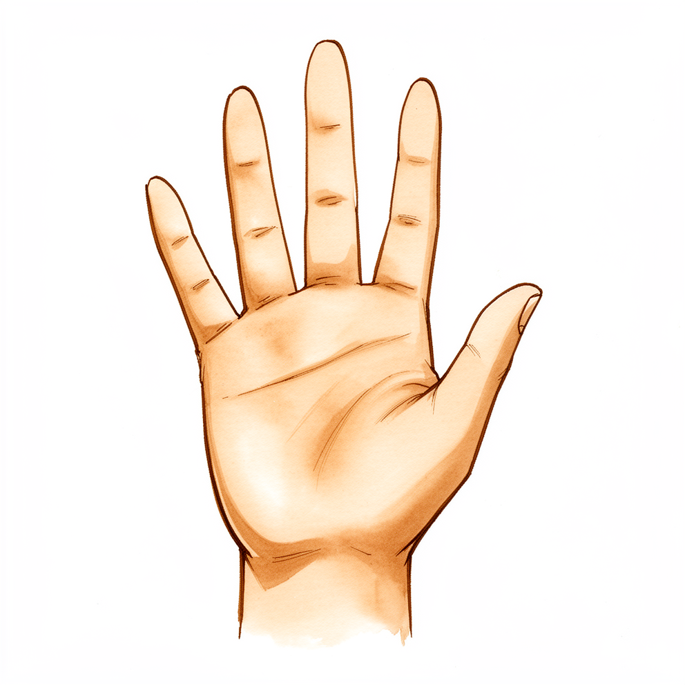
Ang STT joint ay matatagpuan kung saan nagtatagpo ang scaphoid na buto ng pulso at ang dalawang maliliit na buto na tinatawag na trapezium at trapezoid, sa likod mismo ng joint sa base ng iyong hinlalaki. Ang arthritis dito ay nagdudulot ng sakit o kirot sa base ng hinlalaki at sa panig ng hinlalaki ng pulso. Lumalala ito kapag humahawak nang mahigpit (grip) at nagpi-pinch.
Maaari itong ma-trigger ng mga pang-araw-araw na gawain: pagpihit ng susi, pagbubukas ng garapon, pagsusulat, pagdadala ng bag ng pinamili o paghawak ng isang tasa ng tsaa.
Karaniwang makita sa X-ray ang pagkapudpod ng joint na ito, lalo na sa mga nakatatandang babae. Maraming tao na may ganitong mga pagbabago sa X-ray ang walang anumang sakit. Hindi rin mahusay na gabay ang X-ray kung ang joint na ito nga ang nagdudulot ng iyong sakit: maaari nitong maliitin o palakihin kung gaano kalaki ang pagkapudpod. Ito ang dahilan kung bakit maaaring imungkahi ng iyong surgeon ang isang injection ng local anaesthetic at steroid sa loob ng joint, gamit ang gabay ng X-ray o ultrasound. Kung napakalma ng injection ang sakit, tumuturo ito sa STT joint bilang pinagmumulan.
Ang STT arthritis ay madalas na may kasamang arthritis ng joint sa base ng hinlalaki, at maaaring mahirap paghiwalayin ang dalawa. Kapag parehong pudpod ang dalawang joint, tinatawag itong pantrapezial arthritis. Mahalagang malaman kung aling mga joint ang apektado, dahil hinuhubog nito kung aling mga operasyon ang may saysay.
Sa ilang taong may STT arthritis, ang mga buto ng pulso ay tumatagilid paatras, isang pattern na tinatawag na DISI (maikli para sa dorsal intercalated segment instability). Sinusuri ito ng iyong surgeon sa X-ray bago pumili ng operasyon, dahil maaari nitong baguhin ang plano.
Ang pudpod na joint ay nasa tabi mismo ng tendon ng isang kalamnan ng pulso na tinatawag na flexor carpi radialis. Ang tendon na ito ay maaaring maging inflamed at masakit. Napakabihira, maaari itong mapatid.
Magpatingin sa iyong GP kung hindi humuhupa ang iyong mga sintomas, lumalala sa loob ng ilang linggo, ginigising ka sa gabi, o pumipigil sa iyong pagtatrabaho o paggamit ng iyong kamay. Pumunta sa emergency department sa araw ding iyon kung ang iyong kamay o braso ay naging mainit, mapula, namamaga at masakit, lalo na kung may lagnat, dahil maaaring ito ay isang impeksyon na nangangailangan ng pangangalaga sa araw ding iyon.
Ano ang aktwal na nangyayari
Ang arthritis ay pagkapudpod ng lining ng isang joint. Sa kondisyong ito, ang pudpod na joint ay ang STT joint: ang lugar kung saan nagtatagpo ang scaphoid, isa sa maliliit na buto sa kailaliman ng iyong pulso, at ang trapezium at trapezoid, dalawang maliliit na buto na tumutulong bumuo sa base ng iyong hinlalaki. Isipin ang mga butong ito bilang isang maikling hanay ng mga bloke na nagpapahintulot sa iyong hinlalaki na umugoy, humawak nang mahigpit at mag-pinch. Kung saan nagkikiskisan ang mga bloke, maaaring manipis ang makinis na surface. Kapag nangyari ito, nagkikiskisan ang hilaw na buto sa ilalim habang gumagalaw, at ang pagkikiskisang iyon ang kirot na nararamdaman mo sa base ng iyong hinlalaki at sa panig ng hinlalaki ng iyong pulso.
Pinagdugtong ang joint ng isang hanay ng mga strap na tinatawag na ligament. Kung mabanat o mapinsala ang mga strap na ito, ang scaphoid ay maaaring tumagilid palabas ng normal nitong posisyon. Binabago nito kung paano tumatanggap ng load ang mga buto, kaya lumalala ang pagkapudpod. Sa ilang tao, humahantong ito sa isang pattern kung saan tumatagilid paatras ang mga buto ng pulso, ang DISI pattern na hinahanap ng iyong surgeon sa X-ray. Ang pudpod na joint ay nasa tabi mismo rin ng isang tendon ng pulso, at ang tendon na iyon ay maaaring maging inflamed at masakit nang mag-isa.
Ang pagkapudpod dito ay madalas na lumilitaw kasabay ng pagkapudpod sa joint sa pinakabase ng hinlalaki. Kapag parehong pudpod ang dalawa, tinatawag itong pantrapezial arthritis. Mahalaga ito dahil napakalapit ng dalawang joint sa isa't isa kaya ang sakit mula sa isa ay maaaring maramdaman na parang sakit mula sa kabila, kaya maaaring gumamit ang iyong surgeon ng guided injection upang matukoy kung aling joint talaga ang nagdudulot ng iyong mga sintomas.
Ano ang maaari naming gawin tungkol dito
Si Dr Kieran Hirpara, isang upper-limb surgeon sa Mater Private Hospital Rockhampton, ay nagsisimula sa mga opsyon na hindi gaanong invasive na angkop sa iyong kondisyon. Karaniwang nagsisimula ang gamutan nang walang surgery. Ang isang splint na nagpapanatiling hindi gumagalaw ang hinlalaki at pulso ay maaaring magpakalma sa kirot, lalo na kapag marami kang ginagawa gamit ang iyong mga kamay. Nakatutulong din ang pagbabago sa paraan ng paggawa mo ng mga bagay: ang pagbabawas sa mga gawaing nangangailangan ng mahigpit na paghawak at pag-pinch, o ang paghahanap ng ibang paraan upang magbukas ng garapon at magdala ng mga bagay. Layunin ng hand therapy na mapanatiling gumagalaw ang hinlalaki at pulso at palakasin ang mga kalamnan sa paligid ng mga ito. Karaniwan naming hinihiling na bigyan mo ng sapat na pagsubok ang mga hakbang na ito sa loob ng ilang linggo bago pag-usapan ang anumang karagdagang hakbang.
Ang simpleng pain relief at anti-inflammatory na gamot, ang uri na nagpapahupa ng pamamaga, ay makapagpapagaan ng sakit. Ang isang steroid injection sa loob ng joint, na ginagabayan ng X-ray o ultrasound, ay makatutulong upang matukoy kung ang STT joint ang pinagmumulan ng iyong sakit. Kung napakalma nito ang sakit, tumuturo iyon sa joint. Maaaring tumagal nang ilang panahon ang ginhawa, at maaari itong ulitin kung mawala ang bisa nito.
Pumapasok sa usapan ang surgery kapag hindi nagbigay sa iyo ng sapat na pagbuti ang mga hakbang na ito at nililimitahan pa rin ng sakit ang iyong mga ginagawa. Kung aling operasyon ang may saysay ay nakadepende sa kung aling mga joint ang pudpod. Kung ang STT joint lamang ang pudpod, maaari naming alisin ang pudpod na dulo ng scaphoid, o maglagay ng isang maliit na pyrocarbon spacer sa joint upang pumalit sa pudpod na surface. Kung pudpod din ang joint sa base ng iyong hinlalaki, ang karaniwang operasyon ay nag-aalis sa trapezium kasama ang bahagi ng trapezoid. Bago pumili, sinusuri namin sa X-ray kung tumagilid na ang mga buto ng iyong pulso, dahil maaari nitong baguhin ang plano.
Ano ang dapat asahan
Ang STT arthritis ay isang pangmatagalang kondisyon sa halip na isang lumilipas na pinsala. Ang kirot sa base ng iyong hinlalaki ay may tendensiyang pabalik-balik. Madalas itong sumisidhi kapag mabigat ang paghawak o pag-pinch, pagkatapos ay humuhupa kapag ipinahinga mo ang kamay. Kung walang gamutan, karaniwang nananatili o paulit-ulit na bumabalik ang sakit, at maaari nitong dahan-dahang limitahan ang mga bagay na pakiramdam mo ay kaya mong gawin gamit ang iyong kamay.
Karamihan sa mga tao ay bumubuti sa mga hakbang na hindi nangangailangan ng surgery. Ang isang splint, mga pagbabago sa paraan ng paggawa ng mga gawain, simpleng pain relief at isang steroid injection ay maaaring magpakalma sa sakit nang ilang panahon. Maaaring mawala ang ginhawa mula sa injection, at maaari itong ulitin. Kung hindi nagbigay sa iyo ng sapat na pagbuti ang mga hakbang na ito pagkalipas ng ilang linggo, sulit nang pag-usapan ang surgery.
Kung magpapa-opera ka, bahagi ng plano ang hand therapy pagkatapos. Ang iyong therapy ay kasama si Ruby Doolan sa Extend Rehabilitation. Si Ruby ay isang hand therapist: gagabayan niya ang iyong mga ehersisyo sa paggaling at gagawa ng anumang splint na kakailanganin mo sa daan.
Unti-unti ang paggaling. Asahan na gagaan ang kirot sa loob ng mga linggo sa halip na mga araw, at asahan na mabubuo ang lakas at kumpiyansa sa kamay sa loob ng mga buwan. Mas matagal ang kailangan ng ilang tao kaysa sa iba, at ang layunin ay isang kamay na nagagawa ang kailangan mo nang may mas kaunting sakit, hindi isang kamay na pakiramdam ay bago.
Kailan dapat magpatingin
Karamihan sa kirot sa base ng hinlalaki ay humuhupa sa pamamagitan ng pahinga, splint at simpleng pain relief. Magpatingin sa iyong GP kung ang iyong sakit ay hindi humuhupa sa loob ng ilang linggo, lumalala, ginigising ka sa gabi, o pumipigil sa iyong pagtatrabaho o paggamit ng iyong kamay. Humingi ng specialist review kung patuloy na bumabalik ang sakit kapag humahawak nang mahigpit at nagpi-pinch, o kung mas madalas kang nakabibitaw ng mga bagay. Maaaring imungkahi ng iyong surgeon ang isang guided injection upang matukoy kung aling joint ang nagdudulot ng sakit, dahil ang pagkapudpod ng joint na ito ay madalas na lumilitaw kasabay ng pagkapudpod sa base ng hinlalaki.
Pumunta sa emergency department sa araw ding iyon kung ang iyong kamay o braso ay naging mainit, mapula, namamaga at masakit, lalo na kung may lagnat. Maaaring ito ay isang impeksyon, at nangangailangan ito ng pangangalaga sa araw ding iyon. Hindi mo kailangan ng referral upang matingnan.
Mas malalim na pagtalakay
Advanced reading: the deeper science (optional)
Ang seksyong ito ay lumalampas sa kailangan mo para sa iyong sariling mga desisyon sa paggamot. Sulit ang karagdagang pagbabasa tungkol sa STT arthritis dahil sa dalawang dahilan: hindi mahusay na gabay ang X-ray kung ang joint na ito nga ang masakit, at bawat operasyon para dito ay ipinagpapalit ang ginhawa mula sa sakit sa isang pagbabago sa pagkakaayos ng maliliit na buto ng pulso.
Gaano ito kakaraniwan, at kung bakit nakalilinlang ang X-ray
Ang pagkapudpod ng STT joint ay madalas na natutuklasan sa X-ray. Sa 700 magkakasunod na pasyente ng hand clinic na na-X-ray para sa anumang reklamo, 111 (16%) ang nagpakita ng STT arthritis; mas malamang ito habang tumatanda, kapag may puwang sa pagitan ng scaphoid at lunate, at kapag may arthritis sa joint sa base ng hinlalaki [1]. Sa isang random na sample ng mga residenteng Hapon na may edad 50 hanggang 89, 5.3% ng mga pulso ang nagpakita nito, na muling iniugnay sa arthritis sa base ng hinlalaki, pagiging babae at edad [2]. Karamihan sa mga taong ito ay walang sintomas mula sa STT joint.
Hindi maaasahan ang X-ray sa parehong direksyon. Nang direktang tingnan ng mga surgeon ang joint habang nag-oopera sa base ng hinlalaki, 62% ng mga hinlalaki ang may arthritis sa pagitan ng scaphoid at trapezoid, ngunit 44% lamang sa mga ito ang natukoy ng X-ray [3]. Isang mas malaking serye ng 257 na pasyente ang nakatagpo ng end-stage na pagkapudpod sa 31% sa oras ng operasyon, na may X-ray sensitivity na 47% at specificity na 94% [4]. Ang katamtamang pagkapudpod ng STT na hindi ginalaw sa oras ng operasyon sa base ng hinlalaki ay hindi lumala o nagdulot ng mga sintomas sa loob ng mean na 8 taon sa isang pangmatagalang serye [5]. Ito ang dahilan kung bakit ang desisyon ay nakasalalay sa kung saan ang pananakit kapag diniinan, at madalas sa isang guided injection, sa halip na sa X-ray lamang.
Ang pagtagilid ng mga buto ng pulso
Iniuugnay ang STT arthritis sa isang partikular na pagtagilid ng mga buto ng pulso, na tinatawag na dorsal intercalated segment instability (DISI): tumatagilid paatras ang lunate habang mas patayo ang posisyon ng scaphoid. Sa 135 na pulso na may STT arthritis, naroon ang DISI sa 5% ng mga katamtamang kaso at 60% ng mga malubha; hindi tulad ng pagtagilid na sumusunod sa punit na scapholunate ligament, nanatiling normal ang puwang sa pagitan ng scaphoid at lunate at hindi napudpod ang pangunahing joint ng pulso [6]. Inilalarawan na ngayon ng ilang awtor ang isang pattern ng pagbagsak na dulot ng STT arthritis (SOAC wrist) bilang naiiba sa mas kilalang SLAC wrist [7].
Mahalaga ito para sa surgery dahil ang isang operasyon na nag-aalis ng buto ay maaaring magpalala sa pagtagilid. Ang isang pulso na unstable na sa gitnang hanay ng mga buto ay maaaring bumagsak pagkatapos ng simpleng excision, gaya ng sa isang naiulat na kaso na nangailangan ng fusion sa kalaunan [8]. Kaya hinahanap ng mga surgeon ang pagtagilid at ang midcarpal instability bago pumili ng operasyon.
Distal scaphoid excision: ginhawa, kapalit ng pagtagilid
Ang pag-aalis ng dulo ng scaphoid ay nagpapaginhawa ng sakit sa karamihan ng mga serye, at karaniwang tumatagilid ang mga buto ng pulso pagkatapos. Sa isa sa mga unang serye, 12 sa 21 na pulso ang nagkaroon ng DISI, walang nagkaroon ng karagdagang paglala ng joint, at pareho ang satisfaction gaya ng sa mga pulsong hindi tumagilid [9]. Isang mas huling serye ang nakatagpo ng DISI sa 19 sa 21 na pulso (90%; 95% ang iniulat ng papel), na tumaas ang anggulo ng lunate nang mean na 13°, at walang kaugnayan ang pagtagilid sa mga functional score sa mean na 3 taon [10].
Mahalaga kung gaano karaming buto ang inaalis. Pagkatapos ng arthroscopic excision sa 17 na pulso, bumaba ang pain score mula 6.1 patungong 1.7 sa 10 at tumaas ang pinch strength mula 2.5 kg patungong 4.4 kg; mas malamang ang pagtagilid kapag higit sa 3 mm ng buto ang inalis [11]. Sa mga cadaver na pulso, ang pag-aalis ng isang-kapat ng scaphoid ay halos hindi nagbago sa paggalaw ng mga buto, ang kalahati ay nagdulot ng malinaw na pagtagilid, at ang tatlong-kapat ay nagpa-unstable sa pulso [12]. Isang anatomical na pag-aaral ang nakatagpo na ang malalim na bahagi (deep band) ng scaphotrapezial ligament ay nakakabit humigit-kumulang 3.3 mm mula sa dulo ng scaphoid, na nagbibigay ng anatomical na dahilan upang limitahan ang resection sa humigit-kumulang 3 mm [13]. Isang cadaver na pag-aaral ng parehong operasyon ang sumukat na umiikot paatras ang capitate nang humigit-kumulang 14° at ang lunate nang 8° [14]. Bihira, ang matalim na gilid na naiwan sa scaphoid ay nakapudpod hanggang maputol ang isang flexor tendon [15].
Kapag pudpod din ang base ng hinlalaki
Kapag parehong pudpod ang base ng hinlalaki at ang STT joint, ang karaniwang operasyon ay nag-aalis sa trapezium at sa pudpod na dulo ng trapezoid. Sa 37 na hinlalaki, ang pagdaragdag ng partial trapezoid excision ay hindi nagdulot ng karagdagang problema kumpara sa pag-aalis lamang ng trapezium [3]. Ang pinagtatalunang punto ay kung gaano karaming trapezoid ang aalisin. Sa 79 na kamay na sinundan sa loob ng median na 92 buwan, ang pag-aalis ng bahagi o ng buong trapezoid ay nagpalaki sa posibilidad ng paatras na pagtagilid ng lunate na 15° o higit pa (29% pagkatapos ng partial at 26% pagkatapos ng complete na pag-aalis), at mas mababa ang score ng mga pulsong may ganoong pagtagilid; nangyari ang may-sintomas na paglubog ng index metacarpal sa 18% pagkatapos ng complete at 10% pagkatapos ng partial na pag-aalis, at pinayuhan ng mga awtor na huwag gawin ang complete na pag-aalis [16]. Isang cadaver na pag-aaral ang nakatagpo na 4 hanggang 6 mm ng trapezoid ang kailangang alisin bago bumaba ang presyon sa pagitan ng scaphoid at trapezoid sa pagbaluktot at sa paggalaw patagilid [17]. Sa isang serye ng 150 na hinlalaki na sinundan sa loob ng mean na 13.5 taon, ang trapeziectomy na may partial trapezoidectomy at tendon reconstruction ay nagbigay ng mean na DASH disability score na 8.7 at walang revision [18].
Mga pyrocarbon implant
Layunin ng isang pyrocarbon spacer na pagaanin ang sakit habang pinananatili ang taas ng scaphoid, at sa gayon ay limitahan ang pagtagilid. Sa Pyrocardan disc, 20 na pasyente (22 na implant) na sinundan sa loob ng mean na 3.8 taon ang nagkaroon ng mas kaunting sakit, mas mahusay na mga score at mas malakas na pinch, na may kaunting pagkawala ng wrist extension at walang dislocation; bumuti sa halip na lumala ang capitolunate angle [19]. Isang serye na sinundan sa loob ng mean na 5 taon ang nag-ulat na 95% ng mga implant ay walang reoperation, isang walang-sakit na dislocation, calcium sa paligid ng joint sa 4 na pulso, at pagtagilid na nanatili sa 3 at lumitaw sa 1 [20]. Kumpara sa STT fusion sa isang non-randomised na pag-aaral, magkatulad ang mga resulta maliban sa mas mahusay na wrist extension sa implant (58° laban sa 38°); 22% ng mga pulsong may implant ang nagkaroon ng pagtagilid, at 9% ng mga fusion ang hindi nagdugtong [21]. Karamihan sa mga seryeng ito ay galing sa iilang sentro sa France.
Hindi gaanong pare-pareho ang mga resulta sa ibang mga disenyo. Sa isang ibang pyrocarbon implant na hinugis upang palitan ang dulo ng scaphoid (ang STPI), 7 sa 29 na implant ang ni-revise, pangunahin dahil sa sakit, at dumoble ang bilang ng mga pulsong may pagtagilid mula 6 patungong 12 sa loob ng median na 68 buwan [22]. Isang case report ang naglalarawan ng isang Pyrocardan na dalawang beses nabasag sa isang manlalaro ng tennis, na siyang dahilan kung bakit tinatalakay bago ang surgery ang mabigat na pagbibigay ng load sa pulso habang naka-extend ito [23].
STT fusion, para sa paghahambing
Pinagdudugtong ng fusion ang tatlong buto upang maging isa. Sa 1,429 na operasyon sa isang systematic review, hindi nagdugtong ang fusion sa 6.3%, 4.2% ang ginawang full wrist fusion, at ang grip strength ay nasa average na 76% ng kabilang kamay [24]. Sa 58 na pulso na sinundan sa loob ng 10 taon, 10% ang nangailangan ng karagdagang operasyon at 7% ang nagkaroon ng masakit na non-union [25]. Karaniwang napananatili ng fusion ang humigit-kumulang tatlong-kapat ng paggalaw ng pulso, at sapat na karaniwan ang sakit mula sa pagsabit ng radial styloid sa na-fuse na scaphoid kaya tinatabas ng ilang surgeon ang styloid sa parehong pagkakataon [26].
Ang hindi pa alam
Walang randomised trial na naghahambing sa mga operasyong ito. Isang survey ng mga hand surgeon sa Europa ang nakatagpo ng malawak na pagkakaiba-iba ng mga piniling operasyon para sa parehong problema [27], at nagkokonklud ang mga kamakailang review na walang opsyon ang may malinaw na ebidensya ng pagiging mas mahusay; ang pagpili ay nakadepende sa kung ang STT joint lamang ang pudpod, kung pudpod din ang base ng hinlalaki, at kung nakatagilid na ang mga buto ng pulso [28] [29]. Karamihan sa mga serye ay sumusunod sa mas kaunti sa 30 na pasyente sa loob ng mas mababa sa 10 taon, kaya bukas pang mga tanong ang pangmatagalang epekto ng pagtagilid pagkatapos ng excision, at ang pangmatagalang tibay ng mga pyrocarbon implant.
Mga Sanggunian
[1] Scordino LE, Bernstein J, Nakashian M, et al. Radiographic prevalence of scaphotrapeziotrapezoid osteoarthrosis. J Hand Surg Am. 2014;39(9):1677-82. https://doi.org/10.1016/j.jhsa.2014.05.033
[2] Kitamura Y, Kato H, Hayashi M, et al. Prevalence and associated factors for primary osteoarthritis of the scaphotrapeziotrapezoid, radiocarpal, and distal radioulnar joints in the Japanese general elderly population. J Hand Surg Am. 2025;50(1):103.e1-103.e10. Epub 2023. https://doi.org/10.1016/j.jhsa.2023.05.009
[3] Tomaino MM, Vogt M, Weiser R. Scaphotrapezoid arthritis: prevalence in thumbs undergoing trapezium excision arthroplasty and efficacy of proximal trapezoid excision. J Hand Surg Am. 1999;24(6):1220-4. https://doi.org/10.1053/jhsu.1999.1220
[4] Akoon A, Grandizio LC, Mookerjee GG, et al. Comparison of radiographic and intraoperative visual assessment of scaphotrapezoid joint arthritis in patients with end-stage carpometacarpal arthritis of the thumb base. Hand (N Y). 2019;14(5):609-13. Epub 2018. https://doi.org/10.1177/1558944718765246
[5] Glickel SZ, Kornstein AN, Eaton RG. Long-term follow-up of trapeziometacarpal arthroplasty with coexisting scaphotrapezial disease. J Hand Surg Am. 1992;17(4):612-20. https://doi.org/10.1016/0363-5023(92)90303-7
[6] Hattori Y, Sasaki J, Sakamoto S, et al. Radiographic characteristics of carpal instability associated with scaphotrapeziotrapezoid osteoarthritis. J Hand Surg Am. 2026;51(3):259-67. https://doi.org/10.1016/j.jhsa.2025.11.018
[7] Rodriguez Fontan F, Netscher DT. Scaphotrapeziotrapezoid joint osteoarthritis: differentiating SOAC wrist from SLAC wrist. J Am Acad Orthop Surg. 2026. https://doi.org/10.5435/jaaos-d-25-01408
[8] Corbin C, Warwick D. Midcarpal instability after excision arthroplasty for scapho-trapezial-trapezoid (STT) arthritis. J Hand Surg Eur Vol. 2009;34(4):537-8. https://doi.org/10.1177/1753193408098903
[9] Garcia-Elias M, Lluch AL, Farreres A, et al. Resection of the distal scaphoid for scaphotrapeziotrapezoid osteoarthritis. J Hand Surg Br. 1999;24(4):448-52. https://doi.org/10.1054/jhsb.1999.0169
[10] Brouwers D, Decramer A, Cornu P, Le Viet D. Does the DISI matter after distal scaphoidectomy with tendon interposition for STT osteoarthritis? Hand Surg Rehabil. 2020;39(4):284-90. https://doi.org/10.1016/j.hansur.2020.03.005
[11] Iida A, Omokawa S, Kawamura K, et al. Arthroscopic distal scaphoid resection for isolated scaphotrapeziotrapezoid osteoarthritis. J Hand Surg Am. 2019;44(4):337.e1-337.e5. https://doi.org/10.1016/j.jhsa.2018.06.021
[12] Kadar A, Grewal R, Padmore CE, et al. How much scaphoid can be safely resected? A biomechanical analysis of the effects of distal scaphoid resection. Hand (N Y). 2022;17(5):885-92. Epub 2020. https://doi.org/10.1177/1558944720966717
[13] Yoshimura Y, Yokota A, Fujino K, et al. Anatomy of the scaphotrapezial ligament complex and its implications for distal scaphoid resection. J Hand Surg Am. 2026;51(1):25.e1-25.e8. https://doi.org/10.1016/j.jhsa.2025.09.010
[14] Paeffgen L, Riederer J, Adler T, et al. Carpal instability after partial trapeziectomy, total trapeziectomy and the resection of the distal scaphoid pole: a cadaveric study. Arch Orthop Trauma Surg. 2024;144(3):1443-51. Epub 2023. https://doi.org/10.1007/s00402-023-05134-z
[15] Deren ME, Mitchell CH, Weiss AC. Flexor tendon ruptures after distal scaphoid excision for scaphotrapeziotrapezoid osteoarthritis. Hand (N Y). 2017;12(5):NP152-6. https://doi.org/10.1177/1558944717708028
[16] Renfree KJ, Odgers R, Tillinghast C, Zhang N. Effect of partial and complete trapezoid excision on radiographic and functional results after abductor pollicis longus suspensionplasty. J Hand Surg Am. 2020;45(4):364.e1-364.e9. https://doi.org/10.1016/j.jhsa.2019.10.006
[17] Anz H, Roylance J, Ismaily S, et al. Correlation of scaphotrapezoid contact pressures with degree of trapezoid resection: does eliminating contact matter? J Hand Surg Glob Online. 2026;8(4):101049. https://doi.org/10.1016/j.jhsg.2026.101049
[18] Melone CP, Lovy AJ, Ciaglia M, Qin B. Trapeziectomy, partial trapezoidectomy, and abductor pollicis longus dual loop ligament reconstruction for advanced basal joint arthritis: long-term outcome of 150 cases. Hand (N Y). 2023;18(3):491-500. Epub 2022. https://doi.org/10.1177/15589447221092060
[19] Gauthier E, Truffandier M, Gaisne E, Bellemère P. Treatment of scaphotrapeziotrapezoid osteoarthritis with the Pyrocardan implant: results with a minimum follow-up of 2 years. Hand Surg Rehabil. 2017;36(2):113-21. https://doi.org/10.1016/j.hansur.2017.01.003
[20] Cholley-Roulleau M, Bouju Y, Lecoq FA, et al. Pyrocardan scaphotrapeziotrapezoid joint arthroplasty for isolated osteoarthritis: results after a mean follow-up of 5 years. J Wrist Surg. 2022;11(3):262-8. Epub 2021. https://doi.org/10.1055/s-0041-1733877
[21] Cholley-Roulleau M, Dautel G, Dap F, et al. Comparison between scaphotrapeziotrapezoid arthrodesis and Pyrocardan implant for isolated scaphotrapeziotrapezoid osteoarthritis. Orthop Traumatol Surg Res. 2025;111(5):103867. https://doi.org/10.1016/j.otsr.2024.103867
[22] Zander M, Ibsen-Sörensen A, Nilsson A, Björkman A. Retrospective analysis of scaphoid trapezium pyrocarbon implant intervention in STT arthritis: a 3-year follow-up study. J Plast Surg Hand Surg. 2024;59:40-5. https://doi.org/10.2340/jphs.v59.34985
[23] Sarigiannis CN, Axelsson P. Game, set… revision! A case report of a tennis player who smashed his scaphotrapeziotrapezoid-joint Pyrocardan implant twice. J Hand Surg Glob Online. 2026;8(3):100964. https://doi.org/10.1016/j.jhsg.2026.100964
[24] Stephens AR, Garcia BN, Rogers MJ, et al. Scaphotrapeziotrapezoid arthrodesis: systematic review. J Hand Surg Am. 2022;47(3):218-227.e2. https://doi.org/10.1016/j.jhsa.2021.09.029
[25] Rogers MJ, Lu C, Stephens AR, et al. Scaphotrapeziotrapezoid arthrodesis: a 10-year follow-up study of complications in 58 wrists. Hand (N Y). 2022;17(5):919-25. Epub 2020. https://doi.org/10.1177/1558944720964972
[26] Deans VM, Naqui Z, Muir LT. Scaphotrapeziotrapezoidal joint osteoarthritis: a systematic review of surgical treatment. J Hand Surg Asian Pac Vol. 2017;22(1):1-9. https://doi.org/10.1142/s0218810417300017
[27] Berkhout MJL, Yin Q, Ritt MJPF. Current trends in operative treatment of scaphotrapeziotrapezoid osteoarthritis: a survey among European hand surgeons. J Wrist Surg. 2020;9(2):94-9. https://doi.org/10.1055/s-0039-3402796
[28] Bellemère P, Ardouin L. Surgical options and outcomes for treatment of osteoarthritis of the scaphotrapeziotrapezoidal joint. J Hand Surg Eur Vol. 2025;50(1):58-67. https://doi.org/10.1177/17531934241295345
[29] Alder KD, Feroe AG, Karim KE. Management of scaphotrapeziotrapezoid osteoarthritis: a critical analysis review. JBJS Rev. 2023;11(10):e23.00093. https://doi.org/10.2106/JBJS.RVW.23.00093
Evidence & references
This is the clinical evidence summary written for health professionals. It is technical, and it lists the research this page was built from. You do not need to read it to understand your treatment or to make a decision about it.
Anatomy & Pathophysiology
Osseous Skeleton
- The skeleton of the hand and wrist consists of 27 bones, of which 19 are long bones [3].
- The skeleton is divided into five rays, each comprising a polyarticulated chain of metacarpals and phalanges [3].
- The base of each metacarpal articulates with the distal row of the carpus [3].
- The carpus articulates with the skeleton of the forearm through its proximal row [3].
- The radioulnocarpal articulation has two axes of movement, to which a third axis of pronation and supination from the forearm is added [3].
- The wrist has three axes of movement, permitting the hand to be positioned in any spatial configuration [3].
- The radial ray (first ray) is the shortest, made up of a metacarpal and two phalanges [3].
- The first ray continues the external column of the carpus formed by the scaphoid and trapezium [3].
- The trapezium is angled out in front of the carpal plane so that the first metacarpal makes an angle of about 45 degrees with the second metacarpal in the sagittal plane [3].
- The thumb metacarpal is the shortest, while the index metacarpal is by far the longest [3].
- The proximal and middle phalanges of the long and ring fingers are longer than those of the index finger [3].
- The epiphyseal plates are located at the proximal ends of the phalanges and the first metacarpal [3].
- The epiphyseal plates are located at the distal ends of the other metacarpals [3].
- The hand presents a longitudinal and transverse concavity, giving it the shape of a cup with a palmar concavity when the thumb is placed next to the index finger [3].
- The transverse axis of the palm corresponds to the metacarpophalangeal articulations and is not perpendicular to the longitudinal axis [3].
- The transverse axis forms an acute angle of approximately 75 degrees with the longitudinal axis [3].
Musculature
- Extrinsic muscles have their origin outside of the hand and their insertion on the hand or carpus [2].
- Intrinsic muscles have both origin and insertion within the hand [2].
- The extrinsic extensors run through six different fibroosseous retinacular compartments at the wrist level [2].
- The first extensor compartment contains the abductor pollicis longus and the extensor pollicis brevis [2].
- The abductor pollicis longus inserts at the base of the thumb metacarpal and radially abducts the thumb [2].
- The extensor pollicis brevis inserts on the dorsum of the proximal aspect of the proximal phalanx of the thumb and actively extends the metacarpophalangeal joint of the thumb [2].
- The second extensor compartment contains the extensor carpi radialis longus and the extensor carpi radialis brevis [2].
- The extensor carpi radialis longus inserts on the index metacarpal, dorsiflexes and radially deviates the wrist [2].
- The extensor carpi radialis brevis inserts into the base of the middle metacarpal and provides balanced wrist dorsiflexion [2].
- The third compartment contains the extensor pollicis longus, which turns abruptly radialward about Lister tubercle [2].
- The extensor pollicis longus inserts on the distal phalanx and provides forceful extension of the thumb interphalangeal joint [2].
- The fourth extensor compartment contains the extensor indicis proprius lying deep to the four tendons of the extensor digitorum communis [2].
- The fifth compartment contains the extensor digiti quinti [2].
- The principal bony insertion of the extrinsic digital extensors is on the dorsal proximal aspect of the middle phalanx [2].
- Metacarpophalangeal joint extension is provided by extrinsic extensor force transmitted through the sagittal bands [2].
- Distal interphalangeal joint extension is achieved through the conjoined lateral bands composed of tendinous slips from the extrinsic and intrinsic tendons [2].
- The extensor carpi ulnaris tendon runs through the sixth compartment and inserts at the base of the little finger metacarpal [2].
- The extensor carpi ulnaris provides wrist extension and ulnar deviation [2].
- The extensor digitorum communis tendons of the middle, ring, and little fingers are tethered together by juncturae tendinum over the dorsum of the hand proximal to the metacarpophalangeal joint [2].
- The digital extensor tendons are stabilized over the mid-line of the metacarpophalangeal joint by their attachment to sagittal band fibers [2].
- The sagittal band fibers insert onto the volar proximal phalanx and onto the lateral borders of the volar plate [2].
- The sagittal bands keep the extrinsic extensor as far as possible away from the center of rotation of the metacarpophalangeal joint, giving it the greatest mechanical efficiency [2].
- With rupture or attenuation of the sagittal band fibers, the extrinsic extensor tendon can sublux to the ulnar side of the metacarpal head causing ulnar deviation of the finger [2].
- The extrinsic finger flexors are the flexor digitorum profundus and the flexor digitorum superficialis [2].
- The flexor digitorum profundus inserts on the proximal volar aspect of the distal phalanx [2].
- The flexor digitorum profundus flexes the distal interphalangeal joint as well as the proximal interphalangeal and metacarpophalangeal joints [2].
- The flexor digitorum superficialis acts as a flexor of the proximal interphalangeal and metacarpophalangeal joints [2].
- There are seven interosseous muscles, four dorsal and three volar [6].
- The dorsal interossei are abductors [6].
- The dorsal interossei lie to the radial side of the index and middle fingers and the ulnar side of the middle and ring fingers [6].
- The little finger is abducted by the abductor digiti quinti [6].
- The volar interossei are adductors and lie to the ulnar side of the index finger and the radial side of the ring and little fingers [6].
- The middle finger has two dorsal interossei (abductors) and no volar interossei (adductors) because the central axis of the hand lies within it [6].
- Each dorsal interosseous muscle, with the exception of the third, has two muscle heads [6].
- The superficial head of the dorsal interosseous muscles abducts and weakly flexes the proximal phalanx [6].
- The deep head of each dorsal interosseous muscle forms a lateral tendon, or lateral band, at the level of the MP joint [6].
- The deep head flexes and weakly abducts the proximal phalanx while extending the middle and distal phalanges [6].
- Transverse fibers arch dorsally from each lateral band to join each other over the dorsum of the finger, flexing the proximal phalanx [6].
- Oblique fibers (spiral fibers) from the lateral bands insert onto the lateral tubercles at the base of the middle phalanx and extend the middle phalanx (PIP joint) [6].
- The lateral bands are joined by the lateral slips of the extensor tendon to form the conjoined lateral band [6].
- The two conjoined lateral bands to each finger unite at the distal third of the middle phalanx to form the terminal tendon [6].
- The terminal tendon inserts at the base of the distal phalanx to extend it [6].
- The flexor digiti quinti brevis is structurally and functionally similar to the deep head of the dorsal interossei, forming the ulnar lateral band of the little finger [6].
- Each volar interosseous muscle has only one muscle head and none of them insert onto the proximal phalanx [6].
- The volar interossei form the ulnar lateral band of the index finger and the radial lateral band of the ring and little fingers [6].
- The abductor digiti quinti inserts onto the ulnar lateral tubercle at the base of the proximal phalanx of the little finger [6].
- The flexor digiti quinti forms the ulnar lateral band [6].
- The opponens digiti quinti arises from the pisohamate ligament and the hook of the hamate and inserts onto the ulnar side of the diaphysis of the fifth metacarpal [6].
- The opponens digiti quinti flexes and supinates the fifth metacarpal [6].
- The flexor digitorum profundus originates from the proximal ulna and the interosseous membrane [13].
- The flexor digitorum profundus and the flexor pollicis longus muscles form the deep compartment of the volar forearm [13].
- As the flexor digitorum profundus and flexor pollicis longus tendons travel through the carpal tunnel, they occupy the floor of the carpal tunnel [13].
- The tenosynovial sheath of the flexor pollicis longus is continuous with the radial bursa [13].
- The tenosynovial sheath to the little finger is continuous with the ulnar digital bursa [13].
- In some patients, the radial and ulnar bursae communicate, allowing a horseshoe abscess to spread between the thumb and little finger if infection occurs in the flexor tendon sheath of either one of these digits [13].
- The lumbricals originate from the radial side of the index, middle, ring, and little fingers in the palm [13].
- The profundus tendon passes through the bifurcation of the flexor digitorum superficialis before inserting into the proximal palmar base of the distal phalanx [13].
- The innervation of the flexor digitorum profundus of the index and middle fingers is through the anterior interosseous branch of the median nerve [13].
- The profundus of the ring and little fingers is innervated by the ulnar nerve [13].
- The flexor digitorum superficialis has two heads: a radial head originating from the proximal shaft of the radius, and a humeral ulnar head originating from the medial humeral epicondyle and coronoid process of the ulna [13].
- As the superficialis tendons pass through the carpal tunnel, the tendons of the middle and ring fingers are more superficial and central than those of the index and little fingers [13].
- In the proximal aspect of the finger, the flexor digitorum superficialis tendon bifurcates around the flexor digitorum profundus at the beginning of the A2 pulley [13].
- The flexor digitorum superficialis tendon slips reunite distally at the Camper chiasm, with approximately half of the fibers staying on the ipsilateral side and half crossing to the contralateral side of the finger [13].
- The flexor digitorum superficialis inserts via radial and ulnar slips into the proximal metaphysis of the middle phalanx [13].
- The entire flexor digitorum superficialis muscle receives innervation from the median nerve [13].
- The primary function of the superficialis is digital flexion at the proximal interphalangeal joint [13].
- The flexor pollicis longus originates from two heads: a radial head from the proximal radius and interosseous membrane, and an accessory head from the coronoid process of the ulna and medial epicondyle of the humerus [13].
- In the palm, the flexor pollicis longus tendon transverses between the abductor pollicis brevis and the flexor pollicis brevis [13].
- The flexor pollicis longus inserts into the proximal base of the thumb distal phalanx [13].
- The flexor pollicis longus is innervated by the anterior interosseous branch of the median nerve [13].
- The flexor pollicis longus flexes both the interphalangeal and metacarpophalangeal joints of the thumb [13].
Ligaments and Fascia
- The second to fifth metacarpals are all bound together by various fibrous structures, the most distal of which is the deep transverse intermetacarpal ligament [9].
- The deep transverse intermetacarpal ligament is better named the interglenoid ligament because it ties together the anterior "glenoid ligaments" of the metacarpophalangeal articulations, known as the "volar plates" [9].
- The volar plates are interconnected by the transverse interglenoid ligament [9].
- The stability of the metacarpophalangeal joints is essential to the support of the longitudinal arch as well as of the transverse metacarpal arch [9].
- The superficial palmar fascia lies in a coronal plane beneath the palmar subcutaneous tissue [16].
- The superficial palmar fascia covers a triangular area of the central palm, with the proximal corner facing directly proximal [16].
- The palmaris longus tendon, when present, terminates in continuity with the fibers of this proximal corner [16].
- From this common point, four central bands of fascia extend distally toward each of the fingers [16].
- There is no central band for the thumb [16].
- At the distal palmar crease level, the central bands are bridged transversely by the superficial transverse palmar ligament [16].
- Fibers from the central bands pass above, below, and through the superficial transverse palmar ligament [16].
- Superficial fibers of the central bands stay superficial and merge with vertical retinacular fibers at the dermis’s undersurface in the distal palm in areas between skin flexion creases [16].
- Intermediate fibers split transversely into two sections, which extend toward the lateral border of the digit base [16].
- These intermediate fibers are the spiral band that tracks around the neurovascular bundle [16].
- Proximally, the spiral band is central and superficial to the neurovascular bundle; distally, it is lateral and deep beneath it [16].
- Spiral neurovascular bundles develop when spiral bands shorten [16].
- Deep fibers continue dorsally to merge with sagittal interosseous fascia fibers and pierce the transverse deep intermetacarpal ligament to merge with fibers of the sagittal bands of the extensor mechanism [16].
- The digital neurovascular structures are surrounded by a diffuse network of thin transverse oblique fibers [16].
- Fibers dorsal to the neurovascular bundle are collectively called Cleland ligament [16].
- Fibers palmar to the neurovascular bundle are called Grayson ligament [16].
- Undisturbed, these "ligaments" are a mesh of nonparallel individual fibers [16].
- The appearance of a ligament is a dissection artifact from releasing skin attachments [16].
- These fibers have a common origin in continuity with the fingers’ retinacular ligaments, the floor of the flexor tendon sheath, and retinacular fibers attaching the palmar digital skin to deeper structures [16].
- On the small finger’s ulnar border, lateral fascial attachments are in continuity with the abductor digiti minimi fascia and tendon [16].
- A subdermal fascial layer borders the periphery of the web spaces from roughly the radial thumb sesamoid to the ulnar side of the small finger’s proximal flexion crease [16].
- These border fibers form the distal first web space ligament between thumb and index in continuity with the natatory ligament that spans the remaining web spaces [16].
- Fibers from the natatory ligament extend distally at each finger’s lateral base in continuity with Grayson ligament fibers and the lateral digit dermis [16].
Vascular Anatomy
- The "princeps pollicis" artery, the terminal branch of the radial artery, crosses the first intermetacarpal space [10].
- The "princeps pollicis" artery runs along the ulnar side of the first metacarpal bone and along the volar surface of the adductor muscle [10].
- The "princeps pollicis" artery emerges onto the subcutaneous palmar tissue at the level of the cutaneous flexion crease of the metacarpophalangeal joint [10].
- At the metacarpophalangeal joint level, the "princeps pollicis" divides into two terminal rami, namely the collateral palmar arteries of the thumb [10].
- The collateral palmar arteries run along the digital tunnel symmetrically and are of equal caliber [10].
- The collateral palmar arteries head distally to finally unite in the pulp arcade [10].
- During their transit in the digital tunnel, the collateral palmar arteries break off into numerous collateral branches, either cutaneous, articular or osseous [10].
- An arcade located deep in the flexor tendon joins together the two arteries at the level of the distal metaphysis of the first phalanx [10].
- From this arcade, vessels originate which enter the "vincula" and irrigate the flexor tendon [10].
- In anatomical studies, only 15% of dissections fall into the classical category of palmar artery layout [10].
- In
Clinical Presentation
- Inflammatory arthritis is a collection of different diseases that cause inflammation in the joints [20].
- The diagnosis of inflammatory arthritis requires a complete review of the disease process profile [20].
- There are no simple clinical tests that can be used to diagnose inflammatory arthritis diseases [20].
- The rheumatologic history and physical examination play a critical role in the diagnosis and treatment of inflammatory arthritis [20].
- The location of the problems is an important disease feature for diagnosing inflammatory arthritis [20].
- The symmetry of presentation is an important disease feature for diagnosing inflammatory arthritis [20].
- Rheumatoid arthritis has a predilection for the wrists and the proximal joints of the hands and feet [20].
- Psoriatic arthritis involves the distal interphalangeal joint of the hands [20].
- The physical findings with rheumatoid arthritis tend to be symmetrical [20].
- Other inflammatory arthritis conditions do not tend to present with symmetrical physical findings [20].
- The onset of the disease is an important feature for diagnosing inflammatory arthritis [20].
- The chronology of the disease is an important feature for diagnosing inflammatory arthritis [20].
- The onset of rheumatoid arthritis tends to present in a subacute manner [20].
- Septic arthritis has a rapid onset in several hours [20].
- Age can aid in the diagnosis of inflammatory arthritis [20].
- Gender can aid in the diagnosis of inflammatory arthritis [20].
- Precipitating factors can aid in the diagnosis of inflammatory arthritis [20].
Investigations
- Clinical evaluation of the hand and wrist requires combining patient history with a careful physical examination to narrow the scope of possible pathologic processes [1].
- Diagnostic tests such as imaging and serum laboratory studies are useful in determining pathology but can be expensive, time-consuming, and often nonspecific [1].
- A careful physical examination is essential to direct care and future testing if indicated [1].
- The radioulnocarpal articulation has two axes of movement, to which a third axis—pronation and supination from the forearm—is added [3].
- The transverse axis of the palm forms an acute angle of approximately 75 degrees with the longitudinal axis [3].
- An 8-MHz Doppler tone assessment may be used to identify superficially displaced neurovascular bundles when Dupuytren cords lie beneath soft fleshy prominences [19].
- False-negatives are possible with 8-MHz Doppler tone assessment for identifying neurovascular bundles [19].
- MR assessment of Dupuytren’s disease is hindered by the resolution of current equipment, orientation issues due to multiplanar deformities of the fingers, and lack of intraoperative availability [19].
- MRI is probably most useful in identifying additional pathology such as flexor tendon bowstringing [19].
- MRI may be helpful in providing a quantitative noninvasive measure of cellularity of affected areas, which is an index of biologic activity [19].
References
[1] Orthopaedic Knowledge Update 13 Ebook Without Multimedia. Anatomy, Evaluation, Clinical Examination, and Imaging > Evaluation and Clinical Examination: Current Concepts.
[2] A Lange Medical Book Current Diagnosis Treatment In Orthopedics Fifth Edition. 9Hand Surgery > Image DISORDERS OF THE MUSCULATURE OF THE HAND.
[3] Exam Of The Hand Wrist 2Ed. 1.1 SKELETON OF THE HAND > The osseous skeleton.
[6] Green S Operative Hand Surgery. Interosseous and Hypothenar Muscles.
[9] Exam Of The Hand Wrist 2Ed. The arches of the hand > The metacarpal arch.
[10] Exam Of The Hand Wrist 2Ed. Techniques of investigation of the arterial supply by J P Melki > Vascularization of the thumb > Palmar aspect.
[13] A Lange Medical Book Current Diagnosis Treatment In Orthopedics Fifth Edition. 9Hand Surgery > FLEXOR TENDON INJURY.
[16] Green S Operative Hand Surgery. PERTINENT ANATOMY.
[19] Dupuytren S Disease And Related Hyperproliferative Disorders. 54. The Future of Dupuytren’s Research and Treatment > 54.4 Mechanical Measurements and Procedures > 54.4.4 Imaging.
[20] Orthopaedic Knowledge Update 13 Ebook Without Multimedia. Degenerative Conditions of the Knee > Inflammatory Arthritis.