Lý do phẫu thuật này được đề xuất
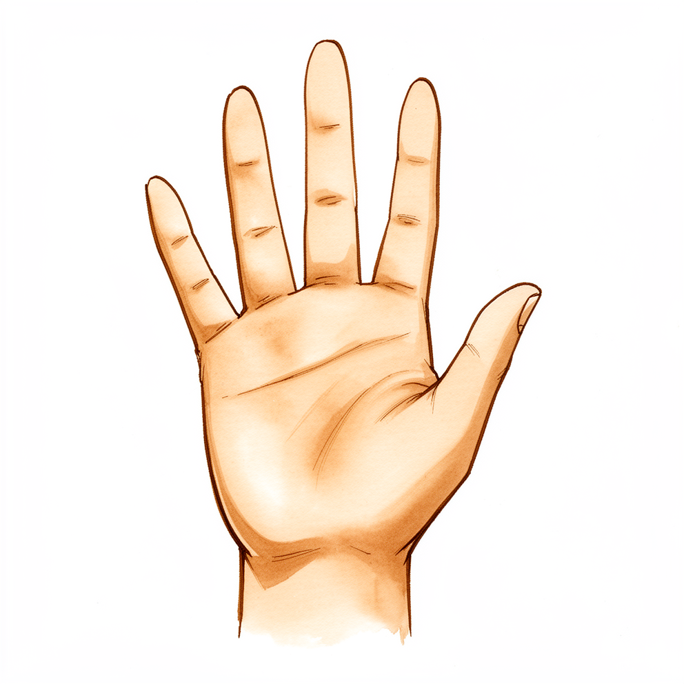
Bác sĩ Kieran Hirpara, bác sĩ phẫu thuật chi trên tại Bệnh viện tư nhân Mater Rockhampton, lựa chọn phương pháp điều trị phù hợp với chấn thương cụ thể của bạn. Gãy xương bàn tay là tình trạng gãy một trong các xương dài của bàn tay. Ca mổ này giữ xương gãy ở đúng vị trí trong khi xương lành. Chúng tôi thường đề xuất phẫu thuật này khi một xương bàn tay bị gãy đã lệch khỏi vị trí, bị xoay hoặc bị ngắn lại, có nhiều xương cùng bị gãy, hoặc xương không giữ được thẳng hàng trong bột. Một số đường gãy đi vào mặt khớp hoặc vỡ thành nhiều mảnh, và những trường hợp này thường cần phẫu thuật để giữ vững.
Hầu hết các ca gãy xương ở bàn tay ban đầu được điều trị không phẫu thuật, bằng nẹp hoặc bó bột và trị liệu bàn tay. Phẫu thuật là bước tiếp theo khi các biện pháp đó không giữ được xương ở vị trí tốt. Thông thường, bệnh nhân được bác sĩ đa khoa giới thiệu đến phòng khám chúng tôi; nếu nhà vật lý trị liệu khuyên bạn nên gặp chúng tôi, bạn vẫn cần có giấy giới thiệu từ bác sĩ đa khoa để được hưởng mức hoàn trả chi phí từ Medicare. Trong buổi khám, chúng tôi hỏi bệnh sử, khám bàn tay của bạn và chỉ định chụp phim nếu cần. Sau đó chúng ta cùng nhau quyết định liệu một ca mổ có phù hợp với chấn thương và mục tiêu của bạn hay không. Mục tiêu là một bàn tay cử động tốt, có cảm giác vững chắc và cho phép bạn sử dụng mà không đau.
Trước khi phẫu thuật
Bác sĩ phẫu thuật sẽ hướng dẫn cụ thể cho bạn, nhưng có một vài điều áp dụng cho hầu hết mọi người. Hãy ngừng ăn và uống bảy giờ trước ca mổ. Chúng tôi yêu cầu bảy giờ để ca mổ của bạn có thể được đẩy sớm hơn nếu lịch mổ chạy nhanh hơn dự kiến. Bạn có thể cần tạm ngừng một số loại thuốc thường dùng trước phẫu thuật; bác sĩ phẫu thuật sẽ cho bạn biết đó là những thuốc nào và cần ngừng trong bao lâu. Hãy mang theo danh sách ghi ra giấy tất cả các loại thuốc bạn đang dùng, và mặc quần áo rộng rãi, thoải mái. Hãy sắp xếp người lái xe đưa bạn về nhà sau đó. Cần chụp X-quang bàn tay để lên kế hoạch cho ca mổ, và đôi khi cần chụp thêm MRI hoặc siêu âm. Nếu bạn có các bệnh lý khác, bạn có thể cần xét nghiệm máu hoặc được bác sĩ gây mê khám trước.
Vào ngày phẫu thuật
Bạn đến khu tiếp nhận phẫu thuật của bệnh viện, nơi bạn được làm thủ tục nhập viện và chuẩn bị cho phòng mổ. Bạn sẽ gặp bác sĩ gây mê, người bác sĩ chăm lo cho giấc ngủ và sự dễ chịu của bạn trong suốt ca mổ. Phẫu thuật này được thực hiện dưới gây mê toàn thân. Bạn sẽ ngủ hoàn toàn trong suốt ca mổ. Một số bệnh nhân cũng có thể được gây tê vùng (phong bế thần kinh) để giảm đau sau mổ; bác sĩ gây mê sẽ quyết định vào ngày mổ dựa trên hoàn cảnh cụ thể của bạn.
Sau đó bạn được đưa vào phòng mổ, nơi ca phẫu thuật được thực hiện. Sau mổ, bạn tỉnh dậy ở khu hồi tỉnh, nơi các điều dưỡng theo dõi bạn trong khi thuốc mê hết tác dụng. Khi tình trạng đã ổn định, bạn sẽ được chuyển về khoa hoặc về nhà. Điều nào xảy ra tùy thuộc vào loại thủ thuật và tiến triển hồi phục của bạn.
Quy trình phẫu thuật
Có hai cách để bác sĩ phẫu thuật thực hiện ca mổ này, và việc lựa chọn tùy thuộc vào kiểu gãy của bạn. Với phương pháp kín, xương được nắn thẳng mà không mở da. Sau đó các đinh hoặc một con vít được đưa vào qua các vết chọc nhỏ để giữ xương. Với phương pháp mở, bác sĩ phẫu thuật rạch một đường trên xương gãy để có thể nhìn thấy các mảnh gãy và ráp chúng lại với nhau. Phương pháp này được dùng khi đường gãy đi vào khớp, khi xương vỡ thành nhiều mảnh, hoặc khi xương không tự thẳng hàng được.
Khi xương đã thẳng hàng, cần có thứ gì đó giữ nó vững trong khi lành. Bác sĩ phẫu thuật chọn dụng cụ cố định phù hợp với hình dạng và vị trí của đường gãy. Có thể xuyên các đinh nhỏ qua xương. Có thể đặt một con vít nhỏ vào bên trong lòng xương, một kỹ thuật gọi là cố định nội tủy, phù hợp với một số kiểu gãy thân và cổ xương. Có thể đặt các vít nhỏ bắt ngang qua đường gãy chéo dài hoặc đường gãy xoắn, tức là khi đường gãy chạy vòng quanh xương. Hoặc có thể dùng một nẹp nhỏ bắt vít để giữ các mảnh gãy lại với nhau.
Các đinh được để thò ra ngoài da thường được rút tại phòng khám sau vài tuần, khi xương đã vững. Vít và nẹp nằm dưới da thường được để nguyên trừ khi chúng gây ra vấn đề. Vết mổ được khâu lại, và một lớp băng được đặt lên trên.
Sau khi phẫu thuật
Bạn sẽ tỉnh dậy ở khu hồi tỉnh, nơi các điều dưỡng theo dõi bạn trong khi thuốc mê hết tác dụng. Bàn tay của bạn sẽ được băng và đặt nẹp, và bạn có thể được đeo đai treo tay cho dễ chịu. Thuốc giảm đau sẽ được sắp xếp để bạn luôn cảm thấy dễ chịu. Bạn có thể đi lại khi cảm thấy đủ sức, và cần có người ở cùng bạn trong 24 giờ đầu. Sau phẫu thuật này, bạn có thể về nhà trong ngày hoặc nằm viện một đêm. Cả hai trường hợp đều thường gặp, và đội ngũ điều trị sẽ trao đổi với bạn về lựa chọn phù hợp.
Nếu bạn được gây tê phong bế thần kinh, bàn tay của bạn có thể cảm thấy tê hoặc nặng trong khoảng 24 giờ. Điều này là bình thường. Hãy gọi cho phòng khám nếu bạn không thể cử động các ngón tay sau khi thuốc tê phong bế đã hết tác dụng. Hãy gọi cho phòng khám ngay trong ngày nếu bạn bị sốt, có vùng da đỏ lan rộng quanh vết mổ, hoặc có dịch hay mủ rỉ ra từ vết mổ, hoặc nếu cơn đau tiếp tục nặng hơn dù đã dùng thuốc giảm đau. Hãy đến khoa cấp cứu nếu các ngón tay, bàn tay hoặc ngón cái của bạn trở nên nhợt nhạt, lạnh, trắng bệch, tím tái hoặc sẫm màu.
Chúng tôi để nguyên băng khoảng 10 ngày; xin đừng tự tháo băng trước thời điểm đó trừ khi chúng tôi yêu cầu. Chúng tôi sẽ thay hoặc tháo băng khi gặp bạn.
Quá trình hồi phục
Bàn tay của bạn sẽ đau và sưng trong những ngày và tuần đầu. Điều này là bình thường và sẽ giảm dần khi xương lành. Kê cao bàn tay khi nghỉ ngơi giúp giảm sưng, và thuốc giảm đau đã được sắp xếp cho bạn giúp làm dịu cảm giác khó chịu.
Bàn tay của bạn sẽ được bảo vệ trong một nẹp do chuyên viên trị liệu bàn tay của bạn, Ruby Doolan tại Extend Rehabilitation, làm. Cô ấy hướng dẫn quá trình trị liệu của bạn và làm bất kỳ nẹp nào bạn cần. Bạn sẽ tập các bài tập cử động đều đặn như một phần của trị liệu bàn tay. Các bài tập này bắt đầu sớm và tăng dần khi xương lành. Mục tiêu là giữ cho các ngón tay cử động được trong khi chỗ gãy ổn định dần. Bạn có thể dùng bàn tay cho các việc nhẹ hằng ngày trong giới hạn mà chuyên viên trị liệu đặt ra. Bạn không được lái xe khi còn đeo nẹp, vì nẹp khiến bạn không thể cầm vô lăng an toàn. Khi đã tháo nẹp và bác sĩ phẫu thuật cho phép, bạn có thể lái xe trở lại; hãy xem trang của chúng tôi về lái xe sau phẫu thuật chi trên.
Khi khả năng cử động trở lại, các việc hằng ngày sẽ dễ dàng hơn. Bạn sẽ nhận thấy sức nắm và sức mạnh dần trở lại theo thời gian. Công việc nặng hơn, chơi thể thao và tập gym sẽ đến sau, khi chuyên viên trị liệu và bác sĩ phẫu thuật cùng đồng ý rằng xương đã sẵn sàng. Bạn có thể ngủ ở bất kỳ tư thế nào thấy thoải mái; không có tư thế đặc biệt nào bạn phải giữ.
Quá trình hồi phục khác nhau ở mỗi người. Thời gian của bạn có thể khác, và bác sĩ phẫu thuật cùng chuyên viên trị liệu sẽ hướng dẫn bạn ở mỗi lần khám.
Những biến chứng có thể xảy ra
Hầu hết bệnh nhân hồi phục tốt, nhưng đôi khi vẫn có thể xảy ra vấn đề. Bác sĩ phẫu thuật và đội ngũ điều trị theo dõi bạn chặt chẽ để phát hiện sớm mọi vấn đề.
Đôi khi xương có thể dịch chuyển khỏi vị trí đã được nắn chỉnh trước khi lành. Bạn có thể nhận thấy ngón tay bắt đầu trông bị xoay hoặc cong, hoặc có thay đổi về cách các ngón tay thẳng hàng khi bạn nắm tay lại. Hãy nêu điều này ở lần tái khám tiếp theo, hoặc gọi cho phòng khám nếu thay đổi có vẻ xảy ra đột ngột.
Đôi khi đinh hoặc vít có thể dịch chuyển khỏi vị trí đã đặt. Đinh nằm gần khớp có thể gây đau và khiến bàn tay khó sử dụng hơn. Nếu bạn cảm thấy một cơn đau nhói mới xuất hiện, hoặc ngón tay không cử động được như trước, hãy gọi cho phòng khám.
Ngón tay cũng có thể bị lệch xoay. Bạn có thể nhận thấy một ngón tay chồng lên ngón khác khi bạn gập bàn tay thành nắm đấm, hoặc ngón tay nằm lệch so với các ngón bên cạnh. Hãy nêu điều này ở lần tái khám để được kiểm tra.
Nhiễm trùng là một nguy cơ của bất kỳ ca mổ nào. Hãy gọi cho phòng khám ngay trong ngày nếu bạn bị sốt, có vùng da đỏ lan rộng quanh vết mổ, hoặc có dịch hay mủ rỉ ra từ vết mổ, hoặc nếu cơn đau tiếp tục nặng hơn dù đã dùng thuốc giảm đau. Nếu bạn không liên lạc được với phòng khám, hãy đến khoa cấp cứu gần nhất.
Đinh để thò qua da có thể gây kích ứng mô xung quanh. Vùng da nơi đinh đi vào có thể trông đỏ hoặc rỉ dịch. Hãy gọi cho phòng khám nếu điều này xảy ra, để chúng tôi kiểm tra trước khi nó trở thành một vấn đề sâu hơn.
Hiếm khi, một gân có thể bị đứt, nghĩa là một trong những sợi dây làm cử động ngón tay đã bị rách. Bạn sẽ nhận thấy mất cử động đột ngột, chẳng hạn không thể duỗi thẳng hoặc gập một ngón tay vốn vẫn cử động bình thường vào ngày hôm trước. Hãy gọi cho phòng khám nếu điều này xảy ra.
Các dây thần kinh chạy sát các xương của bàn tay. Tổn thương thần kinh có thể gây tê hoặc ngứa ran ở một phần bàn tay. Tê trong 24 giờ đầu sau khi gây tê phong bế thần kinh là điều bình thường. Sau khi thuốc tê phong bế đã hết tác dụng, hãy gọi cho phòng khám nếu tình trạng tê hoặc yếu vẫn tiếp diễn.
Một số người bị đau và tăng nhạy cảm kéo dài ở bàn tay, có vẻ không tương xứng với mức độ chấn thương. Bàn tay có thể đau khi chạm vào hoặc khi sử dụng rất lâu sau thời điểm lẽ ra đã phải dịu đi. Hãy nêu điều này ở lần tái khám.
Bảng biến chứng trên trang này liệt kê các tỷ lệ điển hình nếu bạn muốn biết chi tiết.
Khi nào nên gọi cho chúng tôi
Hầu hết các vấn đề xuất hiện sớm, và chúng tôi thà được nghe về chúng còn hơn không biết. Hãy gọi cho phòng khám ngay trong ngày nếu bạn bị sốt, có vùng da đỏ lan rộng quanh vết mổ, hoặc có dịch hay mủ rỉ ra từ vết mổ, hoặc nếu cơn đau tiếp tục nặng hơn dù đã dùng thuốc giảm đau. Hãy đến khoa cấp cứu nếu bắp chân bị sưng hoặc đau, nếu bạn bị khó thở, hoặc nếu bạn bị đau ngực. Đây có thể là dấu hiệu của cục máu đông. Hãy đến khoa cấp cứu nếu các ngón tay, bàn tay hoặc ngón cái của bạn trở nên nhợt nhạt, lạnh, trắng bệch, tím tái hoặc sẫm màu. Hãy gọi cho phòng khám nếu bạn không thể cử động các ngón tay sau khi thuốc tê phong bế thần kinh đã hết tác dụng, khoảng 24 giờ sau phẫu thuật. Tê trong 24 giờ đầu là điều bình thường. Nếu bạn không liên lạc được với phòng khám, hãy đến khoa cấp cứu gần nhất.
Evidence & references
This is the clinical evidence summary written for health professionals. It is technical, and it lists the research this page was built from. You do not need to read it to understand your treatment or to make a decision about it.
Overview
- The history of operative fixation for metacarpal and phalangeal fractures is limited to the twentieth century [6].
- Low-profile metacarpal plates may be sufficient for fixation, provided patient compliance and supplemental stabilization with a cast or splint are considered [1].
- Intramedullary screw (IMS) fixation is a practicable surgical option for metacarpal fractures [2].
- IMS fixation is a time-saving and safe minimally invasive solution for metacarpal fractures, associated with a low rate of complications and promising functional and patient-rated results [3].
- Miniature plates and screws are applicable for selected unstable metacarpal fractures of certain configurations [4].
- External fixation using K-wires and methylmethacrylate is an effective and simple method for treating unstable metacarpal fractures [5].
- Percutaneous intramedullary fixation is cost-effective and appropriate for transverse and oblique fractures of the shafts of the little and ring finger metacarpals [7].
- Further research is necessary to determine whether bioresorbable implants provide adequate stabilization for unstable metacarpal fractures [8].
- Self-correcting intramedullary Kirschner wire fixation of metacarpal shaft fractures has been associated with no malunions, nonunions, or extensor tendon injuries [9].
- Complications occur commonly with plate fixation of metacarpal fractures, leading to a high incidence of unsatisfactory results despite technical advances in plate design and instrumentation [10].
- Retrograde percutaneous pinning is a stable, compressive fixation method that provides effective reduction of displaced fifth metacarpal neck fractures [11].
- Interosseous nylon sutures have been used for the successful repair of oblique metacarpal shaft fractures in patients with metal allergies [12].
- Closed reduction internal fixation (CRIF) and open reduction internal fixation (ORIF) of simple metacarpal fractures can be executed successfully using wide awake local anesthesia and no tourniquet (WALANT) with good functional results and without increased morbidity compared to monitored anesthesia care [13].
Anatomy & Pathophysiology
Bony Anatomy
- The skeleton of the hand and wrist consists of 27 bones, of which 19 are long bones [18].
- The skeleton is divided into five rays, each ray making up a polyarticulated chain comprising the metacarpals and phalanges [18].
- The base of each metacarpal articulates with the distal row of the carpus [18].
- The thumb metacarpal is the shortest, and the index metacarpal is by far the longest [18].
- The proximal and middle phalanges of the long and ring fingers are longer than those of the index finger [18].
- The trapezium is clearly angled out in front of the carpal plane so that the first metacarpal makes an angle of about 45 degrees with the second metacarpal in the sagittal plane [18].
- The epiphyseal plates are located at the proximal ends of the phalanges and the first metacarpal, whereas they are located at the distal ends of the other metacarpals [18].
- The index metacarpal is the most firmly fixed [24].
- The ring metacarpal has about 10 degrees of mobility in flexion and extension [24].
- The fifth metacarpal has a range of flexion–extension of approximately 20 degrees [24].
- The second to fifth metacarpals are all bound together by various fibrous structures, the most distal of which is the deep transverse intermetacarpal ligament [24].
- The deep transverse intermetacarpal ligament is better named the interglenoid ligament because it ties together the anterior “glenoid ligaments” of the metacarpophalangeal articulations, known as the “volar plates” [24].
- The metacarpophalangeal joints are the keystones of the longitudinal arches of the hand [24].
- The metacarpophalangeal joints are stabilized by collateral ligaments and by the thick volar articular capsule, the volar plate [24].
- The sagittal bands of the extensor apparatus insert on the volar plate [24].
- The first annular segment of the pulley of the flexor tendons inserts on the volar plate [24].
Soft Tissue Anatomy
- Control of digital posture requires a complex balance of extrinsic and intrinsic muscle forces [17].
- Extrinsic muscles have their origin outside of the hand and their insertion on the hand or carpus, whereas intrinsic muscles have both origin and insertion within the hand [17].
- The extrinsic extensors run through six different fibroosseous retinacular compartments at the wrist level [17].
- The first (most radial) compartment contains the abductor pollicis longus and the extensor pollicis brevis [17].
- The abductor pollicis longus has multiple slips that insert at the base of the thumb metacarpal and radially abducts the thumb [17].
- The extensor pollicis brevis inserts on the dorsum of the proximal aspect of the proximal phalanx of the thumb and actively extends the metacarpophalangeal joint of the thumb [17].
- The second extensor compartment contains the extensor carpi radialis longus and the extensor carpi radialis brevis [17].
- The extensor carpi radialis longus inserts on the index metacarpal, dorsiflexes and radially deviates the wrist [17].
- The extensor carpi radialis brevis inserts into the base of the middle metacarpal and provides balanced wrist dorsiflexion [17].
- The third compartment contains the extensor pollicis longus, which runs longitudinally down the forearm through the third compartment and turns abruptly radialward about Lister tubercle [17].
- The extensor pollicis longus inserts on the distal phalanx and provides forceful extension of the thumb interphalangeal joint [17].
- The fourth extensor compartment contains the extensor indicis proprius lying deep to the four tendons of the extensor digitorum communis [17].
- The fifth compartment contains the extensor digiti quinti [17].
- The principal bony insertion of the extrinsic digital extensors is on the dorsal proximal aspect of the middle phalanx [17].
- Metacarpophalangeal joint extension is provided by extrinsic extensor force transmitted through the sagittal bands [17].
- Distal interphalangeal joint extension is achieved through the conjoined lateral bands that are composed of tendinous slips from the extrinsic and intrinsic tendons [17].
- The extensor indicis proprius inserts on the index finger ulnar to the extensor digitorum communis [17].
- The extensor digitorum communis inserts on the index, middle, ring, and, in some cases, little fingers [17].
- The extensor digiti quinti tendon inserts on the little finger ulnar to the extensor digitorum communis insertion [17].
- The extensor carpi ulnaris tendon runs through the sixth compartment and inserts at the base of the little finger metacarpal [17].
- The extensor carpi ulnaris provides wrist extension and ulnar deviation [17].
- The extensor digitorum communis tendons of the middle, ring, and little fingers are tethered together by juncturae tendinum over the dorsum of the hand proximal to the metacarpophalangeal joint [17].
- The digital extensor tendons are stabilized over the mid-line of the metacarpophalangeal joint by their attachment to sagittal band fibers [17].
- The sagittal band fibers insert onto the volar proximal phalanx and onto the lateral borders of the volar plate [17].
- The sagittal band fibers form a sling that allows proximal extrinsic extensor tension to be transmitted to the proximal phalanx, permitting metacarpophalangeal joint extension without a tendinous insertion onto the proximal phalanx [17].
- With rupture or attenuation of the sagittal band fibers, the extrinsic extensor tendon can sublux to the ulnar side of the metacarpal head causing ulnar deviation of the finger [17].
- The extrinsic finger flexors are the flexor digitorum profundus and the flexor digitorum superficialis [17].
- The flexor digitorum profundus inserts on the proximal volar aspect of the distal phalanx, flexing the distal interphalangeal joint as well as the proximal interphalangeal and metacarpophalangeal joints [17].
- The flexor digitorum superficialis acts as a flexor of the proximal interphalangeal and metacarpophalangeal joints [17].
- There are seven interosseous muscles, four dorsal and three volar [21].
- The dorsal interossei are abductors [21].
- The anatomic axis of the hand coincides with the axis of the third metacarpal [21].
- The dorsal interossei lie to the radial side of the index and middle fingers and the ulnar side of the middle and ring fingers [21].
- The little finger is abducted by the abductor digiti quinti [21].
- The volar interossei are adductors and lie to the ulnar side of the index finger and the radial side of the ring and little fingers [21].
- The middle finger has two dorsal interossei (abductors) and no volar interossei (adductors) because the central axis of the hand lies within it [21].
- Each dorsal interosseous muscle, with the exception of the third, has two muscle heads [21].
- The superficial head of each dorsal interosseous muscle arises most dorsally from the shaft of the contiguous metacarpals and is inserted deeply by a medial tendon onto the lateral tubercle of the base of the proximal phalanx [21].
- The superficial head abducts and weakly flexes the proximal phalanx [21].
- The deep head of each dorsal interosseous muscle forms a lateral tendon, or lateral band, at the level of the MP joint [21].
- The deep head flexes and weakly abducts the proximal phalanx while extending the middle and distal phalanges [21].
- At the level of the middle of the proximal phalanx, transverse fibers arch dorsally from each lateral band to join each other over the dorsum of the finger, flexing the proximal phalanx [21].
- Oblique fibers (spiral fibers) from the lateral bands sweep over the distal third of the proximal phalanx to insert onto the lateral tubercles at the base of the middle phalanx [21].
- The oblique fibers extend the middle phalanx (PIP joint) [21].
- The lateral bands are joined by the lateral slips of the extensor tendon to form the conjoined lateral band [21].
- The two conjoined lateral bands to each finger unite at the distal third of the middle phalanx to form the terminal tendon [21].
- The terminal tendon inserts at the base of the distal phalanx to extend it [21].
- The flexor digiti quinti brevis is structurally and functionally similar to the deep head of the dorsal interossei, forming the ulnar lateral band of the little finger [21].
- The three volar interossei arise from adjacent surfaces of contiguous metacarpal shafts [21].
- Each volar interosseous muscle has only one muscle head and none of them insert onto the proximal phalanx [21].
- The volar interossei form the ulnar lateral band of the index finger and the radial lateral band of the ring and little fingers [21].
- The abductor digiti quinti and flexor digiti quinti brevis are similar in both structure and function to the superficial and deep heads of the dorsal interossei, respectively, and arise from the fifth metacarpal [21].
- The abductor digiti quinti inserts onto the ulnar lateral tubercle at the base of the proximal phalanx of the little finger [21].
- The flexor digiti quinti forms the ulnar lateral band [21].
- The opponens digiti quinti lies deepest and arises from the pisohamate ligament and the hook of the hamate [21].
- The opponens digiti quinti inserts onto the ulnar side of the diaphysis of the fifth metacarpal, which it flexes and supinates [21].
- The flexor digitorum profundus originates from the proximal ulna and the interosseous membrane [28].
- In the forearm, the flexor digitorum profundus divides into two muscle groups: the most radial component supplying the index finger and the ulnar component supplying the middle, ring, and little fingers [28].
- The flexor digitorum profundus and the flexor pollicis longus muscles form the deep compartment of the volar forearm [28].
- As the flexor digitorum profundus and flexor pollicis longus tendons travel through the carpal tunnel, they occupy the floor of the carpal tunnel [28].
- The tenosynovial sheath of the flexor pollicis longus is continuous with the radial bursa [28].
- The tenosynovial sheath to the little finger is continuous with the ulnar digital bursa [28].
- In some patients, the radial and ulnar bursae communicate, allowing a so-called horseshoe abscess to spread between the thumb and little finger if infection occurs in the flexor tendon sheath of either one of these digits [28].
- The lumbricals originate from the radial side of the index, middle, ring, and little fingers in the palm [28].
- The profundus tendon passes through the bifurcation of the flexor digitorum superficialis before inserting into the proximal palmar base of the distal phalanx [28].
- The innervation of the flexor digitorum profundus of the index and middle fingers is through the anterior interosseous branch of the median nerve [28].
- The profundus of the ring and little fingers is innervated by the ulnar nerve [28].
- The flexor digitorum profundus provides digital flexion at both the proximal and distal interphalangeal joints [28].
- The flexor digitorum superficialis has two heads: The radial head originates from the proximal shaft of the radius, and the humeral ulnar head originates from the medial humeral epicondyle and coronoid process of the ulna [28].
- Each digit has a corresponding independent superficialis muscle [28].
- As the superficialis tendons pass through the carpal tunnel, the tendons of the middle and ring fingers are more superficial and central than those of the index and little fingers [28].
- In the proximal aspect of the finger, the flexor digitorum superficialis tendon bifurcates around the flexor digitorum profundus at the beginning of the A2 pulley [28].
- The flexor digitorum superficialis tendon slips then reunite distally at the Camper chiasm, with approximately half of the fibers staying on the ipsilateral side and half crossing to the contralateral side of the finger [28].
- The tendon then inserts via radial and ulnar slips into the proximal metaphysis of the middle phalanx [28].
- The entire flexor digitorum superficialis muscle receives innervation from the median nerve [28].
- The primary function of the superficialis is digital flexion at the proximal interphalangeal joint [28].
- The flexor pollicis longus originates from two heads: The radial head takes origin from the proximal radius and interosseous membrane, and an accessory head originates from the coronoid process of the ulna and from the medial epicondyle of the humerus [28].
- In the palm, the flexor pollicis longus tendon transverses between the abductor pollicis brevis and the flexor pollicis brevis [28].
- The flexor pollicis longus inserts into the proximal base of the thumb distal phalanx and is innervated by the anterior interosseous branch of the median nerve [28].
- The flexor pollicis longus flexes both the interphalangeal and metacarpophalangeal joints of the thumb [28].
- As the flexor tendons pass distal to the metacarpal neck, they enter the fibroosseous tunnel, or digital flexor sheath [28].
- The fibroosseous tunnel extends distally to the proximal aspect of the distal phalanx [28].
- The tendinous sheath consists of annular pulleys, which provide mechanical stability, and cruciate pulleys, which provide flexibility [28].
- The first, third, and fifth annular pulleys (A1, A3, and A5) are located over the metacarpophalangeal, proximal interphalangeal, and distal interphalangeal joints, respectively [28].
- The second and fourth pulleys (A2 and A4) are situated over the middle portion of the proximal and middle phalanges [28].
- The A2 and A4 pulleys are the most essential in maintaining the mechanical advantage of the flexor tendons [28].
- The tenosynovium that lines the fibroosseous tunnel supplies both nutrition and lubrication to the poorly vascularized flexor tendons [28].
- Proximal to the sheath, the tendons are well vascularized by the peritenon [28].
- Within the sheath, tendon vascularity is supplied via the vincula system: the vinculum longus and brevis [28].
- The superficial palmar fascia lies in a coronal plane beneath the palmar subcutaneous tissue [31].
- The superficial palmar fascia covers a triangular area of the central palm, the proximal corner facing directly proximal [31].
- The palmaris longus tendon, when present, terminates in continuity with the fibers of this proximal corner [31].
- From this common point, four central bands of fascia extend distally toward each of the fingers [31].
- There is no central band for the thumb [31].
- Confluent proximally, the central bands separate and diverge just distal to the transverse retinacular ligament, each following the underlying ray [31].
- At the distal palmar crease level, the central bands are bridged transversely by the superficial transverse palmar ligament [31].
- Although the superficial transverse palmar ligament appears to lie deep beneath the central bands, fibers from the central bands pass above, below, and through it [31].
- At this level, each central band branches in the following three directions: superficial fibers stay superficial and merge with vertical retinacular fibers at the dermis’s undersurface in the distal palm in areas between skin flexion creases [31].
- Intermediate fibers split transversely into two sections, which extend toward the lateral border of the digit base [31].
- These intermediate fibers are the spiral band that tracks around the neurovascular bundle [31].
- Proximally, the spiral band is central and superficial to the bundle; distally, it is lateral and deep beneath it [31].
- Spiral neurovascular bundles develop when spiral bands shorten [31].
- Deep fibers continue dors
Classification
- IMS fixation is a time-saving and safe minimally invasive solution for metacarpal and phalangeal fractures, associated with a low rate of complications and promising functional and patient-rated results [3].
- Miniature plates and screws are applicable for selected unstable metacarpal and phalangeal fractures of certain configurations [4].
- External fixation using K-wires and methylmethacrylate is an effective and simple method for treating unstable metacarpal or phalangeal fractures [5].
- Self-correcting intramedullary Kirschner wire fixation for metacarpal shaft fractures is associated with no reported malunions, nonunions, or extensor tendon injuries [9].
- Complications occur commonly with plate fixation of metacarpal and phalangeal fractures, leading to a high incidence of unsatisfactory results despite technical advances in plate design and instrumentation [10].
Clinical Presentation
- Complications occur commonly with metacarpal and phalangeal fractures, leading to a high incidence of unsatisfactory results despite technical advances in plate design and instrumentation [10].
Investigations
- Clinical evaluation of the injured or dysfunctional hand and wrist requires combining patient history with a careful physical examination to pinpoint or narrow the scope of possible pathologic processes [16].
- Diagnostic tests such as imaging and serum laboratory studies are useful in determining pathology but can be expensive, time consuming, and often nonspecific [16].
- A careful physical examination is essential to direct care and future testing if indicated [16].
- The transverse axis of the palm, which corresponds to the metacarpophalangeal articulations, is oblique, more distal at the metacarpophalangeal joint of the index finger and more proximal at the fifth metacarpophalangeal joint [18].
- The transverse axis forms an acute angle of approximately 75 degrees with the longitudinal axis [18].
Treatment
Intramedullary Fixation
- Intramedullary screw (IMS) fixation is a practicable surgical option for metacarpal fracture fixation [2].
- IMS fixation is a time-saving and safe minimally invasive solution for metacarpal and phalangeal fractures [3].
- IMS fixation has a low rate of complications and promising functional and patient-rated results [3].
- Self-correcting intramedullary Kirschner wire fixation is associated with no malunions, nonunions, or extensor tendon injuries [9].
Plate Fixation
- Low-profile plates may be sufficient for metacarpal fixation, although patient compliance and the use of supplemental stabilization with a cast or splint should be considered [1].
- Complications occur commonly with metacarpal and phalangeal fractures treated with plate fixation, leading to a high incidence of unsatisfactory results [10].
External Fixation
- External fixation using K-wires and methylmethacrylate is an effective and simple method for the treatment of unstable fractures involving the metacarpal or phalangeal bones [5].
Alternative and Anesthetic Considerations
- Closed reduction internal fixation (CRIF) and open reduction internal fixation (ORIF) of simple metacarpal fractures can be executed successfully using wide awake local anesthesia and no tourniquet (WALANT) with good functional results [13].
- WALANT for metacarpal fracture fixation does not increase morbidity compared to monitored anesthesia care [13].
Complications
- Complications occur commonly with metacarpal and phalangeal fractures despite technical advances in plate design and instrumentation, including lower-profile titanium plates [10].
- The high incidence of complications with metacarpal and phalangeal fracture fixation leads to a high incidence of unsatisfactory results [10].
- Intramedullary screw (IMS) fixation for metacarpal and phalangeal fractures is associated with a low rate of complications [3].
- No malunions, nonunions, or extensor tendon injuries are associated with self-correcting intramedullary Kirschner wire fixation of metacarpal shaft fractures [9].
- Internal fixation of simple metacarpal fractures using wide awake local anesthesia and no tourniquet (WALANT) does not result in increased morbidity compared to monitored anesthesia care [13].
Recovery
- Intramedullary screw (IMS) fixation is a time-saving and safe minimally invasive solution for metacarpal fractures with a low rate of complications [3].
- IMS fixation for metacarpal fractures yields promising functional and patient-rated results [3].
- Complications occur commonly with metacarpal and phalangeal fracture plate fixation, leading to a high incidence of unsatisfactory results [10].
- Self-correcting intramedullary Kirschner wire fixation of metacarpal shaft fractures is associated with no reported malunions, nonunions, or extensor tendon injuries [9].
- Internal fixation of simple metacarpal fractures using wide awake local anesthesia and no tourniquet (WALANT) achieves good functional results without increased morbidity compared to monitored anesthesia care [13].
Key Evidence
- [L5] Thus, the smaller, low-profile plates may be sufficient for metacarpal fixation, although patient compliance and the use of supplemental stabilization with a cast or splint should be considered. [1] (10.1053/jhsu.1999.0928)
- [L4] The use of IMS in metacarpal fracture fixation is a practicable surgical option. [2] (10.1142/s2424835524500231)
- [L4] According to the findings of this systematic review, IMS fixation is a time-saving and safe minimally invasive solution for both metacarpal and phalangeal fractures, with a low rate of complications and promising functional and patient-rated results. [3] (10.1016/j.hansur.2021.04.009)
- [L4] We conclude that this technique is applicable for selected unstable metacarpal and phalangeal fractures of certain configurations. [4] (10.1016/s0363-5023(86)80072-7)
- [L4] External fixation using K-wires and methylmethacrylate fixation is an effective and simple method for the treatment of unstable fractures involving the metacarpal or phalangeal bones. [5] (10.1016/0363-5023(91)90030-f)
- [L5] The entire history of operative fixation for metacarpal and phalangeal fractures is limited to the twentieth century. [6] (10.1016/s0363-5023(85)80268-9)
- [L4] The technique is cost-effective and appropriate for transverse and oblique fractures of the shafts of the little and ring finger metacarpals. [7] (10.1016/s0266-7681(99)90039-6)
- [Paper] Further research is necessary to determine whether this level of fixation is adequate to stabilize unstable metacarpal fractures. [8] (10.1053/jhsu.2001.24145)
- [L4] They are aware of no malunions, nonunions, or extensor tendon injuries associated with this method of fixation. [9] (10.1097/bth.0b013e3182821233)
- [L4] Despite technical advances in plate design and instrumentation, including lower-profile titanium plates, complications occur commonly with metacarpal and phalangeal fractures, leading to a high incidence of unsatisfactory results. [10] (10.1016/s0363-5023(98)80157-3)
- [L4] In our experience, retrograde percutaneous pinning is a stable, compressive fixation method that provides effective reduction of displaced fifth metacarpal neck fractures. [11] (10.1016/j.hansur.2017.04.004)
- [L5] The successful repair of two oblique metacarpal shaft fractures with interosseous nylon sutures is reported. [12] (10.1016/0363-5023(89)90067-1)
- [L4] CRIF and ORIF of simple metacarpal fractures can be executed successfully using WALANT with good functional results without increased morbidity compared to monitored anesthesia care. [13] (10.1016/j.hansur.2020.01.003)
References
[1] Low-profile versus conventional metacarpal plating systems: A comparison of construct stiffness and strength. The Journal of Hand Surgery. 1999. DOI: 10.1053/jhsu.1999.0928
[2] Metacarpal Fracture Fixation with Intramedullary Screws. The Journal of Hand Surgery (Asian-Pacific Volume). 2024. DOI: 10.1142/s2424835524500231
[3] Intramedullary screw fixation of metacarpal and phalangeal fractures – A systematic review of 837 patients. Hand Surgery and Rehabilitation. 2021. DOI: 10.1016/j.hansur.2021.04.009
[4] Fixation of metacarpal and phalangeal fractures with miniature plates and screws. The Journal of Hand Surgery. 1986. DOI: 10.1016/s0363-5023(86)80072-7
[5] External fixation of metacarpal and phalangeal fractures. The Journal of Hand Surgery. 1991. DOI: 10.1016/0363-5023(91)90030-f
[6] Carpenter's nails, phonograph needles, piano wires, and safety pins: The history of operative fixation of metacarpal and phalangeal fractures. The Journal of Hand Surgery. 1985. DOI: 10.1016/s0363-5023(85)80268-9
[7] Percutaneous Intramedullary Fixation of Metacarpal Shaft Fractures. Journal of Hand Surgery. 1999. DOI: 10.1016/s0266-7681(99)90039-6
[8] Mechanical testing of bioresorbable implants for use in metacarpal fracture fixation. The Journal of Hand Surgery. 2001. DOI: 10.1053/jhsu.2001.24145
[9] Self-correcting Intramedullary Kirschner Wire Fixation of Metacarpal Shaft Fractures. Techniques in Hand & Upper Extremity Surgery. 2013. DOI: 10.1097/bth.0b013e3182821233
[10] Complications and range of motion following plate fixation of metacarpal and phalangeal fractures. The Journal of Hand Surgery. 1998. DOI: 10.1016/s0363-5023(98)80157-3
[11] Elastic retrograde intramedullary percutaneous pinning for fifth metacarpal neck fractures: A series of 32 patients. Hand Surgery and Rehabilitation. 2017. DOI: 10.1016/j.hansur.2017.04.004
[12] Metacarpal fracture fixation with interosseous nylon suture in a patient with metal allergies. The Journal of Hand Surgery. 1989. DOI: 10.1016/0363-5023(89)90067-1
[13] Internal fixation of metacarpal fractures using wide awake local anesthesia and no tourniquet. Hand Surgery and Rehabilitation. 2020. DOI: 10.1016/j.hansur.2020.01.003
[16] Orthopaedic Knowledge Update 13 Ebook Without Multimedia. Anatomy, Evaluation, Clinical Examination, and Imaging > Evaluation and Clinical Examination: Current Concepts.
[17] A Lange Medical Book Current Diagnosis Treatment In Orthopedics Fifth Edition. 9Hand Surgery > Image DISORDERS OF THE MUSCULATURE OF THE HAND.
[18] Exam Of The Hand Wrist 2Ed. 1.1 SKELETON OF THE HAND > The osseous skeleton.
[21] Green S Operative Hand Surgery. Interosseous and Hypothenar Muscles.
[24] Exam Of The Hand Wrist 2Ed. The arches of the hand > The metacarpal arch.
[28] A Lange Medical Book Current Diagnosis Treatment In Orthopedics Fifth Edition. 9Hand Surgery > FLEXOR TENDON INJURY.
[31] Green S Operative Hand Surgery. PERTINENT ANATOMY.