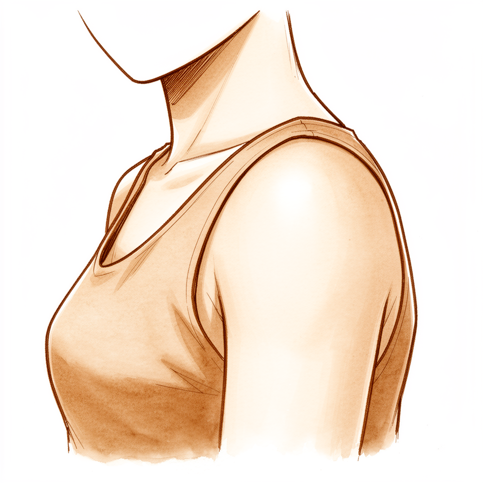
Những triệu chứng bạn đang gặp phải
Rối loạn vận động xương bả vai nghĩa là xương bả vai của bạn nằm hoặc chuyển động khác với bình thường khi bạn nâng lên và hạ cánh tay xuống. Đây là một dấu hiệu của vấn đề ở vai chứ không phải một bệnh lý riêng biệt. Bạn có thể nhận thấy bờ trong của xương bả vai nhô ra, đặc biệt khi bạn nâng cánh tay lên. Xương bả vai có thể nâng lên quá sớm, hoặc nhún lên về phía tai, thay vì trượt nhẹ nhàng.
Cơn đau thường nằm quanh đỉnh vai và ngay tại xương bả vai. Nhiều người cũng cảm thấy đau âm ỉ dọc bên cổ, nơi có cơ thang. Nâng cánh tay lên quá đầu thường làm cơn đau nặng hơn. Với tay lên kệ cao, phơi quần áo, hoặc nhấc đồ lên giá có thể khó khăn. Nằm nghiêng về bên đó thường gây đau, và cơn đau âm ỉ có thể khiến bạn thức giấc vào ban đêm. Vai có thể cứng nhất khi mới thức dậy và đỡ dần khi bạn cử động.
Vấn đề ở xương bả vai thường xuất hiện cùng với những vấn đề khác ở vai, chẳng hạn như hội chứng chèn ép (các gân ở đỉnh vai bị kẹp lại), rách chóp xoay, mất vững khớp vai, hoặc gãy xương đòn. Việc nó là nguyên nhân gây đau hay chỉ là phản ứng của cơ thể trước cơn đau thường không rõ ràng. Tình trạng này cũng thường gặp ở những người hoàn toàn không bị đau, kể cả các vận động viên ném hoặc nâng tạ qua đầu.
Nếu triệu chứng của bạn không thuyên giảm, nặng dần lên trong nhiều tuần, khiến bạn thức giấc vào ban đêm, hoặc khiến bạn không thể làm việc hay sử dụng cánh tay, hãy đi khám bác sĩ đa khoa hoặc đề nghị được bác sĩ chuyên khoa đánh giá. Nếu cơn đau lan xuống cánh tay kèm theo tê, ngứa ran hoặc yếu, hãy đi khám bác sĩ để kiểm tra cổ và các dây thần kinh.
Chuyện gì đang thực sự xảy ra
Xương bả vai của bạn không được gắn vào xương sườn bằng một khớp. Nó trượt trên mặt sau của thành ngực nhờ một lớp mô mỏng trơn láng, giống như một ngăn kéo trượt trên ray của nó. Xương bả vai được giữ đúng chỗ và được di chuyển hoàn toàn nhờ các cơ, bao gồm cơ thang ở phía sau cổ và vai, và cơ răng trước ở mặt bên của xương sườn.
Khi bạn nâng cánh tay, phần chỏm cầu của khớp vai đảm nhận phần lớn chuyển động, nhưng xương bả vai cũng cần xoay lên trên và nghiêng ra sau cùng lúc. Hãy hình dung nó như một bệ đỡ vững chắc xoay sang bên để nhường chỗ khi cánh tay nâng lên. Chuyển động đó nâng phần mái xương phía trên khớp vai lên, để các gân bên dưới có chỗ trượt mà không bị kẹp lại. Khoảng hai phần ba chuyển động nâng tay đến từ chính khớp vai, và khoảng một phần ba đến từ xương bả vai.
Khi sự phối hợp này bị rối loạn, xương bả vai có thể nằm quá ra phía trước, không xoay lên trên được, hoặc nhún lên thay vì trượt. Khi đó phần mái phía trên khớp vai không được nâng lên đủ, và các gân bên dưới có thể bị chèn ép khi bạn nâng cánh tay. Chính sự chèn ép đó gây ra cơn đau bạn cảm thấy ở đỉnh vai và quanh xương bả vai.
Bạn nên biết rằng đây là một dấu hiệu chứ không phải một bệnh lý tự thân. Nó thường xuất hiện cùng với các vấn đề khác ở vai, chẳng hạn như gân bị kẹp, gân bị rách, khớp vai lỏng lẻo, hoặc gãy xương đòn. Đôi khi vấn đề ở xương bả vai góp phần gây ra những rắc rối đó. Đôi khi nó chỉ đơn giản là cách cơ thể bạn bảo vệ một bờ vai đang đau, giống như bạn đi khập khiễng để đỡ cho một bên mắt cá chân bị đau. Điều gì xảy ra trước thường không rõ ràng. Tình trạng này cũng thường gặp ở những người hoàn toàn không bị đau, vì vậy chỉ riêng việc xương bả vai trông khác thường không có nghĩa là vai của bạn bị tổn thương.
Những biện pháp chúng tôi có thể áp dụng
Bác sĩ Kieran Hirpara, bác sĩ phẫu thuật chi trên tại Bệnh viện tư nhân Mater Rockhampton, sẽ bắt đầu bằng các phương pháp điều trị ít xâm lấn nhất phù hợp với tình trạng của bạn. Với hầu hết mọi người, điều đó có nghĩa là vật lý trị liệu, và đây là phương pháp điều trị chính cho vấn đề này. Chuyên viên vật lý trị liệu sẽ lập chương trình tập luyện. Chương trình tập trung vào khả năng kiểm soát và sức mạnh của các cơ quanh xương bả vai, kéo giãn các cơ bị co rút ở phía trước ngực, và xem xét tư thế của bạn. Mục tiêu là khôi phục vị trí và chuyển động bình thường của xương bả vai, để nó giữ vững trong khi bạn sử dụng cánh tay qua đầu. Tập luyện giúp giảm đau và cải thiện chức năng. Vì vấn đề về cơ của mỗi người mỗi khác, chuyên viên vật lý trị liệu sẽ điều chỉnh mục tiêu và bài tập theo những gì họ phát hiện khi khám cho bạn. Song song với việc này, chúng tôi điều trị vấn đề ở vai đi kèm, chẳng hạn như gân bị kẹp hoặc gân bị rách. Thông thường, bệnh nhân được bác sĩ đa khoa giới thiệu đến phòng khám chúng tôi; nếu vật lý trị liệu viên khuyên bạn nên đến gặp chúng tôi, bạn vẫn cần có giấy giới thiệu từ bác sĩ đa khoa để được hưởng mức hoàn trả chi phí từ Medicare.
Phẫu thuật nhằm vào vấn đề nền ở vai, chứ không nhằm vào bản thân xương bả vai. Nếu triệu chứng của bạn vẫn kéo dài dù đã tập vật lý trị liệu, chúng tôi sẽ đánh giá lại cho bạn. Tùy theo triệu chứng và kết quả thăm khám, việc này có thể bao gồm siêu âm hoặc chụp MRI để kiểm tra chỗ đã khâu sửa, các xét nghiệm thần kinh, hoặc kiểm tra cổ.
Những điều có thể mong đợi
Không có một mốc thời gian chung cho vấn đề này, vì đây là dấu hiệu của một vấn đề ở vai chứ không phải một bệnh lý riêng biệt. Tình trạng kéo dài bao lâu phần lớn phụ thuộc vào vấn đề ở vai đi kèm. Một số người thấy chuyển động bất thường của xương bả vai giảm dần khi cơn đau vai thuyên giảm. Ở những người khác, nó vẫn còn kéo dài, hoặc lúc có lúc không, ngay cả khi cơn đau đã dịu đi.
Nếu được điều trị tốt, diễn tiến thường gặp là tiến triển đều đặn trong nhiều tuần đến nhiều tháng chứ không phải khỏi ngay lập tức. Vật lý trị liệu tác động đến cơn đau và chức năng của vai, và đó là điều hầu hết mọi người nhận thấy đầu tiên. Bản thân chuyển động của xương bả vai thường thay đổi chậm hơn so với cơn đau. Sau phẫu thuật vai, kể cả phẫu thuật khâu chóp xoay, xương bả vai thường chuyển động bất thường cả trước và sau ca mổ. Tình trạng này thường cải thiện qua nhiều tháng khi vai hồi phục, nhưng có thể chưa trở lại hoàn toàn bình thường sau một năm. Sáu tháng phục hồi chức năng sau phẫu thuật khâu chóp xoay cũng thường chưa đủ để giải quyết hoàn toàn tình trạng này. Những người có chuyển động xương bả vai vẫn bất thường thường có kết quả kém hơn một chút so với những người có chuyển động hồi phục.
Thành thật mà nói, bằng chứng ở đây chưa thống nhất. Tập luyện giúp giảm đau và cải thiện chức năng một cách đáng tin cậy, nhưng việc tập luyện có làm thay đổi cách bản thân xương bả vai chuyển động hay không thì kém rõ ràng hơn. Một số kiểu chuyển động bất thường cải thiện sau phẫu thuật điều trị vấn đề nền, và những người có chuyển động hồi phục thường có chức năng tốt hơn. Ở những người khác, kiểu chuyển động này vẫn tồn tại mà không gây hại rõ ràng. Tình trạng này cũng thường gặp ở những người hoàn toàn không có triệu chứng, vì vậy chỉ riêng việc xương bả vai trông khác thường không có nghĩa là vai của bạn đang xấu đi.
Để mặc không điều trị thường không khiến tình trạng trở nên nguy hiểm, nhưng cũng không làm cơn đau âm ỉ biến mất. Nếu triệu chứng của bạn không thuyên giảm, nặng dần lên trong nhiều tuần, khiến bạn thức giấc vào ban đêm, hoặc khiến bạn không thể làm việc hay sử dụng cánh tay, hãy đi khám bác sĩ đa khoa hoặc đề nghị được bác sĩ chuyên khoa đánh giá. Nếu cơn đau lan xuống cánh tay kèm theo tê, ngứa ran hoặc yếu, hãy đi khám bác sĩ để kiểm tra cổ và các dây thần kinh.
Khi nào nên đi khám bác sĩ
Hầu hết các vấn đề về xương bả vai không cấp bách. Nhưng một số dấu hiệu cho thấy đã đến lúc cần được giúp đỡ thay vì tiếp tục chờ đợi.
Hãy đi khám bác sĩ đa khoa nếu triệu chứng của bạn không thuyên giảm, nặng dần lên trong nhiều tuần, khiến bạn thức giấc vào ban đêm, hoặc khiến bạn không thể làm việc hay sử dụng cánh tay. Hãy đề nghị được bác sĩ chuyên khoa đánh giá nếu bờ trong của xương bả vai liên tục nhô ra, hoặc tình trạng vai nhún lên khi bạn nâng cánh tay không đỡ khi nghỉ ngơi.
Cơn đau lan xuống cánh tay kèm theo tê, ngứa ran hoặc yếu cho thấy vấn đề nằm ở cổ hoặc một dây thần kinh chứ không phải ở xương bả vai. Hãy đi khám bác sĩ để được kiểm tra.
Nếu xương bả vai đột ngột nhô ra khỏi thành ngực, đặc biệt khi kèm theo đau hoặc yếu, hãy đề nghị bác sĩ đa khoa đánh giá sớm thay vì chờ đợi nhiều tuần.
Phân tích sâu hơn
Advanced reading: the deeper science (optional)
Phần này đi sâu hơn mức bạn cần để đưa ra quyết định điều trị cho bản thân. Rối loạn vận động xương bả vai đáng để đọc thêm vì nó rất thường bị hiểu sai: nó thường gặp ở những người không có triệu chứng, hiếm khi tự nó giải thích được một vấn đề ở vai, và sau phẫu thuật vai, nó cải thiện chậm hơn so với bản thân khớp vai.
Một dấu hiệu, không phải một chẩn đoán
Đồng thuận quốc tế "scapular summit" (hội nghị chuyên đề về xương bả vai) mô tả rối loạn vận động xương bả vai là sự thay đổi ở vị trí nghỉ hoặc chuyển động của xương bả vai, gặp ở một tỷ lệ cao các chấn thương vai. Đồng thuận này cũng kết luận rằng vai trò chính xác của nó trong việc gây ra hoặc làm nặng thêm các vấn đề ở vai chưa được xác định rõ, và tốt nhất nên xem nó là một suy giảm có thể có của chức năng vai chứ không phải một chẩn đoán [1]. Một tổng quan trước đó gọi nó là một phản ứng không đặc hiệu với một bờ vai đau chứ không phải đặc điểm của riêng bệnh lý nào [2].
Tình trạng này cũng thường gặp khi không có triệu chứng. Trong 12 nghiên cứu trên 1,401 vận động viên, nó được ghi nhận ở 61% vận động viên chơi các môn qua đầu và 33% vận động viên khác [3]. Việc nó có dự báo chấn thương hay không vẫn còn đáng ngờ: trong một phân tích gộp trên 923 vận động viên được theo dõi tiến cứu, nó có mặt ở 46%, và không làm tăng đáng kể nguy cơ chấn thương vai về sau (tỷ số nguy cơ 1.07, 95% CI 0.85 đến 1.34) [4].
Cách đánh giá và giới hạn của các xét nghiệm
Nghiệm pháp lâm sàng thường dùng là quan sát xương bả vai từ phía sau trong khi người bệnh nâng lên và hạ cánh tay xuống vài lần, thường cầm tạ nhẹ, rồi phân loại chuyển động là bình thường hoặc bất thường [5]. Mức độ đồng thuận giữa những người khám ở mức trung bình đến tốt: ở 40 bệnh nhân bị hội chứng chèn ép, hệ số kappa giữa hai người đánh giá là 0.59 (đồng thuận 86%). Trong cùng nghiên cứu đó, những người có và không có rối loạn vận động không khác nhau về điểm số vai ở thời điểm ban đầu hay sau ba tháng [6]. Đây là lý do các bác sĩ phẫu thuật xem một nghiệm pháp dương tính là một phần của việc thăm khám, chứ không phải một chẩn đoán.
Xương bả vai nhô cánh do tổn thương thần kinh là một vấn đề riêng biệt. Ở 96 bệnh nhân bị xương bả vai nhô cánh do yếu cơ răng trước đã được chứng minh qua xét nghiệm thần kinh, 88% có tổn thương thần kinh ngực dài, thường gặp nhất là do teo cơ do đau thần kinh (47) hoặc chấn thương (33). Trong số những người bị teo cơ do đau thần kinh, 22 người hồi phục mà không cần điều trị trong vòng ba năm, nhưng không ai hồi phục hoàn toàn khi tình trạng nhô cánh đã kéo dài hơn ba năm [7].
Rách chóp xoay: nguyên nhân hay hậu quả?
Một tổng quan hệ thống gồm 16 nghiên cứu (335 trường hợp rách) cho thấy xương bả vai chuyển động bất thường trong mọi nghiên cứu về rách chóp xoay, nhưng không theo một kiểu nhất quán nào. Các vết rách lớn hơn cho thấy nhiều thay đổi hơn, và các vết rách gây đau nhiều thay đổi hơn so với các vết rách không đau; các vết rách không đau không khác biệt so với vai khỏe mạnh. Tổng quan này không thể kết luận thay đổi nào là thích nghi và thay đổi nào là có hại [8]. Một so sánh cho thấy xương bả vai xoay lên trên ít hơn, và cơ nâng vai hoạt động nhiều hơn, ở các vết rách gây đau so với các vết rách không đau, trong khi cơ thang phần trên hoạt động nhiều hơn so với vai khỏe mạnh [9]. Ở các vết rách rất lớn đã lấy lại được khả năng cử động nhờ vật lý trị liệu, chụp PET cho thấy cơ nâng vai và các cơ trám hoạt động mạnh hơn, phù hợp với việc các cơ xương bả vai bù trừ cho chóp xoay [10].
Điều gì xảy ra sau phẫu thuật khâu chóp xoay
Các nghiên cứu không thống nhất với nhau, và chính sự không thống nhất đó là thông điệp chính.
- Tình trạng thường cải thiện. Ở 35 bệnh nhân được đánh giá bằng CT trước và sau phẫu thuật khâu, 23 (66%) có rối loạn vận động từ trước. 12 trong số 23 người cải thiện sau phẫu thuật, chủ yếu là kiểu giảm độ nghiêng ra sau. Sự cải thiện đi kèm với khả năng nâng tay ra trước tốt hơn và điểm Constant cao hơn, và không có trường hợp khâu nào không liền ở nhóm cải thiện [11].
- Tình trạng có thể trở về bình thường. Ở 26 bệnh nhân được theo dõi bằng cảm biến điện từ trong một năm, chuyển động của xương bả vai dần tiến về kiểu chuyển động của vai bên kia, và khả năng dạng tay cải thiện 20° [12].
- Tình trạng có thể vẫn tồn tại. Ở 21 bệnh nhân được đo bằng cảm biến đeo trên người ở thời điểm 12 tháng trở lên, khả năng cử động và điểm Constant cải thiện, nhưng nhịp chuyển động của xương bả vai không thay đổi đáng kể [13]. Trong một nghiên cứu nhỏ khác, sáu tháng phục hồi chức năng sau phẫu thuật khâu không khôi phục hoàn toàn chuyển động bình thường của xương bả vai [14].
- Tình trạng này ảnh hưởng đến kết quả. Ở 48 bệnh nhân được theo dõi trong một năm phục hồi chức năng sau phẫu thuật khâu, sự bù trừ của xương bả vai giảm đi nhưng vẫn nhiều hơn so với bên kia. Những người mà chuyên viên vật lý trị liệu quan sát thấy có bù trừ có điểm số kém hơn, cử động ít hơn và sức mạnh kém hơn ở mọi thời điểm [15]. Ở 110 bệnh nhân từ 6 đến 18 tháng sau phẫu thuật khâu, rối loạn vận động đi kèm với chức năng kém hơn, và sức bền của các cơ xương bả vai dự báo được kết quả [16].
Phục hồi chức năng có mục tiêu có thể thay đổi được tình trạng này không? Ở 60 bệnh nhân bị rối loạn vận động sau phẫu thuật khâu, việc bổ sung tập mạnh các cơ xương bả vai yếu cụ thể (được xác định qua điện cơ EMG) cho điểm Constant là 87.2 so với 83.9, và khả năng nâng tay ra trước là 137° so với 128°, ở tuần thứ 16. Khoảng chênh lệch đó không lớn, và nghiên cứu không phải là thử nghiệm ngẫu nhiên [17]. Một phân tích gộp năm 2026 cho thấy phục hồi chức năng nhắm vào xương bả vai giúp giảm đau và cải thiện chức năng trong điều trị chóp xoay nói chung, nhưng bằng chứng sau phẫu thuật quá ít để có thể gộp lại [18]. Nói rộng hơn, tập luyện giúp giảm đau và cải thiện chức năng một cách đáng tin cậy trong hội chứng chèn ép [19], nhưng bằng chứng cho thấy tập luyện làm thay đổi chuyển động của bản thân xương bả vai thì còn mâu thuẫn [20].
Một nghiên cứu trên động vật đặt ra khả năng rằng xương bả vai chuyển động bất thường có thể làm ảnh hưởng đến quá trình lành của gân đã được khâu: ở chuột, việc cắt các dây thần kinh điều khiển xương bả vai trước khi khâu cơ trên gai dẫn đến chỗ khâu yếu hơn và kém tổ chức hơn [21]. Điều này chưa được chứng minh ở người.
Sau các phẫu thuật vai khác
- Phẫu thuật Latarjet. Rối loạn vận động được ghi nhận ở 5 trên 20 bệnh nhân sau phẫu thuật Latarjet và không ai trong 20 bệnh nhân sau phẫu thuật làm vững bằng ghép xương không động đến mỏm quạ. Những người bị rối loạn vận động có điểm số thấp hơn [22]. Một nhóm nghiên cứu sau đó ghi nhận tình trạng này ở 25% sau phẫu thuật tiêu chuẩn và không ai trong 20 bệnh nhân sau khi cơ ngực bé được khâu trở lại vào mỏm quạ. 4 trên 5 trường hợp đã ổn định sau sáu tháng phục hồi chức năng chuyên biệt [23].
- Thay khớp vai. Sau thay khớp vai đảo ngược, xương bả vai đảm nhận phần lớn hơn công việc nâng cánh tay so với một khớp vai bình thường (một phân tích gộp trên 464 bệnh nhân) [24]. Ở 85 ca thay khớp vai giải phẫu và đảo ngược, việc xương bả vai đóng góp nhiều hơn sau phẫu thuật đi kèm với đau nhiều hơn và chức năng kém hơn [25].
- Xương đòn và khớp cùng đòn. Rối loạn vận động được ghi nhận ở 9 trên 24 người sau gãy xương đòn di lệch, và những người này đau nhiều hơn và có điểm số kém hơn. Tình trạng này gặp ở 1 trên 12 người được điều trị bằng phẫu thuật và 8 trên 12 người được điều trị không phẫu thuật [26]. Sau phẫu thuật điều trị trật khớp cùng đòn cấp tính, tình trạng này được ghi nhận ở 4 trên 34 người [27].
Đau cơ thang và dấu hiệu nhún vai
Có rất ít nghiên cứu về tình trạng đau âm ỉ ở cơ thang hoặc bên cổ sau phẫu thuật vai. Chưa có nghiên cứu nào đo lường tần suất xảy ra của nó. Bằng chứng gần nhất là "dấu hiệu nhún vai", khi toàn bộ đai vai nâng lên lúc cánh tay được nâng. Ở 982 bệnh nhân liên tiếp được khám trước phẫu thuật, dấu hiệu này có mặt ở 51%, thường gặp nhất trong viêm dính bao khớp vai (95%). Nó đi kèm với cứng khớp, yếu và đau về đêm, và không đặc hiệu cho rách chóp xoay [28]. Điều đó phù hợp với một cách giải thích đơn giản cho tình trạng đau cơ thang trong khi vai đang hồi phục: các cơ nâng xương bả vai phải làm việc nhiều hơn để nâng một cánh tay bị cứng hoặc yếu, như đã thấy ở các vết rách chóp xoay gây đau [9]. Dây đeo của đai treo tay cũng có thể góp phần. Những cách giải thích này hợp lý, nhưng chưa được kiểm chứng. Cơn đau lan xuống cánh tay kèm theo tê, ngứa ran hoặc yếu thì lại cho thấy vấn đề ở cổ hoặc một dây thần kinh và cần được kiểm tra.
Tài liệu tham khảo
[1] Kibler WB, Ludewig PM, McClure PW, et al. Clinical implications of scapular dyskinesis in shoulder injury: the 2013 consensus statement from the 'scapular summit'. Br J Sports Med. 2013;47(14):877-85. https://doi.org/10.1136/bjsports-2013-092425
[2] Kibler WB, Sciascia A. Current concepts: scapular dyskinesis. Br J Sports Med. 2010;44(5):300-5. Epub 2009. https://doi.org/10.1136/bjsm.2009.058834
[3] Burn MB, McCulloch PC, Lintner DM, et al. Prevalence of Scapular Dyskinesis in Overhead and Nonoverhead Athletes. Orthop J Sports Med. 2016;4(2):2325967115627608. https://doi.org/10.1177/2325967115627608
[4] Hogan C, Corbett JA, Ashton S, et al. Scapular Dyskinesis Is Not an Isolated Risk Factor for Shoulder Injury in Athletes: A Systematic Review and Meta-analysis. Am J Sports Med. 2021;49(10):2843-53. Epub 2020. https://doi.org/10.1177/0363546520968508
[5] Tate AR, McClure P, Kareha S, et al. A Clinical Method for Identifying Scapular Dyskinesis, Part 2: Validity. J Athl Train. 2009;44(2):165-73. https://doi.org/10.4085/1062-6050-44.2.165
[6] Christiansen DH, Møller AD, Vestergaard JM, et al. The scapular dyskinesis test: Reliability, agreement, and predictive value in patients with subacromial impingement syndrome. J Hand Ther. 2017;30(2):208-13. https://doi.org/10.1016/j.jht.2017.04.002
[7] Ng CY, Wu F. Scapular winging secondary to serratus anterior dysfunction: analysis of clinical presentations and etiology in a consecutive series of 96 patients. J Shoulder Elbow Surg. 2021;30(10):2336-43. https://doi.org/10.1016/j.jse.2021.02.012
[8] Barcia AM, Makovicka JL, Spenciner DB, et al. Scapular motion in the presence of rotator cuff tears: a systematic review. J Shoulder Elbow Surg. 2021;30(7):1679-92. https://doi.org/10.1016/j.jse.2020.12.012
[9] Ishikawa H, Muraki T, Morise S, et al. Differences in scapular motion and parascapular muscle activities among patients with symptomatic and asymptomatic rotator cuff tears, and healthy individuals. JSES Int. 2021;5(2):238-46. https://doi.org/10.1016/j.jseint.2020.10.014
[10] Chuang HC, Chiu NT, Liu ZW, et al. Compensatory Activation of Periscapular Muscles Aids Active Abduction in Patients With Massive Rotator Cuff Tears. Clin Orthop Relat Res. 2025;483(12):2295-305. https://doi.org/10.1097/corr.0000000000003556
[11] Song HE, Oh K, Yoon JP, et al. Improvement in scapular dyskinesis after rotator cuff repair and subacromial decompression. Knee Surg Sports Traumatol Arthrosc. 2021;29(12):3961-70. https://doi.org/10.1007/s00167-021-06681-x
[12] Kolk A, de Witte PB, Henseler JF, et al. Three-dimensional shoulder kinematics normalize after rotator cuff repair. J Shoulder Elbow Surg. 2016;25(6):881-9. https://doi.org/10.1016/j.jse.2015.10.021
[13] Mercurio M, Cofano E, Mancuso C, et al. Persistent scapular dyskinesis after arthroscopic rotator cuff repair: a prospective study. JSES Int. 2026;10(2):101420. https://doi.org/10.1016/j.jseint.2025.101420
[14] Nyőgér Z, Molnár CA, Szakály N, et al. Scapula dyskinesis in medium‐sized full‐thickness rotator cuff tear after subacromial Lidocaine infiltration and rotator cuff reconstruction. J Exp Orthop. 2025;12(1):e70154. https://doi.org/10.1002/jeo2.70154
[15] Baumgarten KM, Osborn R, Schweinle WE, et al. Scapular substitution after rotator cuff repair correlates with postoperative patient outcome. Int J Sports Phys Ther. 2018;13(4):687-99. https://doi.org/10.26603/ijspt20180687
[16] Alzahrani WM, Baltow BA, Alanazi ZA, et al. Scapular endurance, dyskinesis, and functional outcomes after rotator cuff repair: A cross-sectional mediation analysis. PLoS One. 2026;21(9):e0357765. https://doi.org/10.1371/journal.pone.0357765
[17] Sun JN, Zheng QY, Wang RS, et al. An additional rehabilitation program to improve postoperative outcomes in patients with rotator cuff tear and scapular dyskinesis: a propensity score-matched study. BMC Musculoskelet Disord. 2025;26(1):338. https://doi.org/10.1186/s12891-025-08596-x
[18] Alshehri AF, Samarkandi WO, Alshahrani KA, et al. Clinical Value of Scapular-Targeted Rehabilitation Within the Rotator Cuff Care Continuum: A Systematic Review and Meta-analysis of Conservative and Postoperative Evidence. Orthop Rev (Pavia). 2026;18. https://doi.org/10.52965/001c.165083
[19] Zhong Z, Zang W, Tang Z, et al. Effect of scapular stabilization exercises on subacromial pain (impingement) syndrome: a systematic review and meta-analysis of randomized controlled trials. Front Neurol. 2024;15:1357763. https://doi.org/10.3389/fneur.2024.1357763
[20] Nodehi Moghadam A, Rahnama L, Noorizadeh Dehkordi S, Abdollahi S. Exercise therapy may affect scapular position and motion in individuals with scapular dyskinesis: a systematic review of clinical trials. J Shoulder Elbow Surg. 2020;29(1):e29-e36. https://doi.org/10.1016/j.jse.2019.05.037
[21] Reuther KE, Tucker JJ, Thomas SJ, et al. Effect of scapular dyskinesis on supraspinatus repair healing in a rat model. J Shoulder Elbow Surg. 2015;24(8):1235-42. https://doi.org/10.1016/j.jse.2014.12.029
[22] Carbone S, Moroder P, Runer A, et al. Scapular dyskinesis after Latarjet procedure. J Shoulder Elbow Surg. 2016;25(3):422-7. https://doi.org/10.1016/j.jse.2015.08.001
[23] Candela V, Scacchi M, De Meo D, et al. The Pectoralis Minor–Repairing Latarjet: A Modified Technique to Prevent Postoperative Scapular Dyskinesis After the Latarjet Procedure. Am J Sports Med. 2025;53(7):1716-20. https://doi.org/10.1177/03635465251332270
[24] Johnson MA, Cogsil T, White AE, et al. Impact of reverse total shoulder arthroplasty on scapulohumeral rhythm: a systematic review and meta-analysis. J Shoulder Elbow Surg. 2026;35(4):1117-28. https://doi.org/10.1016/j.jse.2025.08.010
[25] Khawaja SR, Hardrick JC, Und Aspang JS, et al. Total shoulder arthroplasty and scapulohumeral rhythm: the clinical impact of achieving native shoulder mechanics. J Shoulder Elbow Surg. 2026. https://doi.org/10.1016/j.jse.2026.07.013
[26] Shields E, Behrend C, Beiswenger T, et al. Scapular dyskinesis following displaced fractures of the middle clavicle. J Shoulder Elbow Surg. 2015;24(12):e331-e336. https://doi.org/10.1016/j.jse.2015.05.047
[27] Murena L, Canton G, Vulcano E, Cherubino P. Scapular dyskinesis and SICK scapula syndrome following surgical treatment of type III acute acromioclavicular dislocations. Knee Surg Sports Traumatol Arthrosc. 2013;21(5):1146-50. Epub 2012. https://doi.org/10.1007/s00167-012-1959-9
[28] Jia X, Ji JH, Petersen SA, et al. Clinical Evaluation of the Shoulder Shrug Sign. Clin Orthop Relat Res. 2008;466(11):2813-9. https://doi.org/10.1007/s11999-008-0331-3