Lý do phẫu thuật này được đề xuất
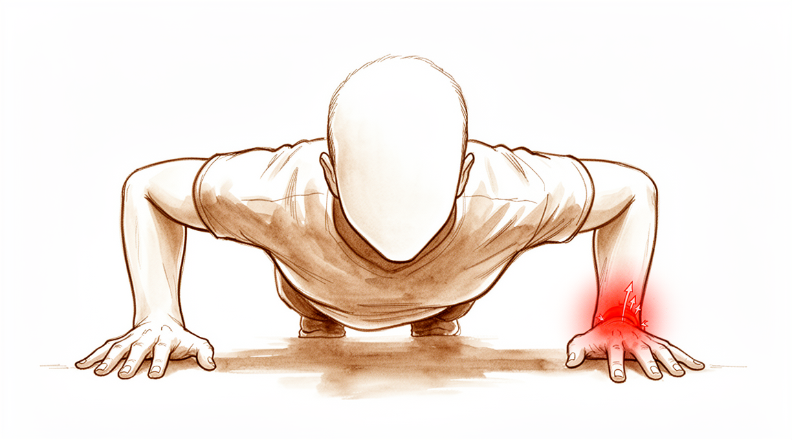
Bác sĩ Kieran Hirpara, bác sĩ phẫu thuật chi trên tại Bệnh viện tư nhân Mater Rockhampton, sẽ bắt đầu bằng các phương pháp điều trị ít xâm lấn nhất phù hợp với tình trạng của bạn. TFCC (phức hợp sụn sợi tam giác) là một cấu trúc gồm sụn và dây chằng nằm ở phía ngón út của cổ tay. Khâu sửa nghĩa là khâu cấu trúc này trở lại vị trí nơi nó bị rách. Chúng tôi thường thử trị liệu tay, nẹp cố định và thay đổi hoạt động trước, và cân nhắc phẫu thuật khi tình trạng đau hoặc mất vững vẫn tiếp diễn sau đó. Thông thường, bệnh nhân được bác sĩ đa khoa giới thiệu đến phòng khám chúng tôi; nếu vật lý trị liệu viên khuyên bạn nên đến gặp chúng tôi, bạn vẫn cần có giấy giới thiệu từ bác sĩ đa khoa để được hưởng mức hoàn trả chi phí từ Medicare. Buổi khám tại phòng khám, với việc hỏi bệnh sử, thăm khám và chụp hình ảnh khi cần thiết, sẽ giúp xác định chẩn đoán.
Ca phẫu thuật này được đề nghị cho những vết rách có thể khâu sửa được, khi khớp không có thay đổi do thoái hóa. Mục tiêu là giảm đau, khôi phục độ vững cho khớp giữa hai xương cẳng tay, và giữ cho cổ tay và cẳng tay của bạn cử động được. Việc khâu sửa được thực hiện bằng phẫu thuật nội soi, thường là phẫu thuật trong ngày dưới gây mê toàn thân. Khoảng ba trong bốn người trở lại hoạt động đầy đủ; một số người thì không. Chúng tôi sẽ trao đổi với bạn xem ca phẫu thuật này có phù hợp với bạn hay không, và cùng nhau quyết định.
Trước khi phẫu thuật
Khi ca mổ đã được đặt lịch, phòng khám của chúng tôi sẽ xác nhận thông tin nhập viện và hướng dẫn nhịn ăn cho bạn. Bạn cần ngừng ăn và uống bảy giờ trước ca mổ, để chúng tôi có thể đưa ca mổ của bạn lên sớm hơn nếu lịch mổ diễn ra sớm hơn dự kiến. Hãy cho chúng tôi biết về bất kỳ thuốc chống đông máu hoặc thuốc thường dùng nào bạn đang uống, vì một số loại cần tạm ngưng trước đó. Hãy mang theo danh sách ghi rõ các loại thuốc bạn đang dùng, và mặc quần áo rộng rãi, thoải mái, có tay áo đủ rộng để luồn qua cổ tay đang băng bó. Bạn sẽ cần người đưa về nhà, vì gây mê toàn thân khiến việc lái xe không an toàn trong suốt thời gian còn lại của ngày hôm đó. Các hình ảnh chẩn đoán như X-quang hoặc MRI có thể đã được thực hiện, hoặc chúng tôi có thể sắp xếp chụp để lên kế hoạch cho ca mổ. Nếu bạn mắc các bệnh lý khác, bạn có thể cần xét nghiệm máu hoặc được bác sĩ gây mê khám trước.
Vào ngày phẫu thuật
Bạn sẽ đến khu vực tiếp nhận bệnh nhân phẫu thuật của bệnh viện, nơi nhân viên làm thủ tục nhập viện và chuẩn bị cho bạn trước ca mổ. Bạn sẽ gặp bác sĩ gây mê – người gây mê cho bạn và chăm sóc bạn trong suốt ca phẫu thuật. Ca phẫu thuật này được thực hiện dưới gây mê toàn thân. Bạn sẽ hoàn toàn ngủ say trong suốt quá trình phẫu thuật. Một số bệnh nhân cũng có thể được chặn dây thần kinh vùng để giảm đau sau mổ; quyết định này do bác sĩ gây mê đưa ra vào ngày phẫu thuật dựa trên tình trạng cụ thể của từng người. Sau đó, bạn được đưa vào phòng mổ để tiến hành phẫu thuật.
Khi ca mổ kết thúc, bạn sẽ tỉnh dậy tại khu vực hồi sức. Các điều dưỡng sẽ theo dõi tình trạng của bạn cho đến khi tác dụng của thuốc mê hết. Khi sức khỏe ổn định, tùy thuộc vào loại phẫu thuật và diễn biến hồi phục của bạn, bạn sẽ được chuyển sang phòng bệnh hoặc về nhà.
Quy trình phẫu thuật
Bác sĩ phẫu thuật bắt đầu bằng phẫu thuật nội soi. Một camera rất nhỏ được đưa vào cổ tay qua các đường rạch nhỏ ở mu cổ tay, để có thể nhìn rõ vết rách. Cách khâu sửa cụ thể phụ thuộc vào những gì quan sát được, vì vậy không thể cam kết trước về phương pháp.
Nếu vết rách nằm ở vành ngoài của TFCC, nó được khâu trở lại vào bao khớp, tức là lớp lót của khớp. Nếu vết rách nằm ở chỗ dây chằng bị bứt khỏi xương trụ, xương nhỏ ở phía ngón út của cẳng tay, nó sẽ được gắn lại vào xương. Việc này có thể được thực hiện bằng một neo nhỏ đặt trong xương, hoặc qua một đường hầm nhỏ được khoan trong xương, đôi khi qua một đường mổ mở ngắn. Vết rách không thể khâu sửa được sẽ được làm gọn thay vào đó, nghĩa là các mép xơ xác được cắt gọt đi. Bác sĩ phẫu thuật sẽ giải thích những gì đã được thực hiện khi bạn tỉnh dậy.
Các đường rạch được khâu lại bằng chỉ và phủ băng gạc.
Sau khi phẫu thuật
Bạn sẽ tỉnh dậy tại khu hồi sức với các điều dưỡng ở gần trong khi thuốc mê hết tác dụng. Đây thường là ca mổ trong ngày, vì vậy bạn có thể dự kiến về nhà ngay trong ngày, mặc dù đôi khi bệnh nhân phải ở lại qua đêm. Cổ tay của bạn sẽ được bó bột dưới khuỷu hoặc đeo nẹp cứng, với cẳng tay xoay ngửa một phần và khuỷu tay được tự do cử động. Cơn đau được kiểm soát bằng thuốc mà bạn có thể dùng ở nhà; cảm giác hơi khó chịu là bình thường. Hãy kê cao bàn tay trên gối khi nghỉ ngơi để giúp giảm sưng. Hãy cử động nhẹ các ngón tay khi có thể. Nếu bạn đã được chặn dây thần kinh, tê và yếu trong 24 giờ đầu là điều bình thường; khi thuốc hết tác dụng, hãy gọi cho phòng khám nếu bạn bị tê hoặc không thể cử động bàn tay hay các ngón tay. Nên có người ở cùng bạn trong 24 giờ đầu tiên. Chúng tôi để nguyên băng gạc khoảng 10 ngày; xin đừng tháo ra trước thời gian đó trừ khi chúng tôi yêu cầu. Chúng tôi sẽ thay hoặc tháo băng khi gặp bạn.
Quá trình hồi phục
Cổ tay của bạn sẽ được bó bột dưới khuỷu hoặc đeo nẹp cứng trong sáu tuần, với cẳng tay xoay ngửa một phần và khuỷu tay được tự do. Điều này bảo vệ chỗ khâu sửa trong khi nó lành lại. Bạn không thể lái xe khi còn bó bột hoặc đeo nẹp. Khi đã tháo bỏ và bác sĩ phẫu thuật cho phép, bạn có thể lái xe trở lại; bản thân chiếc nẹp nhẹ có thể tháo rời mà bạn đeo sau đó không ngăn cản bạn lái xe. Để biết thêm chi tiết, hãy xem Lái xe sau phẫu thuật chi trên.
Sau sáu tuần, bạn sẽ chuyển sang đeo nẹp có thể tháo rời trong khi cổ tay lấy lại khả năng cử động và sức mạnh. Chương trình phục hồi chức năng của bạn là trị liệu tay, không phải vật lý trị liệu. Chương trình này do Ruby Doolan tại Extend Rehabilitation hướng dẫn. Ruby là chuyên gia trị liệu tay: cô ấy hướng dẫn các bài tập của bạn và làm bất kỳ nẹp nào bạn cần.
Công việc văn phòng và các công việc nhẹ hằng ngày thường được tiếp tục sau sáu đến tám tuần. Lao động chân tay, thể thao và tập gym cần nhiều thời gian hơn, khoảng bốn đến sáu tháng. Kết quả cuối cùng, khi tình trạng đau và sức mạnh không còn cải thiện thêm, đến vào khoảng 12 tháng.
Cơn đau thường cải thiện trước sức mạnh và chức năng. Cổ tay của bạn có thể cảm thấy dễ sử dụng hơn từ rất lâu trước khi nó cảm thấy khỏe, vì vậy hãy kiên nhẫn với những công việc nặng hơn.
Gần một nửa số người gặp một đợt thụt lùi tạm thời sau khi đã đạt mức hoạt động cao nhất. Tình trạng này sẽ qua theo thời gian và không làm thay đổi kết quả cuối cùng. Biết trước điều này có thể xảy ra sẽ giúp bạn bớt lo lắng nếu nó xảy ra với bạn.
Những biến chứng có thể xảy ra
Không có ca phẫu thuật nào hoàn toàn không có rủi ro, và bạn nên biết những gì có thể xảy ra.
Chỗ khâu sửa có thể bị rách lại, hoặc khớp giữa hai xương cẳng tay có thể vẫn mất vững hoặc trở nên mất vững trở lại. Bất kỳ tình huống nào trong số này đều có thể đồng nghĩa với việc phải phẫu thuật thêm. Một số người vẫn bị cứng hoặc yếu cổ tay, ngay cả khi bản thân chỗ khâu sửa đã lành.
Dây thần kinh nhỏ ở mu bàn tay, phía ngón út, có thể bị kích ứng do phẫu thuật. Điều này gây ngứa ran hoặc tê ở vùng da đó. Tình trạng này thường tự hết trong vòng vài tháng.
Nếu việc khâu sửa của bạn sử dụng đường hầm xuyên qua xương, nút chỉ khâu đôi khi có thể nằm dưới da và gây kích ứng. Điều này có thể cần một ca tiểu phẫu để lấy nút chỉ ra. Các gân ở phía ngón út của cổ tay cũng có thể bị kích ứng.
Nhiễm trùng hiếm gặp. Hãy gọi cho phòng khám ngay trong ngày nếu bạn bị sốt, có vùng đỏ lan rộng quanh vết mổ, hoặc có dịch hay mủ rỉ ra từ vết mổ, hoặc nếu cơn đau ngày càng tăng dù đã dùng thuốc giảm đau.
Khi nào nên liên hệ với chúng tôi
Hầu hết các vấn đề sau ca phẫu thuật này đều dễ điều trị khi được phát hiện sớm. Hãy gọi cho chúng tôi nếu bạn lo lắng về bất cứ điều gì.
Hãy đến khoa cấp cứu nếu bạn bị sưng hoặc đau bắp chân, hoặc khó thở hay đau ngực. Đây có thể là dấu hiệu của cục máu đông. Hãy đến khoa cấp cứu nếu các ngón tay của bạn chuyển sang nhợt nhạt, lạnh, trắng, xanh tím hoặc sẫm màu, vì đây có thể là vấn đề về tuần hoàn máu.
Hãy gọi cho phòng khám ngay trong ngày nếu bạn bị sốt, vùng đỏ lan rộng quanh vết mổ, hoặc dịch hay mủ rỉ ra từ vết mổ. Cũng hãy gọi ngay trong ngày nếu cơn đau ngày càng tăng dù đã dùng thuốc giảm đau.
Tê và yếu trong 24 giờ đầu sau khi chặn dây thần kinh là điều bình thường. Khi tác dụng chặn dây thần kinh đã hết, hãy gọi cho phòng khám nếu bạn bị tê hoặc không thể cử động bàn tay hay các ngón tay.
Nếu bạn không liên lạc được với phòng khám, ngoài giờ làm việc hoặc vào cuối tuần, hãy đến phòng cấp cứu gần nhất.
Phân tích sâu hơn
Advanced reading: the deeper science (optional)
Phần này đi sâu hơn mức cần thiết để bạn tự đưa ra quyết định điều trị. Việc khâu sửa TFCC đáng để tìm hiểu thêm, vì những lựa chọn gây tranh luận nhiều nhất lại không cho thấy khác biệt trong các nghiên cứu so sánh, trong khi quá trình hồi phục diễn ra chậm hơn và không đều hơn so với những gì việc giảm đau sớm gợi ý.
Nội soi hay mổ mở, neo hay đường hầm
Đối với các vết rách ở hõm xương trụ (fovea), một phân tích tổng hợp năm 2026 gồm 3 nghiên cứu và 157 bệnh nhân không tìm thấy khác biệt có ý nghĩa thống kê giữa khâu sửa nội soi và khâu sửa mổ mở về chức năng, mức độ đau, lực cầm nắm hay biến chứng [1]. Số lượng bệnh nhân ít là hạn chế chính của kết luận này.
Neo khâu và đường hầm xương đã được so sánh trên 904 bệnh nhân trong một tổng quan có hệ thống. Cả hai đều cải thiện chức năng, mức độ đau và lực cầm nắm, và tỷ lệ phải mổ lại đều không cao ở cả hai nhóm [2]. Điều khác biệt là loại vấn đề xảy ra sau đó. Với neo khâu, biến chứng thường gặp nhất là kích ứng dây thần kinh cảm giác nhỏ ở mu bàn tay, thường tự hết trong vòng 2 đến 4 tháng. Với đường hầm, đó là nút chỉ khâu gây kích ứng [2]. Trong một thử nghiệm ngẫu nhiên gồm 60 bệnh nhân, 5 trên 30 (16,7%) người ở nhóm đường hầm cần phẫu thuật lần hai để lấy nút chỉ ra [3].
Một thử nghiệm ngẫu nhiên gồm 40 bệnh nhân đã bổ sung khâu bao khớp vào phương pháp khâu qua đường hầm. Lực cầm nắm hồi phục đạt 94,2% so với tay bên kia, so với 84,2% khi chỉ khâu qua đường hầm. Mức độ đau, điểm DASH và điểm cổ tay không khác biệt có ý nghĩa [4]. Đây chỉ là một thử nghiệm nhỏ duy nhất, và lực cầm nắm là chỉ số duy nhất cho thấy khác biệt.
Chờ đợi dường như không khép lại cơ hội
Có vẻ tự nhiên khi cho rằng khâu sửa muộn sẽ cho kết quả kém hơn, nhưng bằng chứng không mạnh đến vậy. Ở 80 bệnh nhân bị rách tại hõm xương trụ, kết quả là tương tự nhau dù phẫu thuật được thực hiện trong vòng 6 tháng, trong khoảng 6 đến 12 tháng, hay sau 12 tháng, và 78 (97%) người có khớp vững sau 2 năm [5]. Các tác giả lưu ý rằng nghiên cứu không đủ năng lực thống kê để chứng minh sự tương đương. Trong 210 ca khâu sửa tại hõm xương trụ, khoảng thời gian từ khi chấn thương đến khi phẫu thuật không dự đoán được khả năng cử động, lực cầm nắm hay mức độ đau [6].
Một nhóm lớn hơn nhiều cho thấy chiều ngược lại, nhưng chỉ một chút. Trong 274 ca khâu sửa mổ mở, mỗi tháng có triệu chứng thêm làm tăng 0,14 điểm trên thang điểm cổ tay 100 điểm tại thời điểm 12 tháng [7]. Điều đó có thật về mặt thống kê, nhưng nhỏ: chậm trễ một năm sẽ làm tăng chưa đến hai điểm.
Cổ tay có xương trụ dài hơn (biến thiên xương trụ dương)
Xương trụ dài hơn sẽ dồn tải lên phía ngón út của cổ tay, vì vậy có thể cho rằng nó sẽ làm giảm hiệu quả của việc khâu sửa. Trong 50 ca khâu sửa nội soi các vết rách ở ngoại vi, biến thiên xương trụ dương không ảnh hưởng đáng kể đến kết quả. Một vết rách thứ hai ở đĩa trung tâm thì có ảnh hưởng: đó là yếu tố nguy cơ đáng kể duy nhất dẫn đến thất bại, dù với khoảng tin cậy rất rộng, vì chỉ có 4 ca thất bại [8]. Trong 36 ca khâu sửa tại hõm xương trụ, những cổ tay có xương trụ dài hơn cũng cho kết quả tốt tương tự, và các tác giả kết luận rằng chỉ riêng biến thiên xương trụ có thể không đủ lý do để làm thêm phẫu thuật rút ngắn xương trụ [9].
Bức tranh thay đổi nếu vết rách chỉ được làm gọn. Trong 163 ca cắt lọc làm gọn, 40% người có biến thiên xương trụ dương sau đó phải phẫu thuật rút ngắn xương trụ, với nguy cơ tăng 1,8 lần cho mỗi milimét [10]. Tỷ lệ phải mổ lại nói chung là 19%, vì vậy ca phẫu thuật bổ sung này đáng được thảo luận khi xương trụ dài và sụn đã bị mòn.
Quá trình hồi phục chậm hơn mức độ đau gợi ý
Trong một loạt ca năm 2026 gồm 46 ca khâu sửa tại hõm xương trụ qua đường hầm xương, mức độ đau đã cải thiện đáng kể sau 12 tuần, nhưng chức năng cánh tay và chức năng thể chất thì chưa. Những chức năng đó cần nhiều thời gian hơn, và tất cả đều đã cải thiện vào thời điểm trung vị là 32 tháng [11]. Vào thời điểm đó, 77% người đã trở lại thể thao và hoạt động đầy đủ, nghĩa là khoảng một trong bốn người chưa trở lại được.
Cùng loạt ca này đã ghi nhận một điều hiếm khi được báo cáo: 45% người gặp một đợt thụt lùi tạm thời sau khi đạt mức hoạt động cao nhất. Các tác giả hiện nay dặn bệnh nhân nên lường trước điều này, vì những đợt thụt lùi này chỉ diễn ra trong một thời gian và không ảnh hưởng đến kết quả cuối cùng [11].
Thời gian cần bảo vệ chỗ khâu sửa vẫn chưa được xác định
Các phác đồ chăm sóc sau mổ đã công bố dao động từ 2 đến 9 tuần, thường không nêu lý do [12]. Một phân tích tổng hợp trên 288 bệnh nhân không tìm thấy khác biệt đáng kể về mức độ đau, điểm DASH hay lực cầm nắm giữa việc bất động có hạn chế cả khuỷu tay và việc bất động để khuỷu tay tự do. Hạn chế xoay cẳng tay trong khoảng 4 đến 6 tuần cho kết quả tốt hơn so với cho xoay ngay lập tức [13]. Bó bột cẳng tay ngắn ở tư thế ngửa một phần hạn chế động tác sấp tay tốt như bó bột dài trên 40 bệnh nhân, đồng thời ít cản trở các hoạt động hằng ngày hơn [14]. Đó là cách làm được áp dụng tại phòng khám này, trong sáu tuần.
Sự cân bằng không hề đơn giản. Trong 210 ca khâu sửa nêu trên, việc bất động cẳng tay lâu hơn dự báo khả năng cử động kém hơn, trong khi thời gian bất động cổ tay ngắn hơn dự báo nguy cơ rách lại chỗ khâu sửa cao hơn, điều đã xảy ra ở 22 (10%) ca [6]. Các tác giả đề xuất rằng việc xoay cẳng tay có thể được bắt đầu trước khi cử động cổ tay. Một thử nghiệm ngẫu nhiên so sánh 3 với 6 tuần sau khâu sửa vết rách ngoại vi đã được đăng ký [12], vì vậy lời khuyên này có thể thay đổi.
Độ lỏng lẻo của khớp cũng có ảnh hưởng. Trong số 120 ca khâu sửa tại hõm xương trụ, tình trạng mất vững tái phát ở 17,7% người có các khớp lỏng lẻo toàn thân và 5,8% ở những người khác [15].
Tài liệu tham khảo
[1] Pan B, Chen W, Luo H. Arthroscopic versus open repair for triangular fibrocartilage complex foveal tears: a systematic review and meta-analysis. EFORT Open Rev. 2026;11(10):1151-8. https://doi.org/10.1530/EOR-2026-0088
[2] Ma H, Wang J, Yang C. Effectiveness of suture anchor and transosseous suture technique in arthroscopic foveal repair of the triangular fibrocartilage complex: a systematic review. J Orthop Surg Res. 2024;19(1). https://doi.org/10.1186/s13018-024-04530-4
[3] Afifi A, Abdel-Ati EA, Abdel-Wahed M, et al. Arthroscopic-assisted foveal reattachment of triangular fibrocartilage complex tears with distal radioulnar joint instability: a comparison of suture anchors and transosseous sutures. J Hand Surg Am. 2022;47(6):507-16. https://doi.org/10.1016/j.jhsa.2022.01.021
[4] Liao L, Xiong F, Gu F, et al. Combined transosseous and capsular repair improves grip strength in triangular fibrocartilage complex tears: a randomized controlled trial. Arthroscopy. 2025;41(11):4505-14. https://doi.org/10.1016/j.arthro.2025.05.005
[5] Park JH, Lim JW, Kwon YW, et al. Functional outcomes are similar after early and late arthroscopic one-tunnel transosseous repair of triangular fibrocartilage complex foveal tears. Arthroscopy. 2020;36(7):1845-52. https://doi.org/10.1016/j.arthro.2020.03.026
[6] McCarron L, Coombes BK, Bindra R, et al. Predictive factors for patient recovery following triangular fibrocartilage foveal repair surgery: a retrospective case-series. Hand (N Y). 2025;21(4):645-56. https://doi.org/10.1177/15589447251325821
[7] Feitz R, Stip D, van der Oest M, et al. Prognostic factors in open triangular fibrocartilage complex (TFCC) repair. J Hand Surg Glob Online. 2021;3(4):176-81. https://doi.org/10.1016/j.jhsg.2021.05.005
[8] Kwon BC, Lee JH, Lee SY. What is the effect of the ulnar-plus variance on the outcomes of arthroscopic repair of the peripheral ulnar-side triangular fibrocartilage complex tear? Arthroscopy. 2020;36(9):2415-22. https://doi.org/10.1016/j.arthro.2020.05.012
[9] Won JH, Shin JW, Kwon J, et al. Effect of ulnar positive variance on outcomes after arthroscopic transosseous repair of palmer 1B TFCC foveal tears. J Orthop Surg Res. 2026;21(1). https://doi.org/10.1186/s13018-026-07010-z
[10] Verhiel SHWL, Ritt MJPF, Chen NC. Predictors of secondary ulnar shortening and reoperation after arthroscopic TFCC debridement. Hand (N Y). 2021;17(6):1147-53. https://doi.org/10.1177/1558944720977534
[11] Hong DY, Bailey KN, Goldfarb CA. Intermediate-term patient-reported outcomes after foveal TFCC repair: timing of recovery and successful return to sports or work. J Hand Surg Am. 2026. https://doi.org/10.1016/j.jhsa.2026.06.007
[12] Paraj D, Bhat AK, Acharya A, Kunder MA. Comparison of the clinical and functional outcomes of two immobilisation protocols after arthroscopic peripheral triangular fibrocartilage complex (TFCC) repair in adults: a single-centre, double-blinded randomised controlled trial protocol. BMJ Open. 2025;15(5):e095855. https://doi.org/10.1136/bmjopen-2024-095855
[13] Lee JK, Lee TC, Lee S, et al. Postoperative immobilization after foveal triangular fibrocartilage complex repair: a systematic review and meta-analysis of comparative studies. J Hand Surg Am. 2026;51(5):512.e1-512.e11. https://doi.org/10.1016/j.jhsa.2026.01.029
[14] Jung H, Park J, Park H, et al. Postoperative immobilization using a short-arm cast in the semisupination position is appropriate after arthroscopic triangular fibrocartilage complex foveal repair. Bone Joint J. 2022;104-B(2):249-56. https://doi.org/10.1302/0301-620x.104b2.bjj-2021-0592.r2
[15] Kim J, Kim K, Lee S, et al. Generalized joint laxity increases the risk of recurrence of distal radioulnar joint instability after arthroscopic foveal repair of the triangular fibrocartilage complex. Arthroscopy. 2024;41(7):2309-16. https://doi.org/10.1016/j.arthro.2024.10.040
Evidence & references
This is the clinical evidence summary written for health professionals. It is technical, and it lists the research this page was built from. You do not need to read it to understand your treatment or to make a decision about it.
Anatomy & Pathophysiology
Anatomical Components
- The triangular fibrocartilage complex (TFCC) consists of the dorsal and volar radioulnar ligaments, the articular disc, a meniscus homologue, the extensor carpi ulnaris (ECU) tendon, and the ulnolunate and ulnotriquetral ligaments [13].
- The TFCC is subjected to considerable axial loading and shear stresses [9].
- The ulnar attachment of the TFCC is a three-dimensional complex comprising proximal radioulnar ligaments, a distal hammock structure (fibrocartilage disk, meniscus homologue, and ulnocarpal ligaments), and a functional ulnar collateral ligament (UCL) consisting of the ECU tendon subsheath and thickened ulnar capsule [9].
- The distal hammock structure and the UCL constitute the distal component of the TFCC (dc-TFCC), while the radioulnar ligament represents the proximal component (pc-TFCC) [9].
- The dorsal and volar radioulnar ligaments span from the dorsal and volar corners of the distal radius to a broad area of the fovea at the base of the ulnar styloid [9].
- A more superficial component of the radioulnar ligaments runs obliquely and distally to the ulnar styloid [9].
- The deep foveal components are considered the true radioulnar ligaments that stabilize the distal radioulnar joint (DRUJ) [9].
- Frank DRUJ instability can occur when the proximal foveal component is injured, even if the distal component remains intact [9].
- The ulnar styloid provides attachments for portions of the ulnocarpal ligaments, the ECU tendon sheath, and superficial limbs of the radioulnar ligaments [12].
- The deep limbs of the radioulnar ligaments insert into the fovea of the ulnar head [12].
- The tip of the ulnar styloid is devoid of soft tissue attachments [12].
- The ulnotriquetral (UT) ligament arises from the palmar radioulnar ligament and attaches to the palmar and ulnar triquetrum, forming an essential element of the TFCC [6].
Biomechanics and Load Distribution
- In a wrist with neutral variance, 80% of compressive loads are transmitted through the radius and 20% through the ulna [13].
- In a wrist with +2 mm ulnar variance, 60% of compressive loads are transmitted through the radius and 40% through the ulna [13].
- The most important stabilizers of the DRUJ during pronosupination are the palmar and dorsal radioulnar ligaments [4].
- Damage to either or both of the palmar and dorsal radioulnar ligaments generates pain and instability during forearm rotation [4].
- Biomechanical analysis indicates that traumatic disruptions of the distal radioulnar ligaments from the fovea occur predominantly from tension, though a combination of forces is required to destabilize the joint [4].
Injury Mechanisms and Clinical Presentation
- TFCC injuries typically occur with extension and pronation of the axially loaded wrist [9].
- The most common mechanism of TFCC injury is a fall on an outstretched hand [9].
- Peripheral tears of the articular disk are common athletic injuries involving racket sports, golf, or activities requiring rapid twisting and loading to the ulnar side of the wrist [9].
- Peripheral ulnar-sided tears of the articular disk are common work injuries, often resulting from traction or torquing of the forearm when a motorized drill bit binds [9].
- Patients with peripheral TFCC tears complain of deep diffuse pain across the ulnar side of the wrist [9].
- Patients with peripheral TFCC tears frequently report weakness when gripping and a clicking sensation with forearm rotation [9].
- Pain in peripheral TFCC tears is accentuated by passive pronation and supination of the wrist [9].
- Local tenderness in peripheral TFCC tears is located over the dorsal TFCC between the dorsoulnar head and triquetrum and/or over the ulnar snuffbox in supination at the foveal area [9].
- A dorsally prominent ulnar head relative to the radius can be seen in foveal detachments [9].
- Ballottement of the DRUJ reveals increased translation of the joint associated with pain in TFCC injuries [9].
- A class 1A tear typically manifests as ulnar-sided wrist pain aggravated by power grip, especially with ulnar deviation or forearm rotation [7].
- Class 1A tears are confined to the disc, located 2 to 3 mm ulnar to its radial attachment, and oriented volar to dorsal [7].
- Class 1A tears do not cause DRUJ instability [7].
- Symptoms and physical findings associated with a class 1B tear are similar to those of a class 1A tear, although a click is usually absent [12].
- Point tenderness in class 1B tears is characteristically volar to the ulnar styloid, coinciding anatomically with the fovea [12].
- Tenderness at the fovea is termed the fovea sign [12].
- Evaluation of DRUJ stability in class 1B tears is likely to produce pain even if the DRUJ is stable [12].
- A positive ulnar fovea sign was reported to be 90% sensitive and 88% specific in detecting a split tear of the UT ligament [8].
Classification
- The Palmer classification categorizes TFCC tears into traumatic (class 1) or degenerative (class 2) [1].
- Subtypes in the Palmer classification are based on the specific location within the TFCC [1].
- The class and location of the tear have important implications for treatment [1].
- Type I TFCC tears denote traumatic injuries, while type II lesions are degenerative in origin [9].
- Type I injuries are further classified into four types [9].
- Except for type 1A tears, which imply a central articular disk perforation with a stable margin, the other three types of Type I injuries are considered peripheral tears that may cause instability of the DRUJ or ulnocarpal joint, or both [9].
- Estrella and Ho described a dorsal type of tear located at the junction of the dorsal radioulnar ligament and the joint capsule just radial to the ECU tendon subsheath [9].
- Most peripheral lesions of the TFCC are traumatic in origin, though they may also occur in degenerative status due to chronic repetitive loading [9].
- Type 1B tears are among the most common peripheral tears of the TFCC leading to chronic ulnar wrist pain and impairment [9].
- Type 1B tears broadly include all injuries of the TFCC at the ulnar attachment, with or without an accompanied ulnar styloid fracture [9].
- Class 1A injuries are characterized by central perforation or tear [1].
- Class 1B injuries are characterized by ulnar avulsion with or without ulnar styloid fracture [1].
- Class 1C injuries are characterized by distal avulsion involving the origins of the ulnolunate and ulnotriquetral ligaments [1].
- Class 1D injuries are characterized by radial avulsion involving the dorsal and/or volar radioulnar ligaments [1].
- Class 2A degenerative tears are characterized by TFCC wear or thinning [1].
- Class 2B degenerative tears are characterized by TFCC wear plus lunate and/or ulnar chondromalacia [1].
- Class 2C degenerative tears are characterized by TFCC perforation plus lunate and/or ulnar chondromalacia [1].
- Class 2D degenerative tears are characterized by TFCC perforation, lunate and/or ulnar chondromalacia, and lunotriquetral ligament disruption [1].
- Class 2E degenerative tears are characterized by TFCC perforation, lunate and/or ulnar chondromalacia, lunotriquetral ligament disruption, and ulnocarpal and DRUJ arthritis [1].
- A class 1B injury is a partial or complete avulsion of the TFCC from its ulnar attachments, with or without an ulnar styloid fracture [12].
- A class 1D tear is a partial or complete avulsion of the TFCC from the radius, with or without a bone fragment, and may involve one or both radioulnar ligaments [10].
- Class 1C tears involve the distal attachment of the articular disk to the lunate, triquetrum, and lunotriquetral ligaments [6].
- Complete tears of the ulnocarpal ligaments in class 1C injuries can result in ulnar carpal instability and/or volar translocation of the ulnar carpus in relation to the radius [6].
Vascularity and Healing Potential
- The central area of the TFCC is devoid of vascularity and unable to heal [1].
- The peripheral rim of the TFCC is well vascularized, akin to the meniscus within the knee [1].
- Studies showed that the outer 10% to 40% of the articular disk was well perfused and suggested a healing potential of these injured areas upon repair [9].
- Failure to reattach the TFCC to the foveal footprint may induce synovitis, which inhibits the healing of the foveal fibers, setting up a vicious cycle and promoting the persistence of painful DRUJ instability [11].
Classification
- TFCC tear subtypes are defined based on the specific location within the TFCC [1].
- The class and location of a TFCC tear have important implications for treatment [1].
- Class 1A traumatic TFCC injury is characterized by central perforation or tear [1].
- Class 1B traumatic TFCC injury is characterized by ulnar avulsion with or without ulnar styloid fracture [1].
- Class 1C traumatic TFCC injury is characterized by distal avulsion involving the origins of the ulnolunate and ulnotriquetral ligaments [1].
- Class 1D traumatic TFCC injury is characterized by radial avulsion involving the dorsal and/or volar radioulnar ligaments [1].
- Class 2A degenerative TFCC tear is characterized by TFCC wear or thinning [1].
- Class 2B degenerative TFCC tear is characterized by Class 2A features plus lunate and/or ulnar chondromalacia [1].
- Class 2C degenerative TFCC tear is characterized by TFCC perforation plus lunate and/or ulnar chondromalacia [1].
- Class 2D degenerative TFCC tear is characterized by Class 2C features plus lunotriquetral ligament disruption [1].
- Class 2E degenerative TFCC tear is characterized by Class 2D features plus ulnocarpal and distal radioulnar joint arthritis [1].
- Arthroscopy is the gold standard for detection of TFCC tears [1].
- The arthroscopic trampoline test assesses TFCC resiliency by balloting the central portion with a small probe [1].
- The arthroscopic hook test can be used to demonstrate peripheral detachment of the TFCC [1].
- The arthroscopic suction test can show laxity of the TFCC when peripherally scarred in or foveal detachment when the distal radioulnar joint is clinically unstable [1].
- MRI is controversial for TFCC pathology, but newer innovations suggest value in detection and localization [1].
Indications
- Acute traumatic Palmar type 1A TFCC tears are an indication for arthroscopic TFCC debridement [3].
- Degenerative central tears of the TFCC with ulnar neutral or negative variance are an indication for arthroscopic TFCC debridement [3].
- Failure to respond to conservative treatment with splint and medication for more than 3 months is an indication for arthroscopic TFCC debridement [3].
Preoperative Evaluation
- Clinical correlation with provocative signs on the ulnar wrist is required during preoperative evaluation [3].
- Ulnar variance must be checked during preoperative evaluation [3].
- Wrist MRI is used to check for edema of the lunate in cases of ulnar positive variance or suspected impaction [3].
Investigations
Classification and Diagnostic Modalities
- Subtypes within the Palmer classification are based on the specific location of the tear within the TFCC [1].
- MRI is considered controversial for TFCC pathology, but newer innovations suggest value in detection and localization [1].
- Arthroscopy is the gold standard for the detection of TFCC tears [1].
Arthroscopic Diagnostic Tests
- The arthroscopic suction test can show laxity of the TFCC when it is peripherally scarred in or foveal detachment when the DRUJ is clinically unstable [1].
- Observation for swelling of the DRUJ when injecting saline at the 3-4 portal before inserting the arthroscope suggests a free flow perforation at the TFCC [3].
- The ulnar fovea sign is a provocative maneuver aimed at eliciting tenderness at the ulnar fovea [6].
Preoperative Evaluation
- Preoperative evaluation for arthroscopic TFCC debridement includes clinical correlation with provocative signs on the ulnar wrist [3].
- Preoperative evaluation for arthroscopic TFCC debridement includes checking ulnar variance [3].
- MRI of the wrist is used to check for edema of the lunate in cases of ulnar positive variance or suspected impaction [3].
- Preoperative evaluation for TFCC reconstruction with tendon graft includes clinical symptoms and signs of DRUJ instability [5].
- Preoperative evaluation for TFCC reconstruction with tendon graft includes radiographs to check ulnar variance and forearm alignment [5].
- Preoperative evaluation for TFCC reconstruction with tendon graft includes diagnostic arthroscopy or high-resolution MRI to evaluate the potential for TFCC repair [5].
- Preoperative evaluation for Class 1B TFCC tear repair includes positive provocative tests for peripheral TFCC tear [8].
- Preoperative evaluation for Class 1B TFCC tear repair includes DRUJ instability on ballottement test [8].
- Preoperative evaluation for Class 1B TFCC tear repair includes X-ray of the wrist to rule out ulnar styloid fracture [8].
- Preoperative evaluation for Class 1B TFCC tear repair includes MRI of the wrist [8].
Clinical Presentation and Physical Findings
- Class 1A tears typically manifest as ulnar-sided wrist pain aggravated by power grip, especially with ulnar deviation or forearm rotation [7].
- Class 1A tears may cause pain and mechanical symptoms such as clicking but do not cause DRUJ instability [7].
- Class 1A tears are typically confined to the disc, located 2 to 3 mm ulnar to its radial attachment, and oriented volar to dorsal [7].
- Patients with Class 1C tears complain of tenderness over the ulnar snuffbox and pain with grip [6].
- The arthroscopic appearance of UT ligament split tears is predominantly noted superficially as synovitis [8].
- Debridement of synovitis reveals the longitudinal split tear in the long axis of the UT ligament [8].
Treatment
Indications and Contraindications
- TFCC reconstruction with tendon graft is indicated in irreparable TFCC injuries with symptomatic DRUJ instability [4].
- TFCC reconstruction with tendon graft is indicated in neglected chronic injuries [4].
- TFCC reconstruction with tendon graft is indicated after suboptimal healing following nonoperative or surgical repair [4].
- Skeletal malalignment responsible for DRUJ instability, including ulnar incongruity, malunion of the sigmoid notch, and dorsal tilt or coronal shift of the radius, should be addressed concomitantly with TFCC reconstruction [4].
- Osteoarthritis of the DRUJ is a contraindication to TFCC reconstruction [4].
- Axial instability of the forearm due to interosseous membrane injury is a contraindication to TFCC reconstruction [4].
Biomechanical Rationale
- Biomechanical analysis by Adams et al. concluded that traumatic disruptions of the distal radioulnar ligaments from the fovea occurred predominantly from tension [4].
- Adams et al. proposed that a combination of forces was required to destabilize the DRUJ [4].
- The method aims to restore normal DRUJ kinematics by using a single tendon graft with uniform tension [4].
Operative Technique: General Setup and Arthroscopy
- The procedure is performed under general or regional anesthesia [4].
- An image intensifier is used to locate the isometric point for tunnel drilling [4].
- An arm tourniquet is placed [4].
- Radiocarpal joint arthroscopy is conducted through the standard 3-4 viewing and 4-5 working portals to evaluate and debride the TFCC lesion and associated synovitis [4].
- If the central part of the articular disk is intact, a central perforation of 4-5 mm is created with an arthroscopic knife or RF ablation to expose the ulnar head [4].
- The wrist is taken off the traction tower for subsequent tendon graft and bone tunnel preparation [4].
Operative Technique: Volar Incision and Tendon Harvest
- A 2 cm volar longitudinal incision is made midway between the palmaris longus tendon and the radial border of the FCU tendon, extending proximally from the proximal wrist crease [4].
- The palmaris longus tendon is harvested in full length with a tendon stripper [4].
- The flexor tendons and median nerve are retracted radially [4].
- The FCU, ulnar nerve, and artery are retracted ulnarly to expose the distal radius [4].
- The distal border of the pronator quadratus marks the approximate level of the radial tunnel [4].
Operative Technique: Dorsal Incision and Radial Tunnel
- A 2 cm dorsal longitudinal incision is made extending proximally from the 4-5 portal [4].
- The extensor retinaculum is split longitudinally over the fifth dorsal compartment [4].
- The EDM is exposed and retracted as a cotton sling [4].
- The septum of the fourth extensor compartment is stripped subperiosteally to expose the edge of the sigmoid notch [4].
- A 1.1 mm guide pin is inserted on the dorsal surface of the distal radius radial to the sigmoid notch, 5 mm proximal to the articular surface of lunate to fossa and 5 mm radial to the sigmoid fossa [4].
- The guide pin is inserted with a volar tilt of 10 to 15 degrees, parallel to the surface of the lunate fossa [4].
- Tendons and neurovascular structures on the volar side are gently retracted while the guide pin is advanced with direct visualization of the tip [4].
- Guide pin position is confirmed by fluoroscopy [4].
- The tunnel is enlarged from 2.0 to 2.5 mm using cannulated drill bits and soft tissue protection at both entry and exit sites [4].
Operative Technique: Ulnar Incision and Foveal Tunnel
- A 3 cm longitudinal incision is made over the subcutaneous border of ulna beginning 1 cm proximal to the tip of ulnar styloid [4].
- A 1.1 mm guide pin is inserted at the middiaphyseal point from 1.5 to 2 cm proximal to the tip of ulnar styloid [4].
- The guide pin is inserted at an acute angle of about 60 degrees targeting the fovea of the ulnar head and parallel to the ulna shaft in the sagittal plane under fluoroscopic guidance [4].
- The surgeon can place their thumb on the ulnar styloid as a reference to facilitate entry point decision [4].
- The wrist can be suspended in a vertical position with the forearm in neutral rotation position to monitor the foveal exit site of the guide pin under arthroscopy [4].
- Arthroscopic monitoring ensures the guide pin exits at the isometric point of the insertion of the distal radioulnar ligament [4].
References
[1] Miller S Review Of Orthopaedics. DISTAL RADIOULNAR JOINT, TRIANGULAR FIBROCARTILAGE COMPLEX, AND WRIST ARTHROSCOPY > 2. TFCC tears.
[3] Green S Operative Hand Surgery. AUTHOR'S PREFERRED METHOD OF TREATMENT: ARTHROSCOPIC PARTIAL WRIST FUSION > Arthroscopic TFCC Debridement.
[4] Green S Operative Hand Surgery. AUTHOR'S PREFERRED METHOD OF TREATMENT: ARTHROSCOPIC PARTIAL WRIST FUSION > TFCC Reconstruction with Tendon Graft.
[5] Green S Operative Hand Surgery. AUTHOR'S PREFERRED METHOD OF TREATMENT: ARTHROSCOPIC PARTIAL WRIST FUSION > Arthroscopic TFCC Reconstruction With Tendon Graft.
[6] Green S Operative Hand Surgery. AUTHOR'S PREFERRED METHOD OF TREATMENT: ARTHROSCOPIC PARTIAL WRIST FUSION > Arthroscopic-Assisted Suture Anchor Technique (Atzei) > Arthroscopic Repair of Class IC TFCC Tears.
[7] Green S Operative Hand Surgery. Chronic Perilunate Instability (SL Plus LT Instability) > Palmer’s Classification of TFCC Lesions > Class 1A Tear.
[8] Green S Operative Hand Surgery. AUTHOR'S PREFERRED METHOD OF TREATMENT: ARTHROSCOPIC PARTIAL WRIST FUSION > Arthroscopic Repair of Class 1B TFCC Tears.
[9] Green S Operative Hand Surgery. AUTHOR'S PREFERRED METHOD OF TREATMENT: ARTHROSCOPIC PARTIAL WRIST FUSION > TFCC Repair > Classification, Functional Anatomy, Clinical Features, and Indication for Repair.
[10] Green S Operative Hand Surgery. Chronic Perilunate Instability (SL Plus LT Instability) > Palmer’s Classification of TFCC Lesions > Class 1D Tear.
[11] Green S Operative Hand Surgery. AUTHOR'S PREFERRED METHOD OF TREATMENT: ARTHROSCOPIC PARTIAL WRIST FUSION > Arthroscopic Repair of Class 1B TFCC Capsular Tears > Arthroscopic Reattachment of Class 1B TFCC Foveal Tear.
[12] Green S Operative Hand Surgery. Chronic Perilunate Instability (SL Plus LT Instability) > Palmer’s Classification of TFCC Lesions > Class 1B Tear.
[13] Miller S Review Of Orthopaedics. DRUJ, TFCC, WRIST ARTHROSCOPY.