Những triệu chứng bạn đang gặp phải
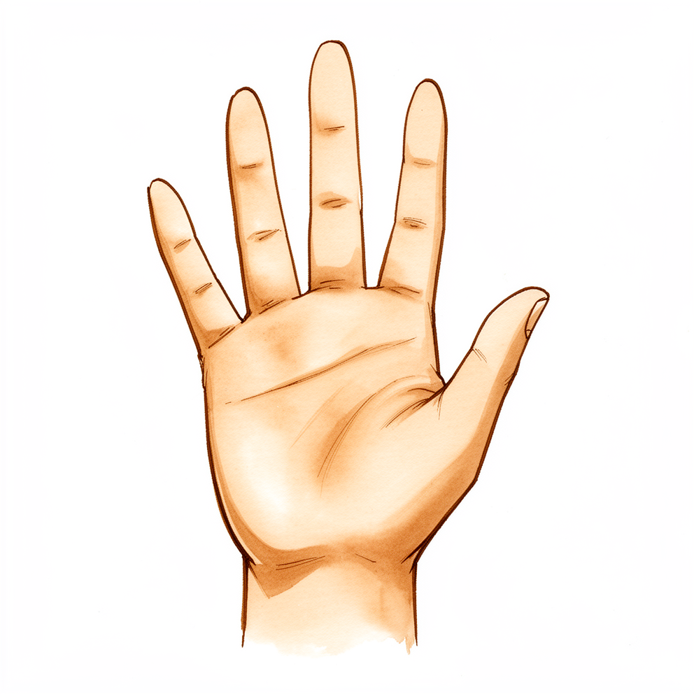
Khớp STT nằm ở nơi xương thuyền của cổ tay gặp hai xương nhỏ gọi là xương thang và xương thê, ngay phía sau khớp ở gốc ngón tay cái. Viêm khớp ở đây gây đau hoặc nhức ở gốc ngón tay cái và ở phía ngón cái của cổ tay. Cơn đau nặng hơn khi nắm và kẹp.
Những việc hằng ngày có thể gây ra cơn đau: vặn chìa khóa, mở nắp lọ, viết, xách túi đồ đi chợ hoặc cầm một tách trà.
Hao mòn ở khớp này thường thấy trên phim X-quang, đặc biệt ở phụ nữ lớn tuổi. Nhiều người có những thay đổi này trên phim X-quang mà hoàn toàn không đau. Phim X-quang cũng không phải là chỉ dẫn tốt để biết liệu khớp này có phải là khớp gây đau cho bạn hay không: nó có thể đánh giá thấp hơn hoặc cao hơn mức độ hao mòn thực tế. Đây là lý do bác sĩ phẫu thuật có thể đề nghị tiêm thuốc tê tại chỗ và steroid vào khớp, dưới hướng dẫn của X-quang hoặc siêu âm. Nếu mũi tiêm làm dịu cơn đau, điều đó cho thấy khớp STT là nguồn gây đau.
Viêm khớp STT thường đi kèm với viêm khớp ở khớp gốc ngón tay cái, và hai tình trạng này có thể khó phân biệt. Khi cả hai khớp đều bị mòn, tình trạng này được gọi là viêm khớp quanh xương thang (pantrapezial arthritis). Biết được những khớp nào bị ảnh hưởng là điều quan trọng, vì nó quyết định những phẫu thuật nào là hợp lý.
Ở một số người bị viêm khớp STT, các xương cổ tay bị nghiêng ra sau, một kiểu gọi là DISI (viết tắt của dorsal intercalated segment instability, mất vững đoạn xen giữa kiểu nghiêng mu tay). Bác sĩ phẫu thuật kiểm tra điều này trên phim X-quang trước khi chọn phẫu thuật, vì nó có thể làm thay đổi kế hoạch.
Khớp bị mòn nằm ngay cạnh gân của một cơ cổ tay gọi là cơ gấp cổ tay quay (flexor carpi radialis). Gân này có thể bị viêm và đau. Rất hiếm khi nó có thể bị đứt.
Hãy đến gặp bác sĩ đa khoa nếu các triệu chứng của bạn không thuyên giảm, nặng dần lên qua nhiều tuần, làm bạn thức giấc vào ban đêm, hoặc khiến bạn không thể làm việc hay sử dụng bàn tay. Hãy đến phòng cấp cứu ngay trong ngày nếu bàn tay hoặc cánh tay của bạn trở nên nóng, đỏ, sưng và đau, đặc biệt khi kèm theo sốt, vì đây có thể là nhiễm trùng cần được chăm sóc ngay trong ngày.
Chuyện gì đang thực sự xảy ra
Viêm khớp là sự hao mòn lớp lót của khớp. Trong tình trạng này, khớp bị mòn là khớp STT: vị trí nơi xương thuyền, một trong những xương nhỏ nằm sâu trong cổ tay, gặp xương thang và xương thê, hai xương nhỏ góp phần tạo nên gốc ngón tay cái. Hãy hình dung những xương này như một chuỗi ngắn các khối cho phép ngón tay cái xoay, nắm và kẹp. Ở nơi các khối cọ xát vào nhau, bề mặt trơn láng có thể bị mòn mỏng. Khi đó, phần xương trần bên dưới cọ xát khi cử động, và sự cọ xát đó chính là cơn đau nhức bạn cảm thấy ở gốc ngón tay cái và phía ngón cái của cổ tay.
Khớp được giữ lại với nhau bằng một hệ thống dải gọi là dây chằng. Nếu những dải này bị giãn hoặc tổn thương, xương thuyền có thể nghiêng ra khỏi vị trí bình thường. Điều đó làm thay đổi cách các xương chịu tải, nên sự hao mòn nặng thêm. Ở một số người, điều này dẫn đến một kiểu mà các xương cổ tay nghiêng ra sau, chính là kiểu DISI mà bác sĩ phẫu thuật tìm kiếm trên phim X-quang. Khớp bị mòn cũng nằm ngay cạnh một gân cổ tay, và bản thân gân đó cũng có thể bị viêm và đau.
Hao mòn ở đây thường xuất hiện cùng với hao mòn ở khớp ngay tại gốc ngón tay cái. Khi cả hai đều bị mòn, tình trạng này được gọi là viêm khớp quanh xương thang. Điều này quan trọng vì hai khớp nằm rất gần nhau đến mức cơn đau từ khớp này có thể giống như cơn đau từ khớp kia, đó là lý do bác sĩ phẫu thuật có thể dùng một mũi tiêm có hướng dẫn để xác định khớp nào thực sự gây ra các triệu chứng của bạn.
Những biện pháp chúng tôi có thể áp dụng
Bác sĩ Kieran Hirpara, bác sĩ phẫu thuật chi trên tại Bệnh viện tư nhân Mater Rockhampton, luôn bắt đầu bằng các phương pháp ít xâm lấn nhất phù hợp với tình trạng của bạn. Điều trị thường bắt đầu mà không cần phẫu thuật. Một chiếc nẹp giữ yên ngón tay cái và cổ tay có thể làm dịu cơn đau nhức, đặc biệt khi bạn làm nhiều việc bằng tay. Thay đổi cách làm việc cũng giúp ích: giảm bớt các việc cần nắm và kẹp, hoặc tìm cách khác để mở nắp lọ và mang vác đồ. Trị liệu tay nhằm giữ cho ngón tay cái và cổ tay cử động được và tăng cường các cơ xung quanh. Chúng tôi thường đề nghị bạn kiên trì áp dụng các biện pháp này trong vài tuần trước khi bàn đến bất kỳ điều gì khác.
Thuốc giảm đau thông thường và thuốc chống viêm, loại thuốc làm giảm sưng, có thể làm dịu bớt cơn đau. Một mũi tiêm steroid vào khớp, dưới hướng dẫn của X-quang hoặc siêu âm, có thể giúp xác định liệu khớp STT có phải là nguồn gây đau hay không. Nếu mũi tiêm làm dịu cơn đau, điều đó cho thấy chính khớp này. Hiệu quả giảm đau có thể kéo dài một thời gian, và có thể tiêm lại nếu hết tác dụng.
Phẫu thuật được tính đến khi các biện pháp này chưa mang lại đủ cải thiện và cơn đau vẫn hạn chế những việc bạn làm. Phẫu thuật nào hợp lý phụ thuộc vào những khớp nào bị mòn. Nếu chỉ riêng khớp STT bị mòn, chúng tôi có thể cắt bỏ đầu xương thuyền bị mòn, hoặc đặt một miếng đệm pyrocarbon nhỏ vào khớp để thay thế bề mặt bị mòn. Nếu khớp ở gốc ngón tay cái cũng bị mòn, phẫu thuật thường được thực hiện là cắt bỏ xương thang cùng với một phần xương thê. Trước khi lựa chọn, chúng tôi kiểm tra trên phim X-quang xem các xương cổ tay của bạn có bị nghiêng hay không, vì điều đó có thể làm thay đổi kế hoạch.
Những điều có thể mong đợi
Viêm khớp STT là một tình trạng lâu dài chứ không phải một chấn thương thoáng qua. Cơn đau nhức ở gốc ngón tay cái thường lúc có lúc không. Nó thường bùng lên khi nắm hoặc kẹp mạnh, rồi dịu đi khi bạn cho bàn tay nghỉ ngơi. Nếu không điều trị, cơn đau thường kéo dài hoặc cứ tái đi tái lại, và có thể dần dần hạn chế những gì bạn cảm thấy có thể làm với bàn tay.
Hầu hết mọi người cải thiện với các biện pháp không cần phẫu thuật. Nẹp, thay đổi cách làm việc, thuốc giảm đau thông thường và một mũi tiêm steroid có thể làm dịu cơn đau trong một thời gian. Hiệu quả của mũi tiêm có thể hết dần, và có thể tiêm lại. Nếu những bước này chưa mang lại đủ cải thiện sau vài tuần, phẫu thuật là điều đáng để trao đổi.
Nếu bạn phẫu thuật, trị liệu tay sau đó là một phần của kế hoạch. Việc trị liệu của bạn do Ruby Doolan tại Extend Rehabilitation thực hiện. Ruby là chuyên gia trị liệu tay: cô ấy sẽ hướng dẫn các bài tập hồi phục và làm nẹp nếu bạn cần trong suốt quá trình.
Quá trình hồi phục diễn ra từ từ. Hãy dự kiến cơn đau nhức giảm dần qua nhiều tuần chứ không phải vài ngày, và dự kiến sức mạnh cũng như sự tự tin với bàn tay được xây dựng lại qua nhiều tháng. Một số người cần thời gian lâu hơn những người khác, và mục tiêu là một bàn tay làm được những gì bạn cần với ít đau hơn, chứ không phải một bàn tay như mới.
Khi nào nên đi khám bác sĩ
Hầu hết các cơn đau nhức ở gốc ngón tay cái dịu đi khi nghỉ ngơi, đeo nẹp và dùng thuốc giảm đau thông thường. Hãy đến gặp bác sĩ đa khoa nếu cơn đau không thuyên giảm qua nhiều tuần, đang nặng lên, làm bạn thức giấc vào ban đêm, hoặc khiến bạn không thể làm việc hay sử dụng bàn tay. Hãy đề nghị được bác sĩ chuyên khoa thăm khám nếu cơn đau cứ quay lại khi nắm và kẹp, hoặc nếu bạn làm rơi đồ thường xuyên hơn. Bác sĩ phẫu thuật có thể đề nghị một mũi tiêm có hướng dẫn để xác định khớp nào đang gây đau, vì hao mòn ở khớp này thường xuất hiện cùng với hao mòn ở gốc ngón tay cái.
Hãy đến phòng cấp cứu ngay trong ngày nếu bàn tay hoặc cánh tay của bạn trở nên nóng, đỏ, sưng và đau, đặc biệt khi kèm theo sốt. Đây có thể là nhiễm trùng, và cần được chăm sóc ngay trong ngày. Bạn không cần giấy giới thiệu để được khám.
Phân tích sâu hơn
Advanced reading: the deeper science (optional)
Phần này đi sâu hơn mức bạn cần để tự đưa ra quyết định điều trị. Viêm khớp STT đáng để tìm hiểu thêm vì hai lý do: phim X-quang không phải là chỉ dẫn tốt để biết liệu khớp này có phải là khớp gây đau hay không, và mọi phẫu thuật cho tình trạng này đều đánh đổi việc giảm đau với một sự thay đổi trong cách các xương nhỏ của cổ tay nằm.
Mức độ phổ biến, và vì sao phim X-quang gây hiểu lầm
Hao mòn ở khớp STT là một phát hiện thường gặp trên phim X-quang. Trong số 700 bệnh nhân liên tiếp tại phòng khám bàn tay được chụp X-quang vì bất kỳ lý do nào, 111 (16%) có viêm khớp STT; tình trạng này gặp nhiều hơn theo tuổi, khi có khe hở giữa xương thuyền và xương nguyệt, và khi có viêm khớp ở khớp gốc ngón tay cái [1]. Trong một mẫu ngẫu nhiên cư dân Nhật Bản từ 50 đến 89 tuổi, 5,3% số cổ tay có tình trạng này, một lần nữa liên quan đến viêm khớp gốc ngón tay cái, giới nữ và tuổi tác [2]. Hầu hết những người này không có triệu chứng nào từ khớp STT.
Phim X-quang không đáng tin cậy theo cả hai chiều. Khi các bác sĩ phẫu thuật quan sát trực tiếp khớp trong lúc phẫu thuật gốc ngón tay cái, 62% số ngón tay cái có viêm khớp giữa xương thuyền và xương thê, nhưng phim X-quang chỉ phát hiện được 44% trong số đó [3]. Một loạt ca lớn hơn trên 257 bệnh nhân phát hiện hao mòn giai đoạn cuối ở 31% khi phẫu thuật, với độ nhạy của X-quang là 47% và độ đặc hiệu là 94% [4]. Hao mòn STT mức độ trung bình được để nguyên khi phẫu thuật gốc ngón tay cái đã không tiến triển hay gây triệu chứng trong thời gian trung bình 8 năm ở một loạt ca theo dõi dài hạn [5]. Đây là lý do quyết định dựa vào vị trí đau khi ấn, và thường dựa vào một mũi tiêm có hướng dẫn, chứ không chỉ dựa vào phim X-quang.
Sự nghiêng của các xương cổ tay
Viêm khớp STT có liên quan đến một kiểu nghiêng đặc biệt của các xương cổ tay, gọi là mất vững đoạn xen giữa kiểu nghiêng mu tay (DISI): xương nguyệt nghiêng ra sau trong khi xương thuyền đứng thẳng hơn. Trong 135 cổ tay bị viêm khớp STT, DISI xuất hiện ở 5% các trường hợp mức độ trung bình và 60% các trường hợp nặng; khác với kiểu nghiêng xảy ra sau khi đứt dây chằng thuyền – nguyệt, khe hở giữa xương thuyền và xương nguyệt vẫn bình thường và khớp cổ tay chính không bị mòn [6]. Một số tác giả hiện mô tả một kiểu sụp đổ do viêm khớp STT gây ra (cổ tay SOAC) là khác biệt với kiểu cổ tay SLAC được biết đến nhiều hơn [7].
Điều này quan trọng đối với phẫu thuật vì một phẫu thuật cắt bỏ xương có thể làm tăng độ nghiêng. Một cổ tay vốn đã mất vững ở hàng xương giữa có thể bị sụp sau một ca cắt bỏ đơn giản, như trong một ca được báo cáo phải làm cứng khớp sau đó [8]. Vì vậy, các bác sĩ phẫu thuật tìm kiếm độ nghiêng và tình trạng mất vững khớp giữa cổ tay trước khi chọn phẫu thuật.
Cắt bỏ đầu xa xương thuyền: giảm đau, với cái giá là sự nghiêng
Cắt bỏ đầu xương thuyền giúp giảm đau trong hầu hết các loạt ca, và các xương cổ tay thường bị nghiêng sau đó. Trong một trong những loạt ca đầu tiên, 12 trong 21 cổ tay xuất hiện DISI, không có cổ tay nào bị thoái hóa khớp thêm, và mức độ hài lòng tương tự như ở những cổ tay không bị nghiêng [9]. Một loạt ca sau đó phát hiện DISI ở 19 trong 21 cổ tay (90%; bài báo ghi là 95%), với góc của xương nguyệt tăng trung bình 13°, và không có liên quan giữa độ nghiêng và điểm chức năng sau thời gian trung bình 3 năm [10].
Lượng xương được cắt bỏ là quan trọng. Sau khi cắt bỏ qua nội soi ở 17 cổ tay, điểm đau giảm từ 6,1 xuống 1,7 trên thang 10 và lực kẹp tăng từ 2,5 kg lên 4,4 kg; tình trạng nghiêng dễ xảy ra hơn khi cắt bỏ hơn 3 mm xương [11]. Trên cổ tay tử thi, cắt bỏ một phần tư xương thuyền hầu như không làm thay đổi cách các xương cử động, cắt bỏ một nửa gây nghiêng rõ rệt, và cắt bỏ ba phần tư làm cổ tay mất vững [12]. Một nghiên cứu giải phẫu phát hiện rằng dải sâu của dây chằng thuyền – thang bám cách đầu xương thuyền khoảng 3,3 mm, đây là lý do giải phẫu để giới hạn phần cắt bỏ ở khoảng 3 mm [13]. Một nghiên cứu trên tử thi về cùng phẫu thuật này đo được xương cả xoay ra sau khoảng 14° và xương nguyệt 8° [14]. Hiếm khi, một cạnh sắc còn lại trên xương thuyền đã làm mòn đứt một gân gấp [15].
Khi gốc ngón tay cái cũng bị mòn
Khi cả gốc ngón tay cái và khớp STT đều bị mòn, phẫu thuật thường được thực hiện là cắt bỏ xương thang và đầu bị mòn của xương thê. Ở 37 ngón tay cái, việc cắt bỏ thêm một phần xương thê không gây thêm vấn đề nào so với chỉ cắt bỏ xương thang [3]. Nên cắt bỏ bao nhiêu xương thê là điểm còn tranh luận. Ở 79 bàn tay được theo dõi trong thời gian trung vị 92 tháng, cắt bỏ một phần hoặc toàn bộ xương thê làm tăng khả năng xương nguyệt nghiêng ra sau từ 15° trở lên (29% sau cắt bỏ một phần và 26% sau cắt bỏ toàn bộ), và những cổ tay có độ nghiêng đó có điểm số kém hơn; tình trạng xương bàn tay ngón trỏ bị lún gây triệu chứng xảy ra ở 18% sau cắt bỏ toàn bộ và 10% sau cắt bỏ một phần, và các tác giả khuyên không nên cắt bỏ toàn bộ [16]. Một nghiên cứu trên tử thi phát hiện rằng phải cắt bỏ 4 đến 6 mm xương thê thì áp lực giữa xương thuyền và xương thê mới giảm khi gập và khi cử động sang hai bên [17]. Trong một loạt 150 ngón tay cái được theo dõi trong thời gian trung bình 13,5 năm, cắt bỏ xương thang kèm cắt bỏ một phần xương thê và tái tạo bằng gân cho điểm hạn chế chức năng DASH trung bình là 8,7 và không có ca nào phải phẫu thuật lại [18].
Khớp nhân tạo pyrocarbon
Một miếng đệm pyrocarbon nhằm giảm đau trong khi giữ được chiều cao của xương thuyền, và nhờ đó hạn chế sự nghiêng. Với đĩa Pyrocardan, 20 bệnh nhân (22 khớp nhân tạo) được theo dõi trong thời gian trung bình 3,8 năm đã bớt đau, có điểm số tốt hơn và lực kẹp mạnh hơn, với một phần mất khả năng duỗi cổ tay và không có trường hợp trật khớp nào; góc cả – nguyệt được cải thiện thay vì xấu đi [19]. Một loạt ca được theo dõi trong thời gian trung bình 5 năm báo cáo 95% số khớp nhân tạo không phải phẫu thuật lại, một trường hợp trật khớp không đau, vôi hóa quanh khớp ở 4 cổ tay, và tình trạng nghiêng vẫn tồn tại ở 3 và mới xuất hiện ở 1 [20]. So sánh với làm cứng khớp STT trong một nghiên cứu không ngẫu nhiên, kết quả tương tự nhau ngoại trừ khả năng duỗi cổ tay tốt hơn với khớp nhân tạo (58° so với 38°); 22% số cổ tay có khớp nhân tạo xuất hiện nghiêng, và 9% số ca làm cứng khớp không liền xương [21]. Hầu hết các loạt ca này đến từ một số ít trung tâm ở Pháp.
Kết quả với các thiết kế khác kém nhất quán hơn. Với một loại khớp nhân tạo pyrocarbon khác có hình dạng để thay thế đầu xương thuyền (STPI), 7 trong 29 khớp nhân tạo phải phẫu thuật lại, chủ yếu vì đau, và số cổ tay bị nghiêng tăng gấp đôi từ 6 lên 12 trong thời gian trung vị 68 tháng [22]. Một báo cáo ca bệnh mô tả một khớp Pyrocardan bị vỡ hai lần ở một người chơi quần vợt, đó là lý do việc dồn tải nặng lên cổ tay ở tư thế duỗi được trao đổi trước khi phẫu thuật [23].
Làm cứng khớp STT, để so sánh
Làm cứng khớp hàn ba xương lại thành một. Trên 1.429 ca phẫu thuật trong một tổng quan hệ thống, xương không liền ở 6,3%, 4,2% phải chuyển sang làm cứng toàn bộ cổ tay, và lực nắm trung bình đạt 76% so với bàn tay bên kia [24]. Ở 58 cổ tay được theo dõi trong 10 năm, 10% cần phẫu thuật thêm và 7% bị không liền xương gây đau [25]. Làm cứng khớp thường giữ được khoảng ba phần tư cử động của cổ tay, và đau do mỏm trâm quay va vào xương thuyền đã được làm cứng khá thường gặp, đến mức một số bác sĩ phẫu thuật cắt gọt mỏm trâm trong cùng lần mổ [26].
Những điều chưa biết
Không có thử nghiệm ngẫu nhiên nào so sánh các phẫu thuật này. Một khảo sát các bác sĩ phẫu thuật bàn tay ở châu Âu phát hiện sự khác biệt lớn về phẫu thuật được ưa chuộng cho cùng một vấn đề [27], và các tổng quan gần đây kết luận rằng không có phương án nào có bằng chứng rõ ràng về sự vượt trội; lựa chọn phụ thuộc vào việc chỉ riêng khớp STT bị mòn, gốc ngón tay cái có bị mòn hay không, và các xương cổ tay đã bị nghiêng hay chưa [28] [29]. Hầu hết các loạt ca theo dõi ít hơn 30 bệnh nhân trong thời gian dưới 10 năm, vì vậy ảnh hưởng lâu dài của sự nghiêng sau khi cắt bỏ, và độ bền lâu dài của khớp nhân tạo pyrocarbon, vẫn là những câu hỏi còn bỏ ngỏ.
Tài liệu tham khảo
[1] Scordino LE, Bernstein J, Nakashian M, et al. Radiographic prevalence of scaphotrapeziotrapezoid osteoarthrosis. J Hand Surg Am. 2014;39(9):1677-82. https://doi.org/10.1016/j.jhsa.2014.05.033
[2] Kitamura Y, Kato H, Hayashi M, et al. Prevalence and associated factors for primary osteoarthritis of the scaphotrapeziotrapezoid, radiocarpal, and distal radioulnar joints in the Japanese general elderly population. J Hand Surg Am. 2025;50(1):103.e1-103.e10. Epub 2023. https://doi.org/10.1016/j.jhsa.2023.05.009
[3] Tomaino MM, Vogt M, Weiser R. Scaphotrapezoid arthritis: prevalence in thumbs undergoing trapezium excision arthroplasty and efficacy of proximal trapezoid excision. J Hand Surg Am. 1999;24(6):1220-4. https://doi.org/10.1053/jhsu.1999.1220
[4] Akoon A, Grandizio LC, Mookerjee GG, et al. Comparison of radiographic and intraoperative visual assessment of scaphotrapezoid joint arthritis in patients with end-stage carpometacarpal arthritis of the thumb base. Hand (N Y). 2019;14(5):609-13. Epub 2018. https://doi.org/10.1177/1558944718765246
[5] Glickel SZ, Kornstein AN, Eaton RG. Long-term follow-up of trapeziometacarpal arthroplasty with coexisting scaphotrapezial disease. J Hand Surg Am. 1992;17(4):612-20. https://doi.org/10.1016/0363-5023(92)90303-7
[6] Hattori Y, Sasaki J, Sakamoto S, et al. Radiographic characteristics of carpal instability associated with scaphotrapeziotrapezoid osteoarthritis. J Hand Surg Am. 2026;51(3):259-67. https://doi.org/10.1016/j.jhsa.2025.11.018
[7] Rodriguez Fontan F, Netscher DT. Scaphotrapeziotrapezoid joint osteoarthritis: differentiating SOAC wrist from SLAC wrist. J Am Acad Orthop Surg. 2026. https://doi.org/10.5435/jaaos-d-25-01408
[8] Corbin C, Warwick D. Midcarpal instability after excision arthroplasty for scapho-trapezial-trapezoid (STT) arthritis. J Hand Surg Eur Vol. 2009;34(4):537-8. https://doi.org/10.1177/1753193408098903
[9] Garcia-Elias M, Lluch AL, Farreres A, et al. Resection of the distal scaphoid for scaphotrapeziotrapezoid osteoarthritis. J Hand Surg Br. 1999;24(4):448-52. https://doi.org/10.1054/jhsb.1999.0169
[10] Brouwers D, Decramer A, Cornu P, Le Viet D. Does the DISI matter after distal scaphoidectomy with tendon interposition for STT osteoarthritis? Hand Surg Rehabil. 2020;39(4):284-90. https://doi.org/10.1016/j.hansur.2020.03.005
[11] Iida A, Omokawa S, Kawamura K, et al. Arthroscopic distal scaphoid resection for isolated scaphotrapeziotrapezoid osteoarthritis. J Hand Surg Am. 2019;44(4):337.e1-337.e5. https://doi.org/10.1016/j.jhsa.2018.06.021
[12] Kadar A, Grewal R, Padmore CE, et al. How much scaphoid can be safely resected? A biomechanical analysis of the effects of distal scaphoid resection. Hand (N Y). 2022;17(5):885-92. Epub 2020. https://doi.org/10.1177/1558944720966717
[13] Yoshimura Y, Yokota A, Fujino K, et al. Anatomy of the scaphotrapezial ligament complex and its implications for distal scaphoid resection. J Hand Surg Am. 2026;51(1):25.e1-25.e8. https://doi.org/10.1016/j.jhsa.2025.09.010
[14] Paeffgen L, Riederer J, Adler T, et al. Carpal instability after partial trapeziectomy, total trapeziectomy and the resection of the distal scaphoid pole: a cadaveric study. Arch Orthop Trauma Surg. 2024;144(3):1443-51. Epub 2023. https://doi.org/10.1007/s00402-023-05134-z
[15] Deren ME, Mitchell CH, Weiss AC. Flexor tendon ruptures after distal scaphoid excision for scaphotrapeziotrapezoid osteoarthritis. Hand (N Y). 2017;12(5):NP152-6. https://doi.org/10.1177/1558944717708028
[16] Renfree KJ, Odgers R, Tillinghast C, Zhang N. Effect of partial and complete trapezoid excision on radiographic and functional results after abductor pollicis longus suspensionplasty. J Hand Surg Am. 2020;45(4):364.e1-364.e9. https://doi.org/10.1016/j.jhsa.2019.10.006
[17] Anz H, Roylance J, Ismaily S, et al. Correlation of scaphotrapezoid contact pressures with degree of trapezoid resection: does eliminating contact matter? J Hand Surg Glob Online. 2026;8(4):101049. https://doi.org/10.1016/j.jhsg.2026.101049
[18] Melone CP, Lovy AJ, Ciaglia M, Qin B. Trapeziectomy, partial trapezoidectomy, and abductor pollicis longus dual loop ligament reconstruction for advanced basal joint arthritis: long-term outcome of 150 cases. Hand (N Y). 2023;18(3):491-500. Epub 2022. https://doi.org/10.1177/15589447221092060
[19] Gauthier E, Truffandier M, Gaisne E, Bellemère P. Treatment of scaphotrapeziotrapezoid osteoarthritis with the Pyrocardan implant: results with a minimum follow-up of 2 years. Hand Surg Rehabil. 2017;36(2):113-21. https://doi.org/10.1016/j.hansur.2017.01.003
[20] Cholley-Roulleau M, Bouju Y, Lecoq FA, et al. Pyrocardan scaphotrapeziotrapezoid joint arthroplasty for isolated osteoarthritis: results after a mean follow-up of 5 years. J Wrist Surg. 2022;11(3):262-8. Epub 2021. https://doi.org/10.1055/s-0041-1733877
[21] Cholley-Roulleau M, Dautel G, Dap F, et al. Comparison between scaphotrapeziotrapezoid arthrodesis and Pyrocardan implant for isolated scaphotrapeziotrapezoid osteoarthritis. Orthop Traumatol Surg Res. 2025;111(5):103867. https://doi.org/10.1016/j.otsr.2024.103867
[22] Zander M, Ibsen-Sörensen A, Nilsson A, Björkman A. Retrospective analysis of scaphoid trapezium pyrocarbon implant intervention in STT arthritis: a 3-year follow-up study. J Plast Surg Hand Surg. 2024;59:40-5. https://doi.org/10.2340/jphs.v59.34985
[23] Sarigiannis CN, Axelsson P. Game, set… revision! A case report of a tennis player who smashed his scaphotrapeziotrapezoid-joint Pyrocardan implant twice. J Hand Surg Glob Online. 2026;8(3):100964. https://doi.org/10.1016/j.jhsg.2026.100964
[24] Stephens AR, Garcia BN, Rogers MJ, et al. Scaphotrapeziotrapezoid arthrodesis: systematic review. J Hand Surg Am. 2022;47(3):218-227.e2. https://doi.org/10.1016/j.jhsa.2021.09.029
[25] Rogers MJ, Lu C, Stephens AR, et al. Scaphotrapeziotrapezoid arthrodesis: a 10-year follow-up study of complications in 58 wrists. Hand (N Y). 2022;17(5):919-25. Epub 2020. https://doi.org/10.1177/1558944720964972
[26] Deans VM, Naqui Z, Muir LT. Scaphotrapeziotrapezoidal joint osteoarthritis: a systematic review of surgical treatment. J Hand Surg Asian Pac Vol. 2017;22(1):1-9. https://doi.org/10.1142/s0218810417300017
[27] Berkhout MJL, Yin Q, Ritt MJPF. Current trends in operative treatment of scaphotrapeziotrapezoid osteoarthritis: a survey among European hand surgeons. J Wrist Surg. 2020;9(2):94-9. https://doi.org/10.1055/s-0039-3402796
[28] Bellemère P, Ardouin L. Surgical options and outcomes for treatment of osteoarthritis of the scaphotrapeziotrapezoidal joint. J Hand Surg Eur Vol. 2025;50(1):58-67. https://doi.org/10.1177/17531934241295345
[29] Alder KD, Feroe AG, Karim KE. Management of scaphotrapeziotrapezoid osteoarthritis: a critical analysis review. JBJS Rev. 2023;11(10):e23.00093. https://doi.org/10.2106/JBJS.RVW.23.00093
Evidence & references
This is the clinical evidence summary written for health professionals. It is technical, and it lists the research this page was built from. You do not need to read it to understand your treatment or to make a decision about it.
Anatomy & Pathophysiology
Osseous Skeleton
- The skeleton of the hand and wrist consists of 27 bones, of which 19 are long bones [3].
- The skeleton is divided into five rays, each comprising a polyarticulated chain of metacarpals and phalanges [3].
- The base of each metacarpal articulates with the distal row of the carpus [3].
- The carpus articulates with the skeleton of the forearm through its proximal row [3].
- The radioulnocarpal articulation has two axes of movement, to which a third axis of pronation and supination from the forearm is added [3].
- The wrist has three axes of movement, permitting the hand to be positioned in any spatial configuration [3].
- The radial ray (first ray) is the shortest, made up of a metacarpal and two phalanges [3].
- The first ray continues the external column of the carpus formed by the scaphoid and trapezium [3].
- The trapezium is angled out in front of the carpal plane so that the first metacarpal makes an angle of about 45 degrees with the second metacarpal in the sagittal plane [3].
- The thumb metacarpal is the shortest, while the index metacarpal is by far the longest [3].
- The proximal and middle phalanges of the long and ring fingers are longer than those of the index finger [3].
- The epiphyseal plates are located at the proximal ends of the phalanges and the first metacarpal [3].
- The epiphyseal plates are located at the distal ends of the other metacarpals [3].
- The hand presents a longitudinal and transverse concavity, giving it the shape of a cup with a palmar concavity when the thumb is placed next to the index finger [3].
- The transverse axis of the palm corresponds to the metacarpophalangeal articulations and is not perpendicular to the longitudinal axis [3].
- The transverse axis forms an acute angle of approximately 75 degrees with the longitudinal axis [3].
Musculature
- Extrinsic muscles have their origin outside of the hand and their insertion on the hand or carpus [2].
- Intrinsic muscles have both origin and insertion within the hand [2].
- The extrinsic extensors run through six different fibroosseous retinacular compartments at the wrist level [2].
- The first extensor compartment contains the abductor pollicis longus and the extensor pollicis brevis [2].
- The abductor pollicis longus inserts at the base of the thumb metacarpal and radially abducts the thumb [2].
- The extensor pollicis brevis inserts on the dorsum of the proximal aspect of the proximal phalanx of the thumb and actively extends the metacarpophalangeal joint of the thumb [2].
- The second extensor compartment contains the extensor carpi radialis longus and the extensor carpi radialis brevis [2].
- The extensor carpi radialis longus inserts on the index metacarpal, dorsiflexes and radially deviates the wrist [2].
- The extensor carpi radialis brevis inserts into the base of the middle metacarpal and provides balanced wrist dorsiflexion [2].
- The third compartment contains the extensor pollicis longus, which turns abruptly radialward about Lister tubercle [2].
- The extensor pollicis longus inserts on the distal phalanx and provides forceful extension of the thumb interphalangeal joint [2].
- The fourth extensor compartment contains the extensor indicis proprius lying deep to the four tendons of the extensor digitorum communis [2].
- The fifth compartment contains the extensor digiti quinti [2].
- The principal bony insertion of the extrinsic digital extensors is on the dorsal proximal aspect of the middle phalanx [2].
- Metacarpophalangeal joint extension is provided by extrinsic extensor force transmitted through the sagittal bands [2].
- Distal interphalangeal joint extension is achieved through the conjoined lateral bands composed of tendinous slips from the extrinsic and intrinsic tendons [2].
- The extensor carpi ulnaris tendon runs through the sixth compartment and inserts at the base of the little finger metacarpal [2].
- The extensor carpi ulnaris provides wrist extension and ulnar deviation [2].
- The extensor digitorum communis tendons of the middle, ring, and little fingers are tethered together by juncturae tendinum over the dorsum of the hand proximal to the metacarpophalangeal joint [2].
- The digital extensor tendons are stabilized over the mid-line of the metacarpophalangeal joint by their attachment to sagittal band fibers [2].
- The sagittal band fibers insert onto the volar proximal phalanx and onto the lateral borders of the volar plate [2].
- The sagittal bands keep the extrinsic extensor as far as possible away from the center of rotation of the metacarpophalangeal joint, giving it the greatest mechanical efficiency [2].
- With rupture or attenuation of the sagittal band fibers, the extrinsic extensor tendon can sublux to the ulnar side of the metacarpal head causing ulnar deviation of the finger [2].
- The extrinsic finger flexors are the flexor digitorum profundus and the flexor digitorum superficialis [2].
- The flexor digitorum profundus inserts on the proximal volar aspect of the distal phalanx [2].
- The flexor digitorum profundus flexes the distal interphalangeal joint as well as the proximal interphalangeal and metacarpophalangeal joints [2].
- The flexor digitorum superficialis acts as a flexor of the proximal interphalangeal and metacarpophalangeal joints [2].
- There are seven interosseous muscles, four dorsal and three volar [6].
- The dorsal interossei are abductors [6].
- The dorsal interossei lie to the radial side of the index and middle fingers and the ulnar side of the middle and ring fingers [6].
- The little finger is abducted by the abductor digiti quinti [6].
- The volar interossei are adductors and lie to the ulnar side of the index finger and the radial side of the ring and little fingers [6].
- The middle finger has two dorsal interossei (abductors) and no volar interossei (adductors) because the central axis of the hand lies within it [6].
- Each dorsal interosseous muscle, with the exception of the third, has two muscle heads [6].
- The superficial head of the dorsal interosseous muscles abducts and weakly flexes the proximal phalanx [6].
- The deep head of each dorsal interosseous muscle forms a lateral tendon, or lateral band, at the level of the MP joint [6].
- The deep head flexes and weakly abducts the proximal phalanx while extending the middle and distal phalanges [6].
- Transverse fibers arch dorsally from each lateral band to join each other over the dorsum of the finger, flexing the proximal phalanx [6].
- Oblique fibers (spiral fibers) from the lateral bands insert onto the lateral tubercles at the base of the middle phalanx and extend the middle phalanx (PIP joint) [6].
- The lateral bands are joined by the lateral slips of the extensor tendon to form the conjoined lateral band [6].
- The two conjoined lateral bands to each finger unite at the distal third of the middle phalanx to form the terminal tendon [6].
- The terminal tendon inserts at the base of the distal phalanx to extend it [6].
- The flexor digiti quinti brevis is structurally and functionally similar to the deep head of the dorsal interossei, forming the ulnar lateral band of the little finger [6].
- Each volar interosseous muscle has only one muscle head and none of them insert onto the proximal phalanx [6].
- The volar interossei form the ulnar lateral band of the index finger and the radial lateral band of the ring and little fingers [6].
- The abductor digiti quinti inserts onto the ulnar lateral tubercle at the base of the proximal phalanx of the little finger [6].
- The flexor digiti quinti forms the ulnar lateral band [6].
- The opponens digiti quinti arises from the pisohamate ligament and the hook of the hamate and inserts onto the ulnar side of the diaphysis of the fifth metacarpal [6].
- The opponens digiti quinti flexes and supinates the fifth metacarpal [6].
- The flexor digitorum profundus originates from the proximal ulna and the interosseous membrane [13].
- The flexor digitorum profundus and the flexor pollicis longus muscles form the deep compartment of the volar forearm [13].
- As the flexor digitorum profundus and flexor pollicis longus tendons travel through the carpal tunnel, they occupy the floor of the carpal tunnel [13].
- The tenosynovial sheath of the flexor pollicis longus is continuous with the radial bursa [13].
- The tenosynovial sheath to the little finger is continuous with the ulnar digital bursa [13].
- In some patients, the radial and ulnar bursae communicate, allowing a horseshoe abscess to spread between the thumb and little finger if infection occurs in the flexor tendon sheath of either one of these digits [13].
- The lumbricals originate from the radial side of the index, middle, ring, and little fingers in the palm [13].
- The profundus tendon passes through the bifurcation of the flexor digitorum superficialis before inserting into the proximal palmar base of the distal phalanx [13].
- The innervation of the flexor digitorum profundus of the index and middle fingers is through the anterior interosseous branch of the median nerve [13].
- The profundus of the ring and little fingers is innervated by the ulnar nerve [13].
- The flexor digitorum superficialis has two heads: a radial head originating from the proximal shaft of the radius, and a humeral ulnar head originating from the medial humeral epicondyle and coronoid process of the ulna [13].
- As the superficialis tendons pass through the carpal tunnel, the tendons of the middle and ring fingers are more superficial and central than those of the index and little fingers [13].
- In the proximal aspect of the finger, the flexor digitorum superficialis tendon bifurcates around the flexor digitorum profundus at the beginning of the A2 pulley [13].
- The flexor digitorum superficialis tendon slips reunite distally at the Camper chiasm, with approximately half of the fibers staying on the ipsilateral side and half crossing to the contralateral side of the finger [13].
- The flexor digitorum superficialis inserts via radial and ulnar slips into the proximal metaphysis of the middle phalanx [13].
- The entire flexor digitorum superficialis muscle receives innervation from the median nerve [13].
- The primary function of the superficialis is digital flexion at the proximal interphalangeal joint [13].
- The flexor pollicis longus originates from two heads: a radial head from the proximal radius and interosseous membrane, and an accessory head from the coronoid process of the ulna and medial epicondyle of the humerus [13].
- In the palm, the flexor pollicis longus tendon transverses between the abductor pollicis brevis and the flexor pollicis brevis [13].
- The flexor pollicis longus inserts into the proximal base of the thumb distal phalanx [13].
- The flexor pollicis longus is innervated by the anterior interosseous branch of the median nerve [13].
- The flexor pollicis longus flexes both the interphalangeal and metacarpophalangeal joints of the thumb [13].
Ligaments and Fascia
- The second to fifth metacarpals are all bound together by various fibrous structures, the most distal of which is the deep transverse intermetacarpal ligament [9].
- The deep transverse intermetacarpal ligament is better named the interglenoid ligament because it ties together the anterior "glenoid ligaments" of the metacarpophalangeal articulations, known as the "volar plates" [9].
- The volar plates are interconnected by the transverse interglenoid ligament [9].
- The stability of the metacarpophalangeal joints is essential to the support of the longitudinal arch as well as of the transverse metacarpal arch [9].
- The superficial palmar fascia lies in a coronal plane beneath the palmar subcutaneous tissue [16].
- The superficial palmar fascia covers a triangular area of the central palm, with the proximal corner facing directly proximal [16].
- The palmaris longus tendon, when present, terminates in continuity with the fibers of this proximal corner [16].
- From this common point, four central bands of fascia extend distally toward each of the fingers [16].
- There is no central band for the thumb [16].
- At the distal palmar crease level, the central bands are bridged transversely by the superficial transverse palmar ligament [16].
- Fibers from the central bands pass above, below, and through the superficial transverse palmar ligament [16].
- Superficial fibers of the central bands stay superficial and merge with vertical retinacular fibers at the dermis’s undersurface in the distal palm in areas between skin flexion creases [16].
- Intermediate fibers split transversely into two sections, which extend toward the lateral border of the digit base [16].
- These intermediate fibers are the spiral band that tracks around the neurovascular bundle [16].
- Proximally, the spiral band is central and superficial to the neurovascular bundle; distally, it is lateral and deep beneath it [16].
- Spiral neurovascular bundles develop when spiral bands shorten [16].
- Deep fibers continue dorsally to merge with sagittal interosseous fascia fibers and pierce the transverse deep intermetacarpal ligament to merge with fibers of the sagittal bands of the extensor mechanism [16].
- The digital neurovascular structures are surrounded by a diffuse network of thin transverse oblique fibers [16].
- Fibers dorsal to the neurovascular bundle are collectively called Cleland ligament [16].
- Fibers palmar to the neurovascular bundle are called Grayson ligament [16].
- Undisturbed, these "ligaments" are a mesh of nonparallel individual fibers [16].
- The appearance of a ligament is a dissection artifact from releasing skin attachments [16].
- These fibers have a common origin in continuity with the fingers’ retinacular ligaments, the floor of the flexor tendon sheath, and retinacular fibers attaching the palmar digital skin to deeper structures [16].
- On the small finger’s ulnar border, lateral fascial attachments are in continuity with the abductor digiti minimi fascia and tendon [16].
- A subdermal fascial layer borders the periphery of the web spaces from roughly the radial thumb sesamoid to the ulnar side of the small finger’s proximal flexion crease [16].
- These border fibers form the distal first web space ligament between thumb and index in continuity with the natatory ligament that spans the remaining web spaces [16].
- Fibers from the natatory ligament extend distally at each finger’s lateral base in continuity with Grayson ligament fibers and the lateral digit dermis [16].
Vascular Anatomy
- The "princeps pollicis" artery, the terminal branch of the radial artery, crosses the first intermetacarpal space [10].
- The "princeps pollicis" artery runs along the ulnar side of the first metacarpal bone and along the volar surface of the adductor muscle [10].
- The "princeps pollicis" artery emerges onto the subcutaneous palmar tissue at the level of the cutaneous flexion crease of the metacarpophalangeal joint [10].
- At the metacarpophalangeal joint level, the "princeps pollicis" divides into two terminal rami, namely the collateral palmar arteries of the thumb [10].
- The collateral palmar arteries run along the digital tunnel symmetrically and are of equal caliber [10].
- The collateral palmar arteries head distally to finally unite in the pulp arcade [10].
- During their transit in the digital tunnel, the collateral palmar arteries break off into numerous collateral branches, either cutaneous, articular or osseous [10].
- An arcade located deep in the flexor tendon joins together the two arteries at the level of the distal metaphysis of the first phalanx [10].
- From this arcade, vessels originate which enter the "vincula" and irrigate the flexor tendon [10].
- In anatomical studies, only 15% of dissections fall into the classical category of palmar artery layout [10].
- In
Clinical Presentation
- Inflammatory arthritis is a collection of different diseases that cause inflammation in the joints [20].
- The diagnosis of inflammatory arthritis requires a complete review of the disease process profile [20].
- There are no simple clinical tests that can be used to diagnose inflammatory arthritis diseases [20].
- The rheumatologic history and physical examination play a critical role in the diagnosis and treatment of inflammatory arthritis [20].
- The location of the problems is an important disease feature for diagnosing inflammatory arthritis [20].
- The symmetry of presentation is an important disease feature for diagnosing inflammatory arthritis [20].
- Rheumatoid arthritis has a predilection for the wrists and the proximal joints of the hands and feet [20].
- Psoriatic arthritis involves the distal interphalangeal joint of the hands [20].
- The physical findings with rheumatoid arthritis tend to be symmetrical [20].
- Other inflammatory arthritis conditions do not tend to present with symmetrical physical findings [20].
- The onset of the disease is an important feature for diagnosing inflammatory arthritis [20].
- The chronology of the disease is an important feature for diagnosing inflammatory arthritis [20].
- The onset of rheumatoid arthritis tends to present in a subacute manner [20].
- Septic arthritis has a rapid onset in several hours [20].
- Age can aid in the diagnosis of inflammatory arthritis [20].
- Gender can aid in the diagnosis of inflammatory arthritis [20].
- Precipitating factors can aid in the diagnosis of inflammatory arthritis [20].
Investigations
- Clinical evaluation of the hand and wrist requires combining patient history with a careful physical examination to narrow the scope of possible pathologic processes [1].
- Diagnostic tests such as imaging and serum laboratory studies are useful in determining pathology but can be expensive, time-consuming, and often nonspecific [1].
- A careful physical examination is essential to direct care and future testing if indicated [1].
- The radioulnocarpal articulation has two axes of movement, to which a third axis—pronation and supination from the forearm—is added [3].
- The transverse axis of the palm forms an acute angle of approximately 75 degrees with the longitudinal axis [3].
- An 8-MHz Doppler tone assessment may be used to identify superficially displaced neurovascular bundles when Dupuytren cords lie beneath soft fleshy prominences [19].
- False-negatives are possible with 8-MHz Doppler tone assessment for identifying neurovascular bundles [19].
- MR assessment of Dupuytren’s disease is hindered by the resolution of current equipment, orientation issues due to multiplanar deformities of the fingers, and lack of intraoperative availability [19].
- MRI is probably most useful in identifying additional pathology such as flexor tendon bowstringing [19].
- MRI may be helpful in providing a quantitative noninvasive measure of cellularity of affected areas, which is an index of biologic activity [19].
References
[1] Orthopaedic Knowledge Update 13 Ebook Without Multimedia. Anatomy, Evaluation, Clinical Examination, and Imaging > Evaluation and Clinical Examination: Current Concepts.
[2] A Lange Medical Book Current Diagnosis Treatment In Orthopedics Fifth Edition. 9Hand Surgery > Image DISORDERS OF THE MUSCULATURE OF THE HAND.
[3] Exam Of The Hand Wrist 2Ed. 1.1 SKELETON OF THE HAND > The osseous skeleton.
[6] Green S Operative Hand Surgery. Interosseous and Hypothenar Muscles.
[9] Exam Of The Hand Wrist 2Ed. The arches of the hand > The metacarpal arch.
[10] Exam Of The Hand Wrist 2Ed. Techniques of investigation of the arterial supply by J P Melki > Vascularization of the thumb > Palmar aspect.
[13] A Lange Medical Book Current Diagnosis Treatment In Orthopedics Fifth Edition. 9Hand Surgery > FLEXOR TENDON INJURY.
[16] Green S Operative Hand Surgery. PERTINENT ANATOMY.
[19] Dupuytren S Disease And Related Hyperproliferative Disorders. 54. The Future of Dupuytren’s Research and Treatment > 54.4 Mechanical Measurements and Procedures > 54.4.4 Imaging.
[20] Orthopaedic Knowledge Update 13 Ebook Without Multimedia. Degenerative Conditions of the Knee > Inflammatory Arthritis.