Patients › Elbow
Codo de golfista
Golfer’s elbow (medial epicondylitis) — causes, symptoms, and conservative treatment options.
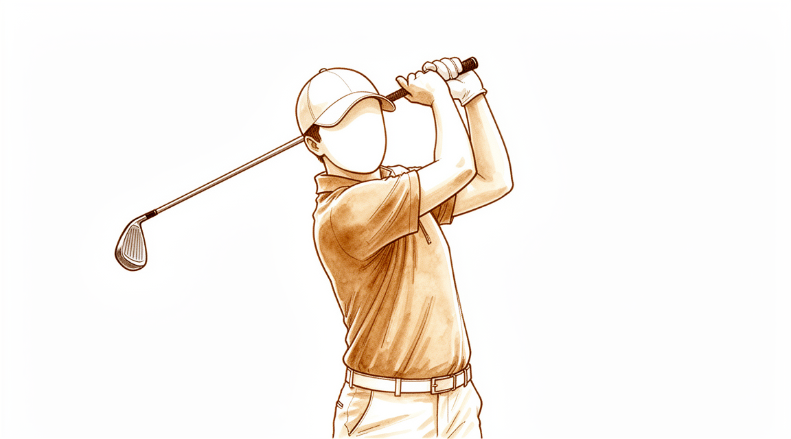
Qué está sintiendo
El dolor se sitúa sobre la protuberancia ósea de la parte interna del codo y a menudo se extiende a los músculos del antebrazo, justo por debajo de ella. Por lo general aparece de forma gradual, sin una lesión evidente, y tiende a persistir incluso cuando descansa. El golf, los lanzamientos, el tenis, el levantamiento de pesas y los bolos pueden desencadenarlo. También puede hacerlo un trabajo que implique un agarre enérgico repetido, levantar cargas de 20 kg o más, o usar herramientas que vibran.
Algunos movimientos cotidianos se vuelven molestos. Girar el pomo de una puerta, estrechar la mano, llevar bolsas de la compra o levantar un hervidor pueden empeorarlo, porque estas acciones cargan los mismos músculos que se insertan en esa protuberancia interna. Es posible que note el agarre más débil que en el otro lado. La mayoría de las personas pueden seguir moviendo el codo y la muñeca en todo su recorrido, aunque algunas notan que el codo se pone algo rígido y no se estira por completo.
El dolor suele intensificarse durante o después de la actividad que lo provocó, y puede ser peor a la mañana siguiente o al agarrar algo por primera vez. Algunas personas lo notan sobre todo al hacer el swing de golf o al lanzar, especialmente en la primera parte del movimiento.
Preste atención a una cosa en particular. El nervio cubital, el nervio que da la sensibilidad a los dedos anular y meñique, pasa justo por detrás de esta parte del codo. Si tiene entumecimiento u hormigueo en esos dos dedos además del dolor en la parte interna del codo, comuníqueselo a su médico de cabecera o pida una valoración por un especialista, porque eso cambia qué tratamiento puede ayudar.
Acuda a su médico de cabecera o pida una valoración por un especialista si el dolor no se calma, empeora a lo largo de las semanas, le despierta por la noche o le impide trabajar o usar la mano o el brazo. Acuda al servicio de urgencias ese mismo día si la mano o el brazo se ponen calientes, enrojecidos, hinchados y dolorosos, sobre todo si tiene fiebre, o si los dedos se ponen pálidos, fríos, blancos o azulados.
¿Qué está ocurriendo realmente?
El codo de golfista es un problema de un tendón, no de la articulación. Un tendón es el cordón resistente que une el músculo al hueso. En la parte interna del codo hay un grupo de músculos del antebrazo que flexionan la muñeca y los dedos y ayudan a girar la palma hacia abajo. Todos ellos se insertan en esa protuberancia ósea a través de un tendón común, un poco como varias cuerdas atadas a un mismo poste de amarre.
Cuando hace un swing, lanza, o agarra y levanta peso de forma repetida, esos músculos tiran del mismo punto una y otra vez. En el tendón se producen pequeñas roturas. El cuerpo intenta repararlas, pero la reparación es irregular y el tejido nuevo es más débil que el original. Con el tiempo, el tendón se engrosa y se desorganiza, como una cuerda que se ha deshilachado y se ha vuelto a anudar muchas veces. Por eso el dolor sigue volviendo incluso después del reposo, y por eso agarrar o levantar peso lo empeora: esos movimientos cargan exactamente el punto donde al tendón le cuesta curarse.
A pesar del nombre, normalmente no hay hinchazón ni inflamación en el punto doloroso. El problema es un desgaste y una curación fallida, no una infección ni una artritis. No hay nada roto y la propia articulación no está dañada.
Hay una estructura cercana que importa aquí. El nervio cubital, que da la sensibilidad a los dedos anular y meñique, pasa justo por detrás de este tendón. Como está tan cerca, la misma irritación puede afectar también al nervio, y por eso algunas personas notan hormigueo en esos dedos además del dolor de codo.
Qué podemos hacer al respecto
El Dr. Kieran Hirpara, cirujano de extremidades superiores en el Mater Private Hospital Rockhampton, comienza con las opciones menos invasivas que se adapten a su condición. La mayoría de los casos de codo de golfista se resuelven sin operación, así que empezamos por ahí. El reposo y cambiar la forma en que usa el brazo importan más que cualquier otra cosa que podamos ofrecerle. Eso significa reducir los agarres, los levantamientos de peso o los movimientos de balanceo que provocan el dolor, y buscar otras formas de hacer las tareas que duelen. Dele una oportunidad real durante al menos 6 meses antes de plantearse la cirugía, porque la cirugía es solo para los síntomas que persisten a pesar de ese tratamiento.
La fisioterapia tiene como objetivo fortalecer los músculos que trabajan junto con el tendón doloroso, para que el tendón soporte menos carga. Lo habitual es un programa que aumente de forma gradual la fuerza del antebrazo, de la muñeca y del agarre. Si el dolor se debe a un deporte, como el golf o los lanzamientos, parte de la recuperación consiste en revisar su técnica, porque los defectos en la forma de hacer el swing o de lanzar pueden seguir cargando el mismo punto.
Los analgésicos y los antiinflamatorios, llamados antiinflamatorios no esteroideos (AINE), pueden ayudarle a mantenerse activo mientras el tendón se calma. No ofrecemos inyecciones para el codo de golfista, así que no se las propondremos como siguiente paso.
Si sus síntomas no han mejorado después de 6 meses o más de un tratamiento no quirúrgico adecuado, podemos hablar de la cirugía. La operación limpia la parte dañada y deshilachada del tendón donde se inserta en el hueso de la parte interna del codo, y deja intacto el tejido sano. Se realiza a través de una pequeña incisión, y usted vuelve a casa el mismo día. La cirugía es una decisión compartida, y solo la recomendaremos cuando estemos de acuerdo en que las medidas más sencillas ya han dado todo lo que podían dar.
Por lo general, los pacientes son derivados a nuestra clínica por su médico de cabecera; si un fisioterapeuta le ha sugerido que nos consulte, igualmente necesitará una derivación de su médico de cabecera para poder acceder al reembolso de Medicare. En su consulta, tomamos su historia clínica, examinamos su codo y solicitamos estudios de imagen, como una ecografía o una resonancia magnética, si van a cambiar lo que le recomendamos. La ecografía suele ser la primera prueba que se utiliza para confirmar el codo de golfista, y la resonancia magnética ofrece una imagen más clara del tendón y del nervio cubital cercano si el diagnóstico no está claro.
Si se opera, la rehabilitación posterior es terapia de la mano con Ruby Doolan, de Extend Rehabilitation, no fisioterapia. Ruby le guiará con los ejercicios y le fabricará cualquier férula que necesite a lo largo del proceso.
Qué esperar
En la mayoría de las personas, el codo de golfista mejora con el tiempo y con el tratamiento adecuado. Lo que suele funcionar es reducir las actividades que lo provocan y después recuperar la fuerza de forma gradual. Muchas personas mejoran a lo largo de meses y no de semanas, así que conviene esperar un cambio lento y constante en lugar de una solución rápida.
Si el dolor ya ha durado meses a pesar del reposo y de los cambios en la forma de usar el brazo, tiende a persistir a menos que algo cambie. Ese es el momento en que hablaríamos de la cirugía. Cuando la cirugía se realiza por síntomas que no se han calmado después de 6 meses o más de un tratamiento no quirúrgico adecuado, la mayoría de las personas refieren menos dolor y una mejor función durante el año siguiente, y la mayoría vuelven a sus actividades habituales. En un grupo de personas que trabajaban antes de la cirugía, 11 de las 12 volvieron al trabajo en un plazo de ocho semanas.
La recuperación no es una línea recta. Algunos días se encontrará mejor que otros, y agarrar o levantar peso todavía puede dar molestias mientras el tendón se reconstruye. Si además tiene hormigueo en los dedos anular y meñique, esa irritación del nervio puede persistir aunque el propio tendón evolucione bien, por lo que conviene comentarlo antes de planificar cualquier tratamiento, no después.
Si sus síntomas no se calman, empeoran a lo largo de las semanas, le despiertan por la noche o le impiden trabajar o usar la mano o el brazo, acuda a su médico de cabecera o pida una valoración por un especialista.
¿Cuándo consultar a un especialista?
La mayoría de los casos de codo de golfista se resuelven con reposo y con cambios en la forma de usar el brazo, así que dele una oportunidad real antes de pedir una cita urgente. Acuda a su médico de cabecera o pida una valoración por un especialista si el dolor no se calma después de varias semanas, empeora o le impide trabajar o usar la mano o el brazo. Lo mismo se aplica si nota el agarre más débil que en el otro lado o si el codo no se estira por completo.
Hay un signo que merece atención antes. Si nota entumecimiento u hormigueo en los dedos anular y meñique junto con el dolor en la parte interna del codo, coméntelo en su consulta. El nervio cubital pasa justo por detrás del tendón doloroso y, cuando también está irritado, cambia qué tratamientos tienen más probabilidades de ayudar.
Acuda a un servicio de urgencias ese mismo día si la mano o el brazo se ponen calientes, enrojecidos, hinchados y dolorosos, sobre todo si tiene fiebre, o si los dedos se ponen pálidos, fríos, blancos o azulados. Estos signos no son propios del codo de golfista, pero necesitan atención ese mismo día.
En profundidad
Advanced reading: the deeper science (optional)
Esta sección va más allá de lo necesario para que usted tome sus propias decisiones terapéuticas. Vale la pena leer más sobre el codo de golfista, ya que habitualmente se lo presenta como la versión interna del codo de tenista; sin embargo, en dos aspectos esa descripción resulta engañosa: los factores que lo predisponen son solo parcialmente mecánicos, y la presencia de otro problema simultáneo modifica los resultados que se pueden obtener con la cirugía.
La mayoría de los casos se resuelven sin cirugía
La cifra principal resulta tranquilizadora: en una revisión sobre el dolor medial del codo, el tratamiento conservador mejora el estado de nueve de cada diez pacientes, mientras que el desbridamiento quirúrgico presenta una tasa de éxito de 80% a 85% [1].
Es importante considerar ambas cifras conjuntamente, no por separado. El tratamiento no quirúrgico tiene una tasa de éxito superior. La cirugía no constituye una versión “mejorada” del mismo tratamiento; es la opción indicada para esa minoría de pacientes en quienes el manejo del tiempo y de la carga física ya no ha surtido efecto, y su tasa de éxito es ligeramente inferior a la del tratamiento de primera línea.
En cuanto al componente de ejercicio, la evidencia respalda que el entrenamiento de fuerza reduce los síntomas en casos de tendinosis; asimismo, las técnicas de manipulación ofrecen un efecto analgésico a corto plazo que podría permitir realizar estiramientos y ejercicios de fortalecimiento más intensos. No obstante, los autores señalan que los resultados aún no son concluyentes [2].
Los factores de riesgo no se limitan únicamente al grado de uso del brazo
La epicondilitis es frecuente en personas en edad laboral; los factores relacionados con la carga física, el tabaquismo y la obesidad son determinantes importantes [3]. El tabaquismo y el peso corporal no son aspectos que la mayoría de la gente asocia con problemas tendinosos; ambos influyen en el aporte sanguíneo al tendón y en su entorno metabólico, más que en la magnitud de la tensión que soporta.
Los datos ocupacionales aportan más información. En un estudio realizado con 1,824 trabajadores, se observaron relaciones estadísticamente significativas entre numerosos factores psicosociales personales y laborales, y tanto la epicondilitis medial como la lateral; dichas relaciones se mantuvieron tras ajustar por variables demográficas y exposiciones físicas laborales. Las asociaciones más fuertes fueron entre el agotamiento físico posterior al trabajo y la epicondilitis lateral, con una odds ratio de 7.04, y entre el agotamiento mental posterior al trabajo y la epicondilitis medial [4].
Ese ajuste es el elemento crucial de la conclusión. No se trata simplemente de que las personas cansadas realicen trabajos más pesados; la relación persiste incluso tras controlar la exposición física. Esto no demuestra que el agotamiento cause tendinopatía, pero sí indica que un plan de tratamiento centrado únicamente en la carga física, sin considerar el grado de agotamiento del paciente tras su jornada laboral, aborda solo una parte del problema.
Por qué el nervio cubital es clave para el resultado
El dolor medial del codo requiere un amplio diagnóstico diferencial: los trastornos del nervio cubital, la radiculopatía cervical y las lesiones ligamentosas provocan dolor en el mismo lugar [1]. Esta afección se origina por cargas excéntricas repetitivas y sobrecarga en valgo; inicialmente se trata mediante modificación de actividades y rehabilitación, reservándose la cirugía para los casos con síntomas persistentes [5]. El nervio cubital es el que más influye en el resultado, ya que discurre justo detrás del origen del tendón que se está tratando.
En los casos en que se realiza desbridamiento, su tasa de éxito puede verse afectada negativamente por una neuritis cubital concomitante [1]. La consecuencia práctica es que el dolor que persiste tras un desbridamiento realizado técnicamente con éxito no implica necesariamente un fracaso quirúrgico; es posible que el nervio, y no el tendón, haya sido responsable de parte de los síntomas desde el principio. Por ello, la presencia de entumecimiento u hormigueo en los dedos anular y meñique, junto con el dolor en la cara interna del codo, debe comunicarse antes de planificar cualquier intervención, no después.
Referencias
[1] Barco R, Antuña SA. Dolor medial del codo. EFORT Open Rev. 2017;2(8):362-71. https://doi.org/10.1302/2058-5241.2.160006
[2] Hoogvliet P, Randsdorp MS, Dingemanse R, Koes BW, Huisstede BMA. ¿La eficacia de la terapia de ejercicios y las técnicas de movilización sirve como guía para el tratamiento de la epicondilitis lateral y medial? Una revisión sistemática. Br J Sports Med. 2013;47(17):1112-9. https://doi.org/10.1136/bjsports-2012-091990
[3] Shiri R, Viikari-Juntura E, Varonen H, Heliovaara M. Prevalencia y determinantes de la epicondilitis lateral y medial: un estudio poblacional. Am J Epidemiol. 2006;164(11):1065-74. https://doi.org/10.1093/aje/kwj325
[4] Thiese MS, Hegmann KT, Kapellusch J, Merryweather A, Bao S, Silverstein B, et al. Factores psicosociales relacionados con la epicondilitis lateral y medial. J Occup Environ Med. 2016;58(6):588-93. https://doi.org/10.1097/JOM.0000000000000701
[5] Amin NH, Kumar NS, Schickendantz MS. Epicondilitis medial: evaluación y manejo. J Am Acad Orthop Surg. 2015;23(6):348-55. https://doi.org/10.5435/JAAOS-D-14-00145
Evidence & references
This is the clinical evidence summary written for health professionals. It is technical, and it lists the research this page was built from. You do not need to read it to understand your treatment or to make a decision about it.
Overview
- Percutaneous common flexor origin release of the medial humeral epicondyle in golfer's elbow is a safe and effective treatment option that provides significant and sustainable improvements in pain and function during a 1-year follow-up period [7].
- Surgical intervention for refractory medial epicondylitis often has a high success rate, with patients generally demonstrating an improvement in patient-reported outcomes and an encouraging number returning to work with limited complications [18].
- Arthroscopic surgical treatment for medial epicondylitis of the elbow provides good outcomes and is safe and effective [35].
- With careful diagnosis and exclusion of other elbow problems, treatment with arthroscopic debridement and focused rehabilitation is highly successful and allows throwing athletes and golfers to return to their previous level of play [5].
- In a review of 21 elbow operations in 17 patients who underwent a percutaneous release for lateral epicondylitis, 20 of 21 elbows resumed normal function and had an Andrews-Carson rating of approximately 198/200 [20].
- Surgical management can be successful in athletes who sustain more significant trauma, who have elbow laxity or instability, or who have significant fracture fragment displacement following medial epicondyle fractures [11].
- Most patients with proximal bony ulnar collateral ligament avulsion fractures of the pediatric medial epicondyle return to activities at about 3 months without surgery, achieving excellent elbow range of motion and Timmerman-Andrews's score, regardless of union or nonunion [51].
- The evaluation of elbow joint instability using fluoroscopy during surgery proved to be valuable for both understanding the pathology and assessing the effectiveness of treatments in pediatric medial epicondyle fractures with collateral ligament injury [10].
- Fragment excision and ligament repair for valgus instability of the elbow due to medial epicondyle nonunion is associated with rapid restoration of elbow stability, minimal surgical morbidity, a high rate of patient satisfaction, and an improvement in objective elbow scores [8].
- Neglected intraarticular entrapment of the medial epicondyle after dislocation of the elbow can be treated with a procedure that restores elbow function, with the patient achieving an active range of motion from 5° to 135° and experiencing no pain at 2 years follow-up [1].
- Open, anatomical reduction is recommended for biepicondylar fracture dislocation of a child's elbow to ensure restoration of elbow stability [3].
- Interposition arthroplasty for untreated chronic dislocation of the elbow can achieve a completely satisfactory result, meeting the objective of a minimum range of motion of 100 in addition to elbow stability [4].
- Medial epicondylectomy has confirmed success rates between 72% and 94% across 12 studies [69].
- Distal medial epicondylectomy for cubital tunnel syndrome has preliminary results comparable with other epicondylectomy techniques with lower complication rates [84].
- Posterior oblique medial epicondylectomy for the treatment of cubital tunnel syndrome may offer advantages over traditional medial epicondylectomy by preserving elbow stability and enabling early active range of motion during the postoperative period [88].
- Arthroscopic extra-articular ulnar nerve release in the setting of a stiff elbow is a surgical technique applicable to posteromedial elbow pathology by 2 medial portals [6].
- It is essential for orthopaedic surgeons to develop a reproducible approach to elbow arthroscopy that allows comprehensive evaluation of both the posterior and anterior compartments of the elbow [31].
Anatomy & Pathophysiology
Bony Anatomy
- The elbow is a trocho-ginglymoid joint consisting of medial and lateral articulations that provide bony stability [95].
- The trochlea articulates with the ulna within the greater sigmoid notch to create the ulnohumeral, hinged, or trochoid portion of the elbow joint [95].
- The ulnohumeral joint exhibits highly congruent anatomy through almost 180° of articular contact, with the exception of a bare area in the greater sigmoid notch devoid of cartilage [95].
- The coronoid process has medial and lateral facets that buttress the trochlea anteriorly [95].
- The sublime tubercle is located just distal and medial to the coronoid process and serves as the attachment site for the anterior bundle of the medial ulnar collateral ligament [95].
- The medial epicondyle forms the attachment site for the origins of the flexor pronator mass and is larger and more posteriorly oriented than the lateral epicondyle [95].
- The capitellum and radial head form the radiocapitellar joint laterally [95].
- The radius is held in close approximation to the ulna at the proximal radioulnar joint by the annular ligament [95].
- The radial head is a concave elliptical structure covered with articular cartilage along the radiocapitellar joint and approximately 270° of the articular margin [95].
- The distal humeral articulation is angled 30° from the longitudinal axis of the humerus [95].
- The axis of rotation is angulated 5° to 7° in the coronal plane relative to the epicondylar axis, with the medial side more distal than the lateral side [95].
- The ulna medially bends approximately 8° at 8 cm from the tip of the olecranon [95].
- The articulation to the tip of the coronoid is approximately 30° from the long axis of the ulna in the sagittal plane [95].
- The articular surface of the distal humerus is angled 30 degrees anterior to the humeral shaft axis [54].
- The normal range of elbow flexion/extension is 0 to 150 degrees [54].
- The normal range of forearm pronosupination is 80 to 85 degrees in each direction [54].
- The functional range of motion for the elbow is 30 to 130 degrees for flexion/extension and 50 degrees for pronosupination [54].
- The normal valgus carrying angle of the elbow is 5 to 10 degrees for men and 10 to 15 degrees for women [54].
- In full extension, 60% of axial load is transmitted through the radiocapitellar joint [54].
- The trochlea has a 300-degree arc of cartilage [56].
- The medial column diverges from the humeral shaft at a 45-degree angle, and the lateral column diverges at a 20-degree angle [56].
- The ulnohumeral joint allows for flexion and extension of the joint, while the radiocapitellar joint allows for forearm rotation [56].
- The medial epicondyle is located medial to the trochlea, where the medial collateral ligament and flexor-pronator group of muscles attach [56].
- The supinator-extensor muscle group attaches to the lateral epicondyle, which is slightly proximal and lateral to the capitellum [56].
- The ulnar nerve passes through the cubital tunnel at the medial column of the elbow and enters the anterior forearm by traveling between the two heads of the flexor carpi ulnaris [56].
Ligamentous Anatomy
- The medial collateral ligament complex comprises three ligaments: the anterior oblique, the posterior oblique, and the transverse [59].
- The anterior oblique ligament is the strongest component and is the primary stabilizer to valgus stress [59].
- The anterior oblique ligament is composed of anterior and posterior bands that provide reciprocal function in resisting valgus stress through the range of flexion-extension motion [59].
- The anterior band of the anterior oblique ligament is taut in extension, while the posterior band is tight in flexion [59].
- The anterior oblique ligament originates on the anterior-inferior edge of the medial epicondyle and inserts on the sublime tubercle of the ulna [59].
- The posterior bundle of the medial collateral ligament is the primary restraint to valgus stress with the elbow in maximal flexion [54].
- Stability in full extension is provided by the medial collateral ligament, joint capsule, and ulnohumeral articulation [54].
- The medial ulnar collateral ligaments are areas of capsular thickening that provide stability to the medial side of the elbow joint [95].
- The lateral ulnar collateral ligamentous complex originates just distal to the lateral epicondyle at the geometric center of the radiocapitellar articulation [95].
- Elbow stability is determined by primary and secondary stabilizers, with the ulnohumeral articulation, medial ulnar collateral ligament, and lateral ulnar collateral ligament complex serving as the three primary stabilizers [32].
- Secondary stabilizers of the elbow include the radiocapitellar articulation, the common flexor tendon, the common extensor tendon, and the joint capsule [32].
- The olecranon stabilizes valgus stress to the elbow, and excessive resection places the medial collateral ligament at risk [59].
- The flexor digitorum superficialis and flexor carpi ulnaris provide a dynamic stabilizing force across the elbow joint and may be protective of the static restraint of the medial collateral ligament [59].
Pathophysiology
- Medial epicondylitis involves pathologic alteration in the musculotendinous origins at the medial epicondyle [50].
- The primary etiology of medial epicondylitis appears to be repetitive overuse of or stress to the flexor pronator muscle origin [121].
- High medial tensile forces and lateral compression forces are generated at the elbow during the late cocking and early acceleration phases of the overhand throwing motion [121].
- Extreme valgus forces are transmitted medially to the ulnar collateral ligament and the flexor pronator muscle group, which acts as an important secondary and dynamic stabilizer of the elbow [121].
- Repetitive overuse results in microscopic tears of the flexor pronator origin with subsequent tendinous repair and replacement with immature reparative tissue [121].
- Histologically, medial epicondylitis tissue is characterized by the absence of inflammatory cells and the presence of fibroblasts, disorganized collagen, and vascular hyperplasia [121].
- This tendon degeneration has been termed angiofibroblastic tendinosis [121].
- In medial epicondylitis, the pathologic tendinosis tissue most commonly involves the pronator teres and the flexor carpi radialis [121].
- In a study of surgical treatment of 50 elbows, degenerated tissue was localized at the flexor carpi radialis–pronator teres interval in 56% of cases and at the flexor carpi ulnaris in 12%, with diffuse changes noted in the common flexor origin in the remaining 32% [121].
- Medial epicondylar tendinopathy is a pathology of the flexor-pronator muscle group at its origin overlying the medial epicondyle [63].
- The etiology of medial epicondylar tendinopathy has been associated with overuse of the flexor-pronator muscle group and has a predilection for certain occupations as well as sports such as golf, baseball, and racquet sports [63].
- Patients with medial epicondylar tendinopathy typically present in the fourth or fifth decades of life [63].
- Histological analysis of medial epicondylar tendinopathy revealed a very brief inflammatory period followed by microtearing, collagen architectural disruption and an incomplete vascular response, and finally angiofibroblastic degeneration [63].
- The pathophysiological process of medial epicondylar tendinopathy is thought to result from an imbalance between persistent microtrauma and a vascular healing response [63].
- Elbow tendinopathy is not an inflammatory condition but rather a tendon degeneration resulting from continued microtrauma and failed attempts at healing [60].
- Electromyographic analysis of amateur and professional golfers demonstrated substantially increased activity in the pronator teres in the trailing arm of amateur golfers during the forward swing phase [121].
- Electromyographic analysis of tennis players showed substantially increased activity of the pronator teres and flexor carpi radialis during the acceleration phase of the overhand serve [121].
- The flexor-pronator mass dynamically stabilizes the elbow against valgus torque [109].
- The medial elbow joint space was significantly reduced under 60-N valgus stress plus 50% maximum voluntary contraction compared to 60-N valgus stress alone [110].
- Incorporating the pronator teres into contraction tasks significantly reduced the medial joint space, emphasizing the important role of the pronator teres in elbow joint stability [111].
- Repetitive baseball pitching reduced elbow valgus stability, attributed to decreased flexor-pronator mass contractile function [147].
- Fragmentation of the medial epicondyle may contribute to compromised medial elbow dynamic stability in adult baseball players [141].
- Increased flexor carpi ulnaris elasticity is a risk factor for medial elbow injury in young baseball players [157].
- High elbow varus torque increases the risk of medial elbow disorder in little league pitchers [128].
- Valgus torque generated at the elbow during throwing maneuvers is highest in the late cocking and early acceleration phases of throwing [59].
- The valgus-hyperextension overloading of the elbow during throwing causes repetitive microtrauma and shear stresses to the medial elbow at the medial epicondyle physis, ulnar collateral ligament, and flexor pronator origin [66].
- In the overhead throwing athlete, pain occurring during the acceleration phase over the medial elbow may indicate medial epicondylosis [63].
- Ulnar neuritis has been reported in up to 60% of patients ultimately requiring surgery for medial epicondylitis [37].
- Patients with medial elbow tendinopathy tend to report a gradual onset of elbow pain localized to the medial epicondyle and over the flexor pronator muscle mass [37].
- Pain in medial elbow tendinopathy is increased with the offending activity such as throwing or playing golf [37].
- Physical examination for medial elbow tendinopathy typically reveals tenderness over the flexor pronator origin anterior and distal to the medial epicondyle [37].
- Pain and weakness on resisted pronation of the forearm have been found to be the most sensitive physical examination findings for medial elbow tendinopathy [37].
- Pain can also be reproduced with resisted wrist flexion in medial elbow tendinopathy [37].
- Grip strength can be decreased in patients with medial elbow tendinopathy [37].
- Plain radiographs are typically normal in medial epicondylitis, although calcifications can sometimes be seen adjacent to the medial epicondyle [37].
- The most specific MRI findings for medial epicondylitis are the presence of intermediate to high T2-weighted signal intensity or high T2-weighted signal intensity within the common flexor tendon and the presence of paratendinous soft-tissue edema [37].
- Ultrasonography has a sensitivity of 95% and specificity of 92% for the diagnosis of clinical medial epicondylitis [37].
- The most common positive ultrasonographic findings in patients with medial epicondylitis were focal hypoechoic regions demonstrating tendinopathy, focal anechoic areas indicating partial common flexor tendon tears, cortical irregularities, and tendon thickening [37].
- Medial-sided elbow pain encompasses a significant differential diagnosis, including ulnar neuritis, tendinopathy, ligamentous instability, intra-articular pathology, and trauma [44].
- In the athlete, medial epicondylitis is typically associated with overhead throwing, golf, or tennis [44].
- Medial epicondylitis is also commonly found in occupational settings involving repetitive forceful grip, manual handling of loads 44 lbs (20 kg), or exposure to constant vibratory forces at the elbow [44].
- Patients with medial epicondylitis typically present with persistent medial-sided elbow pain that is often localized to the medial epicondyle, with radiation into the proximal forearm [44].
- Elbow pain in medial epicondylitis is exacerbated by activity and is particularly bothersome during the late cocking phase in overhead throwing or during early acceleration for the thrower, tennis player, or golfer [44].
- Patient history for medial epicondylitis may include an acute traumatic blow to the elbow resulting in an avulsion of the common flexor tendon [44].
- More commonly, the pain in medial epicondylitis is characterized by an insidious onset, with persistence despite rest [44].
- Physical examination for medial epicondylitis may detect tenderness 5 to 10 mm distal and anterior to the medial epicondyle that is accompanied by soft-tissue swelling [44].
- Resisted wrist flexion, forearm pronation, or forceful grip may be weakened compared with that of the contralateral side and may exacerbate elbow pain in medial epicondylitis [44].
- Patients with medial epicondylitis may present with elbow flexion contracture secondary to pain and guarding [44].
- Most patients with medial epicondylitis present with normal passive and active range of motion at the elbow and wrist [44].
- The pain associated with medial epicondylosis is typically insidious in nature and is made worse with specific activities or upper extremity motions for throwing and swinging [63].
- A history of fluoroquinolone use is associated with increased rates of tendinopathy and rupture [63].
- Focused examination of the medial elbow generally yields pain to palpation over the medial epicondyle in medial epicondylosis [63].
- Pain with resisted forearm pronation has been described as the most sensitive examination finding for medial epicondylosis [63].
- Ultrasonography has a sensitivity of 95% and specificity of 92% for medial epicondylosis, with focal hypoechoic areas and intratendinous calcifications representing the typical findings [63].
- MRI is described as the standard of care for radiographic diagnostic purposes for medial epicondylosis and is helpful in ruling out or identifying concomitant pathology [63].
- A positive finding on T2-weighted MRI for medial epicondylosis will likely demonstrate intermediate to high signal intensity within the proximal flexor-pronator mass [63].
- Nonsurgical treatment of medial epicondylosis is successful in 85% to 90% of cases [63].
- The first step in treatment for medial epicondylosis is rest from the offending repetitive motions and activities that are pain generators [63].
- Counterforce bracing treatment, physical therapy, and sports-specific evaluation to modify technique have been described as providing relief and minimizing symptoms in medial epicondylosis [63].
- Medical management for medial epicondylosis includes systemic and topical NSAIDs, localized injection with corticosteroids, and biologics such as platelet-rich plasma [63].
- In a prospective study comparing corticosteroid with saline injections for medial epicondylosis, the treatment group only saw benefit at the 6-week time point but no added benefit by 3 months following injection [63].
- Age younger than 65 years and obesity (body mass index >30 kg/m2) were risk factors for failing therapeutic injections for medial epicondylosis [63].
- Dry needling of the tendon to generate a bleeding and vascularized response coupled with an injection of autologous blood provided a decrease in visual analog scale scores in 17 of 20 patients at 10 months after the intervention for medial epicondylosis [63].
- After failure of nonsurgical treatment for 4 to 6 months, surgical options may be explored for medial epicondylosis [63].
- In isolated cases of medial epicondylosis without evidence of ulnar neuritis, localized epicondylar débridement is the treatment of choice [63].
- Patients with medial epicondylosis who lacked concomitant ulnar neuritis or had only mild ulnar neuropathy had good to excellent results in 96% of cases [63].
- When moderate to severe ulnar neuropathy was present in patients with medial epicondylosis, the rate of good to excellent results dropped to 40% [63].
- The presence of ulnar nerve symptoms appears to be the most important factor when evaluating outcomes following surgical débridement for medial epicondylosis [63].
- Medial epicondylitis is much more uncommon than lateral epicondylitis, which occurs seven to ten times more frequently [46].
- Medial epicondylitis usually occurs within the fourth and fifth decades of life with equal male and female prevalence [46].
- Seventy-five per cent of patients with medial epicondylitis experience symptoms that are related to repetitive activities [46].
- Operative treatment for recalcitrant medial epicondylitis improved patient function significantly with a mean increase in grip strength of 10kg and a mean decrease in DASH score of 25.7 [
Classification
Medial Epicondylitis
- Accurate diagnosis of medial epicondylitis requires distinguishing it from other elbow conditions [13].
- Treatment of medial epicondylitis is guided by the specific pathologic stage of the tendon [13].
- In a series of 33 patients undergoing double-row repair for recalcitrant medial epicondylitis, 18 elbows were classified as having type I ME and 15 elbows were classified as having type II ME [189].
- Type II medial epicondylitis elbows are further subclassified into type IIA and type IIB depending on whether they have subjective and/or objective ulnar neuritis symptoms [189].
Pediatric Medial Epicondyle Fractures
- Three types of mechanism have been proposed for acute injuries of the medial humeral epicondyle in children: direct trauma, an avulsion mechanism involving an indirect muscular pull, and a combined association with elbow dislocation [48].
- Smith described five types of injury to the medial epicondyle on the basis of the degree of fracture displacement and entrapment of the fragment in the elbow joint [48].
- Papavasiliou described a four-type classification system based on the Watson-Jones classification for medial epicondylar humerus fractures [48].
- In Papavasiliou's classification, type 1 is defined as a small degree of avulsion of the epicondylar fragment [48].
- In Papavasiliou's classification, type 2 is defined as an avulsed epicondylar fragment on the same level of the joint but not trapped [48].
Elbow Fracture-Dislocations and Distal Humeral Fractures
- The Wrightington classification system is a valuable tool for characterizing the majority of elbow-fracture dislocations and guiding surgical interventions [102].
- The Copenhagen Classification for Distal Humeral Fractures (CCDHF) demonstrated validity and clinical applicability in classifying distal humeral fractures [165].
- The CCDHF showed a moderate level of agreement among observers [165].
- The primary objective of the CCDHF is to distinguish fractures that may not be suitable for open reduction and internal fixation (ORIF), requiring treatment with elbow hemiarthroplasty (EHA) or total elbow arthroplasty (TEA) [175].
Clinical Presentation
History and Symptoms
- Patients with medial elbow tendinopathy report a gradual onset of elbow pain localized to the medial epicondyle and over the flexor pronator muscle mass [37].
- Pain is increased with offending activities such as throwing or playing golf [37].
- In overhead throwing athletes, pain during the acceleration phase over the medial elbow may indicate medial epicondylosis [63].
- Pain is exacerbated by activity and is particularly bothersome during the late cocking phase in overhead throwing or during early acceleration for the thrower, tennis player, or golfer [44].
- The pain is characterized by an insidious onset with persistence despite rest [44].
- Patient history may include an acute traumatic blow to the elbow resulting in an avulsion of the common flexor tendon [44].
- Medial epicondylitis is commonly found in occupational settings involving repetitive forceful grip, manual handling of loads 44 lbs (20 kg), or exposure to constant vibratory forces at the elbow [44].
- Medial-sided elbow pain encompasses a differential diagnosis including ulnar neuritis, tendinopathy, ligamentous instability, intra-articular pathology, and trauma [44].
- Medial epicondylitis has been associated with other sports including football, weightlifting, and bowling [44].
- Patients with medial elbow apophysitis in adolescents often report a history of repetitive throwing, often year-round or on more than one team [116].
- Pain during and after throwing at the medial elbow is seen in medial elbow apophysitis [116].
- Patients with MCL injuries report medial elbow pain during the acceleration phase of throwing, which may occur only when throwing at more than 50% to 75% of maximal effort [59].
- Acute MCL injuries may present suddenly with a pop, sharp pain, and inability to continue throwing [59].
- Patients with valgus extension overload report posteromedial elbow pain that occurs during the deceleration phase of throwing as the elbow reaches terminal extension [23].
- Pain in valgus extension overload may also occur during acceleration [23].
- Loss of terminal elbow extension may occur in valgus extension overload [23].
- Patients with medial epicondylar fragmentation in young baseball players reported medial elbow pain prior to fracture [19].
- Medial elbow pain during the return-to-throwing period after ulnar collateral ligament reconstruction is not uncommon, with up to half of pitchers potentially experiencing pain [70].
Physical Examination
- Physical examination typically reveals tenderness over the flexor pronator origin anterior and distal to the medial epicondyle [37].
- Pain can be reproduced with resisted wrist flexion in medial elbow tendinopathy [37].
- Grip strength can be decreased in medial elbow tendinopathy [37].
- The examination of the athlete with medial elbow pain should include a complete evaluation of the integrity of the ulnar collateral ligament and assessment for ulnar neuritis [37].
- Physical examination may detect tenderness 5 to 10 mm distal and anterior to the medial epicondyle accompanied by soft-tissue swelling [44].
- Resisted wrist flexion, forearm pronation, or forceful grip may be weakened compared with the contralateral side and may exacerbate elbow pain [44].
- Patients may present with elbow flexion contracture secondary to pain and guarding [44].
- Most patients present with normal passive and active range of motion at the elbow and wrist [44].
- Focused examination of the medial elbow generally yields pain to palpation over the medial epicondyle [63].
- Patients should be evaluated for ulnar neuritis as 60% of patients requiring surgery for medial epicondylosis have concomitant ulnar neuritis [63].
- Point tenderness can be noted at the MCL or toward its insertion sites [59].
- Valgus instability is tested with the patient’s elbow flexed between 20° and 30° to unlock the olecranon from its fossa as valgus stress is applied [59].
- The milking maneuver is performed by pulling on the patient’s thumb to create valgus stress while the patient’s forearm is supinated and the elbow is flexed beyond 90° [59].
- A subjective feeling of apprehension, instability, or localized pain at the MCL during the milking maneuver indicates injury [59].
- The moving valgus stress test involves applying valgus stress while the elbow is moved through an arc of flexion or extension [59].
- A subjective feeling of apprehension, instability, or localized pain at the MCL during the moving valgus stress test indicates injury [59].
- Crepitus and tenderness over the posteromedial olecranon may be noted in valgus extension overload [23].
- Pain is reproduced when the elbow is forced into extension in valgus extension overload [23].
- Elbow flexion contracture may be seen in valgus extension overload [23].
- In medial elbow apophysitis, pain with valgus testing is usually less than with direct palpation [116].
- The patient may occasionally present with loss of full elbow extension in medial elbow apophysitis [116].
- Chronic conditions of Little Leaguer’s elbow may produce an increased carrying angle or a flexion contracture [66].
- Point tenderness over the medial epicondyle, sublime tubercle, or flexor mass is noted in Little Leaguer’s elbow [66].
- The moving valgus stress test applies valgus stress with the arm in varied degrees of flexion and extension [66].
- The milking maneuver applies valgus stress with forearm supination and >90° elbow flexion [66].
- In medial epicondyle avulsion, the elbow is generally held in flexion and any motion is painful [36].
- There is tenderness over the medial epicondyle that is exacerbated with valgus stress in medial epicondyle avulsion [36].
- Ulnar nerve dysesthesias may be present in medial epicondyle avulsion [36].
- Medial contusion is common at 24 to 48 hours after medial epicondyle avulsion [36].
- With a direct blow to the medial epicondyle, there may be a great deal of soft-tissue swelling and ecchymosis [48].
- The physical exam is directed by history and location of the patient's pain in the anterior, posterior, medial, or lateral aspect of the elbow [32].
- Loss of full extension is the first motion altered by most pathology and the last to be regained [155].
- The simple extension test has a sensitivity of 97% and a negative predictive value of 98% [155].
- The specificity of the simple extension test is 69%, with a positive predictive value of 63% [155].
- Any significant difference between active and passive ranges of motion suggests pain or motor dysfunction as the cause [155].
- Flexion contractures of less than 45° may have little practical significance [155].
- To perform 90% of required daily activity, 50° of pronation and supination are required [155].
Imaging
- Plain radiographs of the elbow should always be performed as part of the workup for the etiology of medial-sided elbow pain [63].
- Ultrasonography has a sensitivity of 95% and specificity of 92% with focal hypoechoic areas and intratendinous calcifications representing the typical findings during evaluation [63].
- MRI is described as the standard of care for radiographic diagnostic purposes and is helpful if trying to rule out or identify concomitant pathology [63].
- A positive finding on the T2-weighted sequence for medial epicondylosis will likely demonstrate intermediate to high signal intensity within the proximal flexor-pronator mass [63].
- AP, lateral, and axillary views should be obtained to assess for joint space narrowing, osteophytes, and loose bodies in MCL injuries [59].
- Valgus stress radiographs with the elbow in 20° to 30° of flexion and the forearm in full supination may be used to measure medial joint line opening [59].
- Greater than 3 mm medial joint line opening on valgus stress radiographs has been considered diagnostic for valgus instability [59].
- Conventional MRI can help identify thickening within the ligament from chronic injury or more obvious full-thickness tears in MCL injuries [59].
- Magnetic resonance arthrography enhanced with intra-articular gadolinium improves the diagnosis of partial undersurface tears in MCL injuries [59].
- Dynamic ultrasonography can help detect increased laxity with valgus stress, however the diagnostic quality of the results is operator dependent [59].
- AP, lateral, oblique, and axillary views of the elbow may reveal posteromedial olecranon osteophytes and/or loose bodies in valgus extension overload [23].
- CT with two-dimensional reconstruction and three-dimensional surface rendering best visualizes the pathology of valgus extension overload [23].
- MRI may be most helpful in evaluating associated injuries including partial or complete tears of the MCL in valgus extension overload [23].
- Bilateral AP, lateral, and oblique radiographs of the elbow should be obtained for Little Leaguer’s elbow [66].
- Comparison with the unaffected side may help determine whether an irregular appearance of the physis is evident and the degree of displacement [66].
- Fragmentation of the medial epicondyle, trochlea, olecranon, or capitellum may be present in Little Leaguer’s elbow [66].
- Medial epicondyle hypertrophy or radial head hypertrophy also may be present in Little Leaguer’s elbow [66].
- Advanced imaging with MRI is indicated in cases of possible UCL injury or when radiographs and physical examination are inconclusive [66].
- Magnetic resonance arthrogram may be helpful for diagnosis of UCL injury [66].
- MRI from one institution was able to identify the cause of medial elbow pain in 20 (95%) of 21 throwing athletes [62].
- The combined approach with both MR arthrography and US shows higher accuracy than each modality alone for the assessment of medial elbow pain [73].
- Plain radiographs remain the hallmark and the best screening test for elbow evaluation [32].
- CT is helpful when assessing for malunion architecture and the location and pattern of osteophytes and/or loose bodies [33].
- Three-dimensional CT is used to check for heterotopic ossification [33].
- MRI can be used to evaluate ligaments and tendons, but it is rarely indicated for elbow stiffness [33].
- The epicondyle may enlarge, exhibit distal traction related avulsive changes, or may have increased apophyseal cartilage width in medial elbow apophysitis [116].
- Edema at the medial epicondyle or sublime tubercle, and occasionally periosteal thickening or layering, may be seen on MRI in medial elbow apophysitis [116].
- MRI of the elbow and the upper extremity athlete may be employed to identify partial or periosteal sleeve avulsions of the medial epicondyle [36].
- Oblique and axial views have been proposed to improve measurement accuracy of displacement in medial epicondyle fractures [36].
- Radiographs typically show osteophyte formation at the coronoid process, coronoid fossa, radial fossa, radial head, olecranon tip, and olecranon fossa in elbow osteoarthritis [24].
- Joint spaces at the ulnohumeral joint usually are preserved, and those at the radiocapitellar joint are mildly narrowed in elbow osteoarthritis [24].
- Loose bodies may be evident on radiographs, and radiographs typically underestimate the number present in elbow osteoarthritis [24].
- CT may be useful for surgical planning and allows a detailed assessment of osteophytes and the presence of loose bodies in elbow osteoarthritis [24].
- Ulnar neuropathy is present in up to 50% of patients with elbow osteoarthritis [24].
- Pain is usually felt at the end ranges of flexion and extension rather than throughout the arc in elbow osteoarthritis [24].
- Forearm rotation is relatively preserved until later in the disease process in elbow osteoarthritis [24].
Investigations
Clinical Evaluation
- Pain and weakness on resisted pronation of the forearm are the most sensitive physical examination findings for medial elbow tendinopathy [37].
- Pain can be reproduced with resisted wrist flexion in patients with medial elbow tendinopathy [37].
- Accurate diagnosis of medial epicondylitis requires distinguishing it from other elbow conditions, and treatment is guided by the specific pathologic stage of the tendon [13].
- Elbow dislocations associated with fractures of the medial epicondyle with intra-articular entrapment can be difficult to diagnose in the acute phase, and thus a high level of suspicion is required [30].
- Incarceration of the medial epicondyle in the joint often occurs in association with an elbow dislocation and is important to consider to avoid diagnostic mistakes [64].
- Differential diagnosis of lateral elbow instability in patients presenting with tennis elbow should be considered [14].
- The evaluation of elbow joint instability using fluoroscopy during surgery proved to be valuable for both understanding the pathology and assessing the effectiveness of treatments [10].
Imaging: Radiography and CT
- CT with two-dimensional reconstruction and three-dimensional surface rendering best visualizes the pathology of valgus extension overload syndrome [23].
- CT is useful to identify associated osseous injury in acute elbow dislocations [105].
- With an incongruous reduction of the elbow, CT or MRI should be considered to identify potential incarcerated osteocartilaginous fragments [105].
- There is substantial variation in imaging practices across the United States when diagnosing a medial epicondyle fracture, with CT scans more likely in smaller cities and older children [174].
Imaging: Ultrasonography
- Ultrasonography is recommended as an initial imaging method for the diagnosis of clinical medial epicondylitis [180].
- Ultrasonographic soft-tissue evaluation in the elbow is most useful in evaluating the distal biceps and the common flexor and extensor tendons [103].
- Ultrasonography allows dynamic imaging, which may be useful in evaluating for ulnar nerve subluxation or a snapping triceps [103].
- MRI of the elbow and the upper extremity athlete may be employed or used to identify partial or periosteal sleeve avulsions of the medial epicondyle [36].
Imaging: MRI
- MRI is the imaging modality best suited for evaluating soft-tissue structures in the elbow including ligaments, tendons, cartilage, and nerves [103].
- Magnetic resonance arthrography (MRA) is particularly beneficial in the evaluation of osteochondral lesions, loose bodies, and ulnar collateral ligament (UCL) injury in a throwing athlete [103].
- Compared with age-matched control patients, the most specific MRI findings for medial epicondylitis are the presence of intermediate to high T2-weighted signal intensity or high T2-weighted signal intensity within the common flexor tendon and the presence of paratendinous soft-tissue edema [37].
- MR imaging findings may be helpful to distinguish between patients with medial epicondylitis and patients with asymptomatic senescent changes of the common flexor tendon [187].
- MRI studies in symptomatic youth athletes often show edema at the distal humerus or medial epicondyle apophysis [108].
- MRI may be most helpful in evaluating associated injuries including partial or complete tears of the MCL in valgus extension overload syndrome [23].
- A reduction in proximal Hounsfield Unit values on CT may reflect localized structural attenuation that is functionally relevant to medial elbow stability [74].
Treatment
Non-Operative Management
- Nonsurgical treatment is successful in most cases of elbow tendinopathy, with surgical intervention reserved for patients with continued symptoms after 6 months or more of treatment [60].
- Rest and activity modification are paramount in the nonsurgical management of elbow tendinopathy [60].
- For nondisplaced or minimally displaced medial epicondyle fractures (<2 mm) in upper extremity athletes, immobilization in a posterior splint, long-arm cast, or sling for 1 to 2 weeks is recommended [36].
- Following initial immobilization for nondisplaced medial epicondyle fractures, early active range-of-motion exercises are recommended [36].
- A physical therapy program focusing on strengthening of shoulder, elbow, and wrist muscles associated with throwing should begin at 3 to 4 weeks for nondisplaced medial epicondyle fractures [36].
- Wrist flexor strengthening should be avoided for 6 to 8 weeks and any motion causing a valgus moment should be avoided during the rehabilitation of nondisplaced medial epicondyle fractures [36].
- A throwing program can be initiated at 8 to 12 weeks for nondisplaced medial epicondyle fractures based on radiographic and clinical healing [36].
- There should be absolutely no throwing until the patient is pain-free at the fracture site [36].
- Conservative treatment without prohibiting tennis play for medial epicondylar fragmentation in male junior tennis players resulted in an 83% rate of spontaneous bone union [53].
- Elbow pain persisted in 50% of subjects with medial epicondylar fragmentation at re-examination despite conservative treatment [53].
- The outcome of non-operative treatment for fractures of the medial epicondyle is usually satisfactory, as even a fibrous union is compatible with excellent function [158].
- Nonoperative treatment may be appropriate for minimally displaced medial epicondylar apophyseal avulsion fractures in youth throwers [178].
- Nonsurgical treatment options for valgus extension overload syndrome include activity modification with a period of rest from throwing, intra-articular corticosteroid injections, NSAIDs, and a course of dedicated flexor-pronator muscle strengthening [23].
- Pitching instruction should be started to correct flaws in pitching technique that may contribute to valgus extension overload syndrome [23].
- Nonsurgical treatment for elbow osteoarthritis includes rest, NSAIDs, corticosteroid injections, and activity modification [24].
Operative Management: Medial Epicondylitis (Golfer's Elbow)
- Percutaneous common flexor origin release of the medial humeral epicondyle in golfer's elbow appears to be a safe and effective treatment option providing significant and sustainable improvements in pain and function during a 1-year follow-up period [7].
- Open and arthroscopic techniques were very effective and comparable for treating chronic medial epicondylitis [83].
- Operative treatment for recalcitrant medial epicondylitis improved patient function significantly with a mean increase in grip strength of 10kg and a mean decrease (improvement) in DASH score of 25.7 [46].
- All but one patient in a retrospective study of 15 patients (17 elbows) with recalcitrant medial epicondylitis experienced little or no residual elbow discomfort and had excellent Mayo elbow performance scores postoperatively [46].
- Eleven of the twelve patients who were previously in employment were able to return to work within eight weeks of surgery for recalcitrant medial epicondylitis [46].
- Surgical repair with aggressive rehabilitation was shown to be reliable and safe in restoring function and relieving pain in recalcitrant cases of medial epicondylitis [167].
- Overall, 41 (97.6%) out of 42 elbows with medial or lateral epicondylitis that were unresponsive to long-term conservative treatments were managed successfully with a mini-open muscle resection procedure under local anesthesia [77].
- The Nirschl surgical technique for medial epicondylitis involves a skin incision, longitudinal exposure of the flexor-pronator origin, excision of pathologic tissue, leaving normal pronator origin tissue intact, and closure of the defect with absorbable suture [71].
- In a case series of 63 elbows treated with common flexor release, partial epicondylectomy, and multiple drilling for medial epicondylitis, significant improvements were noted in VAS, DASH, Mayo Elbow Performance scores, and grip strength with return to work at 2.8 months and exercise at 4.8 months [41].
- In a case series of 60 patients treated with resection of tendinosis tissue for refractory medial epicondylitis, significant improvements in the Mayo Elbow Performance score and pain were noted at 1-year follow-up [41].
- In a small case series of seven patients treated arthroscopically for medial epicondylitis, significant improvements in VAS and DASH scores were reported at short-term follow-up with the technique allowing safe, early rehabilitation [41].
Operative Management: Medial Epicondyle Fractures and Instability
- Surgical management can be successful in athletes who sustain more significant trauma, who have elbow laxity or instability, or who have significant fracture fragment displacement from medial epicondyle fractures [11].
- At an average of 6.3 years after surgery, the clinical outcomes for operatively treated medial epicondyle fractures in pediatric and adolescent patients were excellent [42].
- Operative treatment affords a significantly higher union rate over the non-operative management of medial epicondyle fractures [142].
- Elbow dislocation was associated with poorer functional outcomes following surgical treatment of medial epicondyle fractures in children [166].
- For valgus instability of the elbow due to medial epicondyle nonunion, treatment by fragment excision and ligament repair is associated with rapid restoration of elbow stability, minimal surgical morbidity, a high rate of patient satisfaction, and an improvement in objective elbow scores [8].
- For symptomatic medial epicondyle nonunion, open reduction and fixation with a tension band construct resulted in complete incorporation of the epicondyle with remodeling and return to throwing sports [90].
- For chronic medial epicondyle avulsion, fragment excision and ligament reconstruction with internal brace augmentation is a surgical option where the patient is immobilized in a posterior 90 splint for 7 to 10 days postoperatively [130].
- Following fragment excision and ligament reconstruction for chronic medial epicondyle avulsion, active and active-assisted range of motions are initiated with physical therapy at the first postoperative visit [130].
- Strengthening is initiated at 6 weeks postoperatively for chronic medial epicondyle avulsion treated with fragment excision and ligament reconstruction [130].
- In overhead throwers treated for chronic medial epicondyle avulsion, an interval throwing program is started at 6 months postoperatively and progresses over 6 weeks [130].
- Most throwers are able to return to full activities in 6 to 8 months after fragment excision and ligament reconstruction for chronic medial epicondyle avulsion [130].
- They recommend open, anatomical reduction to ensure restoration of elbow stability for biepicondylar fracture dislocation of a child's elbow [3].
Operative Management: Ulnar Collateral Ligament (UCL) and Throwing Athletes
- Partial and complete tears of the UCL in high-level throwers typically necessitate surgical repair or reconstruction [43].
- Surgical techniques currently used for MCL reconstruction include the modified Jobe technique, the docking technique, and the hybrid interference screw technique [23].
- A muscle-splitting approach is preferred for MCL reconstruction to limit morbidity to the flexor-pronator mass [23].
- Ulnar nerve transposition is reserved for patients with subluxating nerves or motor weakness in the context of MCL injuries [23].
- The postoperative protocol for combined flexor-pronator and UCL injuries involves keeping the arm in a splint for 1 week, followed by a hinged brace allowing motion from 45° of extension to 90° of flexion for approximately 3 additional weeks [140].
- Motion is slowly advanced to full over the next 5 weeks after the initial hinged brace period for combined flexor-pronator and UCL injuries [140].
- Formal physical therapy begins around 6 weeks postoperatively for combined flexor-pronator and UCL injuries, at which point the brace is no longer used [140].
- Patients typically start an interval throwing program at postoperative month 4 for combined flexor-pronator and UCL injuries [140].
- Players are not allowed to start pitching again competitively until at least 9 months after surgery for combined flexor-pronator and UCL injuries [140].
- Following UCL reconstruction, the athlete’s arm is placed in a posterior splint with the elbow immobilized at 90° of flexion for the first 7 days postoperatively [173].
- The athlete is progressed from the posterior splint to a hinged elbow ROM brace to protect healing tissues from valgus stresses, and the brace is discontinued at the beginning of week 5 [173].
- In the immediate postoperative phase (0–3 weeks) following UCL reconstruction, goals include protecting healing tissue, reducing pain and inflammation, retarding muscular atrophy, and protecting the graft site to allow healing [173].
- In the intermediate phase (weeks 4–7) following UCL reconstruction, goals include gradual increase to full ROM, promoting healing of repaired tissue, regaining and improving muscular strength, and restoring full function of the graft site [173].
- In the advanced strengthening phase (weeks 8–14) following UCL reconstruction, goals include increasing strength, power, and endurance, maintaining full elbow ROM, and gradually initiating sporting activities [173].
- In the return to activity phase (weeks 14–32) following UCL reconstruction, goals include continuing to increase strength, power, and endurance of upper extremity musculature and gradually returning to sport activities [173].
- Surgical outcomes for arthroscopic posteromedial decompression in valgus extension overload syndrome are generally good with a cited return to sport rate between 68% and 85% [23].
- Careful evaluation of possible concomitant MCL injury is required for valgus extension overload syndrome, as treating the secondary effects of MCL insufficiency without treating the underlying MCL pathology will lead to unsatisfactory results and an increased revision surgery rate [23].
- MCL insufficiency is a relative contraindication for isolated olecranon débridement in valgus extension overload syndrome [23].
- To prevent increased strain on the MCL during valgus extension overload surgery, it is important to remove only the osteophyte and not the normal olecranon [23].
- Overaggressive olecranon resection may result in valgus instability of the elbow [23].
Operative Management: Arthroscopy and Debridement
- With careful diagnosis and exclusion of other elbow problems, treatment with arthroscopic debridement and focused rehabilitation is highly successful for posterolateral elbow impingement from lateral synovial plicae in throwing athletes and golfers, allowing these athletes to return to their previous level of play [5].
- The authors present a surgical technique for arthroscopic extra-articular ulnar nerve release applicable to posteromedial elbow pathology by 2 medial portals [6].
- Joint-sparing procedures such as débridement, excision of osteophytes, capsular release, and removal of loose bodies are preferred for elbow osteoarthritis and can be performed arthroscopically [24].
- Osteocapsular arthroplasty refers to the arthroscopic technique for elbow joint débridement involving capsular release, loose body removal, and excision of osteophytes [24].
- Contraindications for arthroscopic procedures in elbow osteoarthritis include severe contracture and periarticular heterotopic ossification [24].
- Relative contraindications for arthroscopic procedures in elbow osteoarthritis include prior ulnar nerve transposition and prior extensive open procedures [24].
- Regardless of the type of procedure used for elbow osteoarthritis, ulnar nerve transposition and release of the posterior bundle of the medial collateral ligament (MCL) should be considered for patients who have less than 90° to 100° of elbow flexion [24].
- Infection is a complication of arthroscopic elbow surgery, with deep infections more common than other joints treated arthroscopically at a rate of 0.8% to 2.2% [24].
- Transient nerve palsies complicate 1% to 3% of arthroscopic elbow surgery cases, with radial and ulnar nerves being the most common [24].
- During arthroscopic surgery for the elbow, joint distention moves the capsule away from bone, but the distance between the neurovascular structures and the capsule remains unchanged, keeping the nerves at risk with capsular work [24].
- Neurovascular structures at risk during portal placement, débridement, and capsular release include the median nerve (anteromedial), the ulnar nerve (posteromedial), and the radial nerve (adjacent to the anterolateral capsule) [24].
- The brachialis muscle protects the median nerve and brachial artery during capsular procedures [24].
- The olecranon fossa is an oval structure that is wider in the medial to lateral dimension [24].
- Olecranon osteophytosis extends medially and laterally and not just at the tip, requiring resection along the medial and lateral aspects of the olecranon to allow maximal extension and prevent impingement [24].
- Coronoid osteophytosis extends medially and not just at the tip, requiring resection to be extended medially if necessary to maximize the restoration of flexion range of motion [24].
- In a review of the American Board of Orthopaedic Surgery database, surgeons with fellowship training in shoulder and elbow or sports medicine were more likely to perform arthroscopic surgery for tennis elbow compared with hand surgeons [43].
- There were no self-reported differences in the complications rates between open (4.4%) and arthroscopic (5.5%) procedures for tennis elbow in the American Board of Orthopaedic Surgery database review [43].
- The breakdown of approaches for tennis elbow surgery in the American Board of Orthopaedic Surgery database was percutaneous tenotomy (6.4%), débridement only (46.3%), and débridement with tendon repair (47.3%) [43].
Operative Management: Arthroplasty and Salvage Procedures
- Total elbow arthroplasty is rarely indicated for elbow osteoarthritis and is not indicated for patients younger than 65 years or physically active patients because of concerns about implant longevity [24].
- The Outerbridge-Kashiwagi arthroplasty is the classic open procedure for elbow osteoarthritis, in which the olecranon fossa is trephinated and osteophytes are removed [24].
- Limitations of the Outerbridge-Kashiwagi procedure include incomplete anterior release and incomplete osteophyte removal anteriorly [24].
- Either a medial or lateral column approach can be used for open débridement, loose body removal, osteophyte resection, and capsulectomy depending upon the location of the pathology and concomitant procedures to be performed [24].
- Both elbow hemi arthroplasty and total elbow arthroplasty provided acceptable elbow function for irreparable distal humeral fractures [2].
- The triceps split technique is the author's preferred method for most total elbow arthroplasty cases [29].
- Strut allograft augmentation restores bone stock in revision elbow arthroplasty, but survivorship free of revision with death as competing risk approaches 75% at 10 years [17].
- Humeral implants of 10 cm-length could be privileged as first intention implant regardless of the indication, if there is no imperative to use a longer stem [152].
- Interposition arthroplasty in untreated chronic dislocation of the elbow achieved a completely satisfactory result, meeting the objective of a minimum range of motion of 100 in addition to elbow stability [4].
- The elbow is maintained in a postsurgical dressing with splint for 5 to 7 days after unilateral interposition arthroplasty of the elbow [135].
- After initial immobilization for interposition arthroplasty, the patient is given a hinged brace and permitted load-free, active motion [135].
- Resisted activities, including lifting and pushing, are permitted at 10 to 12 weeks after interposition arthroplasty [135].
- Elbow arthrodesis is reserved for patients with painful arthritis who are not candidates for total elbow arthroplasty, especially individuals who place high demands on the upper extremities, such as manual laborers [114
Complications
Postoperative Stiffness and Heterotopic Ossification
- The reported incidence of heterotopic ossification (HO) after surgical treatment of distal humerus fractures varies from 0% to 49% [148].
- In a retrospective review of 89 consecutive patients with distal humerus fractures, HO was identified in 37 elbows (42%) [148].
- Heterotopic ossification after distal humerus ORIF was associated with less extension and less overall flexion-to-extension movement [148].
- Seven elbows required surgery to excise heterotopic ossification in a cohort of 89 patients treated for distal humerus fractures [148].
- Risk factors for elbow stiffness and heterotopic ossification include head injury, polytrauma, severe soft tissue injury, delay to surgical intervention, prolonged postoperative immobilization, and open fractures [148].
- The development of heterotopic ossification after distal humerus fracture fixation is associated with the method of fracture fixation (perpendicular plating > parallel plating) and the use of bone graft or substitute [148].
- Most patients with heterotopic ossification do not experience significant functional deficits, so resection is not always necessary [148].
- Surgical excision of symptomatic heterotopic ossification is associated with significantly better gains in range of motion than release of soft tissue only contractures [148].
- The surgical excision of symptomatic heterotopic ossification should be delayed until its growth has ceased and it has become corticated [148].
- Chronic elbow dislocation is a rare complication of tennis elbow surgery [14].
Nerve Injury and Neuropathy
- In a study of 40 elbows treated for medial epicondylitis, coexistent ulnar neuritis was identified preoperatively in 24 elbows [182].
- The overall subjective outcome for medial epicondylitis elbows with coexistent ulnar neuritis was less favorable than for those without (p < 0.05) [182].
- In patients with medial epicondylitis and coexistent ulnar neuritis, symptoms of ulnar neuritis persisted in 15 elbows [182].
- In a comparative study of minimal medial epicondylectomy and anterior subcutaneous transposition for cubital tunnel syndrome, no complications occurred after surgery such as injury to the medial antebrachial cutaneous nerve, valgus instability, or tenderness of the medial epicondyle [93].
- In a study of partial medial epicondylectomy for cubital tunnel syndrome, there was no ulnar nerve palsy, no ulnar nerve subluxation, or medial elbow instability [76].
- In a study of partial medial epicondylectomy for cubital tunnel syndrome, 45% of patients reported mild pain at the 6-month follow-up [76].
- Heavy smoking, elbow flexion contracture, and preoperative disease severity are associated with persistently increased disability after minimal medial epicondylectomy for cubital tunnel syndrome [143].
Instability and Fracture Complications
- Valgus instability of the elbow can occur due to medial epicondyle nonunion [8].
- Treatment of valgus instability from medial epicondyle nonunion by fragment excision and ligament repair is associated with rapid restoration of elbow stability and minimal surgical morbidity [8].
- In a study of 145 elbow dislocations in the skeletally immature group, 80% presented with a fracture [172].
- In skeletally immature patients, the presence of multiple fractures, the need for surgical intervention, and prolonged immobilization were correlated with less than excellent functional outcome scores after elbow dislocation [172].
- In a large study on the reconstruction of medial ulnar collateral ligament tears in overhead athletes, complications were found in 20% of the cohort at a minimum 2-year follow-up [172].
- Major complications in medial ulnar collateral ligament reconstruction included ulnar nerve injuries, medial epicondyle fractures, and revision surgery for osteophyte formation, accounting for 4% of the cohort [172].
- Neglected intraarticular entrapment of the medial epicondyle after dislocation of the elbow is a recognized complication [1].
- In a case of neglected intraarticular entrapment of the medial epicondyle, the patient achieved an active range of motion from 5° to 135° and experienced no pain at 2 years follow-up after surgical treatment [1].
- In a study of ORIF for medial epicondyle fractures in youth overhead athletes, no major surgical complications were reported, although 1 patient underwent elective hardware removal [179].
- In a study of ORIF for medial epicondyle fractures in professional baseball pitchers with a history of UCLR, 73.3% were able to return to sport without a significant decline in most performance variables [92].
Arthroplasty and Implant Complications
- In the Mayo experience of 900 elbow replacements using the Bryan–Morrey triceps-reflecting approach, 25 reoperations were performed for triceps insufficiency [47].
- After 15 years, elbows treated with radial head arthroplasty presented signs of arthritis in the majority of patients [28].
- In a study of strut allograft augmentation in revision elbow arthroplasty, survivorship free of revision with death as a competing risk approaches 75% at 10 years [17].
- No mechanical failure of the Latitude EV radial head was observed at short-term follow-up when implanted in total elbow arthroplasty [72].
- In a review of 21 elbow operations for lateral epicondylitis using percutaneous release, 20 of 21 elbows resumed normal function [20].
- In a study of percutaneous golfer's elbow release under local anesthesia, the procedure provided significant and sustainable improvements in pain and function during a 1-year follow-up period [7].
- In a retrospective study of open surgery for recalcitrant medial epicondylitis, all but one patient experienced little or no residual elbow discomfort [46].
- In a retrospective study of open surgery for recalcitrant medial epicondylitis, 11 of the 12 patients who were previously in employment were able to return to work within eight weeks of surgery [46].
- In a systematic review of surgical techniques for medial epicondylitis, patients generally demonstrated an improvement in patient-reported outcomes with limited complications [18].
- In a study comparing open and arthroscopic débridement for chronic medial epicondylitis, both techniques were very effective and comparable [83].
- In a study of arthroscopic surgical treatment for medial epicondylitis, the procedure provided good outcomes and was safe and effective [35].
- In a study of arthroscopic treatment of posterolateral elbow impingement in throwing athletes and golfers, treatment with arthroscopic debridement and focused rehabilitation allowed athletes to return to their previous level of play [5].
- In a study of interposition arthroplasty for untreated chronic dislocation of the elbow, the result was completely satisfactory, achieving a minimum range of motion of 100 degrees and elbow stability [4].
- In a study of elbow hemi arthroplasty versus total elbow arthroplasty for irreparable distal humeral fractures, both treatments provided acceptable elbow function [2].
- In a study of biepicondylar fracture dislocation in a child's elbow, open anatomical reduction was recommended to ensure restoration of elbow stability [3].
- In a study of greatly delayed complication of medial epicondyle injury, the patient had full range of movement at the elbow with no obvious deformity at 6 weeks and no weakness in the limb [9].
- In a study of medial epicondyle fracture with concomitant flexor-pronator mass avulsion, the patient was pain free and had symmetric range of motion, elbow stability, and function at 1-year follow-up [26].
- In a study of nonoperative treatment for humeral medial epicondylar fragmentation in young baseball players, bone union was associated with a decreased prevalence of elbow pain at 1 year [15].
- In a study of medial epicondylar fragmentation in male junior tennis players, conservative treatment without prohibiting tennis play resulted in an 83% rate of spontaneous bone union, but elbow pain persisted in 50% of subjects at re-examination [53].
- In a study of long-term outcomes of operatively treated medial epicondyle fractures in pediatric and adolescent patients, clinical outcomes were excellent at an average of 6.3 years after surgery [42].
- In a study of acute compression of the median nerve at the elbow by the lacertus fibrosis, complete persistent relief of symptoms was observed following surgical decompression [78].
- In a study of treatment of simple elbow dislocations that are stable throughout an arc of motion following reduction, 2 days of immobilization followed by early motion has been shown to benefit patients to avoid stiffness [43].
- In a review of the American Board of Orthopaedic Surgery database, there were no self-reported differences in complication rates between open (4.4%) and arthroscopic (5.5%) procedures for tennis elbow [43].
Recovery
Operative Recovery
- Following percutaneous common flexor origin release of the medial humeral epicondyle for golfer's elbow, patients experienced significant and sustainable improvements in pain and function during a 1-year follow-up period [7].
- In a review of 21 elbow operations in 17 patients who underwent percutaneous release, 20 of 21 elbows resumed normal function and had an Andrews-Carson rating of approximately 198/200 [20].
- For elbows with medial or lateral epicondylitis unresponsive to long-term conservative treatments, 41 out of 42 elbows (97.6%) were managed successfully following a mini-open muscle resection procedure under local anesthesia [77].
- With careful diagnosis and exclusion of other elbow problems, arthroscopic debridement and focused rehabilitation for posterolateral elbow impingement allows athletes to return to their previous level of play [5].
- Following fragment excision and ligament repair for valgus instability due to medial epicondyle nonunion, the procedure is associated with rapid restoration of elbow stability, minimal surgical morbidity, a high rate of patient satisfaction, and an improvement in objective elbow scores [8].
- At an average of 6.3 years after surgery for medial epicondyle fractures in pediatric and adolescent patients, clinical outcomes were excellent [42].
- At the 1-year follow-up visit for a medial epicondyle fracture with concomitant flexor-pronator mass avulsion, the patient was pain free and had symmetric range of motion, elbow stability, and function when compared with his contralateral extremity [26].
- After open reduction internal fixation of the medial epicondyle in professional pitchers with a history of ulnar collateral ligament reconstruction, 73.3% were able to return to sport without a significant decline in most performance variables when compared with their preoperative performance or matched controls [92, 193].
- Following partial medial epicondylectomy for cubital tunnel syndrome, there was no ulnar nerve palsy, no ulnar nerve subluxation, or medial elbow instability, although 45% of patients reported mild pain at the 6-month follow-up [76].
- In a case of neglected intraarticular entrapment of the medial epicondyle after elbow dislocation, the procedure restored elbow function with the patient achieving an active range of motion from 5° to 135° and experiencing no pain at 2 years follow-up [1].
- For interposition arthroplasty in untreated chronic dislocation of the elbow, the result was completely satisfactory, achieving a minimum range of motion of 100 degrees in addition to elbow stability [4].
- In a patient with a greatly delayed complication of medial epicondyle injury, the patient had full range of movement at the elbow with no obvious deformity at 6 weeks and no weakness in the limb [9].
- Elbow flexion showed satisfactory recovery on the operated side (135 ± 5°) compared to the contralateral side (138 ± 4°) following biceps brachii tendon reattachment, with no statistically significant difference (p 0.212) [81].
- Both elbow hemi arthroplasty and total elbow arthroplasty for irreparable distal humeral fractures provided acceptable elbow function [2].
- At a mean follow-up of 29 months for custom distal humeral replacement in re-revision total elbow arthroplasty, ten of eleven prostheses remained fully in situ and all humeral components remained well fixed [168].
- Despite early success of strut allograft augmentation for revision elbow arthroplasty, survivorship free of revision with death as competing risk approaches 75% at 10 years [17].
Non-Operative Recovery
- At 1 year after initial presentation for humeral medial epicondylar fragmentation before epiphyseal closure in young baseball players, bone union was associated with a decreased prevalence of elbow pain [15].
- Most patients with proximal bony ulnar collateral ligament avulsion fractures of the pediatric medial epicondyle return to activities at about 3 months without surgery with excellent elbow range of motion and Timmerman-Andrews's score, regardless of union or nonunion [51].
- Although conservative treatment without prohibiting tennis play for medial epicondylar fragmentation in male junior tennis players resulted in an 83% rate of spontaneous bone union, elbow pain persisted in 50% of subjects at re-examination [53].
Key Evidence
- [L5] The procedure restored elbow function, with the patient achieving an active range of motion from 5° to 135° and experiencing no pain at 2 years follow-up. [1] (10.1016/s1058-2746(09)80077-3)
- [L1] Both treatments provided acceptable elbow function. [2] (10.1016/j.jse.2022.01.016)
- [L5] They recommend open, anatomical reduction to ensure restoration of elbow stability. [3] (10.1016/s0020-1383(96)00138-6)
- [L4] The result is completely satisfactory, achieving the objective of a minimum range of motion of 100 in addition to elbow stability. [4] (10.5435/jaaosglobal-d-21-00034)
- [L4] With careful diagnosis and exclusion of other elbow problems, treatment with arthroscopic debridement and focused rehabilitation is highly successful and allows these athletes to return to their previous level of play. [5] (10.1177/0363546505281917)
- [L4] The authors present a surgical technique applicable to posteromedial elbow pathology by 2 medial portals. [6] (10.1016/j.eats.2024.103062)
- [L4] Percutaneous common flexor origin release of medial humeral epicondyle in golfer's elbow appears to be a safe and effective treatment option and provides significant and sustainable improvements in pain and function during a 1-year follow-up period. [7] (10.1016/j.rboe.2016.06.007)
- [L4] The procedure is associated with rapid restoration of elbow stability, minimal surgical morbidity, a high rate of patient satisfaction, and an improvement in objective elbow scores. [8] (10.1067/mse.2002.126206)
- [L5] The patient had full range of movement at the elbow with no obvious deformity at 6 weeks and no weakness in the limb. [9] (10.1016/s0020-1383(98)00141-7)
- [Case_report] The evaluation of elbow joint instability using fluoroscopy during surgery proved to be valuable for both understanding the pathology and assessing the effectiveness of treatments. [10] (10.1016/j.jseint.2024.05.014)
- [L4] Surgical management can be successful in athletes who sustain more significant trauma, who have elbow laxity or instability, or who have significant fracture fragment displacement. [11] (10.1177/0363546513480797)
- [L5] Accurate diagnosis requires distinguishing it from other elbow conditions, and treatment is guided by the specific pathologic stage of the tendon. [13] (10.1016/j.csm.2004.04.011)
- [L5] Differential diagnosis of lateral elbow instability in patients presenting with tennis elbow should be considered. [14] (10.1016/j.main.2007.05.002)
- [L3] At 1 year after initial presentation, bone union of the medial epicondylar fragmentation was associated with a decreased prevalence of elbow pain. [15] (10.1177/0363546512443807)
- [L4] Despite early success of this technique for most elbows within the first two tears, survivorship free of revision with death as competing risk approaches 75% at 10 years. [17] (10.1016/j.jseint.2025.101581)
- [L4] Surgical intervention for refractory medial epicondylitis often has a high success rate with patients generally demonstrating an improvement in patient-reported outcomes and an encouraging number returning to work with limited complications. [18] (10.1177/03635465221095565)
- [L3] A large majority of patients reported medial elbow pain prior to fracture, suggesting this severe presentation of Little League elbow may be preventable. [19] (10.1177/2325967121s00275)
- [L4] In a review of 21 elbow operations in 17 patients who underwent a percutaneous release, 20 of 21 elbows resumed normal function and had an Andrews-Carson rating of approximately 198/200. [20] (10.1097/00132589-200112000-00003)
- [L5] At the 1-year follow-up visit, the patient was pain free and had symmetric range of motion, elbow stability, and function when compared with his contralateral extremity. [26] (10.2106/jbjs.cc.19.00417)
- [L4] After 15 years, elbows treated with RHA presented signs of arthritis in the majority of patients. [28] (10.1016/j.jseint.2025.101573)
- [L4] The author's experience indicates it is now his preferred method for most total elbow arthroplasty cases. [29] (10.1097/00132589-200203000-00006)
- [L4] Elbow dislocations associated with fractures of the medial epicondyle with intra-articular entrapment can be difficult to diagnose in the acute phase, and thus a high level of suspicion is required. [30] (10.1016/j.jse.2012.11.009)
- [L5] It is essential for orthopaedic surgeons to develop a reproducible approach to this procedure that allows comprehensive evaluation of both the posterior and anterior compartments of the elbow. [31] (10.1016/j.eats.2025.103463)
- [L4] Arthroscopic surgical treatment for medial epicondylitis of the elbow provides good outcomes and is safe and effective. [35] (10.1016/j.jse.2017.08.019)
- [L4] At an average of 6.3 years after surgery, the clinical outcomes for medial epicondyle fracture were excellent. [42] (10.1016/j.jhsg.2021.02.006)
- [L5] [44] (10.5435/JAAOS-D-14-00145)
- [L4] [46] (10.1308/003588413x13629960048479)
- [L5] In the Mayo experience of 900 elbow replacements, 25 reoperations were performed for triceps insufficiency after the surgical technique described above. [47] (10.1097/00132589-200203000-00007)
- [L5] [48] (10.5435/jaaos-20-04-223)
- [Paper] [50] (10.1097/00130911-200312000-00010)
- [L4] Most patients return to activities at about 3 months without surgery with excellent elbow range of motion and Timmerman-Andrews's score, regardless of union or nonunion. [51] (10.1177/2325967125s00095)
- [L2] Although conservative treatment without prohibiting tennis play resulted in an 83% rate of spontaneous bone union, elbow pain persisted in 50% of subjects at re-examination. [53] (10.1016/j.jse.2014.06.044)
- [L3] MRI from our institution was able to identify the cause of medial elbow pain in 20 (95%) of 21 throwing athletes. [62] (10.1016/s1058-2746(95)80208-8)
- [Case_report] Incarceration of the medial epicondyle in the joint often occurs in association with an elbow dislocation and is important to consider to avoid diagnostic mistakes. [64] (10.1016/j.jse.2011.09.030)
- [L5] The article outlines indications and a technique for medial epicondylectomy, noting that 12 studies have confirmed success rates between 72% and 94%. [69] (10.1016/j.hcl.2007.06.002)
- [L3] Medial elbow pain during the return-to-throwing period after UCLR is not uncommon, with up to half of pitchers potentially experiencing pain. [70] (10.1177/2325967118808782)
- [Paper] No mechanical failure of the Latitude EV radial head was observed at short-term follow-up when implanted in total elbow arthroplasty. [72] (10.1016/j.jseint.2024.08.040)
- [L2] The combined approach with both MR arthrography and US shows higher accuracy than each modality alone for the assessment of medial elbow pain. [73] (10.1148/radiol.2015151256)
- [L3] A reduction in proximal HU values may reflect localized structural attenuation that is functionally relevant to medial elbow stability. [74] (10.1177/23259671261472961)
- [L4] There was no ulnar nerve palsy, no ulnar nerve subluxation, or medial elbow instability, although 45% of patients reported mild pain at the 6-month followup. [76] (10.1097/01.blo.0000201153.36948.29)
- [L4] Overall, 41 (97.6%) out of 42 elbows with medial or lateral epicondylitis, which were unresponsive to long-term conservative treatments, were managed successfully. [77] (10.4055/cios.2009.1.3.123)
- [L4] Common characteristics found in all three cases included: An identifiable forced flexion injury against resistance; severe unremitting pain from the time of injury especially with resisted elbow flexion or direct compression over the antecubital fossa; evidence of partial rupture of the myotendinous junction of the biceps at surgery; and complete persistent relief of symptoms following surgical decompression. [78] (10.1016/s1058-2746(96)80503-9)
- [L4] Elbow flexion showed satisfactory recovery on the operated side (135 ± 5°) compared to the contralateral side (138 ± 4°), with no statistically significant difference (p 0.212). [81] (10.1016/j.jseint.2025.101582)
- [L3] Open and arthroscopic techniques were very effective and comparable for treating chronic medial epicondylitis. [83] (10.1016/j.jse.2022.09.018)
- [L4] The preliminary results are comparable with other epicondylectomy techniques with lower complication rates. [84] (10.1007/s00402-012-1599-z)
- [L4] This technique may offer advantages over traditional medial epicondylectomy by preserving elbow stability and enabling early active range of motion during the postoperative period. [88] (10.1016/j.jhsg.2025.100809)
- [L4] [90] (10.1016/j.jse.2010.12.017)
- [L4] After ORIF of the medial epicondyle in professional pitchers with a history of UCLR, 73.3% were able to return to sport (only 55% at the same level or higher) without a significant decline in most performance variables when compared with their preoperative performance or matched controls. [92] (10.1177/2325967119852896)
- [L3] [93] (10.1016/j.jse.2005.10.007)
- [L4] The Wrightington classification system is a valuable tool for characterizing the majority of elbow-fracture dislocations and guiding surgical interventions. [102] (10.1016/j.jseint.2024.08.035)
- [L5] The flexor-pronator mass dynamically stabilizes the elbow against valgus torque. [109] (10.2106/00004623-200410000-00020)
- [L5] The medial elbow joint space was significantly reduced under 60-N valgus stress plus 50% MVC compared to 60-N valgus stress alone. [110] (10.1016/j.jse.2022.03.027)
- [L4] Incorporating the pronator teres into contraction tasks significantly reduced the medial joint space, emphasizing the important role of the PT in elbow joint stability. [111] (10.1016/j.jse.2024.12.025)
- [L3] High elbow varus torque would increase the risk of medial elbow disorder. [128] (10.1177/2325967121s00748)
- [L4] [130] (10.5435/jaaos-d-17-00446)
- [L5] [135] (10.1016/j.eats.2023.09.010)
- [L4] [140] (10.1177/0363546509351558)
- [L2] Fragmentation of the medial epicondyle may contribute to compromised medial elbow dynamic stability in adult baseball players. [141] (10.1016/j.xrrt.2026.100680)
- [L4] Operative treatment affords a significantly higher union rate over the non-operative management of medial epicondyle fractures. [142] (10.1007/s11832-009-0192-7)
- [L4] Heavy smoking, elbow flexion contracture, and preoperative disease severity are associated with persistently increased disability after minimal medial epicondylectomy for CuTS. [143] (10.1016/j.bjps.2018.05.038)
- [L5] Repetitive baseball pitching reduced elbow valgus stability, attributed to decreased flexor-pronator mass contractile function. [147] (10.1016/j.jse.2023.03.026)
- [L4] Humeral implants of10 cm-length could therefore be privileged as first intention implant regardless of the indication, if there is no imperative to use a longer stem. [152] (10.1016/j.jseint.2025.101575)
- [L2] Increased flexor carpi ulnaris (FCU) elasticity is a risk factor for medial elbow injury. [157] (10.1177/03635465231202028)
- [L4] The outcome of non-operative treatment is usually satisfactory as even a fibrous union is compatible with excellent function. [158] (10.1016/0020-1383(88)90109-x)
- [L4] The CCDHF demonstrated validity and clinical applicability in classifying distal humeral fractures, showing a moderate level of agreement among observers. [165] (10.1016/j.jseint.2024.08.004)
- [L4] Elbow dislocation was associated with poorer functional outcomes following surgical treatment of medial epicondyle fractures in children. [166] (10.1016/j.jhsa.2022.02.008)
- [L4] Surgical repair with aggressive rehabilitation was shown to be reliable and safe in restoring function and relieving pain in recalcitrant cases of medial epicondylitis. [167] (10.1016/j.jse.2015.03.017)
- [L4] At a mean follow up of 29 months, ten of eleven prostheses remain fully in situ; however all humeral components remained well fixed. [168] (10.1016/j.jseint.2024.08.046)
- [L3] There is substantial variation in imaging practices across the United States when diagnosing a medial epicondyle fracture, with CT scans more likely in smaller cities and older children, and MRI more likely in smaller hospitals and younger children. [174] (10.1177/2325967119s00071)
- [L1] [175] (10.1016/j.jseint.2024.08.005)
- [L4] Nonoperative treatment may be appropriate for minimally displaced cases. [178] (10.1177/23259671251365974)
- [L4] [179] (10.1177/2325967120976573)
- [L2] Therefore, ultrasonography is recommended as an initial imaging method for the diagnosis of clinical medial epicondylitis. [180] (10.1016/j.apmr.2007.09.048)
- [L4] [182] (10.2106/00004623-199509000-00014)
- [L4] These MR imaging findings may be helpful to distinguish between patients with medial epicondylitis and patients with asymptomatic senescent changes of the common flexor tendon. [187] (10.1007/s00256-005-0896-9)
- [L4] [189] (10.1177/2325967119885608)
- [L4] Following medial epicondyle ORIF of professional pitchers with a history of UCLR, 73.3% were able to RTS without a significant decline in most performance variables when compared to their pre-operative performance levels, or when compared to matched controls. [193] (10.1016/j.jse.2020.01.038)
References
[1] Neglected intraarticular entrapment of the medial epicondyle after dislocation of the elbow. Journal of Shoulder and Elbow Surgery. 1994. DOI: 10.1016/s1058-2746(09)80077-3
[2] Elbow Hemi Arthroplasty Versus Total Elbow Arthroplasty For Irreparable Distal Humeral Fractures. Preliminary Results Of A Randomized Controlled Trial. Journal of Shoulder and Elbow Surgery. 2022. DOI: 10.1016/j.jse.2022.01.016
[3] Biepicondylar fracture dislocation of a child's elbow. Injury. 1997. DOI: 10.1016/s0020-1383(96)00138-6
[4] Interposition Arthroplasty in Untreated Chronic Dislocation of the Elbow. JAAOS: Global Research and Reviews. 2022. DOI: 10.5435/jaaosglobal-d-21-00034
[5] Arthroscopic Treatment of Posterolateral Elbow Impingement from Lateral Synovial Plicae in Throwing Athletes and Golfers. The American Journal of Sports Medicine. 2006. DOI: 10.1177/0363546505281917
[6] Arthroscopic Extra‐articular Ulnar Nerve Release in the Setting of Stiff Elbow. Arthroscopy Techniques. 2024. DOI: 10.1016/j.eats.2024.103062
[7] Percutaneous golfer's elbow release under local anesthesia: a prospective study. Revista Brasileira de Ortopedia (English Edition). 2017. DOI: 10.1016/j.rboe.2016.06.007
[8] Valgus instability of the elbow due to medial epicondyle nonunion: Treatment by fragment excision and ligament repair—a report of 5 cases. Journal of Shoulder and Elbow Surgery. 2002. DOI: 10.1067/mse.2002.126206
[9] Greatly delayed complication of medial epicondyle injury. Injury. 1998. DOI: 10.1016/s0020-1383(98)00141-7
[10] A case of pediatric medial epicondyle fracture with medial and lateral collateral ligament injury. JSES International. 2024. DOI: 10.1016/j.jseint.2024.05.014
[11] Return to Competitive Sports After Medial Epicondyle Fractures in Adolescent Athletes. The American Journal of Sports Medicine. 2013. DOI: 10.1177/0363546513480797
[13] Diagnosis and treatment of medial epicondylitis of the elbow. Clinics in Sports Medicine. 2004. DOI: 10.1016/j.csm.2004.04.011
[14] Chronic elbow dislocation: a rare complication of tennis elbow surgery. Successful treatment by open reduction and external articular distrator. Chirurgie de la Main. 2007. DOI: 10.1016/j.main.2007.05.002
[15] Outcome of Nonoperative Treatment for Humeral Medial Epicondylar Fragmentation Before Epiphyseal Closure in Young Baseball Players. The American Journal of Sports Medicine. 2012. DOI: 10.1177/0363546512443807
[17] Strut Allograft Augmentation Restores Bone Stock in Revision Elbow Arthroplasty, but is not Universally Successful in Preventing a Second Revision. JSES International. 2026. DOI: 10.1016/j.jseint.2025.101581
[18] Surgical Techniques and Clinical Outcomes for Medial Epicondylitis: A Systematic Review. The American Journal of Sports Medicine. 2022. DOI: 10.1177/03635465221095565
[19] Medial Epicondyle Apophyseal Avulsion Fractures in Youth Throwers: A Severe Variant of Little League Elbow (132). Orthopaedic Journal of Sports Medicine. 2021. DOI: 10.1177/2325967121s00275
[20] Management of Lateral Epicondylitis With Percutaneous Release. Techniques in Shoulder & Elbow Surgery. 2001. DOI: 10.1097/00132589-200112000-00003
[23] Aaos Comprehensive Orthopaedic Review 3. Elbow Injuries in the Athlete* > III. Valgus Extension Overload Syndrome and Posterior Impingement.
[24] Aaos Comprehensive Orthopaedic Review 3. Arthritis and Arthroplasty of the Elbow > I. Osteoarthritis.
[26] Medial Epicondyle Fracture With Concomitant Flexor-Pronator Mass Avulsion From the Fracture Fragment. JBJS Case Connector. 2020. DOI: 10.2106/jbjs.cc.19.00417
[28] What happens to the elbow 15 years after a radial head prosthesis? A clinical and imaging long-term follow-up study. JSES International. 2026. DOI: 10.1016/j.jseint.2025.101573
[29] Triceps Split Technique for Total Elbow Arthroplasty. Techniques in Shoulder & Elbow Surgery. 2002. DOI: 10.1097/00132589-200203000-00006
[30] A rare case of elbow dislocation associated with unrecognized fracture of medial epicondyle and delayed ulnar neuropathy in pediatric age. Journal of Shoulder and Elbow Surgery. 2013. DOI: 10.1016/j.jse.2012.11.009
[31] Basics of Elbow Arthroscopy. Arthroscopy Techniques. 2025. DOI: 10.1016/j.eats.2025.103463
[32] Orthopaedic Knowledge Update 13 Ebook Without Multimedia. Anatomy, Biomechanics, Physical Examination, and Imaging of the Elbow > Summary and Conclusions.
[33] Aaos Comprehensive Orthopaedic Review 3. Elbow Stiffness* > IV. Evaluation.
[35] Arthroscopic surgical treatment of medial epicondylitis. Journal of Shoulder and Elbow Surgery. 2017. DOI: 10.1016/j.jse.2017.08.019
[36] Tachdjian S Pediatric Orthopaedics From The Texas Scottish Rite Hospital For Children E Book. Plate 35.2 Scapulocostal Stabilization for Scapular Winging (Ketenjian Technique) > Medial Elbow (Epicondyle Apophysitis and Avulsion) > Medial Epicondyle Avulsion.
[37] Orthopaedic Knowledge Update Sports Medicine 6. Chronic/Overuse Elbow Disorders > Medial Epicondylitis > Evaluation.
[41] Orthopaedic Knowledge Update Sports Medicine 6. Chronic/Overuse Elbow Disorders > Annotated References.
[42] Long-Term Outcomes of Operatively Treated Medial Epicondyle Fractures in Pediatric and Adolescent Patients. Journal of Hand Surgery Global Online. 2021. DOI: 10.1016/j.jhsg.2021.02.006
[43] Orthopaedic Knowledge Update 13 Ebook Without Multimedia. Tendinopathy, Elbow Ligament Reconstruction, and Throwing Injuries > Summary.
[44] Medial Epicondylitis. Journal of the American Academy of Orthopaedic Surgeons. 2015. DOI: 10.5435/JAAOS-D-14-00145
[46] Operative treatment improves patient function in recalcitrant medial epicondylitis. The Annals of The Royal College of Surgeons of England. 2013. DOI: 10.1308/003588413x13629960048479
[47] Bryan–Morrey Triceps-Reflecting Approach. Techniques in Shoulder & Elbow Surgery. 2002. DOI: 10.1097/00132589-200203000-00007
[48] Medial Epicondyle Fractures in the Pediatric Population. Journal of the American Academy of Orthopaedic Surgeons. 2012. DOI: 10.5435/jaaos-20-04-223
[50] Medial Epicondylitis. Techniques in Hand and Upper Extremity Surgery. 2003. DOI: 10.1097/00130911-200312000-00010
[51] Paper 38: Proximal Bony UCL Avulsion Fractures of the Pediatric Medial Epicondyle: Radiographic Union Rate and Early Clinical Outcomes. Orthopaedic Journal of Sports Medicine. 2025. DOI: 10.1177/2325967125s00095
[53] Characteristics and prognosis of medial epicondylar fragmentation of the humerus in male junior tennis players. Journal of Shoulder and Elbow Surgery. 2014. DOI: 10.1016/j.jse.2014.06.044
[54] Miller S Review Of Orthopaedics. SECTION 16 PATELLAR TRACKING IN TOTAL KNEE ARTHROPLASTY > ELBOW.
[56] A Lange Medical Book Current Diagnosis Treatment In Orthopedics Fifth Edition. 2Musculoskeletal Trauma Surgery > INJURIES AROUND THE ELBOW.
[59] Aaos Comprehensive Orthopaedic Review 3. Elbow Injuries in the Athlete* > II. Medial Collateral Ligament Injuries.
[60] Orthopaedic Knowledge Update Sports Medicine 6. Chronic/Overuse Elbow Disorders > Summary.
[62] Diagnostic utility of MRI in elite throwing athletes with medial elbow pain. Journal of Shoulder and Elbow Surgery. 1995. DOI: 10.1016/s1058-2746(95)80208-8
[63] Orthopaedic Knowledge Update 13 Ebook Without Multimedia. Tendinopathy, Elbow Ligament Reconstruction, and Throwing Injuries > Tendinopathy > Medial Epicondylosis.
[64] Chronic incarceration of the medial epicondyle: a case report. Journal of Shoulder and Elbow Surgery. 2012. DOI: 10.1016/j.jse.2011.09.030
[66] Aaos Comprehensive Orthopaedic Review 3. Musculoskeletal Conditions and Injuries in the Young Athlete > III. Little Leaguer’s Elbow.
[69] Medial Epicondylectomy. Hand Clinics. 2007. DOI: 10.1016/j.hcl.2007.06.002
[70] Medial Elbow Pain During the Return-to-Throwing Period After Ulnar Collateral Ligament Reconstruction in Pitchers. Orthopaedic Journal of Sports Medicine. 2018. DOI: 10.1177/2325967118808782
[71] Campbell S Operative Orthopaedics 4 Volume Set. POSTERIOR SURGICAL APPROACH FOR QUADRILATERAL SPACE SYNDROME > ELBOW CONTRACTURES.
[72] No Mechanical Failure Of The Latitude EV Radial Head At Short-Term Follow-Up When Implanted In Total Elbow Arthroplasty. JSES International. 2024. DOI: 10.1016/j.jseint.2024.08.040
[73] Potential Utility of a Combined Approach with US and MR Arthrography to Image Medial Elbow Pain in Baseball Players. Radiology. 2016. DOI: 10.1148/radiol.2015151256
[74] Association Between Computed Tomography–Derived Hounsfield Units of the Ulnar Collateral Ligament and Valgus Laxity in Professional Baseball Players. Orthopaedic Journal of Sports Medicine. 2026. DOI: 10.1177/23259671261472961
[76] Outcome of Partial Medial Epicondylectomy for Cubital Tunnel Syndrome. Clinical Orthopaedics & Related Research. 2006. DOI: 10.1097/01.blo.0000201153.36948.29
[77] Mini-open Muscle Resection Procedure under Local Anesthesia for Lateral and Medial Epicondylitis. Clinics in Orthopedic Surgery. 2009. DOI: 10.4055/cios.2009.1.3.123
[78] Acute compression of the median nerve at the elbow by the lacertus fibrosis. Journal of Shoulder and Elbow Surgery. 1996. DOI: 10.1016/s1058-2746(96)80503-9
[81] Functional, Isokinetic, and Radiological Evaluation of Biceps Brachii Tendon Reattachment Using an Adjustable Cortical Button Mechanism. JSES International. 2026. DOI: 10.1016/j.jseint.2025.101582
[83] Open procedure vs. arthroscopic débridement for chronic medial epicondylitis. Journal of Shoulder and Elbow Surgery. 2023. DOI: 10.1016/j.jse.2022.09.018
[84] Distal medial epicondylectomy. A modification of partial medial epicondylectomy for cubital tunnel syndrome: preliminary results. Archives of Orthopaedic and Trauma Surgery. 2012. DOI: 10.1007/s00402-012-1599-z
[88] Outcomes of Posterior Oblique Medial Epicondylectomy for the Treatment of Cubital Tunnel Syndrome. Journal of Hand Surgery Global Online. 2025. DOI: 10.1016/j.jhsg.2025.100809
[90] Symptomatic medial epicondyle nonunion: treatment by open reduction and fixation with a tension band construct. Journal of Shoulder and Elbow Surgery. 2011. DOI: 10.1016/j.jse.2010.12.017
[92] Open Reduction Internal Fixation of Medial Epicondyle Fractures After Ulnar Collateral Ligament Reconstruction in Professional Baseball Pitchers. Orthopaedic Journal of Sports Medicine. 2019. DOI: 10.1177/2325967119852896
[93] Comparative study between minimal medial epicondylectomy and anterior subcutaneous transposition of the ulnar nerve for cubital tunnel syndrome. Journal of Shoulder and Elbow Surgery. 2006. DOI: 10.1016/j.jse.2005.10.007
[95] Orthopaedic Knowledge Update 13 Ebook Without Multimedia. Anatomy, Biomechanics, Physical Examination, and Imaging of the Elbow > Anatomy > Bony Anatomy.
[102] Assessing The Wrightington Classification System For Elbow Fracture-Dislocations: An External Reliability Study. JSES International. 2024. DOI: 10.1016/j.jseint.2024.08.035
[103] Orthopaedic Knowledge Update Sports Medicine 6. Magnetic Resonance Imaging of the Elbow > Introduction.
[105] Aaos Comprehensive Orthopaedic Review 3. Acute and Recurrent Elbow Instability* > III. Acute Dislocation.
[108] Orthopaedic Knowledge Update 13 Ebook Without Multimedia. Pediatric Athletic Injuries > Little Leaguer’s Elbow (Medial Epicondyle Apophysitis).
[109] Dynamic Contributions of the Flexor-Pronator Mass to Elbow Valgus Stability. The Journal of Bone & Joint Surgery. 2004. DOI: 10.2106/00004623-200410000-00020
[110] Changes in medial elbow joint space with differences in contraction strength of flexor-pronator muscle under elbow valgus stress. Journal of Shoulder and Elbow Surgery. 2022. DOI: 10.1016/j.jse.2022.03.027
[111] Influence of flexor pronator muscle contraction on medial elbow joint space distance in high school baseball players: a cross-sectional study. Journal of Shoulder and Elbow Surgery. 2025. DOI: 10.1016/j.jse.2024.12.025
[116] Tachdjian S Pediatric Orthopaedics From The Texas Scottish Rite Hospital For Children E Book. Plate 35.2 Scapulocostal Stabilization for Scapular Winging (Ketenjian Technique) > Medial Elbow (Epicondyle Apophysitis and Avulsion) > Medial Epicondyle Apophysitis.
[121] Orthopaedic Knowledge Update Sports Medicine 6. Chronic/Overuse Elbow Disorders > Medial Epicondylitis > Pathophysiology.
[128] Poster 187: The Effect of Elbow Varus Torque on Medial Epicondyle in Little League Pitchers. Orthopaedic Journal of Sports Medicine. 2022. DOI: 10.1177/2325967121s00748
[130] Chronic Medial Epicondyle Avulsion: Technique of Fragment Excision and Ligament Reconstruction With Internal Brace Augmentation. Journal of the American Academy of Orthopaedic Surgeons. 2019. DOI: 10.5435/jaaos-d-17-00446
[135] Unilateral Technique for Interposition Arthroplasty of the Elbow—A Technical Note. Arthroscopy Techniques. 2023. DOI: 10.1016/j.eats.2023.09.010
[140] Combined Flexor-Pronator Mass and Ulnar Collateral Ligament Injuries in the Elbows of Older Baseball Players. The American Journal of Sports Medicine. 2010. DOI: 10.1177/0363546509351558
[141] Influence of medial epicondyle morphology on the contribution of flexor–pronator muscle contractions to dynamic elbow stability. JSES Reviews, Reports, and Techniques. 2026. DOI: 10.1016/j.xrrt.2026.100680
[142] Operative versus non-operative management of pediatric medial epicondyle fractures: A systematic review. Journal of Children's Orthopaedics. 2009. DOI: 10.1007/s11832-009-0192-7
[143] Clinical features affecting the patient-based outcome after minimal medial epicondylectomy for cubital tunnel syndrome. Journal of Plastic, Reconstructive & Aesthetic Surgery. 2018. DOI: 10.1016/j.bjps.2018.05.038
[147] Repetitive pitching decreases the elbow valgus stability provided by the flexor-pronator mass: the effects of repetitive pitching on elbow valgus stability. Journal of Shoulder and Elbow Surgery. 2023. DOI: 10.1016/j.jse.2023.03.026
[148] Rockwood And Green S Fractures In Adults. 29: Principles of Nonunion and Bone Defect Treatment > Postoperative Elbow Stiffness and Heterotopic Ossification.
[152] Influence of the type of implant used on the survival rate of the Coonrad-Morrey total elbow arthroplasty. JSES International. 2026. DOI: 10.1016/j.jseint.2025.101575
[155] Orthopaedic Knowledge Update 13 Ebook Without Multimedia. Anatomy, Biomechanics, Physical Examination, and Imaging of the Elbow > Biomechanics > Motion.
[157] Elasticity of the Forearm Flexor-Pronator Muscles as a Risk Factor for Medial Elbow Injuries in Young Baseball Players: A Prospective Cohort Study of 314 Players. The American Journal of Sports Medicine. 2023. DOI: 10.1177/03635465231202028
[158] Treatment of fractures of the medial epicondyle of the humerus. Injury. 1988. DOI: 10.1016/0020-1383(88)90109-x
[165] Copenhagen Classification For Distal Humeral Fractures Is Useful To Identify Patients Who May Require Hemi- Or Total Elbow Arthroplasty. JSES International. 2024. DOI: 10.1016/j.jseint.2024.08.004
[166] Comparison of Surgical Treatment Outcomes of Pediatric Medial Epicondyle Fractures With and Without Elbow Dislocation. The Journal of Hand Surgery. 2023. DOI: 10.1016/j.jhsa.2022.02.008
[167] An effective approach to diagnosis and surgical repair of refractory medial epicondylitis. Journal of Shoulder and Elbow Surgery. 2015. DOI: 10.1016/j.jse.2015.03.017
[168] Custom Distal Humeral Replacement With Locked Flange To Manage Massive Distal Humeral Bone Loss In Re-Revision Total Elbow Arthroplasty. JSES International. 2024. DOI: 10.1016/j.jseint.2024.08.046
[172] Orthopaedic Knowledge Update Sports Medicine 6. Shoulder and Elbow Injuries in the Skeletally Immature Athlete > Elbow > Instability.
[173] Orthopaedic Knowledge Update Sports Medicine 6. Nonsurgical and Postoperative Rehabilitation for Injuries of the Overhead Athlete’s Elbow > Specific Postoperative Rehabilitation Guidelines > UCL Reconstruction.
[174] HOW DO IMAGING PRACTICES VARY ACROSS THE UNITED STATES WHEN DIAGNOSING A MEDIAL EPICONDYLE FRACTURE?. Orthopaedic Journal of Sports Medicine. 2019. DOI: 10.1177/2325967119s00071
[175] Leukocyte-Poor Platelet Rich Plasma As An Adjuvant Of Arthroscopic Rotator Cuff Repairs Reduces Retears Rates But Does Not Improve Functional. JSES International. 2024. DOI: 10.1016/j.jseint.2024.08.005
[178] Medial Epicondylar Apophyseal Avulsion Fractures in Youth Throwers: A Severe Variant of Little League Elbow. Orthopaedic Journal of Sports Medicine. 2025. DOI: 10.1177/23259671251365974
[179] Clinical Outcomes and Return to Play in Youth Overhead Athletes After Medial Epicondyle Fractures Treated With Open Reduction and Internal Fixation. Orthopaedic Journal of Sports Medicine. 2021. DOI: 10.1177/2325967120976573
[180] Diagnostic Value of Ultrasonography for Clinical Medial Epicondylitis. Archives of Physical Medicine and Rehabilitation. 2008. DOI: 10.1016/j.apmr.2007.09.048
[182] The results of operative treatment of medial epicondylitis.. The Journal of Bone & Joint Surgery. 1995. DOI: 10.2106/00004623-199509000-00014
[187] Magnetic resonance imaging findings in patients with medial epicondylitis. Skeletal Radiology. 2005. DOI: 10.1007/s00256-005-0896-9
[189] Double-Row Repair for Recalcitrant Medial Epicondylitis. Orthopaedic Journal of Sports Medicine. 2019. DOI: 10.1177/2325967119885608
[193] Open Reduction Internal Fixation of Medial Epicondyle Fractures After Ulnar Collateral Ligament Reconstruction in Professional Baseball Pitchers. Journal of Shoulder and Elbow Surgery. 2020. DOI: 10.1016/j.jse.2020.01.038