Patients › Hand
Tumor de células gigantes de la vaina tendinosa (bulto en un dedo o en el pulgar)
A giant cell tumour of tendon sheath is a common, benign (non-cancerous) lump on a finger or thumb. What causes it, how it is diagnosed and removed, and how often it comes back.
Qué está sintiendo
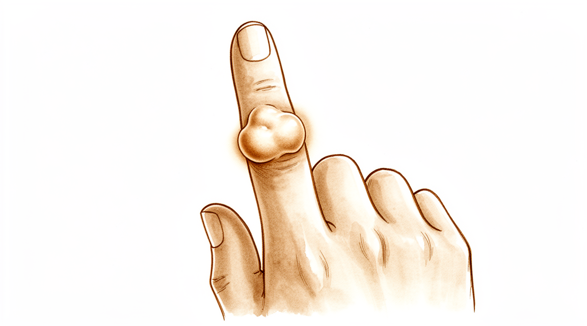
La mayoría de las personas notan un bulto en un dedo o en el pulgar que no duele. Por lo general es firme y crece lentamente. Algunos bultos se sitúan justo al lado de una articulación, a menudo la más cercana a la punta del dedo, y pueden aparecer en la parte de arriba, en el costado o en el lado de la palma del dedo. El dedo índice es la localización más frecuente.
En casi todos los casos, el bulto en sí no duele. Cuando hay molestias, suelen deberse a que el bulto presiona las estructuras cercanas a medida que crece. Un pequeño número de personas notan hormigueo o entumecimiento en el dedo si el bulto presiona un nervio. Algunas personas notan que el dedo se traba o hace un chasquido al moverse, de forma parecida a un dedo en gatillo, en el que el dedo se queda atascado un momento y luego se suelta de golpe. Si el bulto está cerca de una articulación, puede resultar difícil doblar ese dedo por completo porque el bulto estorba.
Las tareas cotidianas en las que se usa ese dedo pueden volverse incómodas. Recoger monedas pequeñas, abrocharse los botones, sujetar un bolígrafo o agarrar la tapa de un frasco pueden resultar torpes si el bulto está justo donde se apoya el agarre. El bulto también puede engancharse en los guantes o en los bolsillos.
A menudo el bulto lleva mucho tiempo ahí antes de que le moleste lo suficiente como para consultar. Muchas personas esperan meses o años, observando cómo cambia lentamente.
Hay algunas cosas sobre esta afección que conviene saber. Es benigna, es decir, no se disemina a otras partes del cuerpo. Es algo más frecuente en las mujeres que en los hombres, y suele aparecer entre los 32 y los 51 años. La mayoría de las personas tienen un solo bulto, aunque algunas tienen más de uno en distintos lugares.
Esté atento a que un dedo o la mano se pongan calientes, rojos, hinchados y dolorosos, sobre todo si hay fiebre. Si eso ocurre, acuda a urgencias ese mismo día. Si el bulto sigue creciendo, empieza a doler o el dedo no funciona como debería, consulte a su médico de cabecera o pida una evaluación especializada.
¿Qué está ocurriendo realmente?
Los tendones son los cordones resistentes que unen el músculo al hueso y permiten que los dedos se doblen. Cada tendón discurre dentro de una funda delgada llamada vaina tendinosa, que funciona de forma parecida a una junta alrededor de una cuerda: mantiene el cordón resbaladizo para que pueda deslizarse cuando el dedo se mueve.
En esta afección, las células de esa vaina empiezan a crecer más de lo que deberían. El resultado es un bulto de tejido blando que se sitúa sobre el tendón o a su lado, a menudo cerca de una articulación. Es benigno, es decir, no se disemina, y crece lentamente. La mayoría de los bultos son pequeños, de alrededor de 1,35 cm en promedio, aunque algunos crecen más.
El bulto está formado por varios elementos agrupados: células inflamatorias, células gigantes (que son simplemente células con varios núcleos) y un pigmento oscuro llamado hemosiderina, la misma sustancia que da color a un moretón antiguo. Ese pigmento es la razón por la que estos bultos pueden verse de color pardo si llegan a observarse durante una cirugía.
Los síntomas que notó en la sección anterior se deben a que el bulto estorba. A medida que crece, puede presionar el tendón, la articulación o un nervio, lo que explica que el dedo se trabe, la rigidez y el hormigueo ocasional. La mayor parte del tiempo no duele en absoluto, y solo alrededor de 16 de cada 100 personas refieren dolor.
Algunos puntos que conviene conocer. La mayoría de las personas tienen un solo bulto, pero alrededor de 21 de cada 100 tienen dos o más bultos separados. El bulto suele situarse al lado de una articulación, y en alrededor de un tercio de los casos en los dedos se encuentra en la articulación más cercana a la punta del dedo. Muy rara vez, el bulto puede presionar el propio hueso y hundirse en él, lo cual se ve en una radiografía.
También existe una forma más rara y difusa de la misma afección, que se extiende a lo largo del tendón en forma de finas "frondas" en lugar de formar un único bulto bien delimitado. Esa forma es mucho menos frecuente en un dedo y suele comportarse de manera diferente; por eso su cirujano querrá saber con claridad qué tipo de bulto es exactamente antes de planificar nada.
¿Qué podemos hacer al respecto?
El Dr. Kieran Hirpara, cirujano de extremidades superiores en el Mater Private Hospital Rockhampton, comienza con las opciones menos invasivas que se adapten a su condición. Por lo general, los pacientes son derivados a nuestra clínica por su médico de cabecera; si un fisioterapeuta le ha sugerido consultarnos, igualmente necesitará una derivación de su médico de cabecera para poder acceder al reembolso de Medicare. En la consulta, tomamos su historia clínica, examinamos su mano y, cuando es necesario, solicitamos estudios de imagen para determinar con exactitud de qué tipo de bulto se trata.
Como este bulto es benigno y crece lentamente, no hacer nada es una opción real. En el caso de un bulto pequeño e indoloro, simplemente vigilarlo puede ser la decisión correcta. Si el bulto estorba, la terapia de la mano puede ayudar con la rigidez y con el atrapamiento del dedo. La terapia tiene como objetivo mantener el dedo en movimiento y facilitar las tareas cotidianas. Por lo general, recomendamos darle una oportunidad razonable antes de pensar en la cirugía.
No existe ningún medicamento que reduzca el tamaño de este bulto, por lo que los analgésicos y los antiinflamatorios solo alivian las molestias que causa el bulto al presionar los tejidos cercanos. No hacen que el bulto se haga más pequeño.
La cirugía es el tratamiento habitual cuando el bulto es grande, duele o impide que el dedo funcione correctamente. El objetivo es extirpar todo el bulto junto con su zona de unión a la vaina del tendón, porque dejar esa zona de unión es lo que permite que vuelva a crecer. Planificamos con cuidado antes de operar, utilizamos aumento óptico durante la operación y extirpamos el bulto por completo. La recurrencia es el principal riesgo de esta afección y suele producirse dentro de los 36 meses posteriores a la extirpación. En un grupo de 605 personas a las que se les extirpó un bulto en un dedo, al 14,8 % les reapareció. Dicho de otro modo: a la mayoría de las personas, alrededor de 85 de cada 100, no les reaparece. Si reaparece, volver a extirparlo con medidas adicionales para evitar que vuelva a crecer todavía puede resolver el problema.
Qué esperar
El pronóstico de este bulto es estable, sin cambios drásticos. Es benigno, por lo que no se disemina a ninguna otra parte del cuerpo. Si se deja sin tratar, un bulto pequeño e indoloro suele seguir creciendo lentamente o simplemente se queda como está. Rara vez desaparece por sí solo, y no se convierte en nada peligroso.
Lo principal que hace esta afección con el tiempo es reaparecer después de la extirpación. La recurrencia es el riesgo principal de este bulto y suele producirse dentro de los 36 meses posteriores a la extirpación. Algunos bultos tienen más probabilidades de reaparecer que otros: los formados por dos o más fragmentos separados que no están unidos entre sí, y los que han crecido dentro del propio tendón o del revestimiento de la articulación. Cuando se extirpa todo el bulto junto con el punto donde está unido, ese riesgo disminuye mucho.
La función tras el tratamiento suele ser buena. Las personas a las que se les ha extirpado uno de estos bultos de un dedo obtienen, en promedio, un 92 % de la función normal de la extremidad en una escala estándar que mide cómo funciona la extremidad. En los casos publicados, las personas han conservado el movimiento completo del dedo, y la forma del hueso del dedo en la radiografía había vuelto a la normalidad en el control de los dos años.
Si el bulto es del tipo más raro y extendido, que rodea el tendón en forma de frondas, es más difícil extirparlo por completo y tiene más probabilidades de reaparecer. En esos casos, la radioterapia dirigida a la zona puede ayudar a controlar el bulto manteniendo el funcionamiento de la mano.
Si decide simplemente vigilar un bulto pequeño e indoloro, lo realista es esperar un bulto que crece lentamente o se mantiene igual, y que con el tiempo puede hacer más difícil doblar el dedo o usarlo para tareas pequeñas. Si empieza a doler, sigue creciendo o el dedo deja de funcionar como debería, ese es el momento en que el tratamiento suele merecer la pena.
Cuándo consultar a un especialista
La mayoría de los bultos como este no son urgentes, y el tratamiento nunca es una carrera. Consulte a su médico de cabecera si un bulto no mejora, crece a lo largo de semanas, empieza a doler o le impide usar el dedo o la mano con normalidad. Pida una evaluación especializada si el dedo se traba o hace un chasquido al moverse, si nota hormigueo o entumecimiento, o si le resulta difícil doblar el dedo por completo porque el bulto estorba. Estas son las situaciones que conviene revisar cuanto antes, porque un bulto que ha crecido dentro del tendón o del revestimiento de la articulación es más difícil de extirpar por completo y tiene más probabilidades de reaparecer.
Hay un conjunto de signos que sí requiere atención el mismo día. Si el dedo o la mano se ponen calientes, rojos, hinchados y dolorosos, sobre todo si hay fiebre, acuda a urgencias. No necesita una derivación previa de su médico de cabecera. Si no puede comunicarse con la clínica fuera del horario de atención o durante un fin de semana, acuda al servicio de urgencias más cercano.
En mayor profundidad
Advanced reading: the deeper science (optional)
Esta sección profundiza más de lo necesario para que usted tome sus propias decisiones de tratamiento. El tumor de células gigantes de la vaina tendinosa merece ser leído con atención, ya que su problema principal es la recurrencia; además, la evidencia indica que dicha recurrencia depende más de la biología del tumor en sí que de cómo se realice su extirpación.
La recurrencia es una propiedad del tumor, no solo de la cirugía
La explicación instintiva para el regreso de un bulto es que quedó algo del mismo. Sin embargo, una revisión sistemática de 605 casos concluyó lo contrario: la biología intrínseca del tumor parece desempeñar un papel más fundamental en la recurrencia que la ubicación del tumor o su invasividad local. Los autores solicitaron la realización de estudios prospectivos más amplios para identificar qué tumores son propensos a la recurrencia [1].
Esta información resulta verdaderamente útil antes de una operación. La recurrencia tras una escisión realizada correctamente constituye un comportamiento característico de este tumor, y no una señal de que algo salió mal.
El tratamiento es opcional; no hacer nada también es una opción válida
Es fácil olvidar esto una vez que se planifica una operación. El principio del tratamiento de primera línea es la resección completa; sin embargo, el tratamiento nunca es urgente, y la indicación para intervenir debe evaluarse en función de los síntomas, la progresión de la enfermedad, la ubicación del tumor y sus propias circunstancias [4].
En el caso de un nódulo pequeño, indoloro y de crecimiento lento, observarlo sin intervenir es una elección legítima. El tumor es benigno y no se disemina; por lo tanto, los motivos para operar se relacionan con la función, el tamaño y el grado de molestia que provoca, no con el riesgo de peligro.
Pero dos factores quirúrgicos sí son relevantes
La biología no lo explica todo. En 941 pacientes con tumor de células gigantes tenosinovial de tipo localizado, los factores asociados a la recurrencia tras la resección fueron un tamaño tumoral mayor y el tratamiento inicial mediante artroscopia. Dadas las tasas de complicaciones relativamente bajas y los buenos resultados funcionales, los autores recomiendan un abordaje abierto con resección completa siempre que sea posible, para reducir la recurrencia en casos de alto riesgo [2].
Por otro lado, se ha demostrado que la excisión artroscópica resulta eficaz para el tipo localizado en cuatro articulaciones, según un estudio realizado en 1,448 pacientes; en cambio, para el tipo difuso, la sinovectomía artroscópica solo ha mostrado eficacia en la rodilla [3].
Para conciliar ambos enfoques: en lesiones pequeñas, localizadas y bien delimitadas, cualquiera de los dos métodos puede ser adecuado. No obstante, a medida que el tamaño aumenta o en el caso del tipo difuso, la excisión abierta completa cuenta con mayor respaldo. La razón es de índole mecánica: este tumor se extiende en forma de “frondas” alrededor de tendones, nervios y articulaciones; las zonas que más fácilmente se pasan por alto son aquellas situadas detrás de estructuras que deben ser levantadas e inspeccionadas directamente.
¿Por qué es importante la nomenclatura?
Actualmente, esta afección se denomina tumor de células gigantes tenosinovial, término que abarca tanto la forma localizada en la mano como la forma difusa intraarticular anteriormente conocida como sinovitis villonodular pigmentada [4]. Se trata de la misma entidad, aunque en distintas localizaciones y con patrones de crecimiento diferentes.
Es importante tener esto en cuenta si investiga sobre el tema, ya que las búsquedas le mostrarán información referente a rodillas y caderas que describe la enfermedad en su forma difusa. Las tasas de recurrencia mencionadas para esta forma difusa son considerablemente más altas que las correspondientes a lesiones digitales localizadas. Aplicar esos datos de rodillas a un nódulo en el dedo podría sobreestimar el riesgo real.
Posición de la radioterapia en el tratamiento
En casos de enfermedad difusa que ha recidivado o que no puede resecarse por completo, a veces se considera la radioterapia adyuvante. Un metaanálisis determinó que la sinovectomía abierta, o la sinovectomía combinada con radioterapia perioperatoria, se asociaba con una tasa reducida de recurrencia en la sinovitis villonodular pigmentada difusa; no obstante, se requieren estudios prospectivos a largo plazo y de gran escala para confirmar dichos resultados [5].
En el caso de tumores digitales localizados, que constituyen la gran mayoría de los casos en la mano, esto no es aplicable. Este tipo de tumor pertenece al extremo del espectro caracterizado por afectación difusa, recurrencia y localización articular; se menciona aquí únicamente porque una búsqueda por el nombre de esta patología arrojará resultados relacionados con ella.
Qué no es
A pesar del nombre, se trata de un tumor benigno. No se disemina a otras partes del cuerpo. La palabra “tumor” tiene una connotación que aquí no le corresponde; la preocupación por su reaparición se refiere a la necesidad de realizar cirugías locales repetidas, a la rigidez articular y a la proximidad con los nervios, no al cáncer.
Referencias
[1] Fotiadis E, Papadopoulos A, Svarnas T, Akritopoulos P, Sachinis NP, Chalidis BE. Tumor de células gigantes del tejido sinovial de los dedos. Una revisión sistemática. Hand (N Y). 2011;6(3):244-9. https://doi.org/10.1007/s11552-011-9341-9
[2] Mastboom M, Staals E, Verspoor F, Rueten-Budde A, Stacchiotti S, Palmerini E, et al. Tratamiento quirúrgico de los tumores de células gigantes sinoviales de tipo localizado en articulaciones grandes: un estudio basado en una base de datos multicéntrica de 31 centros internacionales de sarcoma. J Bone Joint Surg Am. 2019;101(14):1309-18. https://doi.org/10.2106/JBJS.18.01147
[3] Noailles T, Brulefert K, Briand S, Longis P, Andrieu K, Chalopin A, et al. Tumor de células gigantes del tejido sinovial: cirugía abierta o sinovectomía artroscópica. Una revisión sistemática de la literatura. Orthop Traumatol Surg Res. 2017;103(5):809-14. https://doi.org/10.1016/j.otsr.2017.03.016
[4] Gouin F, Noailles T. Formas localizadas y difusas del tumor de células gigantes sinoviales (anteriormente tumor de células gigantes del tejido sinovial y sinovitis villonodular pigmentada). Orthop Traumatol Surg Res. 2017;103(1):S91-S97. https://doi.org/10.1016/j.otsr.2016.11.002
[5] Mollon B, Lee A, Busse JW, Griffin AM, Ferguson PC, Wunder JS, et al. Efecto de la sinovectomía quirúrgica y la radioterapia sobre la tasa de recurrencia de la sinovitis villonodular pigmentada de la rodilla. Bone Joint J. 2015;97-B(4):550-7. https://doi.org/10.1302/0301-620X.97B4.34907
Evidence & references
This is the clinical evidence summary written for health professionals. It is technical, and it lists the research this page was built from. You do not need to read it to understand your treatment or to make a decision about it.
Overview
- The diagnosis and treatment of suspected hand neoplasms should proceed in an orderly fashion similar to accepted methods applied to tumors in other locations of the extremities [1].
- Giant cell tumors of the tendon sheaths in the hand are benign lesions where recurrence is the primary risk [3, 4].
- Recurrence of giant cell tumors of the tendon sheaths typically occurs within 36 months of excision [3].
- The fundamental consideration in the management of hand tumours is to reconcile adequate tumour clearance with the maintenance of function and appearance [20].
- Complete surgical resection remains the treatment of choice for most patients with tenosynovial giant cell tumors [15].
- Diffuse tenosynovial giant cell tumor disease presents challenges due to high recurrence rates [15].
- Curettage, cryosurgery, and cementation performed by experienced surgeons appears to be a safe, effective, and reliable method for treating selected giant cell tumors of the hand [5].
- In cases of infiltrative giant cell tumor of the tendon sheath, radiation therapy may provide local tumor control with preservation of hand function [12].
- En bloc resection of the tumour and reconstruction with a fibular graft where necessary should be considered as the treatment of choice in giant cell tumour of the hand [10].
- En bloc resection of the tumour and reconstruction with an autograft should be considered in the treatment of recurrent giant cell tumour of the hand [8].
- En bloc resection and matched nonvascularized toe phalangeal transfer for Campanacci Grade 2 or 3 giant cell tumor of the phalanges resulted in a functional tumor-free digit with a low complication rate and no recurrences [35].
- Well-designed studies combining the recurrence rates of several hand surgery centers implementing a standardized treatment are needed to better demonstrate the associated risk factors for recurrence [28].
Anatomy & Pathophysiology
Clinical Presentation and Demographics
- Giant cell tumour of tendon sheath (GCTTS) is the second most common benign proliferative tumour in the upper extremities after ganglion cysts [102].
- GCTTS are usually slow-growing, painless, benign soft tissue tumours [102].
- GCTTS are most commonly found in the fingers and among women in their fourth and fifth decades [102].
- The male-to-female ratio for GCTTS of the digits is 1:1.47 [23].
- The mean age range for GCTTS of the digits is 32 to 51 years [23].
- Pain was reported in 15.7% of GCTTS cases and sensory disturbances in 4.57% of cases [23].
- A definite history of trauma was recorded in 5% of GCTTS lesions [23].
- The most frequent tumour location for GCTTS of the digits is the index finger (29.7%) [23].
- In a series of 64 cases, the most frequent location for GCTTS was the long finger (23.5%), followed by the thumb, index finger, and hand (20.3% each) [49].
- Patients with GCTTS arising in the fingers complained of a painless mass in almost every instance [24].
- The duration of symptoms for finger GCTTS ranged from two weeks to fifteen years, with an average duration of thirty-eight months [24].
- GCTTS are usually firm, lobulated, and non-tender masses [24].
- As the tumour grows, patients may present with swelling, pain, and limitation of movement [102].
Anatomical Distribution and Morphology
- Fifty-three xanthomas (GCTTS) arose adjacent to joints in the fingers, whereas twenty-nine bore no relation to joints [24].
- Thirty-one of ninety-one finger GCTTS occurred at the distal joints [24].
- Of the thirty-one distal joint GCTTS, eighteen were on the dorsal aspect and thirteen were distributed evenly on the radial, ulnar, and volar aspects [24].
- Type I GCTTS (single lesions) were more frequently detected (78.7%) than Type II tumours (two or more distinct tumours that were not joined together) (21.3%) [23].
- Type 1 GCTTS includes a nodular or multinodular lesion surrounded by a capsule, while Type 2 describes tumours with no connective tissue membrane and satellite, diffuse, or multicentric nodules [102].
- The average size of GCTTS was 1.35 cm, with a range of 0.3 cm to 5 cm [49].
- Bone erosion was found in 3 patients (4.7%) in a series of 64 GCTTS cases [49].
- Tendon involvement with flexors/extensors ratio of 4:3 was found in 7 cases (10.9%) in a series of 64 GCTTS cases [49].
- Involvement of the neurovascular bundle was presented in 7 patients (10.9%) in a series of 64 GCTTS cases [49].
- A case report describes the first patient with three separate GCTTS developing from both flexor and extensor tendons in a single digit [7].
Histology and Pathogenesis
- Microscopically, all GCTTS contained multinucleated giant cells, histiocytes, and haemosiderin deposits [49].
- GCTTS originates from the synovial membrane, tendon sheath, or synovial bursa [102].
- Inflammation resulting from reactive or regenerative hyperplasia is the generally accepted theory of pathogenesis for GCTTS [102].
- Genetic factors have been observed in previous studies regarding GCTTS pathogenesis [102].
- Fibroma of tendon sheath is histologically distinct from giant cell tumor of tendon sheath, a lesion with which it is commonly confused [16].
Diagnostic Imaging
- Radiography can be helpful in evaluating cortical destruction but is not helpful in the definitive diagnosis of GCTTS [102].
- Magnetic resonance imaging (MRI) is the most useful examination for the diagnosis and treatment planning of GCTTS [102].
- Al-Qattan classified GCTTS with MRI into two types according to the risk of recurrence [102].
Classification
- Giant cell tumors of the synovial sheaths in the hand are benign lesions in which recurrence is the primary risk [4].
- Recurrence for giant cell tumors of the tendon sheaths typically occurs within 36 months of excision [3].
- The Al-Qattan classification divides giant cell tumours of tendon sheath into two main types based on whether the entire tumour is surrounded by one pseudocapsule as assessed by the surgeon during surgery [90].
- Type I tumours are defined as those where the entire tumour is surrounded by one pseudocapsule [90].
- Type II tumours are defined as those where the entire tumour is not surrounded by one pseudocapsule [90].
- Type I tumours are sub-classified according to the thickness of the capsule, lobulation of the tumour, the presence of satellite lesions, and the diffuse or multicenteric nature of the tumour [90].
- Type II tumours are sub-classified according to the thickness of the capsule, lobulation of the tumour, the presence of satellite lesions, and the diffuse or multicenteric nature of the tumour [90].
- In a prospective study of 43 consecutive cases, none of the type I tumours (n=30) recurred [90].
- In a prospective study of 43 consecutive cases, recurrence occurred in five out of 13 type II tumours [90].
- Second recurrences were seen with type II B and C tumours, but not type II A tumours [90].
- Type I tumours (single lesions) were more frequently detected (78.7%) than type II tumours (two or more distinct tumours that were not joined together) (21.3%) in a systematic review of 605 patients [23].
- Type II tumours were associated with higher recurrence rates compared to Type I tumours in a systematic review of 605 patients [23].
- Direct involvement of the extensor tendons, flexor tendons, or joint capsule puts patients in a high-risk category with respect to recurrence [43].
- Preoperative diagnosis and meticulous surgical technique were found to be the only predictive factor of recurrence in a series of 14 cases [34].
- The overall recurrence rate in a systematic review of 605 patients was 14.8% [23].
Clinical Presentation
- Patients with fibrous xanthomas arising in the fingers complained of a painless mass in almost every instance [24].
- The duration of symptoms for finger tumors ranged from two weeks to fifteen years, with an average duration of thirty-eight months [24].
- The tumors were usually firm, lobulated, and non-tender [24].
- Fifty-three xanthomas arose adjacent to joints in the fingers, whereas twenty-nine bore no relation to joints [24].
- Thirty-one of the ninety-one tumors in the fingers occurred at the distal joints [24].
- Eighteen of the thirty-one distal joint tumors were on the dorsal aspect of the joint, and thirteen were distributed about evenly on the radial, ulnar, and volar aspects of the joint [24].
- The most frequent tumour location for giant cell tumour of tendon sheath of the digits was the index finger (29.7%) [23].
- Pain was reported in 15.7% of cases and sensory disturbances in 4.57% of cases [23].
- A definite history of trauma was recorded in 5% of lesions [23].
- Type I tumours (single lesions) were more frequently detected (78.7%) than type II tumours (two or more distinct tumours that were not joined together) (21.3%) [23].
- The male-to-female ratio for giant cell tumour of tendon sheath of the digits was 1:1.47 [23].
- The mean age for giant cell tumour of tendon sheath of the digits ranged from 32 to 51 years [23].
- A tendon tumour can present as a trigger finger [18].
- Trigger wrist caused by a giant cell tumour of tendon sheath has been reported [26].
- The trigger phenomenon in trigger wrist caused by a giant cell tumour of tendon sheath probably occurred when the tumour entered and left the distal carpal tunnel [26].
- Fibroma of tendon sheath is a benign soft tissue tumor that has a predilection for the hand [16].
- Fibroma of tendon sheath may be more common than has been previously recognized [16].
- The differential diagnosis of a soft tissue tumor in the digits and hand is usually limited to a ganglion, an inclusion cyst, or a giant cell tumor of tendon sheath (xanthofibroma) [16].
- Fibroma of tendon sheath should be included in the differential diagnosis of a soft tissue tumor in the digits and hand [16].
- A case of fibroma of tendon sheath presented as a 6-year history of a slowly enlarging palmar mass of the left index finger [16].
- Examination of a fibroma of tendon sheath case demonstrated a soft, compressible, lobulated, nontender mass extending from the second web space to the tip of the finger [16].
- Flexion of the digit was limited by the bulge in a case of fibroma of tendon sheath [16].
- The unusual location of a fibroma of the tendon sheath and atypical nature of the triggering phenomenon can lead to it being reported as a rare cause of median nerve compression at the wrist [50].
- Giant cell tumor of tendon sheath and pigmented villonodular synovitis are benign, locally invasive tumors involving synovium, tendon sheaths, and bursae [47].
- Patients with giant cell tumor of tendon sheath or pigmented villonodular synovitis may present with a discrete mass or with joint swelling, pain, or locking or catching [47].
- The knee and digits of the hand are the most common locations of lesions for giant cell tumor of tendon sheath and pigmented villonodular synovitis [47].
- Multifocal lesions of giant cell tumor of tendon sheath and pigmented villonodular synovitis are rare [47].
- The authors report the uniqueness of a case of multicentric giant cell tumor in the upper extremity [9].
- Radiological changes in the form of bony indentation was seen in only 2 cases of giant cell tumor of tendon sheath in a series of 12 patients [34].
- The most common presentation for giant cell tumor of tendon sheath in a series of 12 patients was with a mass over the hand, with a predilection to the thumb [34].
- A 65-year-old man presented with a slowly enlarging mass on the dorsal aspect of the left thumb consistent with a recurrent giant cell tumor of the tendon sheath [33].
Investigations
- The differential diagnosis of a soft tissue tumor in the digits and hand is usually limited to a ganglion, an inclusion cyst, or a giant cell tumor of tendon sheath [16].
- Fibroma of tendon sheath should be included in the differential diagnosis as it may be more common than has been previously recognized [16].
- Fibro-osseous pseudotumor of the digit should be considered in the differential diagnosis of rapidly enlarging digital lesions [64].
- Although MRI findings and location might help in the diagnosis of tenosynovial giant cell tumors, careful assessment is mandatory, especially in unusual locations [129].
Treatment
General Principles
Surgical Excision
- Complete surgical resection remains the treatment of choice for most patients with tenosynovial giant cell tumors, though diffuse disease presents challenges due to high recurrence rates [15].
- En bloc resection of metacarpal giant cell tumors with reconstruction by iliac graft appears to be an effective treatment [14].
- En bloc resection and matched nonvascularized toe phalangeal transfer resulted in a functional tumor-free digit with a low complication rate and no recurrences for Campanacci Grade 2 or 3 giant cell tumors of the phalanges [35].
- Total surgical excision ensuring removal of the attachment site (flexor sheath/palmar fascia) resulted in no local recurrences at a mean follow-up of 3.2 years for fibroma of tendon sheath [11].
- Local excision of the tumour appears to be a satisfactory method of treatment for Nora’s lesion [109].
- Preoperative planning aided by a tissue diagnosis with fine needle aspiration cytology, wide surgical exposure, and meticulous dissection with help of magnification are imperative for a successful outcome in giant cell tumor of tendon sheath [34].
- In all cases of giant cell tumor of tendon sheath, magnifying loupe or operating microscope was used during surgical excision [49].
- In cases of infiltrative giant cell tumor of tendon sheath, radiation therapy may provide local tumor control with preservation of hand function [12].
Recurrence and Adjuvant Therapy
- Giant cell tumors of the tendon sheaths in the hand are benign lesions where recurrence is the primary risk, typically occurring within 36 months of excision [3].
- In a study of 64 cases of giant cell tumor of tendon sheath, the recurrence rate was 4.7% (n=3) [49].
- In a systematic review of 605 patients with giant cell tumor of tendon sheath of the digits, 14.8% of patients had tumour recurrence [23].
- Repeated curettage with adjuvants eventually resulted in the cure for all patients and is therefore a reasonable treatment for both primary and recurrent giant cell tumor of the small bones of the hands and feet [105].
- Intralesional excision with cautery and methylmethacrylate provides a reliable method of treatment of giant cell tumors with good long-term functional results [71].
- Intralesional excision with local adjuvant therapy is recommended for the treatment of giant cell tumor of bone because it results in a good functional outcome compared to extralesional excision [98].
- Intralesional excision remains a viable, and likely the standard, mode of treatment for most giant cell tumors of the distal radius unless there is extensive bone loss [29].
- Based on data obtained from the number of studies available, intralesional excision appears to be more appropriate for the treatment of local lesions (eg, Grades 1 and 2) than Grade 3 giant cell tumors of the distal radius [52].
- Well-designed studies combining the recurrence rates of several hand surgery centers implementing a standardized treatment are needed to better demonstrate the associated risk factors for recurrence in giant cell tumor of tendon sheath [28].
Reconstruction and Resection Options
- Both curettage and resection/amputation are acceptable treatment options for the rare condition of giant cell tumour of bone in the hand, with a need to individualize treatment decisions based on the site and extent of disease to minimize treatment morbidity while maximizing disease control [6].
- Aggressive and malignant bone tumors of the second through fifth metacarpals generally require en bloc bone excision [17].
- Ray excision is usually best for lesions of the second and fifth metacarpals [17].
- If bone graft has been used for reconstruction, carpometacarpal arthrodesis and MCP ligament reconstruction or silicone arthroplasty may be practical [17].
- Whatever level is chosen for reconstruction, it is essential that an adequate, safe margin of normal tissue first be excised en bloc with the tumor [17].
- Aggressive tumors of the second through fifth metacarpals that have invaded soft tissue or sustained pathologic fractures often require excision of not only the involved ray but also the contiguous ray or rays radial and ulnar to the involved digit [17].
- If a malignant tumor has broken into the midpalm and extends across the metacarpals, removal of all digital rays may be needed to gain an adequate soft tissue margin [17].
- Retention of a sensate and relatively mobile thumb may be considerably more esthetic and functionally satisfactory than a forearm- or wrist-level amputation [17].
- If a tumor extends proximally from the metacarpal level, a more proximal level of hand, wrist, or forearm amputation is required for safe tumor management [17].
- Malignant soft tissue tumors in the palm or carpal tunnel often require at least partial hand amputation [17].
- If treated by only local or limited excision, the chance of recurrence is extremely high particularly in the setting of a positive margin for malignant soft tissue tumors in the palm or carpal tunnel [17].
- Below-elbow amputation is necessary to treat larger tumors of the palm or carpal tunnel [17].
- Wide en bloc excision of soft tissue sarcomas with negative margins is required to achieve local control of the lesion [17].
- At a minimum, aggressive soft tissue tumors, such as bone tumors, require ray resection or removal of multiple rays [17].
- Central palmar lesions more likely require sacrifice of three rays; those on the border are more likely than those in the center to be salvageable by removing just two rays [17].
- In the presence of proximal, broader, and larger lesions, all four digits or the entire hand may have to be sacrificed to save the patient [17].
- The use of a massive biocompatible bipolar unconstrained prosthesis is a viable treatment option for distal radius reconstruction after en-bloc resection of a giant cell tumour, offering rapid functional improvement without donor-site morbidity [68].
- Reconstruction after wide excision by nonvascularized fibular graft is a viable alternative for giant cell tumors of the lower end of radius though it is a challenging procedure and may be accompanied by major complications [111].
- En bloc resection is recommended for aggressive giant cell tumours (GCT) of the bone involving the distal end of the radius to minimize the risk of recurrence [91].
- The distal ulna may be widely resected with or without stabilization of the residual ulnar stump, yielding satisfactory local disease control and functional outcome [48].
- This is a simple and effective modality of reconstruction after resection of distal radial tumors using centralization of the ulna [30].
Complications
- Recurrence is the primary risk associated with giant cell tumors of the tendon sheaths in the hand [3].
- The overall recurrence rate for giant cell tumour of tendon sheath of the digits is 14.8% [23].
- Type II tumours (two or more distinct tumours that were not joined together) are associated with a higher risk of recurrence compared to Type I tumours [23].
- Direct involvement of the extensor tendons, flexor tendons, or joint capsule places patients in a high-risk category for recurrence [43].
- Surgical treatment of pigmented villonodular synovitis led to good functional results with an average Enneking score of 92% of normal limb function [32].
- A tendon tumour arising from the superficialis tendon can present as a trigger finger [18].
- A giant cell tumour of the tendon sheath can cause a trigger wrist phenomenon when the tumour enters and leaves the distal carpal tunnel [26].
- A fibroma of the tendon sheath in an unusual location can cause a triggering phenomenon and median nerve compression at the wrist [50].
- Total surgical excision ensuring removal of the attachment site (flexor sheath/palmar fascia) for fibroma of tendon sheath resulted in no local recurrences at a mean follow-up of 3.2 years [11].
- Patients with giant cell tumor of bone in the hand who are at higher risk of recurrence should be clinically followed more closely [41].
- Both curettage and resection/amputation are acceptable treatment options for giant cell tumour of bone in the hand, with a need to individualize treatment decisions based on the site and extent of disease to minimize treatment morbidity while maximizing disease control [6].
- Below-elbow amputation is necessary to treat larger malignant soft tissue tumors in the palm or carpal tunnel [17].
- Patients who received preoperative radiotherapy for hand tumors had a decrease in functional outcomes and grip strength that averaged 66% compared with the contralateral hand [55].
- None of the patients who underwent single-ray amputation for tumors of the hand, including giant cell tumors of bone, had local recurrences [55].
- A 29-year-old patient with recurrent giant-cell tumor of the digit was treated by phalangeal excision and toe phalanx transplant [54].
Recovery
- Recurrence of giant cell tumors of the tendon sheaths in the hand typically occurs within 36 months of excision [3].
- Total surgical excision ensuring removal of the attachment site (flexor sheath/palmar fascia) resulted in no local recurrences at a mean follow-up of 3.2 years [11].
- In cases of infiltrative GCTTS, radiation therapy may provide local tumor control with preservation of hand function [12].
- At 2-year final follow-up examination, no recurrence was noted, finger range of motion was full, and the x-ray film contour of the phalanx had returned to normal [13].
- Surgical treatment led to good functional results with an average Enneking score of 92% of normal limb function [32].
- En bloc resection and matched nonvascularized toe phalangeal transfer resulted in a functional tumor-free digit with a low complication rate and no recurrences [35].
Key Evidence
- [L5] The diagnosis and treatment of suspected hand neoplasms should proceed in an orderly fashion similar to accepted methods applied to tumors in other locations of the extremities. [1] (10.1016/s0363-5023(83)80277-9)
- [L4] Giant cell tumors of the tendon sheaths in the hand are benign lesions where recurrence is the primary risk, typically occurring within 36 months of excision. [3] (10.1016/j.otsr.2013.03.008)
- [L4] Giant cell tumors of the synovial sheaths in the hand are benign lesions in which recurrence is the primary risk. [4] (10.1016/j.jhsa.2013.08.051)
- [L4] Curettage, cryosurgery, and cementation performed by experienced surgeons appears to be a safe, effective, and reliable method for treating selected giant cell tumors of the hand. [5] (10.1053/jhsu.2001.22525)
- [L4] Both curettage and resection/amputation are acceptable treatment options for the rare condition of giant cell tumour of bone in the hand, with a need to individualize treatment decisions based on the site and extent of disease to minimize treatment morbidity while maximizing disease control. [6] (10.1177/17531934211007820)
- [L5] We believe this case report describes the first patient with three separate GCTTS developing from both flexor and extensor tendons in a single digit. [7] (10.1142/s2424835518720189)
- [L4] En bloc resection of the tumour and reconstruction with an autograft should be considered in the treatment of recurrent giant cell tumour of the hand. [8] (10.1016/s0266-7681(98)80197-6)
- [L5] The authors report the uniqueness of this case of multicentric giant cell tumor in the upper extremity. [9] (10.1016/j.main.2011.11.006)
- [L4] En bloc resection of the tumour and reconstruction with a fibular graft where necessary should be considered as the treatment of choice in giant cell tumour of the hand. [10] (10.1016/s0266-7681(96)80161-6)
- [L4] Total surgical excision ensuring removal of the attachment site (flexor sheath/palmar fascia) resulted in no local recurrences at a mean follow-up of 3.2 years. [11] (10.1177/1753193412469146)
- [L4] In cases of infiltrative GCTTS, radiation therapy may provide local tumor control with preservation of hand function. [12] (10.1016/j.jhsa.2012.01.011)
- [L5] At 2-year final follow-up examination, no recurrence was noted, finger range of motion was full, and the x-ray film contour of the phalanx had returned to normal. [13] (10.1016/s0363-5023(05)80151-0)
- [L5] En bloc resection of metacarpal giant cell tumors with reconstruction by iliac graft appears to be an effective treatment. [14] (10.1016/s1297-3203(03)00042-8)
- [L5] Complete surgical resection remains the treatment of choice for most patients with tenosynovial giant cell tumors, though diffuse disease presents challenges due to high recurrence rates. [15] (10.5435/jaaos-d-24-01255)
- [L5] [16] (10.1016/s0363-5023(84)80031-3)
- [L5] At operation a tumour arising from the superficials tendon was found and removed. [18] (10.1016/0266-7681(86)90283-4)
- [L5] The fundamental consideration in the management of hand tumours is to reconcile adequate tumour clearance with the maintenance of function and appearance. [20] (10.1016/0266-7681(91)90159-l)
- [L1] [23] (10.1007/s11552-011-9341-9)
- [L4] [24] (10.2106/00004623-196951010-00005)
- [L5] [26] (10.1016/s0266-7681(85)80038-3)
- [L3] Well-designed studies combining the recurrence rates of several hand surgery centers implementing a standardized treatment are needed to better demonstrate the associated risk factors for recurrence. [28] (10.1186/s12891-019-2866-8)
- [L3] Intralesional excision remains a viable, and likely the standard, mode of treatment for most giant cell tumors of the distal radius unless there is extensive bone loss. [29] (10.1007/s11999-014-4054-3)
- [L4] This is a simple and effective modality of reconstruction after resection of distal radial tumors. [30] (10.1016/j.jhsa.2022.05.011)
- [L4] Surgical treatment led to good functional results with an average Enneking score of 92% of normal limb function. [32] (10.1097/01.blo.0000224051.01873.fb)
- [L4] [33] (10.1016/j.jhsa.2012.11.001)
- [L4] [34] (10.1007/s12593-010-0020-9)
- [L4] En bloc resection and matched nonvascularized toe phalangeal transfer resulted in a functional tumor-free digit with a low complication rate and no recurrences. [35] (10.1016/j.jhsa.2024.06.013)
- [L4] Our observations suggest there are subsets of patients with giant cell tumor of bone who are at higher risk of recurrence and should be clinically followed more closely. [41] (10.1007/s11999-011-2172-8)
- [L3] Direct involvement of the extensor tendons, flexor tendons, or joint capsule puts patients in a high-risk category with respect to recurrence. [43] (10.1016/j.jhsa.2009.12.004)
- [Case_report] [47] (10.1016/j.jhsa.2014.11.010)
- [L3] The distal ulna may be widely resected with or without stabilization of the residual ulnar stump, yielding satisfactory local disease control and functional outcome. [48] (10.1177/1558944717743598)
- [L4] [49] (10.11138/gchir/2013.34.5.149)
- [L5] The unusual location of the fibroma of the tendon sheath (FGT) and the atypical nature of the triggering phenomenon led to the reporting of this observation as a rare cause of median nerve compression at the wrist. [50] (10.1016/j.main.2006.03.001)
- [L3] Based on data obtained from the number of studies available, intralesional excision appears to be more appropriate for the treatment of local lesions (eg, Grades 1 and 2) than Grade 3 GCTs of the distal radius. [52] (10.1007/s11999-012-2464-7)
- [L5] [54] (10.1016/s0363-5023(79)80134-3)
- [Paper] Fibro-osseous pseudotumor of the digit should be considered in the differential diagnosis of rapidly enlarging digital lesions. [64] (10.1177/15589447261481209)
- [Case_report] The use of a massive biocompatible bipolar unconstrained prosthesis is a viable treatment option for distal radius reconstruction after en-bloc resection of a giant cell tumour, offering rapid functional improvement without donor-site morbidity. [68] (10.1016/j.otsr.2013.04.001)
- [L4] Intralesional excision with cautery and methylmethacrylate provides a reliable method of treatment of giant cell tumors with good long-term functional results. [71] (10.1097/01.blo.0000128280.59965.e3)
- [L3] [90] (10.1054/jhsb.2000.0522)
- [L4] [91] (10.1177/17531934211068622)
- [L3] Intralesional excision with local adjuvant therapy is recommended for the treatment of giant cell tumor of bone because it results in a good functional outcome compared to extralesional excision. [98] (10.1007/s004020100317)
- [L4] [102] (10.1177/17531934231222401)
- [L3] Repeated curettage with adjuvants eventually resulted in the cure for all patients and is therefore a reasonable treatment for both primary and recurrent GCT of the small bones of the hands and feet. [105] (10.1302/0301-620x.95b6.30876)
- [L5] Local excision of the tumour appears to be a satisfactory method of treatment. [109] (10.1016/s0266-7681(97)80269-0)
- [L4] Reconstruction after wide excision by nonvascularized fibular graft is a viable alternative for giant cell tumors of the lower end of radius though it is a challenging procedure and may be accompanied by major complications. [111] (10.1007/s00402-010-1059-6)
- [L4] Although MRI findings and location might help in the diagnosis of a T-GCT, careful assessment is mandatory, especially in unusual locations. [129] (10.1186/s12891-016-1050-7)
References
[1] Tumors in the hand. The Journal of Hand Surgery. 1983. DOI: 10.1016/s0363-5023(83)80277-9
[3] Giant cell tumors of the tendon sheaths in the hand: Review of 96 patients with an average follow-up of 12 years. Orthopaedics & Traumatology: Surgery & Research. 2013. DOI: 10.1016/j.otsr.2013.03.008
[4] Giant Cell Tumors of the Tendon Sheaths in the Hand: Review of 96 Patients With an Average Follow-Up of 12 Years. The Journal of Hand Surgery. 2013. DOI: 10.1016/j.jhsa.2013.08.051
[5] Giant cell tumor of the hand: Superior results with curettage, cryosurgery, and cementation. The Journal of Hand Surgery. 2001. DOI: 10.1053/jhsu.2001.22525
[6] Giant cell tumour of hand bones: outcomes of treatment. Journal of Hand Surgery (European Volume). 2021. DOI: 10.1177/17531934211007820
[7] Multiple Giant Cell Tumor of Tendon Sheath Involving Both Flexor and Extensor Tendons in a Single Digit: A Case Report and Review of the Literatures. The Journal of Hand Surgery (Asian-Pacific Volume). 2018. DOI: 10.1142/s2424835518720189
[8] Recurrent Metacarpal Giant Cell Tumour Treated by En Bloc Resection and Metatarsal Transfer. Journal of Hand Surgery. 1998. DOI: 10.1016/s0266-7681(98)80197-6
[9] Tumeurs à cellules géantes à localisations multiples du membre supérieur. Chirurgie de la Main. 2012. DOI: 10.1016/j.main.2011.11.006
[10] Giant Cell Tumours of the Hand. Journal of Hand Surgery. 1996. DOI: 10.1016/s0266-7681(96)80161-6
[11] Fibroma of tendon sheath of the hand: a series of 20 patients with 23 tumours. Journal of Hand Surgery (European Volume). 2012. DOI: 10.1177/1753193412469146
[12] Radiation Therapy for Infiltrative Giant Cell Tumor of the Tendon Sheath. The Journal of Hand Surgery. 2012. DOI: 10.1016/j.jhsa.2012.01.011
[13] Vascular leiomyoma of the finger causing bone erosion. The Journal of Hand Surgery. 1995. DOI: 10.1016/s0363-5023(05)80151-0
[14] Tumeur à cellules géantes du troisième métacarpien : à propos d’un cas. Chirurgie de la Main. 2003. DOI: 10.1016/s1297-3203(03)00042-8
[15] Tenosynovial Giant Cell Tumor and Pigmented Villonodular Synovitis. Journal of the American Academy of Orthopaedic Surgeons. 2025. DOI: 10.5435/jaaos-d-24-01255
[16] Fibroma of tendon sheath. The Journal of Hand Surgery. 1984. DOI: 10.1016/s0363-5023(84)80031-3
[17] Green S Operative Hand Surgery. BOX 59.1 Ganglions of the Hand and Wrist > Finger Metacarpals.
[18] A tendon tumour presenting as a trigger finger. The Journal of Hand Surgery: Journal of the British Society for Surgery of the Hand. 1986. DOI: 10.1016/0266-7681(86)90283-4
[20] Soft Tissue Tumours of the Hand. Journal of Hand Surgery. 1991. DOI: 10.1016/0266-7681(91)90159-l
[23] Giant Cell Tumour of Tendon Sheath of the Digits. A Systematic Review. HAND. 2011. DOI: 10.1007/s11552-011-9341-9
[24] Fibrous Xanthoma of Synovium (Giant-Cell Tumor of Tendon Sheath, Pigmented Nodular Synovitis). The Journal of Bone & Joint Surgery. 1969. DOI: 10.2106/00004623-196951010-00005
[26] Trigger Wrist Caused by a Giant Cell Tumour of Tendon Sheath. Journal of Hand Surgery. 1985. DOI: 10.1016/s0266-7681(85)80038-3
[28] Giant cell tumor of tendon sheath in the hand: analysis of risk factors for recurrence in 50 cases. BMC Musculoskeletal Disorders. 2019. DOI: 10.1186/s12891-019-2866-8
[29] Is Intralesional Treatment of Giant Cell Tumor of the Distal Radius Comparable to Resection With Respect to Local Control and Functional Outcome?. Clinical Orthopaedics & Related Research. 2015. DOI: 10.1007/s11999-014-4054-3
[30] Functional Outcomes of Centralization of the Ulna as a Method of Reconstruction Following Resection of Campanacci Grade 3 Giant Cell Tumor of the Distal Radius. The Journal of Hand Surgery. 2024. DOI: 10.1016/j.jhsa.2022.05.011
[32] What Affects the Recurrence and Clinical Outcome of Pigmented Villonodular Synovitis?. Clinical Orthopaedics and Related Research. 2006. DOI: 10.1097/01.blo.0000224051.01873.fb
[33] Giant Cell Tumor of the Tendon Sheath. The Journal of Hand Surgery. 2013. DOI: 10.1016/j.jhsa.2012.11.001
[34] Giant Cell Tumor of Tendon Sheath: Case Series and Review of Literature. Journal of Hand and Microsurgery. 2010. DOI: 10.1007/s12593-010-0020-9
[35] Campanacci Grade 2 or 3 Giant Cell Tumor of the Phalanges: En Bloc Resection and Matched Nonvascularized Toe Phalangeal Transfer. The Journal of Hand Surgery. 2025. DOI: 10.1016/j.jhsa.2024.06.013
[41] Giant Cell Tumor of Bone: Are We Stratifying Results Appropriately?. Clinical Orthopaedics & Related Research. 2012. DOI: 10.1007/s11999-011-2172-8
[43] Recurrence of Giant Cell Tumors in the Hand: A Prospective Study. The Journal of Hand Surgery. 2010. DOI: 10.1016/j.jhsa.2009.12.004
[47] Recurrent Pigmented Villonodular Synovitis and Multifocal Giant Cell Tumor of the Tendon Sheath: Case Report. The Journal of Hand Surgery. 2015. DOI: 10.1016/j.jhsa.2014.11.010
[48] Extensor Carpi Ulnaris Tenodesis Versus No Stabilization After Wide Resection of Distal Ulna Giant Cell Tumors. HAND. 2017. DOI: 10.1177/1558944717743598
[49] Giant cell tumor of tendon sheath: study of 64 cases and review of literature. Giornale di Chirurgia - Journal of Surgery. 2013. DOI: 10.11138/gchir/2013.34.5.149
[50] Fibrome de la gaine tendineuse occasionnant un « poignet à ressaut » et un syndrome du canal carpien. Chirurgie de la Main. 2006. DOI: 10.1016/j.main.2006.03.001
[52] Which Treatment is the Best for Giant Cell Tumors of the Distal Radius? A Meta-analysis. Clinical Orthopaedics & Related Research. 2012. DOI: 10.1007/s11999-012-2464-7
[54] Treatment of recurrent giant-cell tumor of the digit by phalangeal excision and toe phalanx transplant: A case report. The Journal of Hand Surgery. 1979. DOI: 10.1016/s0363-5023(79)80134-3
[55] Green S Operative Hand Surgery. Index Finger Ray Amputation.
[64] Recurrent Fibro-Osseous Pseudotumour of the Digit Mimicking Infection: A Narrative Review and Case Report. HAND. 2026. DOI: 10.1177/15589447261481209
[68] Massive wrist prosthesis for giant cell tumour of the distal radius: A case report with a 3-year follow-up. Orthopaedics & Traumatology: Surgery & Research. 2013. DOI: 10.1016/j.otsr.2013.04.001
[71] Results of Giant Cell Tumor of Bone Treated With Intralesional Excision. Clinical Orthopaedics & Related Research. 2004. DOI: 10.1097/01.blo.0000128280.59965.e3
[90] Giant Cell Tumours of Tendon Sheath: Classification and Recurrence Rate. Journal of Hand Surgery. 2001. DOI: 10.1054/jhsb.2000.0522
[91] En bloc resection and vascularized ulnar pedicle graft reconstruction with plate fixation for giant cell tumour of the distal radius. Journal of Hand Surgery (European Volume). 2022. DOI: 10.1177/17531934211068622
[98] Oncologic and functional results after treatment of giant cell tumors of bone. Archives of Orthopaedic and Trauma Surgery. 2001. DOI: 10.1007/s004020100317
[102] The effect of surgical factors on recurrence of tendon sheath giant cell tumours. Journal of Hand Surgery (European Volume). 2024. DOI: 10.1177/17531934231222401
[105] Giant cell tumours of the small bones of the hands and feet. The Bone & Joint Journal. 2013. DOI: 10.1302/0301-620x.95b6.30876
[109] Nora’s Lesion. Journal of Hand Surgery. 1997. DOI: 10.1016/s0266-7681(97)80269-0
[111] Autogenous non-vascularized fibula for treatment of giant cell tumor of distal end radius. Archives of Orthopaedic and Trauma Surgery. 2010. DOI: 10.1007/s00402-010-1059-6
[129] Tenosynovial giant cell tumors in unusual locations detected by positron emission tomography imaging confused with malignant tumors: report of two cases. BMC Musculoskeletal Disorders. 2016. DOI: 10.1186/s12891-016-1050-7