Patients › Hand
Reemplazo de la articulación PIP
PIP joint replacement addresses painful arthritis in the middle finger joint when non-surgical options fail.
¿Por qué se ha recomendado esta operación?
El Dr. Kieran Hirpara, cirujano de extremidades superiores en el Mater Private Hospital Rockhampton, comienza por proponer las opciones menos invasivas que se adapten a su condición. Por lo general, los pacientes son derivados a nuestra clínica por su médico de cabecera; si un fisioterapeuta le ha sugerido que nos consulte, igualmente necesitará una derivación de su médico de cabecera para poder acceder al reembolso de Medicare. En su consulta, tomamos su historia clínica, examinamos su mano y, si es necesario, solicitamos estudios de imagen. Eso nos permite determinar cuál es el problema en la articulación.
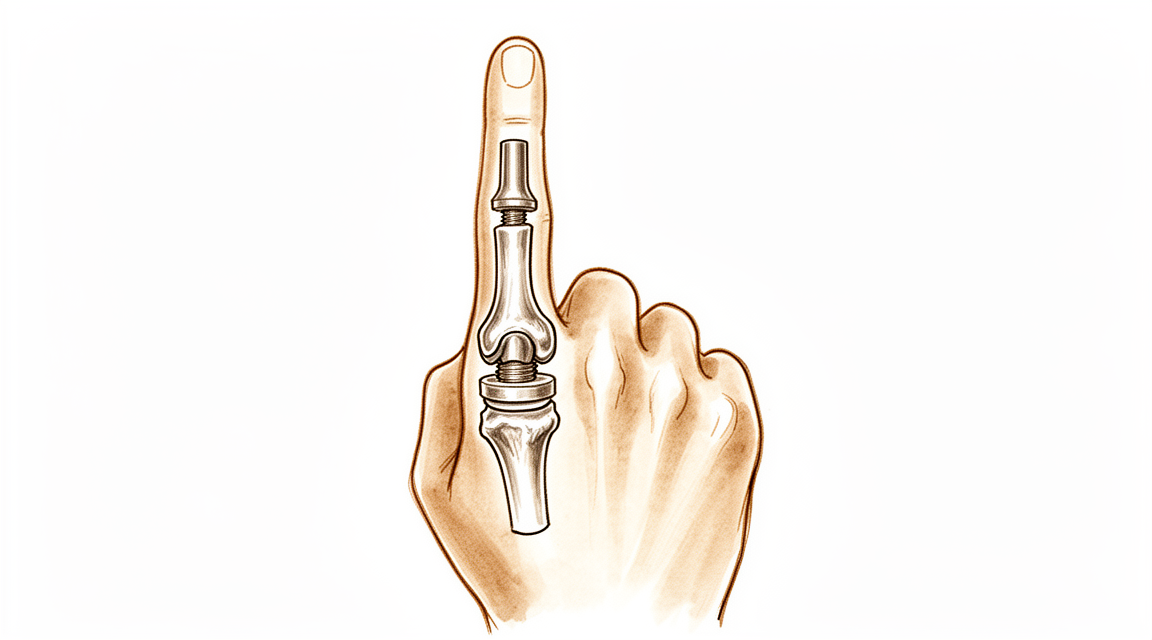
Esta operación consiste en sustituir la articulación intermedia de un dedo por un implante artificial. Generalmente se indica a personas que padecen artritis por desgaste o artritis posterior a una lesión, sobre todo a pacientes mayores y menos activos. El objetivo es mantener el movimiento de la articulación y evitar su fusión, es decir, unir los huesos de forma permanente.
En casos de artritis crónica, normalmente intentamos primero tratamientos no quirúrgicos. Esto puede implicar modificar la forma en que utiliza la mano, realizar terapia de mano o usar férulas. Si estos tratamientos no logran la mejora deseada, entonces analizamos con usted la posibilidad de realizar la cirugía. Los objetivos principales son aliviar el dolor y conseguir una articulación que pueda utilizar.
Antes de la operación
Para planificar su operación, tomamos radiografías de su dedo; a veces también realizamos una resonancia magnética o una ecografía. Estas imágenes muestran el estado de la articulación y nos ayudan a elegir el implante adecuado.
En las semanas previas a la cirugía, prepare algunas cosas prácticas. No coma ni beba durante siete horas antes de la operación. Pedimos que sea siete horas en lugar de seis para poder adelantar su hora si la lista quirúrgica avanza antes de lo previsto. Su cirujano le indicará qué medicamentos habituales debe suspender y cuándo. Lleve una lista por escrito de todos los fármacos que toma. Organice que alguien lo lleve a casa después de la intervención, ya que no podrá conducir usted mismo. Use ropa holgada y cómoda, cuyas mangas se puedan deslizar fácilmente sobre la mano.
Si padece otras enfermedades, es posible que necesite análisis de sangre o una consulta con el anestesista (el médico encargado de administrar la anestesia). La mayoría de las personas no requieren ninguno de estos procedimientos.
El día de la intervención
Llega usted a la unidad de admisiones quirúrgicas del hospital. El personal le registra y le prepara para el quirófano. Posteriormente, conoce al anestesista. Esta operación se realiza bajo anestesia general; usted permanecerá completamente dormido durante todo el procedimiento. Algunos pacientes también pueden recibir un bloqueo nervioso regional para aliviar el dolor postoperatorio; el anestesista decide al respecto ese mismo día, según sus circunstancias personales.
A continuación, le llevan al quirófano, donde se realiza la operación. Una vez finalizada, despierta usted en la sala de recuperación. Allí, las enfermeras le vigilan mientras la anestesia va desapareciendo. Cuando su estado se estabiliza, es trasladado a una sala de hospitalización o se le permite volver a casa el mismo día. Lo que ocurra dependerá del tipo de intervención y de cómo evolucione su recuperación.
Descripción del procedimiento quirúrgico
El Dr. Hirpara realiza esta operación mediante una única incisión en la cara palmar del dedo, justo sobre la articulación afectada. Realizar la incisión por este lado permite dejar intacto el tendón encargado de estirar el dedo, lo cual facilita que usted comience a moverlo poco después de la cirugía.
Una vez abierta la articulación, el cirujano extrae las superficies articulares desgastadas y dañadas. Estas se sustituyen por un implante artificial que crea una nueva superficie lisa entre los dos huesos del dedo. El implante está diseñado para permitir el movimiento de flexión y extensión de la articulación, aliviando al mismo tiempo el dolor causado por las superficies ásperas y desgastadas.
Finalmente, la herida se cierra con puntos de sutura y se coloca un vendaje encima. Usted se irá a casa con ese vendaje puesto; lo revisaremos junto con usted cuando lo retiremos, aproximadamente 10 días después.
Después de la operación
La mayoría de los pacientes permanecen una noche en el hospital tras esta operación, aunque algunos pueden volver a casa el mismo día. Despertará en la sala de recuperación y luego será trasladado a la habitación. Las enfermeras lo revisarán periódicamente y le administrarán analgésicos según sea necesario. Su dedo quedará cubierto con un vendaje; lo dejaremos puesto durante unos 10 días; por favor, no lo retire antes de ese plazo a menos que se lo indiquemos. Lo cambiaremos o lo quitaremos cuando le veamos. Puede levantarse y moverse en cuanto se sienta capaz; las enfermeras le ayudarán. Asegúrese de que alguien permanezca con usted durante las primeras 24 horas después de llegar a casa.
Recuperación
Durante los primeros días, el dedo le dolerá y estará hinchado. Esto mejora gradualmente. Mantener la mano elevada sobre un cojín, incluso mientras está sentado o duerme, ayuda a reducir la hinchazón. El uso de analgésicos sencillos según las indicaciones de su equipo médico aliviará el malestar.
El vendaje se mantiene durante unos 10 días; en esa fecha revisaremos la herida. Posteriormente, su recuperación dependerá de la terapia de mano que le proporcione Ruby Doolan en Extend Rehabilitation. Ruby es terapeuta especializada en mano: ella le indicará los ejercicios y confeccionará cualquier férula que su dedo necesite. Deberá realizar movimientos suaves de flexión y extensión en casa, poco a poco y a menudo. El objetivo es proteger el tendón en proceso de curación mientras la articulación recupera paulatinamente su movilidad; por ello, siga estrictamente el plan que ella le indique y no insista si siente dolor.
Al principio, deberá utilizar la otra mano para la mayoría de las tareas. Podrá vestirse, preparar comidas sencillas y moverse por la casa en cuanto se sienta capaz. No deberá conducir mientras el dedo lleve férula si esta le impide agarrar el volante con seguridad. Una vez retirada la férula y con autorización de su cirujano, podrá volver a conducir; consulte nuestra página sobre conducción tras cirugía de miembro superior.
A medida que disminuya la hinchazón y recupere el movimiento, notará que la articulación se flexiona más y cumple mejor sus funciones. Los hitos de recuperación se definen por estos cambios, no por fechas concretas.
Cada persona sana a su propio ritmo, por lo que su cronograma puede variar. Su cirujano y su terapeuta de mano le guiarán durante todo el proceso.
Qué puede salir mal
La mayoría de los pacientes evolucionan bien, pero en ocasiones pueden surgir problemas. Su cirujano y el equipo lo vigilarán de cerca para detectar cualquier anomalía a tiempo.
En algunas ocasiones, el propio implante genera problemas: puede aflojarse o aparecer una pequeña fisura en el hueso que lo rodea. Es posible que note un dolor nuevo en la articulación, la sensación de que el dedo está menos estable que antes, o un sonido de chasquido o rechinar al moverlo. También puede irritarse e inflamarse el revestimiento de la articulación. Si la articulación vuelve a desviarse de su posición normal, o si el dedo ya no se dobla o estira tanto como antes, hágaselo saber en su próxima consulta. Estos cambios merecen ser evaluados, incluso cuando no causen dolor.
El implante puede romperse. En el caso de un implante de silicona (un espaciador blando y flexible), su rotura suele provocar el regreso del dolor tras un período de mejoría. Si el dedo vuelve a doler, o si la movilidad sigue limitada, con o sin dolor, infórmenos. A veces el dedo desarrolla una inclinación lateral que antes no existía. Cualquiera de estos cambios indica que el implante debe ser evaluado.
La articulación puede volverse inestable: el dedo puede sentirse inestable lateralmente o incluso salirse parcialmente de su posición. Una articulación claramente desalineada requiere atención inmediata; por ello, llame a la clínica en lugar de esperar a su próxima cita.
También puede aparecer rigidez: el dedo podría no doblarse ni estirarse por completo, y la articulación se sentiría tensa. La rigidez persistente acompañada de dolor debe ser revisada.
Las infecciones son poco frecuentes pero graves. Esté atento a un dolor profundo y palpitante que no ceda con analgésicos comunes, enrojecimiento que se extiende desde la herida, calor en la zona o secreción. Si observa alguno de estos signos, comuníquese de inmediato con la clínica o acuda a urgencias si es fuera del horario laboral.
Durante o después de la cirugía pueden producirse fracturas alrededor del implante. El dolor repentino, la hinchazón o un cambio en la forma del dedo posterior a la intervención deben ser reportados.
Si surge algún problema, a veces es necesaria una cirugía adicional. Esto puede implicar reparar o reemplazar el implante; en algunos casos, incluso unir los huesos de forma permanente. Si fuera necesario, le explicaremos todo detalladamente.
En la tabla de complicaciones de esta página se detallan las tasas habituales; si desea conocer los datos específicos, puede consultarla.
Cuándo llamarnos
La mayoría de los problemas se manifiestan de las formas que describimos anteriormente; sin embargo, algunos requieren atención inmediata. Llámenos de inmediato si tiene fiebre, si el enrojecimiento alrededor de la herida se extiende o si aparece secreción nueva. También llámenos si experimenta dolor intenso y repentino, o si el dedo se entumece o no puede moverlo. Acuda a urgencias si presenta hinchazón o dolor en la pantorrilla, o dificultad para respirar, pues estos síntomas pueden indicar la presencia de un coágulo sanguíneo. Si es fuera del horario laboral y está preocupado, acuda a urgencias en lugar de esperar hasta la mañana.
¿Dónde leer más sobre esta afección?
Esta página trata sobre la intervención quirúrgica en sí. La afección que se trata con ella, así como las evidencias sobre cuándo la cirugía resulta beneficiosa y cuándo no, se explican con mayor detalle en la página Artritis de la articulación PIP.
Evidence & references
This is the clinical evidence summary written for health professionals. It is technical, and it lists the research this page was built from. You do not need to read it to understand your treatment or to make a decision about it.
Anatomy & Pathophysiology
General Hand Architecture
- The hand is composed of 19 bones, 17 articulations, and 19 muscles situated entirely within the hand [2].
- The hand contains approximately the same number of tendons activated by the forearm muscles as it has intrinsic muscles [2].
- The hand functions as an organ designed to obtain information and an organ of execution [2].
- The hand moves within a large volume of space with the shoulder as the apex, allowing it to reach any part of the body fairly easily due to the mobility of the shoulder, elbow, and wrist [2].
- The open hand with fingers extended and in contact forms a balanced graceful oval in its longitudinal axis [2].
- The dorsal aspect of the hand is convex, while the anterior, palmar or volar aspect is concave [2].
- The distal half of the hand is separated into five digits which flex toward the palm [2].
- The thumb has a more proximal and lateral position than the four fingers, allowing movement inward and outward from the palm [2].
- The four fingers are the distal extension of the carpometacarpal part of the hand [2].
- The hinges of finger movements are located at the thenar crease and at the transverse distal palmar crease, not at the bases of the digits [2].
- When fingers are extended and separated, their tips lie on the circumference of a circle whose center is the head of the third metacarpal [2].
Osseous and Arch Anatomy
- The metacarpal arch possesses adaptability due to the mobility of the peripheral metacarpals [7].
- The index metacarpal is the most firmly fixed of the metacarpals [7].
- The ring metacarpal has about 10 degrees of mobility in flexion and extension [7].
- The fifth metacarpal is semi-independent and has a range of flexion–extension of approximately 20 degrees [7].
- The second to fifth metacarpals are bound together by fibrous structures, the most distal of which is the deep transverse intermetacarpal ligament [7].
- The deep transverse intermetacarpal ligament is also known as the interglenoid ligament because it ties together the anterior glenoid ligaments of the metacarpophalangeal articulations [7].
- The longitudinal arches of the hand are composed of a fixed carpometacarpal portion and a mobile digital portion [7].
- The metacarpophalangeal articulations serve as the keystones of the longitudinal arches [7].
- The thick anterior glenoid capsules, known as volar plates, prevent hyperextension at the metacarpophalangeal joints [7].
- The stability of the metacarpophalangeal joints is essential to the support of both the longitudinal and transverse metacarpal arches [7].
Intrinsic Musculature and Extensor Mechanism
- There are seven interosseous muscles in the hand, consisting of four dorsal and three volar muscles [4].
- The dorsal interossei are abductors, while the volar interossei are adductors [4].
- The middle finger has two dorsal interossei and no volar interossei because the central axis of the hand lies within it [4].
- Each dorsal interosseous muscle, with the exception of the third, has two muscle heads: a superficial head and a deep head [4].
- The superficial head of the dorsal interosseous muscles abducts and weakly flexes the proximal phalanx [4].
- The deep head of the dorsal interosseous muscles flexes and weakly abducts the proximal phalanx while extending the middle and distal phalanges [4].
- Transverse fibers arch dorsally from each lateral band to join over the dorsum of the finger, flexing the proximal phalanx [4].
- Oblique fibers, also called spiral fibers, from the lateral bands insert onto the lateral tubercles at the base of the middle phalanx to extend the middle phalanx at the PIP joint [4].
- The lateral bands are joined by the lateral slips of the extensor tendon to form the conjoined lateral band [4].
- The two conjoined lateral bands to each finger unite at the distal third of the middle phalanx to form the terminal tendon [4].
- The terminal tendon inserts at the base of the distal phalanx to extend it [4].
- The volar interossei have only one muscle head and do not insert onto the proximal phalanx [4].
- The volar interossei form the ulnar lateral band of the index finger and the radial lateral band of the ring and little fingers [4].
- The flexor digiti quinti brevis is structurally and functionally similar to the deep head of the dorsal interossei, forming the ulnar lateral band of the little finger [4].
Cutaneous Anatomy
- Functional cutaneous units in the hand are similar to those described in the face [3].
- One dorsal cutaneous unit extends from the wrist to the proximal interphalangeal joints of the fingers and the interphalangeal joint of the thumb [3].
- The dorsal covering of the interphalangeal articulations forms a unique cutaneous unit characterized by considerable excess of skin when digits are in extension [3].
- The fine tight skin of the dorsal aspect of the middle phalanx forms a distinct cutaneous unit [3].
- The dorsal integument of the distal phalanx is distinct due to the nail bed with its matrix [3].
- The palm forms a cutaneous unit extending from the distal transverse crease of the wrist up to the transverse crease at the base of the digits [3].
- The palmar integument is subdivided into two zones by the oppositional crease of the thumb, which constitutes the oblique axis of the hand [3].
- The skin of the radial portion of the palm covers the thenar eminence and is relatively well vascularized and mobile [3].
- The skin of the ulnar and distal portion of the palm covers the hypothenar eminence where skin mobility is poor [3].
- The central triangular part of the palm has fixed and poorly vascularized skin that covers almost directly the superficial palmar aponeurosis [3].
- The integument of the palmar face of the digits is subdivided into phalangeal units separated by digital flexion folds [3].
- When a digit is completely flexed, the integument of adjacent phalanges comes into contact in the zones of the flexion creases, establishing areas of cutaneous contact in the form of a diamond [3].
- The sides of the diamond-shaped cutaneous contact zones do not undergo variations in length during flexion and extension movements [3].
- Incisions made along the lines of the diamond-shaped cutaneous contact zones present a minimal chance of retraction [3].
- The web spaces are formed from the union of two nonsymmetrical cutaneous surfaces [3].
- The dorsal slope of the web space has a gradual incline and supple skin that is not adherent to the subjacent region [3].
- The palmar surface of the web space is flat and precipitously interrupted, with skin densely adherent to the commissural skeleton [3].
- The commissural skeleton is formed by the interdigital palmar ligament between the fingers and by the distal transverse ligament at the level of the thumb web [3].
Vascular Anatomy
- The arteries of the thumb vary in both size and number, making surgical reconstruction delicate [8].
- The layout of thumb arteries is the result of innumerable variations regarding origin, transit, connections, and size [8].
- In anatomical studies, only 15% of dissections of the palmar arteries of the thumb fall into the classical "typical" category [8].
- The princeps pollicis artery is the terminal branch of the radial artery that crosses the first intermetacarpal space [8].
- The princeps pollicis artery runs along the ulnar side of the first metacarpal bone and along the volar surface of the adductor muscle [8].
- The princeps pollicis artery emerges onto the subcutaneous palmar tissue at the level of the cutaneous flexion crease of the metacarpophalangeal joint [8].
- At the metacarpophalangeal flexion crease, the princeps pollicis divides into two terminal rami known as the collateral palmar arteries of the thumb [8].
- The collateral palmar arteries run along the digital tunnel symmetrically and are of equal caliber [8].
- An arcade located deep in the flexor tendon joins together the two palmar arteries at the level of the distal metaphysis of the first phalanx [8].
- Vessels entering the vincula and irrigating the flexor tendon originate from the subtendinous anastomosis at the neck of the first phalanx [8].
- In the second segment of the thumb, the main artery is the ulnar collateral artery [8].
- The subtendinous anastomosis situated at the level of the neck of the first phalanx acts as a moderator between the two collateral arteries [8].
- In the pulp segment of the thumb, the two arteries are of similar size and run through thick fatty subcutaneous padding [8].
- The dorsal arteries of the thumb originate from palmar arteries at the level of the first metacarpal [8].
- The ulnar dorso-collateral artery generally stems from the princeps pollicis onto the medial border of the neck of the first metacarpal [8].
Surgical Anatomy and Incisions
- Distal palmar incisions are typically transverse, while proximal palmar incisions tend to be more longitudinal [9].
- Proximal palmar incisions should parallel the thenar crease [9].
- Incisions extended proximal to the wrist should not cross the flexor wrist creases at a right angle [9].
- The most important structure in the thenar area is the recurrent branch (motor) of the median nerve [9].
- Anatomic studies show that there is no single longitudinal incision in the proximal palm that completely avoids the palmar cutaneous branches of the median and ulnar nerves [9].
- The superficial volar neurovascular arch should be protected when deeper exposure is required in the distal palm [9].
- Structures lying between the metacarpal heads in the distal palm are not protected by the palmar fascia [9].
- Midlateral incisions described for fingers are suitable for the thumb, with the radial side being more accessible [9].
- A midlateral thumb incision can be extended by curving its proximal end at the midmetacarpal area to create a flap on the palmar surface of the thumb [9].
- Care must be taken to avoid injury to the dorsal branch of the superficial radial nerve when using a radial midlateral incision on the thumb [9].
Investigations
- Clinical evaluation of the hand and wrist requires combining patient history with a careful physical examination to pinpoint or narrow the scope of possible pathologic processes [1].
- Diagnostic tests such as imaging and serum laboratory studies are useful in determining pathology but can be expensive, time-consuming, and often nonspecific [1].
- A careful physical examination is essential to direct care and future testing if indicated [1].
- The skeleton of the hand and wrist consists of 27 bones, of which 19 are long bones [12].
- The skeleton is divided into five rays, each ray making up a polyarticulated chain comprising the metacarpals and phalanges [12].
- The base of each metacarpal articulates with the distal row of the carpus [12].
- The carpus articulates with the skeleton of the forearm through its proximal row [12].
- The radioulnocarpal articulation has two axes of movement to which is added a third—pronation and supination from the forearm [12].
- The wrist has three axes of movement, permitting the hand to be positioned in any spatial configuration and allowing it to be placed as needed for grasping [12].
- The radial ray or first ray is the shortest and is made up of only three bones—a metacarpal and two phalanges [12].
- The trapezium is clearly angled out in front of the carpal plane so that the first metacarpal makes an angle of about 45 degrees with the second metacarpal in the sagittal plane [12].
- The lengths of the metacarpals vary, with the thumb metacarpal being the shortest and the index finger the longest [12].
- The proximal and particularly the middle phalanges of the middle and ring fingers are longer than those of the index finger [12].
- The epiphyseal plates are located at the proximal ends of the phalanges and the first metacarpal [12].
- The epiphyseal plates are located at the distal ends of the other metacarpals [12].
References
[1] Orthopaedic Knowledge Update 13 Ebook Without Multimedia. Anatomy, Evaluation, Clinical Examination, and Imaging > Evaluation and Clinical Examination: Current Concepts.
[2] Exam Of The Hand Wrist 2Ed. INTRODUCTION.
[3] Exam Of The Hand Wrist 2Ed. Functional cutaneous units.
[4] Green S Operative Hand Surgery. Interosseous and Hypothenar Muscles.
[7] Exam Of The Hand Wrist 2Ed. The arches of the hand > The metacarpal arch.
[8] Exam Of The Hand Wrist 2Ed. Techniques of investigation of the arterial supply by J P Melki > Vascularization of the thumb > Palmar aspect.
[9] Campbell S Operative Orthopaedics 4 Volume Set. RESULTS OF SUTURE OF THE SCIATIC NERVE > PALMAR INCISIONS.
[12] Exam Of The Hand Wrist 2Ed. 1.1 SKELETON OF THE HAND > The osseous skeleton.