Patients › Hand
Fasciectomía de Dupuytren
Dupuytren’s fasciectomy — correcting hand contractures to improve function and daily activities.
¿Por qué se ha recomendado esta operación?
El Dr. Kieran Hirpara, cirujano de extremidad superior en el Mater Private Hospital Rockhampton, comienza por proponer las opciones menos invasivas que se adapten a su condición. La enfermedad de Dupuytren provoca que bandas de tejido en la palma se tensen y obliguen a uno o más dedos a quedar doblados. La fasciectomía consiste en extirpar ese tejido tensado para que el dedo pueda enderezarse de nuevo. Por lo general, los pacientes son derivados a nuestra clínica por su médico de cabecera; si un fisioterapeuta le ha sugerido que nos consulte, igualmente necesitará una derivación de su médico de cabecera para poder acceder al reembolso de Medicare. En su cita, tomamos su historia clínica, examinamos su mano y, si es necesario, solicitamos estudios de imagen.
Normalmente ofrecemos primero opciones no quirúrgicas, como terapia manual, uso de férulas o inyecciones. La cirugía se considera cuando estas medidas no producen mejoría suficiente, o cuando la deformidad ya es considerable. Por lo general la recomendamos cuando la articulación del nudillo (metacarpofalángica) está doblada 30 grados o más, o cuando la articulación media del dedo ya ha comenzado a curvarse. Cuanto más tiempo permanezca un dedo doblado, mayor es la probabilidad de que la contractura se vuelva permanente. El objetivo de la cirugía es enderezar el dedo y restaurar la función de la mano. En el 75 % de los casos se logra una corrección total o casi total. Conversaremos con usted para determinar si esta operación es adecuada para su mano y para sus objetivos.
Antes de la operación
Antes de la cirugía, recibirá instrucciones claras sobre el ayuno. Le pedimos que no ingiera nada, ni comida ni bebida, durante siete horas antes de la operación. Este tiempo es un poco más largo que el que exigen otras clínicas, ya que así podemos adelantar su cirugía si el programa quirúrgico lo permite. Su cirujano le indicará qué medicamentos habituales debe suspender y cuándo hacerlo; por ello, traiga una lista por escrito de todos los fármacos que toma. Organice que alguien lo lleve a casa después de la intervención. Use ropa holgada y cómoda, fácil de poner y quitar. Para planificar la operación, podría ser necesario realizar estudios de imagen como radiografías, ecografías o resonancias magnéticas. Si padece otras enfermedades, es posible que necesite análisis de sangre o una consulta con el anestesista.
El día de la intervención
El día de la operación, acude a la unidad de admisiones quirúrgicas del hospital. Allí se le registrará y se le preparará para el quirófano. Posteriormente, conocerá al anestesista, el médico encargado de velar por su sueño y comodidad durante la cirugía. Esta intervención se realiza bajo anestesia general; usted permanecerá completamente dormido durante todo el procedimiento. En algunos pacientes también se aplica un bloqueo nervioso regional para aliviar el dolor postoperatorio; el anestesista decide al respecto ese mismo día, según sus circunstancias personales.
Después, se le lleva al quirófano, donde se realiza la operación. Una vez finalizada, despierta en la sala de recuperación. Allí, las enfermeras le vigilan mientras la anestesia va desapareciendo. Cuando su estado sea estable, será trasladado a una planta hospitalaria o podrá volver a casa, según el tipo de intervención y cómo evolucione su recuperación.
Descripción del procedimiento quirúrgico
La fasciectomía se realiza mediante una sola incisión en la zona a operar, generalmente en la palma de la mano o a lo largo del dedo afectado. El cirujano extrae las bandas de tejido tensas que mantienen el dedo en posición flexionada. Una vez eliminado ese tejido, el dedo puede enderezarse.
La intervención se adapta específicamente a su mano. Si la enfermedad ha afectado la articulación media de un dedo, el cirujano trabaja con cuidado alrededor de dicha articulación. En ocasiones, una banda de tejido tensa cercana a la articulación impide que el dedo se enderece incluso después de retirar las bandas originales; liberar dicha banda puede facilitar aún más el enderezamiento del dedo.
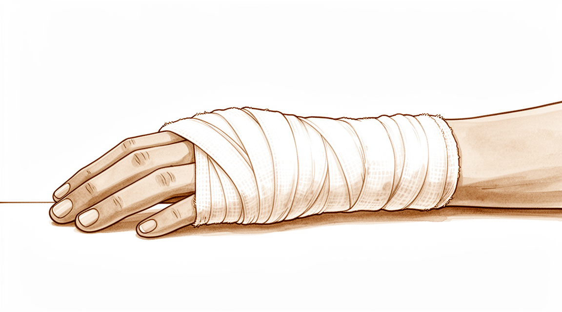
Al final de la operación, la incisión se cierra con puntos de sutura. Se coloca un vendaje sobre la herida antes de que abandone el quirófano. Deberá mantener ese vendaje durante unos 10 días; la sección “Después de la operación” describe lo que ocurrirá a continuación.
Después de la operación
La mayoría de los pacientes permanecen una noche en el hospital tras esta operación, aunque algunos pueden volver a casa el mismo día. Despertará en la sala de recuperación, donde las enfermeras lo vigilarán mientras el efecto de la anestesia desaparece. Su mano estará vendada y podría sentirse rígida o adolorida; se le administrarán analgésicos antes de que se vaya, y el equipo médico le explicará cómo manejar cualquier molestia. Mantenga la mano elevada sobre almohadas durante el descanso, pues esto ayuda a reducir la hinchazón. Alguien debe acompañarlo durante las primeras 24 horas. Dejamos el vendaje puesto durante unos 10 días; por favor, no lo retire antes de ese plazo a menos que se lo indiquemos. Lo cambiaremos o lo retiraremos cuando venga a la consulta. Puede moverse por casa según se sienta capaz, utilizando la otra mano para las tareas cotidianas.
Recuperación
Durante los primeros días, su mano estará adolorida e hinchada. Esto se irá mejorando gradualmente. Mantener la mano elevada sobre almohadas ayuda, al igual que el alivio del dolor que se le proporcionó antes de salir del hospital. Para muchas personas, la molestia es mayor en los primeros días y luego disminuye de forma constante.
Poco después de la cirugía, comenzará a realizar ejercicios de movilidad suaves. La terapia de mano tras esta operación la llevará a cabo Ruby Doolan en Extend Rehabilitation. Ruby es terapeuta especializada en mano: ella le guiará en los ejercicios y confeccionará cualquier férula que necesite. Es más importante realizar estos ejercicios con frecuencia y en pequeñas dosis que hacer sesiones largas. Estos ejercicios ayudan a que el dedo mantenga la rectitud lograda con la operación y contribuyen a reducir la hinchazón.
Al principio, utilizará la otra mano para la mayoría de las tareas cotidianas. Una vez que se retire el vendaje y la herida se sienta cómoda, normalmente podrá volver a conducir, siempre que pueda agarrar y girar el volante sin proteger la mano operada. Consulte nuestra página sobre Conducción tras una cirugía de miembro superior. El regreso al trabajo dependerá de las características de su empleo; su terapeuta le indicará cuándo su mano estará lista.
A medida que recupere la movilidad, notará que el dedo se endereza aún más y que tareas como agarrar una taza o sostener un bolígrafo se vuelven más fáciles. La recuperación varía según cada persona, por lo que su cronograma puede ser distinto. Su cirujano y su terapeuta de mano le brindarán orientación en cada control.
Qué puede salir mal
La mayoría de los pacientes evolucionan bien, pero en ocasiones pueden surgir problemas. Su cirujano y el equipo lo vigilarán de cerca para detectar cualquier anomalía a tiempo.
El tejido que se tensa y provoca la enfermedad de Dupuytren puede volver a desarrollarse. Si el dedo comienza a doblarse nuevamente en el mismo lugar, o si aparece una nueva banda fibrosa cerca, háganoslo saber en su próxima revisión. En algunos casos, será necesario realizar otro procedimiento para liberarla. Es mejor comentarlo a tiempo en lugar de esperar a que el dedo se doble mucho, ya que el tratamiento suele ser más sencillo cuando el problema se detecta precozmente.
Después de la cirugía, la hinchazón bajo la piel puede formar un coágulo de sangre cerca de la herida. Notará un bulto duro y sensible que aparece en los días posteriores a la operación; la piel sobre él podría verse amoratada o sentirse tensa. Si la hinchazón aumenta rápidamente o el dolor empeora en lugar de mejorar, llame a la clínica. Es posible que queramos verlo antes de lo previsto.
Cierto grado de dolor después de la cirugía es normal, pero debería disminuir progresivamente con los analgésicos que se le recetaron antes de irse. Sin embargo, un dolor profundo y pulsátil que no cede, o que empeora después de los primeros días, merece ser reportado. Llame a la clínica para que podamos examinar su mano.
Mientras el vendaje permanezca en su lugar, debe vigilar la herida. Si nota enrojecimiento que se extiende desde la zona quirúrgica, líquido que filtra a través del vendaje, o si la mano se calienta y se vuelve más sensible, comuníquese con nosotros. Estos pueden ser signos de infección, la cual es más fácil de tratar si se detecta a tiempo.
Algunas personas perciben un clic o sensación de “traba” al mover el dedo, o notan que no se endereza tanto como antes tras la cirugía. Comente esto en su próxima revisión. No siempre indica que haya algún problema, pero es útil registrarlo para poder seguir la evolución de su mano con el tiempo.
En la tabla de complicaciones de esta página se detallan las tasas típicas; si desea conocer los datos específicos, puede consultarla.
¿Cuándo deben llamarnos?
Llámenos si observan fiebre, enrojecimiento que se extiende desde la herida o líquido que se filtra a través del vendaje. Llámenos si el dolor empeora repentinamente o no disminuye. Acudan a urgencias si presentan hinchazón en la pantorrilla o dificultad para respirar, pues estos síntomas pueden indicar la presencia de un coágulo sanguíneo. Acudan a urgencias si su mano pierde sensibilidad o no puede moverse en absoluto. Si algo les parece anormal y no están seguros, llamen a la clínica. Preferimos realizar una evaluación temprana antes de que tengan que esperar.
¿Dónde leer más sobre esta enfermedad?
Esta página trata sobre la intervención quirúrgica en sí. La enfermedad que se trata, así como la evidencia científica sobre cuándo la cirugía resulta útil y cuándo no, se explican con mayor detalle en la página de la Enfermedad de Dupuytren.
Evidence & references
This is the clinical evidence summary written for health professionals. It is technical, and it lists the research this page was built from. You do not need to read it to understand your treatment or to make a decision about it.
Anatomy & Pathophysiology
General Hand Architecture
- The hand is composed of 19 bones, 17 articulations, and 19 muscles situated entirely within the hand [2].
- Approximately the same number of tendons activated by forearm muscles are present in the hand [2].
- The hand functions as an organ designed to obtain information and an organ of execution [2].
- The dorsal aspect of the hand is convex, while the anterior, palmar, or volar aspect is concave [2].
- The palmar surface is the functional surface of the hand, whereas the dorsal surface is usually visible and aesthetically important [2].
- The open hand with fingers extended and in contact forms a balanced graceful oval in its longitudinal axis [2].
- The proximal carpometacarpal half of the hand is flattened, presenting two faces with unique anatomical and functional significance [2].
Cutaneous Units and Skin
- Functional cutaneous units exist in the hand similar to those described in the face [3].
- One dorsal cutaneous unit extends from the wrist to the proximal interphalangeal joints of the fingers and the interphalangeal joint of the thumb [3].
- The dorsal covering of the interphalangeal articulations forms a unique cutaneous unit characterized by considerable excess of skin when digits are in extension [3].
- The palm forms a cutaneous unit extending from the distal transverse crease of the wrist up to the transverse crease at the base of the digits [3].
- The palmar integument is subdivided into two separate zones by the oppositional crease of the thumb, which constitutes the oblique axis of the hand [3].
- The skin of the radial portion of the palm covers the thenar eminence and external part of the palm and is relatively well vascularized and mobile [3].
- The skin of the ulnar and distal portion covers the hypothenar eminence where skin has poor mobility [3].
- The central triangular part of the palm has fixed, poorly vascularized skin that covers almost directly the superficial palmar aponeurosis, which inserts into it [3].
- The integument of the palmar face of the digits is subdivided into phalangeal units separated by digital flexion folds [3].
- When a digit is completely flexed, the integument of adjacent phalanges comes into contact in the zones of the flexion creases, establishing areas of cutaneous contact in the form of a diamond [3].
- The sides of the diamond-shaped cutaneous contact zones do not undergo variations in length during flexion and extension movements [3].
- Incisions made along the lines of the diamond-shaped cutaneous contact zones present a minimal chance of retraction [3].
- The web spaces are formed from the union of two nonsymmetrical cutaneous surfaces [3].
- The dorsal slope of the web space has a gradual incline and its supple skin is not adherent to the subjacent region [3].
- The palmar surface of the web space is flat and precipitously interrupted, with skin densely adherent to the commissural skeleton [3].
- The commissural skeleton is formed by the interdigital palmar (natatory) ligament between the fingers and by the distal transverse ligament at the level of the thumb web [3].
- The distal transverse ligament at the level of the thumb web is by far the deepest and the most mobile [3].
Muscular Anatomy
- There are seven interosseous muscles in the hand, consisting of four dorsal and three volar muscles [4].
- The dorsal interossei are abductors [4].
- The anatomic axis of the hand coincides with the axis of the third metacarpal [4].
- Dorsal interossei lie to the radial side of the index and middle fingers and the ulnar side of the middle and ring fingers [4].
- The little finger is abducted by the abductor digiti quinti [4].
- The volar interossei are adductors [4].
- Volar interossei lie to the ulnar side of the index finger and the radial side of the ring and little fingers [4].
- The middle finger has two dorsal interossei (abductors) and no volar interossei (adductors) because the central axis of the hand lies within it [4].
- Each dorsal interosseous muscle, with the exception of the third, has two muscle heads [4].
- The superficial head of the dorsal interosseous muscles arises most dorsally from the shaft of the contiguous metacarpals [4].
- The superficial head inserts deeply by a medial tendon onto the lateral tubercle of the base of the proximal phalanx [4].
- The superficial head abducts and weakly flexes the proximal phalanx [4].
- The superficial head has no direct effect on the middle or distal phalanges [4].
- The deep head of each dorsal interosseous muscle forms a lateral tendon, or lateral band, at the level of the MP joint [4].
- The deep head flexes and weakly abducts the proximal phalanx while extending the middle and distal phalanges [4].
- At the level of the middle of the proximal phalanx, transverse fibers arch dorsally from each lateral band to join each other over the dorsum of the finger [4].
- These transverse fibers flex the proximal phalanx [4].
- Oblique fibers (spiral fibers) from the lateral bands sweep over the distal third of the proximal phalanx to insert onto the lateral tubercles at the base of the middle phalanx [4].
- The oblique fibers extend the middle phalanx at the PIP joint [4].
- The lateral bands are joined by the lateral slips of the extensor tendon to form the conjoined lateral band [4].
- The two conjoined lateral bands to each finger unite at the distal third of the middle phalanx to form the terminal tendon [4].
- The terminal tendon inserts at the base of the distal phalanx to extend it [4].
- The flexor digiti quinti brevis is structurally and functionally similar to the deep head of the dorsal interossei [4].
- The flexor digiti quinti brevis forms the ulnar lateral band of the little finger [4].
- Each volar interosseous muscle has only one muscle head [4].
- None of the volar interossei insert onto the proximal phalanx [4].
- The volar interossei form the ulnar lateral band of the index finger and the radial lateral band of the ring and little fingers [4].
- The abductor digiti quinti and flexor digiti quinti brevis arise from the fifth metacarpal [4].
- The abductor digiti quinti inserts onto the ulnar lateral tubercle at the base of the proximal phalanx of the little finger [4].
- The flexor digiti quinti forms the ulnar lateral band [4].
- The opponens digiti quinti lies deepest among the hypothenar muscles [4].
- The opponens digiti quinti arises from the pisohamate ligament and the hook of the hamate [4].
- The opponens digiti quinti inserts onto the ulnar side of the diaphysis of the fifth metacarpal [4].
- The opponens digiti quinti flexes and supinates the fifth metacarpal [4].
Arches and Stability
- The metacarpal arch is endowed with a great deal of adaptability because of the mobility of the peripheral metacarpals [7].
- The peripheral metacarpals form the sides of the cup or palmar gutter and can deepen the concavity as they approach each other [7].
- The peripheral metacarpals are attached to the fixed element, which is the middle metacarpals [7].
- The thumb metacarpal is independent and articulates with the trapezium [7].
- The middle metacarpals are united to the carpus by the intrinsic interlocking encasement of the bones themselves [7].
- The index metacarpal is the most firmly fixed [7].
- The ring metacarpal is a transitional element to the fifth metacarpal and has about 10 degrees of mobility in flexion and extension [7].
- The fifth metacarpal is semi-independent and articulates with the hamate [7].
- The fifth metacarpal is restrained on its radial side by its articulation with the base of the fourth metacarpal [7].
- The fifth metacarpal has a range of flexion–extension of approximately 20 degrees [7].
- The second to fifth metacarpals are all bound together by various fibrous structures [7].
- The most distal fibrous structure binding the second to fifth metacarpals is the deep transverse intermetacarpal ligament [7].
- The deep transverse intermetacarpal ligament is better named the interglenoid ligament [7].
- The interglenoid ligament ties together the anterior “glenoid ligaments” of the metacarpophalangeal articulations, known as the “volar plates” [7].
- The longitudinal arches are composed of a fixed portion, the carpometacarpal, and a mobile portion, the digits [7].
- For every ray there is a longitudinal arch [7].
- The longitudinal arches diverge distally according to their different obliquities, with the thumb ray being the most divergent [7].
- The keystones of the longitudinal arches are the metacarpophalangeal articulations [7].
- The thick anterior glenoid capsules, known as volar plates, prevent hyperextension at the metacarpophalangeal articulations [7].
- The volar plates are interconnected by the transverse interglenoid ligament [7].
- The stability of the metacarpophalangeal joints is essential to the support of the longitudinal arch as well as of the transverse metacarpal arch [7].
- The five rays of the hand differ in mobility and independence [7].
- Mobility and independence are considerable for the thumb, much less for the fifth ray, and even less for the others [7].
- The index ray has a certain degree of independence at the phalangeal level owing to the arrangement of its flexor and extensor muscles [7].
Vascular Anatomy
- The arteries of the thumb vary in both size and number [8].
- The layout of the thumb arteries is the result of innumerable variations regarding origin, transit, connections, and size [8].
- The most common variations of the palmar arteries can be schematized by dividing the thumb into three segments defined by the metacarpophalangeal and interphalangeal flexion creases [8].
- In anatomical studies, only 15% of dissections fall into the category of the classical layout of palmar thumb arteries [8].
- The “princeps pollicis” artery is the terminal branch of the radial artery [8].
- The “princeps pollicis” artery crosses the first intermetacarpal space [8].
- The “princeps pollicis” artery runs along the ulnar side of the first metacarpal bone and along the volar surface of the adductor muscle [8].
- The “princeps pollicis” artery emerges onto the subcutaneous palmar tissue at the level of the cutaneous flexion crease of the metacarpophalangeal joint [8].
- At the metacarpophalangeal joint level, the “princeps pollicis” artery divides into two terminal rami, namely the collateral palmar arteries of the thumb [8].
- The collateral palmar arteries of the thumb run along the digital tunnel symmetrically and are of equal caliber [8].
- The collateral palmar arteries head distally to finally unite in the pulp arcade [8].
- During their transit in the digital tunnel, the collateral palmar arteries break off into numerous collateral branches, either cutaneous, articular, or osseous [8].
- An arcade located deep in the flexor tendon joins together the two arteries at the level of the distal metaphysis of the first phalanx [8].
- Vessels originating from this arcade enter the “vincula” and irrigate the flexor tendon [8].
- In the first segment of the thumb (between opposition crease and metacarpophalangeal flexion crease), it is rare to find arteries of surgical interest on the volar surface [8].
- The artery in the first segment is located deeply and is more easily accessible from the dorsal surface [8].
- In the second segment, the two arteries run alongside the flexor tendon and behind the collateral nerves [8].
- In the second segment, the main artery is the ulnar collateral artery [8].
- The ulnar collateral artery is sometimes absent, in which case it is replaced by the dorsal artery [8].
- The ulnar collateral artery is more often easier to dissect than the radial collateral artery [8].
- The size of the ulnar collateral artery enables the surgeon to achieve a more reliable microanastomosis [8].
- At the level of the neck of the first phalanx, a subtendinous anastomosis acts as a “moderator” between the two arteries [8].
- In cases where the palmar ulnar collateral artery is absent, the dorsal artery takes its place by means of a branch through the subtendinous arcade [8].
- In the third segment (pulp segment), the two arteries are of similar size [8].
- In the pulp segment, the arteries run through the thick fatty subcutaneous padding [8].
- In the pulp segment, the arteries cross over and convert into the ends of the digital nerves at the level of the median axis [8].
- The posterior area of the thumb is vascularized by two arteries which originate from the palmar arteries (princeps, commissural, or anastomoses of the superficial arcade) at the level of the first metacarpal [8].
- These dorsal arteries run laterally along the metacarpophalangeal joint and continue obliquely from volar to dorsal [8].
- The ulnar dorso-collateral artery generally stems from the “princeps pollicis” onto the medial border of the neck of the first metacarpal [8].
Surgical Considerations
- Distal palmar incisions are transverse as a rule [9].
- In the proximal palm, incisions tend to be more longitudinal, with the distal end curving radially and paralleling the closest major skin crease [9].
- An incision of any desired length can be made across the palm, provided that the underlying digital nerves and other vital structures are protected [9].
- After the skin and underlying fat have been incised, the fat is dissected from the palmar fascia and is carried with the skin flaps [9].
- It may be desirable to preserve small vessels perforating the palmar fascia if wide undermining of the skin flaps is necessary [9].
- Most of the vital structures are deep to the palmar fascia [9].
- In the distal palm, structures lying between the metacarpal heads are not protected by the palmar fascia [9].
- After the skin flaps have been retracted, the fascia can be incised in any direction necessary for ample exposure [9].
- Excision of the fascia may be desirable [9].
- The tendons and, parallel to them, the neurovascular bundles can then be seen [9].
- The superficial volar neurovascular arch should be protected when deeper exposure is required [9].
- Incisions in the more proximal palm should parallel the thenar crease [9].
- When extended proximal to the wrist, incisions should not cross the flexor wrist creases at a right angle [9].
- The most important structure in the thenar area is the recurrent branch (motor) of the median nerve [9].
- The recurrent branch of the median nerve should be exposed and protected if its exact location is in doubt [9].
- Care should be taken to avoid injury to the palmar cutaneous branches of the median and ulnar nerves [9].
- Anatomic studies have shown that there is no single longitudinal incision in the proximal palm that completely avoids the palmar cutaneous branches of the median and ulnar nerves [9].
- Midlateral incisions described for the fingers are also suitable for the thumb [9].
- The radial side of the thumb is more accessible for midlateral incisions [9].
- A radial midlateral incision can be extended by curving its proximal end at the midmetacarpal area and creating a flap on the palmar surface of the thumb [9].
- Care should be taken to avoid the dorsal branch of the superficial radial nerve to the radial side of the thumb [9].
- The radial midlateral incision can be used for tendon grafts without an additional palmar incision because the flap can be developed sufficiently to expose most of the flexor surface [9].
Investigations
Clinical Examination
- A careful physical examination is essential to direct care and future testing if indicated [1].
- Diagnostic tests such as imaging and serum laboratory studies can be expensive, time consuming, and often nonspecific [1].
- A systematic method to approaching the physical examination of the hand and wrist is essential due to the number of structures in a small space [1].
Imaging
- An 8-MHz Doppler tone assessment may be used to identify superficially displaced neurovascular bundles when Dupuytren cords lie beneath soft fleshy prominences [11].
- False-negatives are possible with 8-MHz Doppler tone assessment for identifying neurovascular bundles [11].
- Doppler imaging is a promising improvement for preoperative identification of structures, but higher resolution imaging technology is needed [11].
- MR assessment of Dupuytren’s disease is hindered by the resolution of current equipment [11].
- MR assessment of Dupuytren’s disease is hindered by orientation issues due to multiplanar deformities of the fingers [11].
- MR assessment of Dupuytren’s disease is hindered by the lack of intraoperative availability [11].
- MRI is probably most useful in identifying additional pathology such as flexor tendon bowstringing [11].
- MRI may be helpful in providing a quantitative noninvasive measure of cellularity of affected areas, which is an index of biologic activity [11].
- The potential of MRI as a staging tool based on cellularity has not been investigated yet on a large scale [11].
References
[1] Orthopaedic Knowledge Update 13 Ebook Without Multimedia. Anatomy, Evaluation, Clinical Examination, and Imaging > Evaluation and Clinical Examination: Current Concepts.
[2] Exam Of The Hand Wrist 2Ed. INTRODUCTION.
[3] Exam Of The Hand Wrist 2Ed. Functional cutaneous units.
[4] Green S Operative Hand Surgery. Interosseous and Hypothenar Muscles.
[7] Exam Of The Hand Wrist 2Ed. The arches of the hand > The metacarpal arch.
[8] Exam Of The Hand Wrist 2Ed. Techniques of investigation of the arterial supply by J P Melki > Vascularization of the thumb > Palmar aspect.
[9] Campbell S Operative Orthopaedics 4 Volume Set. RESULTS OF SUTURE OF THE SCIATIC NERVE > PALMAR INCISIONS.
[11] Dupuytren S Disease And Related Hyperproliferative Disorders. 54. The Future of Dupuytren’s Research and Treatment > 54.4 Mechanical Measurements and Procedures > 54.4.4 Imaging.