Patients › Hand
Excisión de ganglio en la vaina del tendón flexor
Excision of a flexor tendon sheath ganglion — when surgery is considered for painful or limiting cysts.
¿Por qué se ha recomendado esta operación?
El Dr. Kieran Hirpara, cirujano de extremidad superior en el Mater Private Hospital Rockhampton, comienza por valorar las opciones menos invasivas que se adapten a su condición. Por lo general, los pacientes son derivados a nuestra clínica por su médico de cabecera; si un fisioterapeuta le ha sugerido que nos consulte, igualmente necesitará una derivación de su médico de cabecera para poder acceder al reembolso de Medicare. En su consulta, tomamos su historia clínica, examinamos su mano y, si es necesario, solicitamos estudios de imagen para confirmar que el bulto es un ganglio.
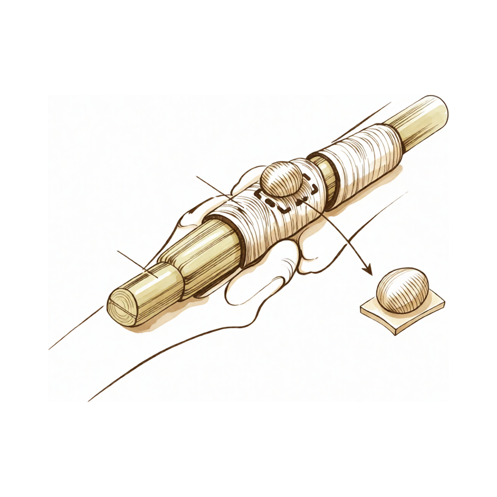
Un ganglio en la vaina del tendón flexor es un bulto pequeño y benigno relleno de líquido espeso. Se encuentra junto al túnel de tejido por donde se deslizan los tendones de su dedo. Esta operación consiste en extirpar dicho bulto mediante una sola incisión realizada en la zona a tratar.
La mayoría de los ganglios se tratan primero sin cirugía. Se puede utilizar una aguja para extraer el líquido, procedimiento que generalmente se intenta antes de proponer una operación. Algunos ganglios también disminuyen de tamaño por sí solos; en niños, un quiste que se estabiliza suele hacerlo en un plazo de 18 meses.
La cirugía se indica cuando el bulto sigue causando problemas tras haber probado estas medidas menos invasivas. Estos problemas pueden ser dolor, debilidad en la fuerza de agarre o un bulto que se engancha al mover el dedo. La extirpación del ganglio tiene como objetivo eliminar ese dolor y permitir que su dedo y mano funcionen con normalidad.
Antes de la operación
Su cirujano confirmará el plan durante la consulta y le indicará cómo prepararse. Se le pedirá que no coma ni beba durante 7 horas antes de la cirugía. Solicitamos 7 horas en lugar de las 6 habituales para poder adelantar su intervención si el programa quirúrgico lo permite. Es posible que deba suspender algunos de sus medicamentos habituales antes de la operación; su cirujano le dará instrucciones precisas al respecto, así que lleve consigo una lista de todos los fármacos que toma. Organice que alguien lo lleve a casa después de la cirugía y vístase con ropa cómoda y holgada. Para planificar la operación, podrían realizarse estudios por imagen como radiografías, ecografías o resonancias magnéticas. Si padece otras enfermedades, es posible que necesite análisis de sangre o una evaluación con el anestesiólogo.
El día de la intervención
Llega usted a la unidad de admisiones quirúrgicas del hospital, donde se le registra y se le prepara para el quirófano. Allí conocerá al anestesista. Esta operación se realiza bajo anestesia general; usted permanecerá completamente dormido durante todo el procedimiento. En algunos pacientes también se aplica un bloqueo nervioso regional para aliviar el dolor postoperatorio; el anestesista decide al respecto ese mismo día según sus circunstancias individuales. A continuación, se le lleva al quirófano, donde se realiza la operación. Después, despierta usted en la sala de recuperación, donde las enfermeras lo vigilan mientras la anestesia va desapareciendo. Una vez que se encuentre estable, será trasladado a la planta de hospitalización o podrá volver a casa, según el tipo de intervención y su recuperación.
Qué implica la operación
En esta operación se extirpa el ganglio mediante una única incisión realizada sobre el bulto. El cirujano trabaja con cuidado alrededor del túnel de tejido por donde se deslizan los tendones de los dedos, y retira el ganglio junto con el pequeño tallo que lo conecta a dicho túnel. Extraer todo el bulto, incluida su base, ayuda a reducir la probabilidad de que vuelva a aparecer.
La incisión se cierra con puntos de sutura. Antes de que abandone el quirófano, se coloca un apósito sobre la herida.
Durante la operación, el cirujano también examinará los tejidos circundantes. Si alguna parte del tendón se ha debilitado por la presión ejercida por el ganglio, dicho tendón se protegerá y se mantendrá intacto.
Después de la operación
Por lo general, se trata de una intervención ambulatoria, por lo que podrá volver a casa el mismo día; aunque en ocasiones los pacientes permanecen una noche en el hospital. Despertará en la sala de recuperación, donde las enfermeras lo vigilarán mientras el efecto de la anestesia desaparece. Su mano tendrá un vendaje suave sobre la herida, y le administraremos analgésicos para que se sienta cómodo. Podrá moverse en cuanto se sienta estable. Alguien debe acompañarlo durante las primeras 24 horas después de llegar a casa. Dejamos el vendaje puesto durante unos 10 días; por favor, no lo retire antes de ese plazo a menos que se lo indiquemos. Lo cambiaremos o lo quitaremos cuando venga a la consulta.
Recuperación
Durante los primeros días, su mano estará adolorida y ligeramente hinchada; esto disminuye gradualmente. Mantener la mano elevada sobre una almohada, incluso mientras está sentado o duerme, ayuda a reducir la hinchazón y la molestia. Los analgésicos que le administramos le permitirán sentirse cómodo mientras pasa lo peor.
Al irse a casa, tendrá un vendaje suave sobre la herida. Lo dejamos puesto durante unos 10 días y lo cambiamos o retiramos cuando venga a la consulta. Su dedo y mano pueden moverse suavemente dentro de dicho vendaje; no necesitará yeso ni férula.
La terapia de mano después de la cirugía la realizará Ruby Doolan en Extend Rehabilitation. Ruby es terapeuta especializada en mano, por lo que le guiará en los ejercicios y confeccionará cualquier férula que pudiera necesitar. Estos ejercicios mantienen el dedo en movimiento e impiden que se vuelva rígido mientras la herida cicatriza; debe hacerlos en casa, poco a poco y con frecuencia.
Las actividades cotidianas volverán poco a poco. Una vez que la herida deje de molestar y pueda agarrar objetos o girar un volante sin proteger la mano, normalmente podrá volver a conducir. Puede leer más en nuestra página sobre cómo conducir tras una cirugía de extremidad superior. En cuanto al trabajo, depende de las funciones propias de su empleo; su terapeuta le indicará cuándo su mano estará lista para retomarlo.
La recuperación varía según cada persona. Su cronograma personal puede ser distinto; su cirujano y terapeuta de mano lo guiarán durante todo el proceso.
Qué puede salir mal
La mayoría de los pacientes evolucionan bien, pero en ocasiones pueden surgir problemas. Su cirujano y el equipo lo vigilarán de cerca para detectar cualquier anomalía a tiempo.
A veces el bulto reaparece en el mismo lugar. Notará o verá una pequeña hinchazón firme cerca de la cicatriz previa. Si observa esto, mencione el hecho en su próxima consulta.
Los nervios pasan muy cerca de la zona operada. Durante la cirugía, un nervio puede sufrir contusión o irritación. Esto se siente como hormigueo, sensación de pinchazos o una zona de piel entumecida cerca de la incisión. En la mayoría de los casos esto mejora, pero a veces queda un pequeño punto sensible o nódulo donde el nervio resultó afectado. Informe a su cirujano en la revisión si presenta entumecimiento o hormigueo que no desaparece.
Los pequeños nervios y vasos sanguíneos que recorren el lateral del dedo se encuentran cerca. Las lesiones en estas estructuras son poco frecuentes, pero su cirujano está atento a ello durante la operación.
La herida puede infectarse. Esté atento a enrojecimiento que se extienda desde la incisión, aumento del dolor, calor en la zona o secreción de líquido. También podría sentir fiebre. Si observa cualquiera de estos signos, llame de inmediato a la clínica o acuda al servicio de urgencias si es fuera del horario laboral.
Después de la cirugía, puede acumularse sangre debajo de la herida. Esto se manifiesta como una hinchazón firme y sensible que aparece en los primeros días. Si es grande o muy dolorosa, podría ser necesario drenarla en el quirófano. Comuníquese con la clínica si nota esto.
La cicatriz puede volverse elevada, gruesa y rojiza durante la cicatrización; esto se denomina cicatriz hipertrófica. Puede ser firme y, a veces, provocar picor. Mencione este hecho en su revisión, ya que existen tratamientos que pueden ayudar.
La vaina del tendón cercana a la herida puede inflamarse. Esto se siente como crujidos o chasquidos al mover el dedo, acompañados de sensibilidad a lo largo del tendón. Comente esto en su próxima consulta.
También es posible que su dedo o muñeca se sientan rígidos durante un tiempo. Los movimientos suaves y los ejercicios de terapia de mano ayudan a prevenirlo. Si la rigidez no mejora, informe a su terapeuta o cirujano.
En la tabla de complicaciones de esta página se detallan las tasas típicas; puede consultarlas si desea información específica.
¿Cuándo deben llamarnos?
La mayoría de los problemas se detectan durante las revisiones, pero algunos requieren atención más pronta. Llámenos si tiene fiebre, enrojecimiento que se extiende alrededor de la incisión o secreción de líquido desde la herida. Llámenos también si el dolor empeora repentinamente, o si aparece una hinchazón firme y sensible cerca de la incisión durante el primer o segundo día. Acuda a urgencias si presenta hinchazón o dolor en la pantorrilla, o dificultad para respirar; estos pueden ser signos de un coágulo sanguíneo. Llámenos igualmente si sus dedos se entumecen, se enfrían o se vuelven pálidos, o si no puede mover el dedo o la mano cuando debería poder hacerlo. Si es fuera del horario laboral, diríjase al servicio de urgencias.
¿Dónde leer más sobre esta afección?
Esta página trata sobre la operación en sí. La afección que se trata con ella, incluyendo lo que demuestran las evidencias sobre cuándo la cirugía es útil y cuándo no, se explica con mayor detalle en la página Ganglio en la vaina del tendón flexor.
Evidence & references
This is the clinical evidence summary written for health professionals. It is technical, and it lists the research this page was built from. You do not need to read it to understand your treatment or to make a decision about it.
Anatomy & Pathophysiology
General Hand Architecture
- The hand is composed of 19 bones, 17 articulations, and 19 muscles situated entirely within the hand [2].
- The hand contains approximately the same number of tendons activated by forearm muscles as it has intrinsic muscles [2].
- The open hand with fingers extended and in contact forms a balanced graceful oval in its longitudinal axis [2].
- The proximal carpometacarpal half of the hand is flattened and presents two faces with unique anatomical and functional significance [2].
- The posterior or dorsal aspect of the hand is convex, while the anterior, palmar, or volar aspect is concave [2].
- The distal half of the hand is separated into five digits that flex toward the palm [2].
- The thumb has a more proximal and lateral position than the four fingers, allowing movement inward and outward from the palm [2].
- The hinges for finger flexion and extension are located at the thenar crease and the transverse distal palmar crease, not at the bases of the digits [2].
- When fingers are extended and separated, their tips lie on the circumference of a circle whose center is the head of the third metacarpal [2].
Cutaneous Units
- The dorsum of the hand contains a cutaneous unit extending from the wrist to the proximal interphalangeal joints of the fingers and the interphalangeal joint of the thumb [3].
- The dorsal covering of the interphalangeal articulations forms a unique cutaneous unit characterized by considerable excess of skin when digits are in extension [3].
- The fine tight skin of the dorsal aspect of the middle phalanx forms a distinct cutaneous unit [3].
- The dorsal integument of the distal phalanx is distinct due to the presence of the nail bed and its matrix [3].
- The palm forms a cutaneous unit extending from the distal transverse crease of the wrist up to the transverse crease at the base of the digits [3].
- The palmar integument is subdivided into two zones by the oppositional crease of the thumb, which constitutes the oblique axis of the hand [3].
- The skin of the radial portion of the palm covers the thenar eminence and external part of the palm and is relatively well vascularized and mobile [3].
- The skin of the ulnar and distal portion covers the hypothenar eminence, where skin mobility is poor [3].
- The central triangular part of the palm has fixed, poorly vascularized skin that covers almost directly the superficial palmar aponeurosis [3].
- The integument of the palmar face of the digits is subdivided into phalangeal units separated by digital flexion folds [3].
- When a digit is completely flexed, the integument of adjacent phalanges comes into contact in the zones of the flexion creases, establishing areas of cutaneous contact in the form of a diamond [3].
- The sides of the diamond-shaped cutaneous contact zones do not undergo variations in length during flexion and extension movements [3].
- Incisions made along the lines of the diamond-shaped cutaneous contact zones present a minimal chance of retraction [3].
- The web spaces are formed from the union of two nonsymmetrical cutaneous surfaces [3].
- The dorsal slope of the web space has a gradual incline and supple skin that is not adherent to the subjacent region [3].
- The palmar surface of the web space is flat, precipitously interrupted, and densely adherent to the commissural skeleton [3].
- The commissural skeleton is formed by the interdigital palmar (natatory) ligament between fingers and by the distal transverse ligament at the level of the thumb web [3].
Intrinsic Muscles
- There are seven interosseous muscles in the hand, consisting of four dorsal and three volar muscles [4].
- The dorsal interossei are abductors [4].
- The volar interossei are adductors [4].
- The middle finger has two dorsal interossei and no volar interossei because the central axis of the hand lies within it [4].
- Each dorsal interosseous muscle, with the exception of the third, has two muscle heads [4].
- The superficial head of the dorsal interosseous muscles arises most dorsally from the shaft of contiguous metacarpals and inserts deeply by a medial tendon onto the lateral tubercle of the base of the proximal phalanx [4].
- The superficial head of the dorsal interosseous muscles abducts and weakly flexes the proximal phalanx [4].
- The deep head of each dorsal interosseous muscle forms a lateral tendon, or lateral band, at the level of the metacarpophalangeal joint [4].
- The deep head of the dorsal interosseous muscles flexes and weakly abducts the proximal phalanx while extending the middle and distal phalanges [4].
- At the level of the middle of the proximal phalanx, transverse fibers arch dorsally from each lateral band to join each other over the dorsum of the finger, flexing the proximal phalanx [4].
- Oblique fibers, or spiral fibers, from the lateral bands sweep over the distal third of the proximal phalanx to insert onto the lateral tubercles at the base of the middle phalanx [4].
- The oblique fibers extend the middle phalanx at the proximal interphalangeal joint [4].
- The lateral bands are joined by the lateral slips of the extensor tendon to form the conjoined lateral band [4].
- The two conjoined lateral bands to each finger unite at the distal third of the middle phalanx to form the terminal tendon [4].
- The terminal tendon inserts at the base of the distal phalanx to extend it [4].
- The flexor digiti quinti brevis is structurally and functionally similar to the deep head of the dorsal interossei, forming the ulnar lateral band of the little finger [4].
- Each volar interosseous muscle has only one muscle head and none insert onto the proximal phalanx [4].
- The volar interossei form the ulnar lateral band of the index finger and the radial lateral band of the ring and little fingers [4].
- The abductor digiti quinti and flexor digiti quinti brevis are similar in structure and function to the superficial and deep heads of the dorsal interossei, respectively [4].
- The abductor digiti quinti arises from the fifth metacarpal and inserts onto the ulnar lateral tubercle at the base of the proximal phalanx of the little finger [4].
- The opponens digiti quinti lies deepest among the hypothenar muscles, arising from the pisohamate ligament and hook of the hamate [4].
- The opponens digiti quinti inserts onto the ulnar side of the diaphysis of the fifth metacarpal, which it flexes and supinates [4].
Arches and Stability
- The metacarpal arch is endowed with a great deal of adaptability because of the mobility of the peripheral metacarpals [7].
- The peripheral metacarpals form the sides of the cup or palmar gutter and can deepen the concavity as they approach each other [7].
- The thumb metacarpal is independent and articulates with the trapezium [7].
- The middle metacarpals are united to the carpus by the intrinsic interlocking encasement of the bones themselves [7].
- The index metacarpal is the most firmly fixed [7].
- The ring metacarpal is a transitional element to the fifth metacarpal and has about 10 degrees of mobility in flexion and extension [7].
- The fifth metacarpal is semi-independent, articulates with the hamate, and is restrained on its radial side by its articulation with the base of the fourth metacarpal [7].
- The fifth metacarpal has a range of flexion–extension of approximately 20 degrees [7].
- The second to fifth metacarpals are bound together by various fibrous structures, the most distal of which is the deep transverse intermetacarpal ligament [7].
- The deep transverse intermetacarpal ligament is better named the interglenoid ligament because it ties together the anterior glenoid ligaments of the metacarpophalangeal articulations, known as the volar plates [7].
- The longitudinal arches are composed of a fixed portion, the carpometacarpal, and a mobile portion, the digits [7].
- The keystones of the longitudinal arches are the metacarpophalangeal articulations [7].
- The thick anterior glenoid capsules, or volar plates, of the metacarpophalangeal joints prevent hyperextension [7].
- The volar plates are interconnected by the transverse interglenoid ligament [7].
- The stability of the metacarpophalangeal joints is essential to the support of the longitudinal arch as well as of the transverse metacarpal arch [7].
Vascular Anatomy
- The arteries of the thumb vary in both size and number, making surgical reconstruction delicate [8].
- The palmar arteries of the thumb can be schematized by dividing the thumb into three segments defined by the metacarpophalangeal and interphalangeal flexion creases [8].
- In the classical layout, the princeps pollicis artery, a terminal branch of the radial artery, crosses the first intermetacarpal space and runs along the ulnar side of the first metacarpal bone [8].
- The princeps pollicis artery runs along the volar surface of the adductor muscle and emerges onto the subcutaneous palmar tissue at the level of the cutaneous flexion crease of the metacarpophalangeal joint [8].
- At the metacarpophalangeal joint level, the princeps pollicis artery divides into two terminal rami, the collateral palmar arteries of the thumb [8].
- The collateral palmar arteries run along the digital tunnel symmetrically and are of equal caliber, heading distally to unite in the pulp arcade [8].
- During transit in the digital tunnel, the collateral palmar arteries break off into numerous collateral branches that are cutaneous, articular, or osseous [8].
- An arcade located deep in the flexor tendon joins the two arteries at the level of the distal metaphysis of the first phalanx [8].
- Vessels originating from the subtendinous arcade enter the vincula and irrigate the flexor tendon [8].
- Only 15% of anatomical dissections of the palmar arteries of the thumb fall into the classical "typical" category [8].
- In the first segment of the thumb (between opposition crease and metacarpophalangeal flexion crease), arteries of surgical interest are rarely found on the volar surface [8].
- In the second segment, the two arteries run alongside the flexor tendon and behind the collateral nerves [8].
- In the second segment, the main artery is the ulnar collateral artery, which is more often easier to dissect than the radial collateral artery [8].
- In the second segment, a subtendinous anastomosis situated at the level of the neck of the first phalanx acts as a moderator between the two arteries [8].
- In the third segment (pulp segment), the two arteries are of similar size and run through thick fatty subcutaneous padding [8].
- In the pulp segment, the arteries cross over and convert into the ends of the digital nerves at the level of the median axis [8].
- The posterior area of the thumb is vascularized by two arteries that originate from palmar arteries at the level of the first metacarpal [8].
- These dorsal arteries run laterally along the metacarpophalangeal joint and continue obliquely from volar to dorsal, heading distally on the side of the two distal phalanges [8].
- At the level of the neck of the first phalanx, an anastomosis originating from palmar arteries can be found for the dorsal supply [8].
- The dorsal arteries are joined by three arcades: one inconstant arcade under the extensor tendon at the neck of the first phalanx, the arcade of the nail matrix, and the arcade of the nailbed [8].
Surgical Anatomy and Incisions
- Distal palmar incisions are transverse, while proximal palmar incisions tend to be more longitudinal with the distal end curving radially to parallel the closest major skin crease [9].
- An incision of any desired length can be made across the palm provided that underlying digital nerves and other vital structures are protected [9].
- After skin and underlying fat are incised, the fat is dissected from the palmar fascia and carried with the skin flaps [9].
- Most vital structures in the palm are deep to the palmar fascia [9].
- In the distal palm, structures lying between the metacarpal heads are not protected by the palmar fascia [9].
- The superficial volar neurovascular arch should be protected when deeper exposure is required [9].
- Incisions in the more proximal palm should parallel the thenar crease [9].
- Incisions extended proximal to the wrist should not cross the flexor wrist creases at a right angle [9].
- The most important structure in the thenar area is the recurrent branch (motor) of the median nerve, which should be exposed and protected if its exact location is in doubt [9].
- Care should be taken to avoid injury to the palmar cutaneous branches of the median and ulnar nerves [9].
- There is no single longitudinal incision in the proximal palm that completely avoids the palmar cutaneous branches of the median and ulnar nerves [9].
- The volar zigzag finger incision does not require mobilizing either neurovascular bundle and directly exposes the volar surface of the flexor tendon sheath [9].
- The volar midoblique incision crosses the flexion creases obliquely in the midline of the finger between the neurovascular bundles [9].
- Midlateral incisions described for fingers are suitable for the thumb, with the radial side being more accessible [9].
- A radial midlateral thumb incision can be extended by curving its proximal end at the midmetacarpal area to create a flap on the palmar surface of the thumb [9].
- Care should be taken to avoid the dorsal branch of the superficial radial nerve to the radial side of the thumb during radial midlateral incisions [9].
Investigations
- Clinical evaluation of the hand and wrist requires combining patient history with a careful physical examination to pinpoint or narrow the scope of possible pathologic processes [1].
- Diagnostic tests such as imaging and serum laboratory studies are useful in determining pathology but can be expensive, time consuming, and often nonspecific [1].
- A careful physical examination is essential to direct care and future testing if indicated [1].
- An 8-MHz Doppler tone assessment may be used to identify superficially displaced neurovascular bundles when Dupuytren cords lie beneath soft fleshy prominences [11].
- False-negatives are possible with 8-MHz Doppler tone assessment for identifying neurovascular bundles [11].
- MRI is probably most useful in identifying additional pathology such as flexor tendon bowstringing [11].
- MRI may be helpful in providing a quantitative noninvasive measure of cellularity of affected areas, which is an index of biologic activity [11].
- The potential of MRI as a staging tool based on cellularity measurement has not been investigated yet on a large scale [11].
References
[1] Orthopaedic Knowledge Update 13 Ebook Without Multimedia. Anatomy, Evaluation, Clinical Examination, and Imaging > Evaluation and Clinical Examination: Current Concepts.
[2] Exam Of The Hand Wrist 2Ed. INTRODUCTION.
[3] Exam Of The Hand Wrist 2Ed. Functional cutaneous units.
[4] Green S Operative Hand Surgery. Interosseous and Hypothenar Muscles.
[7] Exam Of The Hand Wrist 2Ed. The arches of the hand > The metacarpal arch.
[8] Exam Of The Hand Wrist 2Ed. Techniques of investigation of the arterial supply by J P Melki > Vascularization of the thumb > Palmar aspect.
[9] Campbell S Operative Orthopaedics 4 Volume Set. RESULTS OF SUTURE OF THE SCIATIC NERVE > PALMAR INCISIONS.
[11] Dupuytren S Disease And Related Hyperproliferative Disorders. 54. The Future of Dupuytren’s Research and Treatment > 54.4 Mechanical Measurements and Procedures > 54.4.4 Imaging.