Patients › Hand
Liberación de dedo en gatillo
Trigger finger release — understanding the condition, conservative treatments, and surgical options for a stuck or clicking finger.
¿Por qué se ha recomendado esta operación?
El Dr. Kieran Hirpara, cirujano de extremidad superior en el Mater Private Hospital Rockhampton, comienza por valorar las opciones menos invasivas que se adapten a su condición. Por lo general, los pacientes son derivados a nuestra clínica por su médico de cabecera; si un fisioterapeuta le ha sugerido que nos consulte, igualmente necesitará una derivación de su médico de cabecera para poder acceder al reembolso de Medicare. En su consulta, tomamos su historia clínica, examinamos su mano y, si es necesario, solicitamos estudios de imagen. Esto nos permite determinar qué está causando que su dedo o pulgar se trabe o se bloquee.
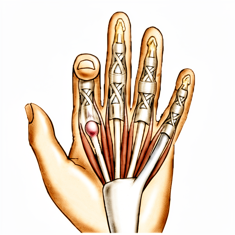
El dedo en gatillo se produce cuando un tendón del dedo o pulgar se atasca al deslizarse por un túnel estrecho en la palma de la mano. En primer lugar, se suelen aplicar tratamientos no quirúrgicos, como el uso de férula, terapia de la mano o una inyección de esteroides (un medicamento que se administra cerca del tendón para reducir la inflamación). Consideramos la cirugía cuando estos tratamientos no logran mejoría suficiente, o cuando el dedo queda atrapado en una posición doblada que ya no se puede enderezar.
La operación consiste en una liberación: se realiza una única incisión en la zona a tratar y se abre la parte estrecha del túnel para que el tendón pueda deslizarse libremente. Aproximadamente el 97 % de los pacientes experimentan una resolución completa del problema tras la cirugía. El objetivo es lograr que el dedo o pulgar se mueva sin dificultad, sin trabarse, bloquearse ni causar dolor.
Antes de la operación
La mayoría de las personas apenas necesitan preparación. Se le indicará que no debe comer ni beber durante siete horas antes de la cirugía. Pedimos que sean siete horas en lugar de seis para poder adelantar su turno si la lista de cirugías avanza antes de lo previsto. Su cirujano le indicará qué medicamentos debe suspender y cuándo, y usted deberá llevar una lista por escrito de todos los fármacos que toma. Organice que alguien lo lleve a casa después de la intervención, y el día de la operación use ropa holgada y cómoda. En ocasiones se solicitan estudios de imagen como radiografías, resonancia magnética o ecografías con antelación para ayudar a planificar la cirugía. Si padece otras enfermedades, es posible que necesite análisis de sangre o una consulta con el anestesista (el médico encargado de administrar la anestesia); sin embargo, la mayoría de las personas no requieren nada de esto.
El día de la intervención
Llega a la unidad de admisiones quirúrgicas del hospital, donde se le registrará y preparará para la cirugía. Conocerá al anestesista (el médico encargado de administrar la anestesia). Esta operación puede realizarse con anestesia local (una inyección que adormece únicamente la zona de la intervención, mientras el paciente permanece despierto) o con anestesia general (el paciente queda completamente dormido). La mayoría de las personas optan por la anestesia local: la recuperación es más rápida y pueden volver a casa poco después. Si prefiere estar dormido, también es una opción válida; hable de ello con su cirujano y con el anestesista.
A continuación, se le llevará al quirófano, donde se realiza la operación. Después, despertará en la sala de recuperación, donde las enfermeras le vigilarán mientras la anestesia va desapareciendo. Una vez que su estado sea estable, será trasladado a la planta de hospitalización o podrá volver a casa, según el tipo de intervención y su recuperación. La mayoría de los pacientes que se someten a esta operación regresan a casa el mismo día. Lleve consigo al conductor que organizó en la sección anterior y siga todas las indicaciones que le den las enfermeras respecto a su mano antes de irse.
¿En qué consiste la operación?
La operación se denomina liberación del dedo en gatillo. El cirujano realiza una única incisión en la zona a operar, generalmente de unos 2 cm de longitud. A través de esta incisión, el cirujano accede a la zona estrecha del túnel en la palma por donde se desliza el tendón. La banda de tejido tensa se abre cuidadosamente, aproximadamente 1 cm a la vez, hasta que el tendón pueda moverse libremente. Posteriormente, el cirujano verifica que el dedo o el pulgar se flexione y extienda sin dificultad ni bloqueos.
En el caso del pulgar en gatillo, solo es necesario liberar una banda tensa. En los dedos, también puede ser preciso abrir una segunda banda cercana. El cirujano actúa con sumo cuidado para proteger los nervios adyacentes y dejar intactas las capas de tejido de soporte más profundas, pues estas son esenciales para el correcto funcionamiento del tendón.
Una vez finalizada la liberación, la herida se cierra con puntos de sutura. A continuación, se coloca un vendaje de compresión sobre la mano. Deberá mantener este vendaje durante unos 10 días; los puntos se retiran entre los días 10 y 14. Se recomienda utilizar el dedo o el pulgar de forma normal desde el primer momento.
Si padece artritis reumatoide (una enfermedad que provoca inflamación articular), el procedimiento podría variar ligeramente. En ese caso, el cirujano podría extirpar una pequeña porción del tendón en lugar de liberar la banda tensa, ya que esta liberación podría hacer que el dedo se desvíe lateralmente con el tiempo. Antes de obtener su consentimiento, el cirujano le explicará el plan específico para su mano.
Después de la operación
Despertará en la sala de recuperación, donde las enfermeras lo vigilarán mientras el efecto de la anestesia desaparece. Este es un procedimiento ambulatorio, por lo que podrá volver a casa el mismo día. En su mano se colocará un vendaje de compresión (un vendaje firme); podrá mover el dedo o el pulgar de inmediato. Es normal sentir algo de molestia durante el primer o segundo día; por lo general, los analgésicos de venta libre recomendados por el farmacéutico o el médico son suficientes. Pídale a alguien que lo acompañe durante las primeras 24 horas. Dejamos el vendaje puesto durante unos 10 días; por favor, no lo retire antes de ese plazo a menos que se lo indiquemos. Lo cambiamos o lo retiramos cuando venga a la consulta. La mayoría de las personas vuelven a conducir en una o dos semanas, una vez que la herida no les causa molestias y pueden agarrar el volante y girarlo sin proteger la mano.
Recuperación
La mayoría de las personas se sorprenden de lo rápido que la mano se recupera. Durante los primeros días, la zona alrededor de la incisión estará adolorida y ligeramente hinchada. Por lo general, los analgésicos de venta libre recomendados por el farmacéutico o el médico son suficientes para mantener el confort; además, mantener la mano elevada sobre una almohada mientras se está sentado o descansando ayuda a reducir la hinchazón. Es posible que la sensibilidad alrededor de la cicatriz persista más tiempo que el resto, pero disminuye a medida que la herida madura.
Podrá mover el dedo o el pulgar de inmediato; de hecho, se recomienda su uso normal desde el principio. No es necesario usar ninguna férula o soporte; solo el vendaje, que dejamos puesto durante unos 10 días. En su siguiente consulta, lo cambiaremos o lo retiraremos y retiraremos los puntos de sutura. La terapia de la mano posterior a la cirugía la llevará a cabo Ruby Doolan en Extend Rehabilitation. Ruby le guiará en ejercicios sencillos que permiten que el dedo se flexione y extienda sin problemas; además, puede confeccionar una férula si su mano la necesita.
Podrá realizar la mayoría de las actividades domésticas en cuanto se sienta capaz: comer, vestirse, teclear y tareas ligeras en casa. Evite agarrar objetos con fuerza, levantar pesos y exponer la mano a entornos sucios o húmedos hasta que la herida haya cicatrizado. Una vez que pueda agarrar objetos y girar el volante sin proteger la mano, normalmente podrá volver a conducir; consulte nuestra página sobre conducción tras una cirugía de miembro superior.
La recuperación varía según cada persona, por lo que su cronograma puede ser distinto. Su cirujano y su terapeuta lo guiarán durante todo el proceso.
Qué puede salir mal
La mayoría de los pacientes evolucionan bien, pero en ocasiones pueden surgir problemas. Su cirujano y el equipo lo vigilarán de cerca para detectar cualquier incidencia a tiempo.
Los problemas más frecuentes son leves y se relacionan con la herida, o bien el dedo puede sentirse rígido durante un tiempo. Es posible que note cierta molestia persistente, hinchazón, o que el dedo no se doble con la libertad esperada en las primeras semanas. Si esto no mejora, coméntele al médico en su próxima revisión.
Las infecciones son poco comunes, pero es importante conocer sus señales. Esté atento a cualquier enrojecimiento que se extienda desde la incisión, aumento del dolor, calor en la zona o secreción. Una infección profunda puede provocar un dolor intenso y pulsátil que no cede con analgésicos comunes, y usted podría sentirse mal. Si observa cualquiera de estos síntomas, llame a la clínica de inmediato en lugar de esperar a su próxima cita.
En ocasiones, un nervio cercano a la zona quirúrgica puede irritarse. Esto se manifiesta como entumecimiento, hormigueo o sensación de “agujas y alfileres” en parte del dedo o el pulgar. Al principio, cierto entumecimiento en el borde de la herida es normal; sin embargo, si persiste o se extiende, hágalo saber en su revisión.
En un pequeño número de personas, el fenómeno de “disparo” puede reaparecer. Si el dedo vuelve a atascarse o “disparar” después de haber mejorado, infórmenos en su seguimiento.
Algunas personas desarrollan rigidez en el dedo, de modo que este no se endereza ni dobla por completo, aun cuando el problema de “disparo” ya haya desaparecido. Los ejercicios de terapia de la mano ayudan a prevenir esto; por ello, siga realizando los movimientos que Ruby le haya enseñado. Si el dedo parece volverse más rígido en lugar de más flexible, avíselo a su terapeuta o cirujano cuanto antes.
Si su dedo presentaba el problema de “disparo” desde hace mucho tiempo, la articulación central del dedo puede seguir doliendo incluso después de la cirugía. Esta molestia suele mejorar de forma gradual, no de inmediato.
Por último, la cirugía para el dedo en gatillo conlleva un riesgo reducido de desarrollar la enfermedad de Dupuytren, una afección en la que se forman cordones duros en la palma que obligan a los dedos a permanecer doblados. Si posteriormente nota engrosamientos o bultos en la palma, haga que lo examinen.
En la tabla de complicaciones de esta página se detallan las tasas típicas, por si desea conocer los datos específicos.
¿Cuándo debemos ser contactados?
Llame a la clínica de inmediato si tiene fiebre, o si el enrojecimiento, el calor o el exudado alrededor de la herida empeoran. Comuníquese con nosotros si el dolor se vuelve repentinamente intenso, o si la entumecimiento o las hormigueos se extienden y no desaparecen. Acuda a urgencias si presenta hinchazón o dolor en la pantorrilla, o dificultad para respirar, ya que estos pueden ser signos de un coágulo sanguíneo. Acuda a urgencias si no puede mover el dedo o el pulgar en absoluto, o si la mano se vuelve fría o pálida.
¿Dónde leer más sobre esta afección?
Esta página trata sobre la intervención quirúrgica en sí. La afección que se trata, incluyendo lo que demuestran las evidencias sobre cuándo la cirugía es útil y cuándo no, se explica con mayor detalle en la página de Dedos en gatillo.
Evidence & references
This is the clinical evidence summary written for health professionals. It is technical, and it lists the research this page was built from. You do not need to read it to understand your treatment or to make a decision about it.
Overview
Operative Technique
- Local anesthetic infiltration in the palm proximal to the incision site is preferred for trigger finger release [5].
- A pneumatic arm tourniquet may be helpful, although a high forearm Esmarch wrap usually is sufficient [5].
- For middle, ring, and small trigger finger releases, a transverse incision about 2 cm long is made several millimeters distal to the distal palmar crease [5].
- For index trigger finger releases, a transverse incision about 2 cm long is made several millimeters distal to the proximal palmar crease [5].
- Trigger thumb releases can be performed through incisions either distal or proximal to the metacarpophalangeal joint flexion crease [5].
- Alternative incisions for fingers can be made obliquely or longitudinally between the metacarpophalangeal and distal palmar creases [5].
- Alternative incisions for the thumb can be made obliquely across the thumb metacarpophalangeal flexion crease [5].
- The digital nerves on the thumb are more palmar and closer to the flexor sheath than might be anticipated [5].
- The thumb radial digital nerve is especially vulnerable during trigger thumb release [5].
- Subcutaneous tissues are spread away from the underlying annular pulley system to ensure digital nerves are safely protected [5].
- Trigger thumbs require release of only the A1 pulley [5].
- Trigger digits require division of the A1 and A0, or proximal palmar pulley [5].
- Pulley division is usually accomplished with an initial opening of the pulley with a No. 15 knife blade and a pair of tenotomy scissors [5].
- For trigger thumb release, cutting too far distally should be avoided to prevent disrupting the oblique pulley [5].
- The sheath is incised from proximal to distal, approximately 1 cm, and reassessed for triggering [5].
- Persistent triggering after initial release implies that either the A1 and palmar pulleys are incompletely released or an alternate site of triggering is present [5].
- When the distal A1 pulley edge is released, the divided pulley leaves are parallel rather than ending in a V-shaped pattern [5].
- The distinction between the A1 and A2 pulleys may not be apparent during surgery [5].
- Other fingers can be found to trigger at the same surgical setting and can be managed at the same time [5].
Postoperative Care
- The compression dressing is removed after 48 hours [5].
- Sutures are removed at 10 to 14 days [5].
- Normal use of the finger or thumb is encouraged postoperatively [5].
Anatomy & Pathophysiology
Demographics & Epidemiology
- Trigger finger occurs in 2% to 3% of the general population [9].
- Women are more commonly affected than men [9].
- Women older than 50 years of age are the primary demographic for trigger finger [1, 2].
- Middle and ring finger involvement is most common in adults [1, 2].
- The digits are affected in the following order of decreasing prevalence: thumb, ring, long, little, and index [9].
- Stenosing tenosynovitis is more common in diabetic patients than in nondiabetic patients [4].
- Trigger finger is more common in patients with diabetes mellitus, with a 10% to 20% lifetime incidence [9].
Comorbidities & Etiology
- Trigger finger is associated with diabetes and inflammatory arthropathy [1, 2].
- Trigger finger is seen in patients with hypothyroidism, sarcoidosis, rheumatoid arthritis, and septic tenosynovitis [9].
- The etiology of trigger finger is possibly associated with repetitive grasping activities [1, 2].
- Gout can present as trigger finger due to monosodium urate precipitation eliciting a fulminant inflammatory reaction in the tenosynovium [9].
- Calcific tendinitis can result in triggering due to calcium salt deposition in the tenosynovium [9].
- Pseudogout can cause triggering via calcium pyrophosphate dihydrate crystal deposition [9].
- Amyloidosis, characterized by beta-2-microglobulin deposition, can cause trigger finger in patients with renal failure undergoing dialysis [9].
- Trigger finger is considered an early indication of Dupuytren’s disease by some authors [12].
- The incidence of concurrent trigger finger and Dupuytren’s disease is higher in the middle and ring fingers than expected by statistical coincidence [11].
Histology & Pathology
- Histology of the affected pulley demonstrates fibrocartilaginous metaplasia of the pulley and/or FDS tendon [1, 2].
- Pathologic examination of affected pulleys demonstrates a proliferation of chondrocytes and increased type III collagen [9].
- The flexor digitorum profundus tendon often demonstrates a pathologic nodule, while the flexor digitorum superficialis is often unaffected [9].
- A nodule or fusiform swelling of the flexor tendon just distal to the first annular pulley may be palpable [7].
- The tendon nodule is usually located just proximal to the anulus at the metacarpophalangeal joint level [7].
- In rheumatoid patients, a nodule distal to the metacarpophalangeal joint level may cause triggering [7].
- A volar retinacular ganglion cyst between the A1 and A2 pulleys may be present [9].
- In congenital trigger digits, the pathologic anatomy includes narrowing and thickening of the sheath with occasional formation of a ganglion cyst [10].
- An intratendinous nodule proximal to the first annular pulley, often referred to as Notta’s nodule, may be present in congenital trigger digits [10].
Clinical Presentation
- Patients present with pain and tenderness in the palm at the proximal edge of the digital A1 pulley [4].
- Patients frequently note catching or triggering of the affected finger or thumb after forceful flexion [4].
- In more severe cases, the opposite hand must be used to force the finger or thumb passively into extension [4].
- In the most severe cases, the finger becomes locked in a flexed position [4].
- Triggering is often more pronounced in the morning than later in the day [4].
- A common complaint is referred pain at the dorsal MCP/PIP area [1, 2].
- Patients frequently state that the problem is in the proximal interphalangeal joint with trigger finger or trigger thumb [7].
- Concomitant trigger finger and carpal tunnel syndrome occurs in 40% to 60% of patients [1, 2].
- Local tenderness may be present but is not a prominent complaint [7].
- Pressure accentuates the apparent snapping or triggering of the more distal joints [7].
Anatomical Variations & Specific Structures
- Newer evidence has found a fourth pulley (variable annular pulley) in 75% of patients with thumb trigger finger, which may contribute to stenosis [1, 2].
- The radial digital nerve is at risk of iatrogenic injury during thumb trigger finger release due to its superficial location [1, 2].
- On the thumb, digital nerves are more palmar and closer to the flexor sheath than might be anticipated [5].
- The thumb radial digital nerve is especially vulnerable during surgical release [5].
- Trigger thumbs require release of only the A1 pulley, whereas trigger digits require division of the A1 and A0 (or proximal palmar) pulley [5].
- The distinction between the A1 and A2 pulleys may not be apparent during surgery; however, when the distal A1 pulley edge is released, the divided pulley leaves are parallel rather than ending in a V-shaped pattern [5].
- The fibers of the A2 pulley must be spared to preserve effective digital flexion [4].
- In patients with rheumatoid arthritis, the entire annular pulley system should be preserved to prevent further ulnar drift of the fingers [4].
Classification
- Green Classification Grade I is defined as pain and tenderness at the A1 pulley [1, 2, 9].
- Green Classification Grade II is defined as catching of the finger [1, 2] or mechanical catching of the digit without locking [9].
- Green Classification Grade III is defined as locking of the finger that is passively correctable [1, 2] or mechanical locking of the digit which is passively correctable [9].
- Green Classification Grade IV is defined as a fixed, locked finger [1, 2, 9].
Classification
- The Green classification of trigger finger consists of four grades [1].
- Grade I is defined as pain and tenderness at the A1 pulley [1].
- Grade II is defined as catching of the finger [1].
- Grade III is defined as locking of the finger that is passively correctable [1].
- Grade IV is defined as a fixed, locked finger [1].
Clinical Presentation
Demographics and Epidemiology
- Trigger finger is most common in women older than 50 years of age [1, 2].
- Trigger finger is more common in patients with systemic diseases such as diabetes mellitus, hypothyroidism, sarcoidosis, rheumatoid arthritis, and septic tenosynovitis [9].
- Diabetes mellitus has a 10% to 20% lifetime incidence of trigger finger [9].
- When multiple digits are involved, the possibility of diabetes should be considered [4].
Etiology and Pathology
- Trigger finger is possibly associated with repetitive grasping activities [1, 2].
- Histology of the affected pulleys demonstrates fibrocartilaginous metaplasia of the pulley and/or FDS tendon [1, 2].
- Pathologic examination of the affected pulleys demonstrates a proliferation of chondrocytes and increased type III collagen [9].
- The flexor digitorus profundus tendon often demonstrates a pathologic nodule, while the flexor digitorus superficialis is often unaffected [9].
- A fourth pulley (variable annular pulley) is found in 75% of patients with thumb trigger finger, which may contribute to stenosis [1, 2].
Symptoms and Signs
- Patients present with pain and tenderness in the distal palm [1, 2].
- Pain and tenderness are located at the proximal edge of the digital A1 pulley [4].
- Symptoms progress to mechanical catching or locking of the finger [1, 2].
- In severe cases, the opposite hand must be used to force the finger or thumb passively into extension [4].
- Patients may note a lump or knot in the palm [7].
- The lump may be a thickened area in the first annular pulley or a nodule/fusiform swelling of the flexor tendon just distal to it [7].
- The tendon nodule can be palpated by the examiner’s fingertip and moves with the tendon [7].
- The tendon nodule is usually just proximal to the anulus at the metacarpophalangeal joint level [7].
- In rheumatoid patients, a nodule distal to the MCP joint level may cause triggering [7].
- Patients frequently state that the problem is in the proximal interphalangeal joint [7].
- Concomitant trigger finger and carpal tunnel syndrome occur in 40% to 60% of patients [1, 2].
Physical Examination Findings
- Tenderness to palpation of the flexor tendon at the level of the A1 pulley is a physical examination finding [9].
- Palpable triggering or pain with flexion and extension of the finger is a physical examination finding [9].
- Nodularity of the flexor tendon just proximal to the A1 pulley is a physical examination finding [9].
- Presence of a volar retinacular ganglion cyst between the A1 and A2 pulleys is a physical examination finding [9].
- Presence of a fixed flexion deformity of the proximal interphalangeal (PIP) joint is a physical examination finding [9].
Classification
- Green classification Grade II is defined as catching of the finger [1, 2].
- Green classification Grade II is defined as mechanical catching of the digit without locking [9].
- Green classification Grade III is defined as locking of the finger that is passively correctable [1, 2].
- Green classification Grade III is defined as mechanical locking of the digit which is passively correctable [9].
Differential Diagnosis Considerations
- Intraarticular disorders such as loose bodies, degenerative joint disease, and fractures can cause symptoms similar to trigger finger [7].
- Common extensor tendon subluxation can cause symptoms similar to trigger finger [7].
- Gout can mimic infectious tenosynovitis with marked pain, erythema, swelling, and warmth [9].
- Calcific tendinitis can resemble an infection and result in triggering [9].
- Pseudogout can present with calcium pyrophosphate dihydrate crystal deposition localized to the triangular fibrocartilage or within the carpal tunnel [9].
- Amyloidosis can present with beta-2-microglobulin deposition along flexor tendons, most commonly in patients with renal failure undergoing dialysis [9].
Investigations
Clinical Presentation and History
- Trigger finger is characterized by pain and tenderness in the palm at the proximal edge of the digital A1 pulley [4].
- Pain and tenderness in the distal palm progress to mechanical catching or locking, and may become fixed [1].
- A common complaint is referred pain at the dorsal MCP or PIP area [1].
Physical Examination Findings
- Physical examination may reveal tenderness to palpation of the flexor tendon at the level of the A1 pulley [9].
- Nodularity of the flexor tendon just proximal to the A1 pulley may be present on examination [9].
- A volar retinacular ganglion cyst between the A1 and A2 pulleys may be present on examination [9].
- A fixed flexion deformity of the proximal interphalangeal (PIP) joint may be present on examination [9].
Classification
Associated Conditions and Demographics
- The lifetime incidence of trigger finger in patients with diabetes mellitus is 10% to 20% [9].
- Calcific tendinitis can result in triggering and is affected five times more frequently in males than females [9].
- Pseudogout involves calcium pyrophosphate dihydrate crystal deposition often localized to the triangular fibrocartilage or within the carpal tunnel [9].
- Amyloidosis is characterized by the deposition of beta-2-microglobulin in thick, plaque-like accumulations along the flexor tendons [9].
Imaging and Histology
- MR imaging findings of trigger thumb have been described [3].
Treatment
Nonoperative
- Corticosteroid injection into the flexor tendon sheath is curative in about 60% of patients initially [1].
- Diabetic patients are generally less responsive to corticosteroid injection [1].
- There is no difference between soluble and insoluble corticosteroid preparations for trigger finger injection [1].
- In a study of 292 corticosteroid injections, repeat injections provided symptomatic relief for a year or more in 50% of patients [7].
- Corticosteroid injections may elevate serum glucose levels for 5 days or more in patients with diabetes mellitus [7].
- Patients with unstable diabetes may be better treated without corticosteroid injection [7].
- Preoperative hypoglycemia increases infection risk after trigger finger injection and release [7].
- Immediate surgical release in the clinic was identified as the most cost-effective treatment strategy for trigger finger in diabetic patients [7].
- Nonoperative methods for trigger digits include stretching, night splinting, and combinations of heat and ice [7].
Operative
- Surgical release of the A1 pulley is curative in digits refractory to steroid injection [4].
- Surgical release reliably relieves the problem for most patients, with approximately 97% of patients having complete resolution after operative treatment [7].
- Persistence of triggering is more common than recurrence after surgical release [7].
- Trigger release should be performed with a local block so that the cessation of triggering can be evaluated intraoperatively [7].
- Adjacent finger triggering may become obvious only after a given finger is released and can be managed at the same surgical setting [7].
- In patients with rheumatoid arthritis, the preference is to excise a slip of the FDS tendon rather than to release the A1 pulley to prevent exacerbation of ulnar drift at the MCP joint [1].
- The fibers of the A2 pulley must be spared during surgical release to preserve effective digital flexion [4].
- Minor complications of surgical release include wound dehiscence, scar tenderness, and decreased range of motion [1].
- Incomplete pulley release and damage to the flexor tendons and digital nerves remain concerns, especially in the index finger and thumb with limited exposure techniques [7].
- Percutaneous release of the A1 pulley may be accomplished with a needle on the middle and ring fingers, especially if they actively lock [4].
- The safety and effectiveness of percutaneous trigger finger release using a needle or a push knife have literature support [7].
- For percutaneous release, an 18- or 19-gauge needle may suffice [8].
- During percutaneous release, the bevel of the needle should be oriented longitudinally parallel to the flexor tendons [8].
- Injection of corticosteroid is optional during percutaneous trigger finger release [8].
- Postoperative compression dressing for open trigger finger release is removed after 48 hours [5].
- Sutures for open trigger finger release are removed at 10 to 14 days [5].
- Normal use of the finger or thumb is encouraged after open trigger finger release [5].
- Active hand and finger use with stretching exercises is encouraged after percutaneous trigger finger release [8].
Complications
Operative
- Digital nerves on the thumb are more palmar and closer to the flexor sheath than might be anticipated, making the thumb radial digital nerve especially vulnerable [5].
- Minor complications of trigger finger release are relatively high and include wound dehiscence, scar tenderness, and decreased range of motion [1].
- Incomplete pulley release remains a concern with limited exposure techniques [7].
- Damage to flexor tendons remains a concern with limited exposure techniques, especially in the index finger and thumb [7].
- Damage to digital nerves remains a concern with limited exposure techniques, especially in the index finger and thumb [7].
- Patients may experience triggering after operative release because of catching of the tendon on the palmar aponeurosis transverse fibers [7].
- Triggering caused by catching on palmar aponeurosis transverse fibers usually resolves with time [7].
- A partially lacerated flexor tendon at the metacarpophalangeal joint level may heal with a nodule sufficiently large to cause triggering [7].
- Persistence of triggering is more common than recurrence after operative treatment [7].
- Preoperative hypoglycemia increases infection risk after trigger finger release [3].
- Preoperative hypoglycemia increases infection risk after trigger finger injection [3].
- In patients with rheumatoid arthritis, release of the A1 pulley carries a chance of exacerbating ulnar drift at the MCP joint [1].
Non-Operative
- Corticosteroid injections may elevate serum glucose levels for 5 days or more [7].
References
[1] Miller S Review Of Orthopaedics. SECTION 16 PATELLAR TRACKING IN TOTAL KNEE ARTHROPLASTY > 2. Flexor tendon injury > 3. Stenosing tenosynovitis (trigger finger).
[2] Miller S Review Of Orthopaedics. 2. Flexor tendon injury > 3. Stenosing tenosynovitis (trigger finger).
[3] Campbell S Operative Orthopaedics 4 Volume Set. EXCISION OF NECROTIC MUSCLES COMBINED WITH NEUROLYSIS OF MEDIAN AND ULNAR NERVES FOR SEVERE CONTRACTURE > REFERENCES > TRIGGER THUMB AND TRIGGER FINGER.
[4] A Lange Medical Book Current Diagnosis Treatment In Orthopedics Fifth Edition. 9Hand Surgery > 2. Flexor Tenosynovitis (Trigger Finger and Trigger Thumb).
[5] Campbell S Operative Orthopaedics 4 Volume Set. EXCISION OF NECROTIC MUSCLES COMBINED WITH NEUROLYSIS OF MEDIAN AND ULNAR NERVES FOR SEVERE CONTRACTURE > TRIGGER FINGER AND THUMB > SURGICAL RELEASE OF TRIGGER FINGER.
[7] Campbell S Operative Orthopaedics 4 Volume Set. EXCISION OF NECROTIC MUSCLES COMBINED WITH NEUROLYSIS OF MEDIAN AND ULNAR NERVES FOR SEVERE CONTRACTURE > TRIGGER FINGER AND THUMB.
[8] Campbell S Operative Orthopaedics 4 Volume Set. EXCISION OF NECROTIC MUSCLES COMBINED WITH NEUROLYSIS OF MEDIAN AND ULNAR NERVES FOR SEVERE CONTRACTURE > PERCUTANEOUS RELEASE OF TRIGGER FINGER.
[9] Aaos Comprehensive Orthopaedic Review 3. Tendinopathy of the Hand and Wrist* > II. Trigger Finger.
[10] Campbell S Operative Orthopaedics 4 Volume Set. MULTIPLE Z-PLASTY RELEASE OF A CONGENITAL RING > TRIGGER FINGERS.
[11] Dupuytren S Disease And Related Hyperproliferative Disorders. 31. The Influence of Dupuytren’s Disease on Trigger Fingers and Vice Versa > 31.5 Discussion.
[12] Dupuytren S Disease And Related Hyperproliferative Disorders. 31. The Influence of Dupuytren’s Disease on Trigger Fingers and Vice Versa > 31.5 Discussion > 31.5.1 Why Is DD Concurring with TF?.