Patients › Elbow
Liberación de codo rígido (artrólisis)
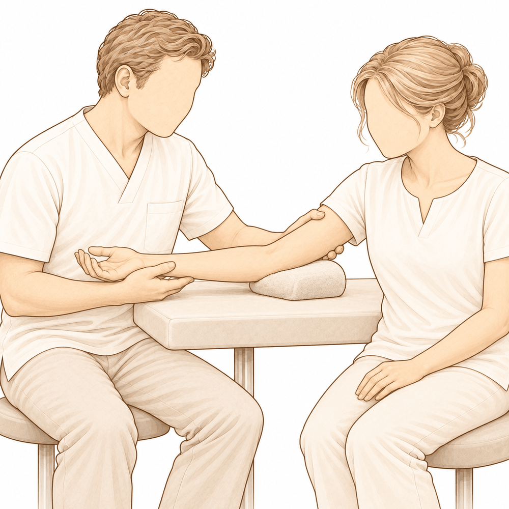
¿Por qué se ha recomendado esta operación?
El Dr. Kieran Hirpara, cirujano de extremidades superiores en el Mater Private Hospital Rockhampton, comienza por ofrecer las opciones menos invasivas adecuadas para su condición. Por lo general, los pacientes son derivados a nuestra clínica por su médico de cabecera; si un fisioterapeuta le ha sugerido que nos consulte, igualmente necesitará una derivación de su médico de cabecera para poder acceder al reembolso de Medicare. En su consulta, tomamos su historia clínica, examinamos su codo y, si es necesario, solicitamos estudios de imagen. Un codo rígido suele mejorar con el tiempo y el uso, por lo que normalmente probamos primero tratamientos no quirúrgicos. Estos pueden incluir fisioterapia, el uso de férulas o inyecciones durante un período de 6 a 12 semanas. La cirugía se considera cuando este tratamiento no produce mejoría suficiente.
La liberación del codo rígido, también conocida como artrólisis, es una operación que permite liberar el tejido cicatricial y tenso que rodea el codo, permitiendo así que la articulación vuelva a moverse. La recomendamos cuando la rigidez sigue limitando sus movimientos tras haber seguido un tratamiento no quirúrgico. Este procedimiento funciona mejor cuando la superficie articular sigue siendo lisa y conserva su forma normal. El objetivo es lograr una mejora duradera en la movilidad y reducir el dolor, facilitando así la realización de las tareas diarias.
Antes de la operación
Una vez programada la cirugía, hay algunos aspectos prácticos que deben resolverse. Se le indicará cuándo debe dejar de comer y beber: esto es siete horas antes de la operación. Pedimos que sea siete horas en lugar de seis para que, si el programa quirúrgico avanza antes de lo previsto, podamos adelantar su intervención. Su cirujano le indicará qué medicamentos habituales debe suspender y cuándo; usted debe llevar consigo una lista completa de los mismos. Organice que alguien lo lleve a casa después de la operación. El día de la intervención, use ropa holgada y cómoda. Siempre se realizan radiografías para planificar la cirugía; además, podría ser necesario un escáner de tomografía computarizada si la rigidez afecta al hueso. Si padece otras enfermedades, es posible que necesite análisis de sangre o una evaluación por parte del anestesista.
El día de la intervención
Llega usted a la unidad de admisiones quirúrgicas del hospital, donde se le registra y se le prepara para la cirugía. Allí conoce al anestesista. Esta operación se realiza bajo anestesia general. En ocasiones se añade un bloqueo nervioso regional para aliviar el dolor postoperatorio; el anestesista hablará con usted al respecto ese mismo día. A continuación, se le lleva al quirófano, donde se lleva a cabo la intervención.
Despierta usted en la sala de recuperación, donde las enfermeras le vigilan mientras la anestesia va desapareciendo. Una vez que se encuentra estable, según el tipo de intervención y su recuperación, será trasladado a la planta de hospitalización o podrá volver a casa.
Qué implica la operación
Existen varias formas de liberar un codo rígido; su cirujano elegirá el método más adecuado para su caso. Algunas liberaciones se realizan mediante cirugía mínimamente invasiva, usando unos cuantos cortes pequeños y una cámara delgada. Otras se efectúan mediante uno o dos cortes abiertos, a veces de 3 a 5 cm de longitud, en la cara interna o externa del codo. En casos de codos muy rígidos, se puede realizar un corte en la parte posterior del codo para acceder a ambos lados.
Una vez dentro, el cirujano libera la cápsula tensa, que es la envoltura de tejido que rodea la articulación. Se eliminan los espolones óseos que impiden el movimiento, así como cualquier fragmento suelto de hueso o cartílago presente en la articulación. Si un nervio situado en la cara interna del codo queda comprimido por tejido cicatricial, se puede liberar de ese túnel de tejido o trasladar a una nueva posición donde no corra riesgo de estirarse. Cuando la superficie articular está muy desgastada, existen otras opciones, como recubrir el hueso con tejido blando o sustituir dichas superficies por piezas metálicas y plásticas. En ocasiones, se coloca un dispositivo articulado en el codo para mantenerlo en una buena posición mientras cicatriza.
Al final, los cortes se cierran con puntos de sutura y se cubren con un vendaje. Deberá mantener ese vendaje puesto durante unos 10 días, tal como se describe en la sección de recuperación.
Después de la operación
Despertará en la sala de recuperación y luego será trasladado a la habitación. Las enfermeras revisarán su codo, su mano y cómo se siente. Se le administrará analgesia según sea necesario; avise a las enfermeras si siente molestias en el codo. Su codo quedará cubierto con un vendaje suave, y podrá volver a casa con el brazo en un cabestrillo para mayor comodidad. Poco después podrá levantarse de la cama y caminar; las enfermeras le ayudarán la primera vez. Su mano, muñeca y hombro podrán moverse libremente mientras el codo se recupera. Alguien debe acompañarle durante las primeras 24 horas después de volver a casa. Su equipo médico le informará si podrá volver a casa el mismo día o si deberá permanecer una noche en el hospital. Dejamos el vendaje puesto durante unos 10 días; por favor, no lo retire antes de ese plazo a menos que se lo indiquemos. Lo cambiaremos o lo retiraremos cuando venga a la consulta.
Recuperación
Durante los primeros días y semanas, el codo le dolerá y se hinchará. Esto es normal después de la intervención. El descanso, mantener la mano elevada y tomar los analgésicos según las indicaciones ayudarán a aliviar la molestia. La hinchazón disminuye gradualmente; a medida que esto ocurre, el codo suele sentirse menos tenso.
Se le dará un cabestrillo para llevar el brazo a casa, con el fin de mayor comodidad. Puede dejar de usarlo cuando ya no sea necesario. Desde el principio, la mano, la muñeca y el hombro deben seguir moviéndose libremente. La terapia de mano posterior a la cirugía la llevará a cabo Ruby Doolan en Extend Rehabilitation. Ruby le guiará en los ejercicios y le confeccionará cualquier férula que necesite. Los ejercicios constituyen el aspecto principal de su recuperación; seguir las indicaciones al pie de la letra es lo más importante. Deberá mantener el vendaje durante unos 10 días; nosotros lo cambiaremos o lo retiraremos en su siguiente consulta.
Las actividades cotidianas volverán poco a poco. Al principio, necesitará ayuda para cocinar, vestirse y llevar objetos. A medida que recupere la movilidad y la hinchazón disminuya, podrá realizar más tareas con el brazo operado. Una vez que su cirujano le autorice a conducir, podrá volver a hacerlo; nuestra guía sobre conducción tras una cirugía de miembro superior explica las normas aplicables, entre ellas no conducir mientras el brazo lleve cabestrillo y no estar bajo el efecto de analgésicos fuertes.
La recuperación varía según cada persona. Su cronograma puede ser distinto; su cirujano y terapeuta le guiarán durante todo el proceso.
Qué puede salir mal
La mayoría de los pacientes evolucionan bien, pero en ocasiones pueden surgir problemas. Su cirujano y el equipo lo vigilarán de cerca para detectar cualquier incidencia a tiempo.
Cuanto más tiempo haya estado rígido el codo, mayor es la probabilidad de que aparezcan problemas tras la liberación. Si su codo ha estado rígido durante mucho tiempo, su cirujano hablará con usted sobre este aspecto antes de que tome una decisión.
Algunas personas notan después de la intervención una sensación nueva de hormigueo, entumecimiento o debilidad en el dedo meñique y el anular. Esto se debe al nervio situado en la parte interna del codo. No siempre es posible predecir quién experimentará esto. Si nota estas sensaciones, mencione esto en su próxima consulta o llame a la clínica si aparecen de forma repentina.
Los codos que ya han sufrido lesiones previamente tienen mayor riesgo de desarrollar una infección profunda posteriormente. Esté atento a si la herida presenta enrojecimiento que se extiende, calor, mayor hinchazón o un dolor profundo y pulsátil que no cede con analgésicos comunes. También podría sentir fiebre o malestar general. Si observa cualquiera de estos signos, llame de inmediato a la clínica o acuda al servicio de urgencias si es fuera del horario laboral.
Si su operación implica la colocación de un marco articulado en el codo, hay algunos aspectos adicionales que debe conocer. Este marco incrementa la pérdida de sangre durante la cirugía, el tiempo en quirófano y la estancia hospitalaria. Además, existe una pequeña probabilidad de que la articulación se vuelva inestable o poco firme. Su cirujano le explicará cómo esto afecta su hospitalización y recuperación.
Si ya se ha sometido a otras operaciones en ese codo, el riesgo de infección profunda vuelve a ser mayor. Lo mismo ocurre cuando la intervención en sí es más compleja. Su cirujano evaluará todo esto junto con usted antes de proceder.
Las lesiones crónicas del codo también pueden modificar el funcionamiento de la articulación. La piel cicatrizada, los huesos que se han soldado fuera de posición, el cartílago dañado, el exceso óseo, los ligamentos flojos o tensos, los nervios comprimidos y el músculo cicatrizado pueden alterar el movimiento del codo. Su cirujano buscará estos factores al planificar la operación.
En la tabla de complicaciones de esta página se detallan las tasas típicas, por si desea conocer los datos específicos.
¿Cuándo deben llamarnos?
Llámenos si observa fiebre, enrojecimiento que se extiende alrededor de la herida, secreción procedente de la herida o un dolor que empeora progresivamente. Acuda a urgencias si experimenta dolor intenso y repentino, hinchazón o dolor en la pantorrilla, o dificultad para respirar. Llámenos de inmediato si su mano o dedos se entumecen o palidecen, o si no puede mover el brazo. Si el hormigueo o entumecimiento en el dedo meñique y el anular aparece de forma súbita, llame a la clínica en lugar de esperar a su próxima cita de seguimiento.
En mayor profundidad
Advanced reading: the deeper science (optional)
Esta sección profundiza más de lo necesario para que usted tome decisiones sobre su propio tratamiento. Merece la pena leerla si desea comprender por qué, al liberar un codo rígido, la evidencia apunta a algo inusual: la técnica quirúrgica que la mayoría de los pacientes preguntan por, ya sea artroscópica u abierta, parece ser menos importante que la extensión de la intervención; además, el verdadero “enemigo” de la operación es el mismo proceso que originó la rigidez desde un principio.
Lo importante no es si se utiliza la técnica abierta o artroscópica
Una revisión sistemática y metaanálisis realizada en 4,311 pacientes con rigidez postraumática del codo comparó la artrolisis abierta con la artroscópica, y determinó que los resultados generales eran comparables [1]. La conclusión fue que la experiencia del cirujano y el estado particular del paciente son factores más relevantes que la técnica empleada [1].
Se trata de una afirmación inusualmente directa para una revisión sistemática; por ello, merece ser tomada al pie de la letra. Se trata de una intervención técnicamente compleja que se realiza cerca de tres nervios principales, en una articulación cuya capacidad de movimiento ya se encuentra limitada por la contractura que se está tratando. La familiaridad del cirujano con la técnica empleada resulta más determinante que la técnica en sí.
Menos cirugía, menos problemas
Una revisión sistemática previa realizada con 798 pacientes arrojó una conclusión que se ha mantenido: el número de complicaciones parece aumentar conforme aumenta la extensión del procedimiento quirúrgico. Por ello, la recomendación es optar por el tratamiento menos invasivo posible [2]. Los autores calificaron dicha recomendación de forma moderada; la evidencia científica no es suficiente para llegar a una conclusión estadística definitiva, pero la tendencia es coherente.
Esta es la tensión central en la práctica quirúrgica: una liberación más completa permite mayor movilidad durante la operación, pero conlleva mayor traumatismo de los tejidos blandos, más sangrado en la articulación y un mayor estímulo inflamatorio, factores que favorecen la reaparición de la rigidez articular.
La osificación heterotópica es lo que hay que prevenir
La osificación heterotópica, que consiste en la formación de hueso en tejidos blandos que deberían permanecer flexibles, es el mecanismo mediante el cual un codo previamente liberado vuelve a quedar rígido. La profilaxis con antiinflamatorios es el procedimiento estándar, aunque la evidencia científica al respecto es menos contundente que la práctica clínica.
Un metaanálisis realizado con 622 pacientes, en el que se compararon los antiinflamatorios selectivos para COX-2 frente a los no selectivos tras cirugía por traumatismo de codo, reveló que ambos tipos reducen eficazmente el riesgo de osificación heterotópica. No obstante, las comparaciones directas entre ellos no mostraron diferencias estadísticamente significativas, y la solidez general de la evidencia resultó ser baja [3]. En conclusión: la profilaxis parece beneficiosa, la elección del fármaco no parece tener gran relevancia, y la certeza de los datos disponibles es limitada.
El contexto clínico ayuda a establecer expectativas realistas. Tras una artroplastia total de codo, la osificación heterotópica es una complicación poco frecuente; en la mayoría de los casos en que se produce, los pacientes permanecen asintomáticos y no requieren intervención quirúrgica alguna. De hecho, la literatura médica no respalda la profilaxis rutinaria tras dicha operación [4]. El riesgo no es uniforme en todas las cirugías de codo; se concentra especialmente en casos de traumatismo y en intervenciones que implican liberaciones extensas de tejidos.
Una intervención que no produce el efecto esperado
El ácido tranexámico reduce el sangrado en muchas cirugías ortopédicas; por ello, reducir el sangrado en un codo recién operado parecería contribuir a disminuir la rigidez articular. Un metaanálisis con 660 pacientes reveló que el ácido tranexámico sí puede reducir el volumen de sangrado durante la artrolisis abierta del codo, pero no influye en el rango final de movimiento ni en las puntuaciones de dolor [5].
Se trata de un claro ejemplo de cómo un mecanismo aparentemente lógico no se traduce en el resultado que realmente importa; además, sirve como recordatorio de que “menos sangrado” es solo un indicador sustitutivo, no el resultado final deseado.
¿Qué determina realmente el resultado final?
Nada en esta literatura científica sugiere la existencia de atajos técnicos. Los indicadores constantes indican que la intervención quirúrgica debe ser lo más reducida posible, que la profilaxis contra la osificación heterotópica resulta beneficiosa, y que el rango de movilidad ganado durante la cirugía solo se mantiene mediante la rehabilitación posterior. El codo tiende de forma notable a volverse rígido; los meses posteriores a la liberación del mismo son al menos tan importantes como la propia intervención.
Referencias
[1] Khorram R, Ghayyad K, Vafadar R, Borazjani R, Nezameslami A, Huffman GR, et al. Tratamientos quirúrgicos para la rigidez postraumática del codo: una revisión sistemática y metaanálisis. J Shoulder Elbow Surg. 2026;35(1):387-407. https://doi.org/10.1016/j.jse.2025.05.004
[2] Kodde IF, van Rijn J, van den Bekerom MP, Eygendaal D. Tratamiento quirúrgico de la rigidez postraumática del codo: una revisión sistemática. J Shoulder Elbow Surg. 2013;22(4):574-80. https://doi.org/10.1016/j.jse.2012.11.010
[3] Ahmad A, Khorram R, Ghayyad K, Amin V, Kachooei AR, Huffman GR, et al. Profilaxis con antiinflamatorios no esteroideos después de la cirugía para prevenir la osificación heterotópica en el codo: una revisión sistemática y metaanálisis comparando inhibidores selectivos y no selectivos de la COX-2. JSES Rev Rep Tech. 2026;6(2):100628. https://doi.org/10.1016/j.xrrt.2025.100628
[4] Liu EY, Hildebrand A, Horner NS, Athwal GS, Khan M, Alolabi B. Osificación heterotópica tras artroplastia total de codo: una revisión sistemática. J Shoulder Elbow Surg. 2019;28(3):587-95. https://doi.org/10.1016/j.jse.2018.10.003
[5] Nejat MH, Khayami A, Daliri M, Ebrahimzadeh MH, Sadeghi M, Moradi A. ¿Reduce el ácido tranexámico la hemorragia y el dolor en la artrolisis abierta de codo? Una revisión sistemática y metaanálisis. BMC Musculoskelet Disord. 2023;24(1). https://doi.org/10.1186/s12891-023-06835-7
Evidence & references
This is the clinical evidence summary written for health professionals. It is technical, and it lists the research this page was built from. You do not need to read it to understand your treatment or to make a decision about it.
Overview
- In the setting of failed nonsurgical treatment of the stiff elbow, surgical release can yield sustained improvement of range of motion and patient quality of life [1].
- Treatment choices for elbow stiffness must consider non-surgical management and various surgical options including arthroscopic or open capsular release, arthroplasty, and elbow replacement [2].
- Arthroscopic capsular release of the elbow is effective for restoring a functional arc of motion in the short term in most patients with extrinsic contractures [3].
- Arthroscopic elbow contracture release can improve function and range of motion, though outcomes may vary based on preoperative patient characteristics [4].
- Current literature provides state-of-the-art guidance on the management regarding prevention, evaluation, and treatment of elbow stiffness [5].
- Patients with stiff elbows who underwent arthroscopic arthrolysis achieved satisfactory clinical outcomes very early postoperatively [7].
- Current treatment options for post-traumatic elbow stiffness range from conservative to surgical, with varying rates of success, invasiveness, and complications [8].
- A multinational initiative provides the first comprehensive clinical practice guideline for open arthrolysis in adult posttraumatic elbow stiffness [9].
- A subset of pediatric patients with persistent stiffness following medial epicondyle fractures may benefit from additional interventions, including intensive therapy, transposition of the ulnar nerve, and open capsular release [13].
- In most cases, the current use of arthroscopy by experienced surgeons will produce results superior to those of open release given the proper indications [14].
- The use of a hinged external fixator in open arthrolysis for posttraumatic elbow stiffness may result in short-term improvements in flexion-extension range of motion [16].
- The use of a hinged external fixator in open arthrolysis for posttraumatic elbow stiffness is accompanied by increased blood loss, longer operative time, extended hospitalization, and higher costs [16].
- The dual mediolateral mini-open technique allows for a safe and effective release of stiff elbows through small incisions of 3–5 cm in length [18].
Anatomy & Pathophysiology
Bony Anatomy
- The elbow is a trocho-ginglymoid joint consisting of medial and lateral articulations that provide bony stability [28].
- The medial articulation involves the trochlea and the ulna within the greater sigmoid notch, forming the ulnohumeral hinged portion [28].
- The ulnohumeral articulation features highly congruent anatomy through almost 180° of articular contact, except for a bare area of the greater sigmoid notch devoid of cartilage [28].
- The coronoid process has medial and lateral facets that buttress the trochlea anteriorly [28].
- The sublime tubercle is located just distal and medial to the coronoid and serves as the attachment site for the anterior bundle of the medial ulnar collateral ligament [28].
- The lateral articulation involves the capitellum and radial head, forming the radiocapitellar joint [28].
- The radius is held in close approximation to the ulna at the proximal radioulnar joint by the annular ligament [28].
- The radial head is a concave elliptical structure covered with articular cartilage along the radiocapitellar joint and approximately 270° of the articular margin [28].
- The distal humeral articulation is angled 30° from the longitudinal axis [28].
- The axis of rotation is angulated 5° to 7° in the coronal plane relative to the epicondylar axis, with the medial side more distal than the lateral side [28].
- The ulna medially bends approximately 8° at 8 cm from the tip of the olecranon [28].
- The articulation to the tip of the coronoid is approximately 30° from the long axis of the ulna in the sagittal plane [28].
Ligaments and Soft Tissue
- Elbow stability is determined by primary stabilizers (ulnohumeral articulation, MUCL, LUCL complex) and secondary stabilizers (radiocapitellar articulation, common flexor tendon, common extensor tendon, joint capsule) [25].
- The medial ulnar collateral ligament is the primary valgus stabilizer of the elbow [29].
- The anterior bundle of the medial ulnar collateral ligament is the most important component for stability [29].
- The posterior bundle of the medial ulnar collateral ligament becomes taut at flexion beyond 120 degrees [29].
- The lateral ulnar collateral ligament serves as the posterolateral stabilizer of the elbow [29].
- Osborne’s ligament stabilizes the ulnar nerve in the cubital tunnel [29].
- The ligament of Struthers is a variant anatomy arising from the supracondylar process to attach to the medial epicondyle and is a potential site of median nerve compression [29].
- The joint capsule allows maximum distension at approximately 70 to 80 degrees of flexion [29].
- The anterior capsule attaches at a point approximately 6 mm distal to the tip of the coronoid [29].
- Tensile forces are present at the medial elbow, while compressive forces are present at the lateral elbow [29].
Muscles
- The brachialis is the strongest elbow flexor and attaches to the coronoid 11 mm distal to the tip [29].
- The biceps brachii inserts at the ulnar margin of the radial tuberosity and acts as a powerful supinator of the forearm [29].
- The triceps brachii is the primary elbow extensor and inserts on the olecranon process [29].
- The mobile wad consists of the brachioradialis, extensor carpi radialis longus, and extensor carpi radialis brevis [29].
- The flexor-pronator mass includes the pronator teres, flexor carpi radialis, palmaris longus, flexor carpi ulnaris, and flexor digitorum superficialis [29].
Pathophysiology and Functional Range
- The normal elbow has a range of motion from 0° to 140° from extension to flexion [25].
- The normal elbow has a range of motion of 75° in pronation and 85° in supination [25].
- A functional arc of motion for the elbow is 100° for flexion and extension [25].
- A functional arc of motion for the elbow is 100° for forearm rotation [25].
- Successful intervention for a stiff elbow depends on the quality of the joint and the extent of soft tissue damage [11].
- The functional arc of motion serves as a frame of reference rather than an absolute goal for stiff elbow treatment [11].
- Treatment of the stiff elbow requires a thorough understanding of normal anatomy and etiological factors to develop effective strategies [10].
Classification
- The S.T.I.F. classification system highlights the aetiology of elbow stiffness [51].
- The S.T.I.F. classification system provides a framework to better understand the natural history of elbow stiffness [51].
- The S.T.I.F. classification system provides a framework to direct surgical management of elbow stiffness [51].
- The S.T.I.F. classification system provides a framework to predict clinical outcomes for elbow stiffness [51].
- Influencing factors must be taken into consideration for proper surgical indication and prognosis when using the S.T.I.F. classification system [51].
- The S.T.I.F. classification system is beneficial for research because identifying homogenous patterns of stiffness allows for more appropriate comparison between different treatment options [51].
- Based on the S.T.I.F. classification system, a treatment algorithm can be proposed that includes the severity of the disorders as a guide to surgical treatment [51].
- Based on the S.T.I.F. classification system, a treatment algorithm can be proposed that includes the severity of the disorders as a guide to the preferred operative approach (open or scope) [51].
- The prevalent types of stiffness identified by the S.T.I.F. classification system could be applied in the future to Artificial Intelligence to help in diagnosing the cause of elbow stiffness [51].
Clinical Presentation
- Elbow stiffness is a challenging problem with no ideal management solution [6].
- Elbow contracture is challenging to treat, and therefore prevention is of paramount importance [17].
- Restoration of joint motion in the posttraumatic stiff elbow can be a difficult, time-consuming, and costly challenge [15].
- Successful intervention for stiff elbow depends on the quality of the joint and the extent of soft tissue damage [11].
- The functional arc of motion serves as a frame of reference rather than an absolute goal for stiff elbow intervention [11].
- Conservative treatment is indicated for patients that have elbow stiffness due to heterotopic ossification for less than six months [22].
- Current non-surgical treatment measures for elbow stiffness due to heterotopic ossification include physical therapy and manipulation under anesthesia in order to restore the range of motion of the involved joint [22].
- Non-surgical options for heterotopic ossification have limited effects and can be used mainly in cases that cause a small limitation of range of motion [22].
- Surgical options are necessary if nonoperative management fails to restore the elbow function and range of motion after 6 months in patients with heterotopic ossification [22].
- Surgical excision of ectopic bone and contracture release, if present, are the most common surgical options in elbow heterotopic ossification management [22].
- Arthroscopic elbow contracture release can improve function and range of motion, although outcomes may vary based on preoperative patient characteristics [4].
- Although both open and arthroscopic techniques may produce satisfactory results, the current use of arthroscopy by experienced surgeons will produce results superior to those of open release given the proper indications [14].
- Operative management of pediatric elbow contractures is effective [23].
- Patients undergoing operative management of elbow stiffness secondary to heterotopic ossification maintained substantial reductions in pain, improvement in elbow range of motion, and increased overall elbow function [12].
- Treatment of bony encasement of the ulnar nerve secondary to heterotopic ossification of the elbow leads to superior range of motion, improved or resolved ulnar neuropathy, and good to excellent long-term functional outcomes [24].
- Delayed-onset anterior interosseous nerve palsy can develop one week after open elbow contracture release, likely due to a stretch injury [26].
Investigations
History and Physical Examination
- The history for elbow stiffness evaluation includes the duration of the contracture, initial injury, previous surgical procedures, trials of splinting/therapy/injections, surgical complications, and patient work/life demands [34].
- Physical examination must assess the function of the shoulder, wrist, and hand [34].
- The soft tissue surrounding the elbow should be examined for previous skin incisions, grafts, eschar, or infection [34].
- Active and passive flexion, extension, supination, and pronation should be evaluated using a goniometer for accurate measurement [34].
- The contralateral elbow should be examined for comparison during range of motion assessment [34].
- If the elbow has less than 90° to 100° of flexion, the posterior bundle of the medial collateral ligament is contracted and must be released to restore flexion [34].
- Pain assessment during the mid-arc or at terminal ends of motion helps distinguish intrinsic disease from contracture, as mid-arc pain is more common with intrinsic disease and may not improve with contracture release alone [34].
- The ulnar nerve is of utmost importance in the neurovascular examination due to its anatomic proximity to the elbow [34].
- Electromyography and nerve conduction velocity studies should be performed if there is any question about neurologic dysfunction [34].
- An assessment for ulnar nerve subluxation should be performed, as subluxation is a relative contraindication for arthroscopic procedures due to the risk of iatrogenic nerve injury [34].
- The surgeon must verify if the ulnar nerve has been transposed if there is a history of prior surgical procedures [34].
- Successful intervention for stiff elbow depends on the quality of the joint and the extent of soft tissue damage, with the functional arc of motion serving as a frame of reference rather than an absolute goal [11].
Imaging
- Radiographs should always be obtained for the evaluation of elbow stiffness [34].
- Standard radiographic views include AP, lateral, and oblique views, with serial radiography used as follow-up when heterotopic ossification is present [34].
- Primary bony landmarks assessed on radiographs include the ulnohumeral joint, coronoid process, radial head, capitellum, radiocapitellar joint, olecranon tip, coronoid/olecranon fossae, and trochlear ridge [34].
- CT is helpful for assessing malunion architecture and the location and pattern of osteophytes or loose bodies [34].
- Three-dimensional CT is used to check for heterotopic ossification [34].
- CT is not necessary when the stiffness is entirely soft-tissue related, but is beneficial if any joint incongruity or abnormal bony anatomy is present [34].
- MRI can be used to evaluate ligaments and tendons but is rarely indicated for elbow stiffness [34].
- The normal elbow has a range of motion from 0° to 140° from extension to flexion and 75° and 85° in pronation and supination respectively [25].
- A functional arc in each plane is 100° for flexion and extension and forearm rotation [25].
- Elbow stability is determined by primary stabilizers (ulnohumeral articulation, MUCL, LUCL) and secondary stabilizers (radiocapitellar articulation, common flexor tendon, common extensor tendon, joint capsule) [25].
Treatment
General Principles and Indications
- Elbow stiffness is a challenging problem with no ideal management solution; however, functional improvements can be achieved with both nonsurgical and surgical strategies [6].
Arthroscopic Release
- Arthroscopic elbow contracture release can improve function and range of motion; however, outcomes may vary based on preoperative patient characteristics [4].
- Although both open and arthroscopic techniques may produce satisfactory results, the authors believe that in most cases the current use of arthroscopy by experienced surgeons will produce results superior to those of open release given the proper indications [14].
- Additional peripheral nerve block combined with a postoperative nerve block catheter in arthroscopic arthrolysis in cases of elbow stiffness may be an opportunity to enhance postoperative outcomes by achieving better functional ROM, perhaps through reduced postoperative pain [48].
Open Release and Techniques
- This multinational initiative provides the first comprehensive clinical practice guideline for open arthrolysis in adult posttraumatic elbow stiffness [9].
- The use of a hinged external fixator in open arthrolysis for posttraumatic elbow stiffness may result in short-term improvements in flexion-extension range of motion but is accompanied by increased blood loss, longer operative time, extended hospitalization, and higher costs [16].
- The most common reason for a failed contracture release is a failure on the part of the surgeon to abandon an orthodox method in favor of a less assured technique or to use a single anatomic approach to the joint when the potential gain in motion is not achieved at the time of surgery [40].
- The operative contracture release requires a progressive, adaptive surgical approach, and the surgeon must safely increase the exposure in order to excise the offending fibrosis on nearly all aspects of the joint, anteroposterior and mediolateral [40].
- If an uncomplicated, simple dislocation has caused a 30-degree flexion contracture, arthroscopic capsular excision may be sufficient [40].
- The best care for a given patient with a contracture is the most minimal necessary surgery, including minimal anatomic exposure, and rehabilitation to achieve the desired result [40].
- A patient with a range of 40 to 105 degrees and heterotopic ossification will usually require exposure of the capsule for complete excision from both the medial and lateral approaches but not necessarily hinged fixation [40].
- In a circumstance with more massive heterotopic ossification, hinged external fixation may be necessary to stabilize the elbow postoperatively and permit immediate motion with intermittent passive stretch [40].
- The surgeon should be prepared to extend the exposure or approach the joint from an additional direction if full passive motion is not achieved on the operating table [40].
- In older patients with suspected cartilage damage or avascular bone, total elbow replacement should be available and discussed before surgery [40].
Outcomes and Complications
- Overall, patients maintained substantial reductions in pain, improvement in elbow range of motion, and increased overall elbow function after operative management of elbow stiffness secondary to heterotopic ossification [12].
- The gain in range of motion for open arthrolysis was 51° [19].
- The gain in range of motion for arthroscopic arthrolysis was 40° [19].
- The gain in range of motion for open arthrolysis with external fixation was 88° [19].
- The gain in range of motion for open arthrolysis with distraction arthroplasty was 56° [19].
- The average percentage of complications for open arthrolysis was 23% [19].
- The average percentage of complications for arthroscopic arthrolysis was 5% [19].
- The average percentage of complications for open arthrolysis with external fixation was 73% [19].
- The average percentage of complications for open arthrolysis with distraction arthroplasty was 58% [19].
Complications
- The use of a hinged external fixator in open arthrolysis is accompanied by increased blood loss [16].
- The use of a hinged external fixator in open arthrolysis is accompanied by longer operative time [16].
- The use of a hinged external fixator in open arthrolysis is accompanied by extended hospitalization [16].
- The use of a hinged external fixator in open arthrolysis is accompanied by higher costs [16].
- Current treatment options for post-traumatic elbow stiffness have varying rates of complications [8].
Recovery
- Surgical release of the stiff elbow can yield sustained improvement of range of motion and patient quality of life in the setting of failed nonsurgical treatment [1].
- Arthroscopic capsular release is effective for restoring a functional arc of motion in the short term in most patients with extrinsic contractures [3].
- Functional improvements can be achieved with both nonsurgical and surgical strategies for elbow stiffness [6].
- Treatment for bony encasement of the ulnar nerve secondary to heterotopic ossification of the elbow leads to superior range of motion, improved or resolved ulnar neuropathy, and good to excellent long-term functional outcomes [24].
Key Evidence
- [L5] In the setting of failed nonsurgical treatment of the stiff elbow, surgical release can yield sustained improvement of ROM and patient quality of life. [1] (10.5435/jaaos-d-14-00051)
- [L5] Treatment choices must consider non-surgical management and various surgical options including arthroscopic or open capsular release, arthroplasty, and elbow replacement. [2] (10.1016/j.jisako.2023.10.009)
- [L5] Arthroscopic capsular release of the elbow is effective for restoring a functional arc of motion in the short term in most patients with extrinsic contractures. [3] (10.5435/00124635-201105000-00004)
- [L4] Arthroscopic elbow contracture release can improve function and range of motion; however, outcomes may vary based on preoperative patient characteristics. [4] (10.1016/j.jseint.2026.101621)
- [L4] This paper reviews the current literature and provides state-of-the-art guidance on the management regarding prevention, evaluation, and treatment of elbow stiffness. [5] (10.1530/eor-23-0039)
- [L4] Elbow stiffness is a challenging problem with no ideal management solution; however, functional improvements can be achieved with both nonsurgical and surgical strategies, and recent advancements in biology and pathology may lead to future breakthroughs in prevention and treatment. [6] (10.1016/j.jhsa.2013.06.007)
- [L1] Patients with stiff elbows who underwent arthroscopic arthrolysis achieved satisfactory clinical outcomes very early postoperatively. [7] (10.1016/j.jse.2024.06.009)
- [L5] Current treatment options for post-traumatic elbow stiffness range from conservative to surgical, with varying rates of success, invasiveness, and complications. [8] (10.1177/1758573218793903)
- [L5] This multinational initiative provides the first comprehensive clinical practice guideline for open arthrolysis in adult posttraumatic elbow stiffness. [9] (10.1016/j.jse.2025.07.015)
- [L5] Treatment of the stiff elbow requires a thorough understanding of normal anatomy and etiological factors to develop effective strategies. [10] (10.1016/j.jisako.2023.10.006)
- [L5] Successful intervention for stiff elbow depends on the quality of the joint and the extent of soft tissue damage, with the functional arc of motion serving as a frame of reference rather than an absolute goal. [11] (10.1016/j.jisako.2023.09.002)
- [L4] Overall, patients maintained substantial reductions in pain, improvement in elbow range of motion, and increased overall elbow function. [12] (10.1016/j.jse.2024.11.019)
- [L4] A subset of pediatric patients with persistent stiffness following medial epicondyle fractures may benefit from additional interventions, including intensive therapy, transposition of the ulnar nerve, and open capsular release. [13] (10.1016/j.jhsg.2023.07.002)
- [L5] Although both open and arthroscopic techniques may produce satisfactory results, the authors believe that in most cases the current use of arthroscopy by experienced surgeons will produce results superior to those of open release given the proper indications. [14] (10.1016/j.jse.2010.11.029)
- [L4] Restoration of joint motion in the posttraumatic stiff elbow can be a difficult, time-consuming, and costly challenge. [15] (10.1016/j.jhsa.2007.09.015)
- [L3] The use of a hinged external fixator in open arthrolysis for posttraumatic elbow stiffness may result in short-term improvements in flexion-extension range of motion but is accompanied by increased blood loss, longer operative time, extended hospitalization, and higher costs. [16] (10.1186/s12891-024-08167-6)
- [L5] Elbow contracture is challenging to treat, and therefore prevention is of paramount importance. [17] (10.1016/j.jhsa.2009.02.020)
- [L4] The dual mediolateral mini-open technique allows for a safe and effective release of stiff elbows through small incisions of 3–5 cm in length. [18] (10.1186/s13018-025-06288-9)
- [L4] [19] (10.1016/j.jse.2012.11.010)
- [L4] [22] (10.3390/life13122358)
- [L1] Operative management of pediatric elbow contractures is effective. [23] (10.1016/j.jhsa.2024.01.010)
- [L4] This treatment approach leads to superior range of motion, improved or resolved ulnar neuropathy, and good to excellent long-term functional outcomes. [24] (10.1016/j.jse.2023.12.003)
- [Case_report] The authors describe a case of delayed-onset anterior interosseous nerve palsy developing one week after open elbow contracture release, likely due to a stretch injury. [26] (10.5397/cise.2022.00899)
- [L3] Additional peripheral nerve block combined with a postoperative nerve block catheter in arthroscopic arthrolysis in cases of elbow stiffness may be an opportunity to enhance postoperative outcomes by achieving better functional ROM, perhaps through reduced postoperative pain. [48] (10.1016/j.jseint.2024.10.009)
- [L5] [51] (10.1016/j.jisako.2023.10.011)
References
[1] Open Surgical Release for Contractures of the Elbow. Journal of the American Academy of Orthopaedic Surgeons. 2015. DOI: 10.5435/jaaos-d-14-00051
[2] Elbow stiffness: Arthritis and heterotopic ossification. Journal of ISAKOS. 2024. DOI: 10.1016/j.jisako.2023.10.009
[3] Arthroscopic Management of the Stiff Elbow. American Academy of Orthopaedic Surgeon. 2011. DOI: 10.5435/00124635-201105000-00004
[4] Preoperative risk factors associated with patient outcomes following arthroscopic elbow contracture release. JSES International. 2026. DOI: 10.1016/j.jseint.2026.101621
[5] Management of the stiff elbow: a literature review. EFORT Open Reviews. 2023. DOI: 10.1530/eor-23-0039
[6] Prevention and Treatment of Elbow Stiffness: A 5-Year Update. The Journal of Hand Surgery. 2013. DOI: 10.1016/j.jhsa.2013.06.007
[7] Does tranexamic acid reduce elbow swelling and improve early function following arthroscopic arthrolysis? A double-blind randomized controlled trial. Journal of Shoulder and Elbow Surgery. 2024. DOI: 10.1016/j.jse.2024.06.009
[8] Post-traumatic elbow stiffness: Pathogenesis and current treatments. Shoulder & Elbow. 2018. DOI: 10.1177/1758573218793903
[9] Clinical guideline on the open arthrolysis for post-traumatic elbow stiffness in adult patients. Journal of Shoulder and Elbow Surgery. 2026. DOI: 10.1016/j.jse.2025.07.015
[10] The examination and treatment of soft tissue contracture of the elbow. Journal of ISAKOS. 2024. DOI: 10.1016/j.jisako.2023.10.006
[11] Elbow stiffness: Interview with professor Bernard Morrey. Journal of ISAKOS. 2024. DOI: 10.1016/j.jisako.2023.09.002
[12] Assessing long-term outcomes after operative management of elbow stiffness secondary to heterotopic ossification. Journal of Shoulder and Elbow Surgery. 2025. DOI: 10.1016/j.jse.2024.11.019
[13] Outcomes of Therapy and Ulnar Nerve Transposition for Elbow Stiffness After Pediatric Medial Epicondyle Fractures. Journal of Hand Surgery Global Online. 2023. DOI: 10.1016/j.jhsg.2023.07.002
[14] Arthroscopic management of the post-traumatic stiff elbow. Journal of Shoulder and Elbow Surgery. 2011. DOI: 10.1016/j.jse.2010.11.029
[15] The Posttraumatic Stiff Elbow: A Review of the Literature. The Journal of Hand Surgery. 2007. DOI: 10.1016/j.jhsa.2007.09.015
[16] Comparative study of open elbow arthrolysis with and without hinge external fixation for the treatment of post-traumatic elbow stiffness. BMC Musculoskeletal Disorders. 2024. DOI: 10.1186/s12891-024-08167-6
[17] Prevention and Treatment of Elbow Stiffness. The Journal of Hand Surgery. 2009. DOI: 10.1016/j.jhsa.2009.02.020
[18] Dual mediolateral mini-open technique for the release of elbow contracture. Journal of Orthopaedic Surgery and Research. 2025. DOI: 10.1186/s13018-025-06288-9
[19] Surgical treatment of post-traumatic elbow stiffness: a systematic review. Journal of Shoulder and Elbow Surgery. 2013. DOI: 10.1016/j.jse.2012.11.010
[22] Heterotopic Ossification around the Elbow Revisited. Life. 2023. DOI: 10.3390/life13122358
[23] Clinical Outcomes Following Surgical Management of Post-Traumatic Elbow Contractures in the Pediatric Age Group: A Meta-Analysis and Systematic Review. The Journal of Hand Surgery. 2025. DOI: 10.1016/j.jhsa.2024.01.010
[24] Bony encasement of the ulnar nerve secondary to heterotopic ossification of the elbow: an evaluation of long-term outcomes. Journal of Shoulder and Elbow Surgery. 2024. DOI: 10.1016/j.jse.2023.12.003
[25] Orthopaedic Knowledge Update 13 Ebook Without Multimedia. Anatomy, Biomechanics, Physical Examination, and Imaging of the Elbow > Summary and Conclusions.
[26] Anterior interosseous nerve palsy in the early postoperative period after open capsular release for elbow stiffness: a case report. Clinics in Shoulder and Elbow. 2023. DOI: 10.5397/cise.2022.00899
[28] Orthopaedic Knowledge Update 13 Ebook Without Multimedia. Anatomy, Biomechanics, Physical Examination, and Imaging of the Elbow > Anatomy > Bony Anatomy.
[29] Miller S Review Of Orthopaedics. SECTION 16 PATELLAR TRACKING IN TOTAL KNEE ARTHROPLASTY > TABLE 2.3 Shoulder Spaces.
[34] Aaos Comprehensive Orthopaedic Review 3. Elbow Stiffness* > IV. Evaluation.
[40] Green S Operative Hand Surgery. Classifying the Contracture and Matching the Operative Plan.
[48] A comparative analysis of short-term results in range of motion following arthroscopic arthrolysis with vs. without peripheral nerve block in cases of elbow stiffness. JSES International. 2025. DOI: 10.1016/j.jseint.2024.10.009
[51] Classification of elbow stiffness. Journal of ISAKOS. 2024. DOI: 10.1016/j.jisako.2023.10.011