Patients › Wrist
Fusión total de la muñeca
Wrist fusion permanently joins wrist bones to relieve pain from arthritis or injury.
¿Por qué se ha recomendado esta operación?
El Dr. Kieran Hirpara, cirujano de extremidad superior en el Mater Private Hospital Rockhampton, comienza por proponer las opciones menos invasivas que se adapten a su condición. Por lo general, los pacientes son derivados a nuestra clínica por su médico de cabecera; si un fisioterapeuta le ha sugerido que nos consulte, igualmente necesitará una derivación de su médico de cabecera para poder acceder al reembolso de Medicare. Una evaluación clínica, que incluye su historia clínica, un examen físico y estudios de imagen cuando es necesario, permite establecer el diagnóstico.
La artritis de la muñeca provoca dolor, debilidad en la prensión y, a veces, una muñeca que no se mantiene estable. Para muchas personas, primero se opta por tratamientos no quirúrgicos: modificación de actividades, terapia manual, uso de férulas o inyecciones. Recomendamos la fusión total de la muñeca cuando estas medidas no hayan logrado una mejora suficiente. La operación consiste en unir los huesos de la muñeca para que se consoliden como una sola pieza sólida; esto elimina el roce doloroso entre las superficies articulares desgastadas. Por lo general, esta intervención se indica en casos de artritis degenerativa grave tras una lesión, o en muñecas dañadas por artritis reumatoide que se han vuelto inestables o deformadas. También puede considerarse si un reemplazo articular previo de la muñeca ha fracasado. El objetivo es obtener una muñeca estable, fuerte y con mucho menos dolor, para que pueda utilizar la mano con confianza.
Antes de la operación
Su cirujano planificará la operación utilizando imágenes de su muñeca, como radiografías o resonancia magnética. La resonancia magnética emplea un imán potente para obtener imágenes detalladas y puede detectar problemas que las radiografías convencionales no muestran. Recibirá instrucciones claras sobre los medicamentos que debe tomar antes de la cirugía. Es posible que sea necesario suspender temporalmente algunos fármacos; su cirujano le indicará cuáles y por cuánto tiempo. No debe ingerir alimentos ni bebidas durante las siete horas previas a la hora de la operación. Pedimos que sea siete horas en lugar de seis para poder adelantar su intervención si el programa quirúrgico lo permite. Organice que alguien lo lleve a casa después de la operación. Lleve consigo una lista de los medicamentos que está tomando actualmente. Use ropa holgada y cómoda. Si padece otras enfermedades, es posible que necesite análisis de sangre o una evaluación con el anestesista.
El día de la intervención
Llega usted a la unidad de admisiones quirúrgicas del hospital, donde se le registra y prepara para la cirugía. Allí conocerá al anestesista. Esta operación se realiza bajo anestesia general; usted permanecerá completamente dormido durante todo el procedimiento. En algunos pacientes también se puede aplicar un bloqueo nervioso regional para aliviar el dolor postoperatorio; el anestesista decide al respecto ese mismo día según sus circunstancias individuales. Posteriormente, se le lleva al quirófano, donde se lleva a cabo la operación.
Después, despierta usted en la sala de recuperación, donde las enfermeras le vigilan mientras la anestesia va desapareciendo. Una vez que su estado sea estable, será trasladado a la planta de hospitalización o podrá volver a casa, según el tipo de intervención y su recuperación.
Descripción del procedimiento quirúrgico
El cirujano realiza una única incisión en la parte posterior de la muñeca. A través de esta abertura, accede a las superficies articulares desgastadas situadas entre los pequeños huesos de la muñeca y el extremo del hueso del antebrazo. Elimina el cartílago liso que aún queda en dichas superficies, así como cualquier tejido óseo endurecido, de modo que quede expuesto hueso fresco y sangrante en ambos lados de cada articulación que se va a unir. Este contacto directo entre huesos es lo que permite que éstos se fusionen formando una sola pieza sólida.
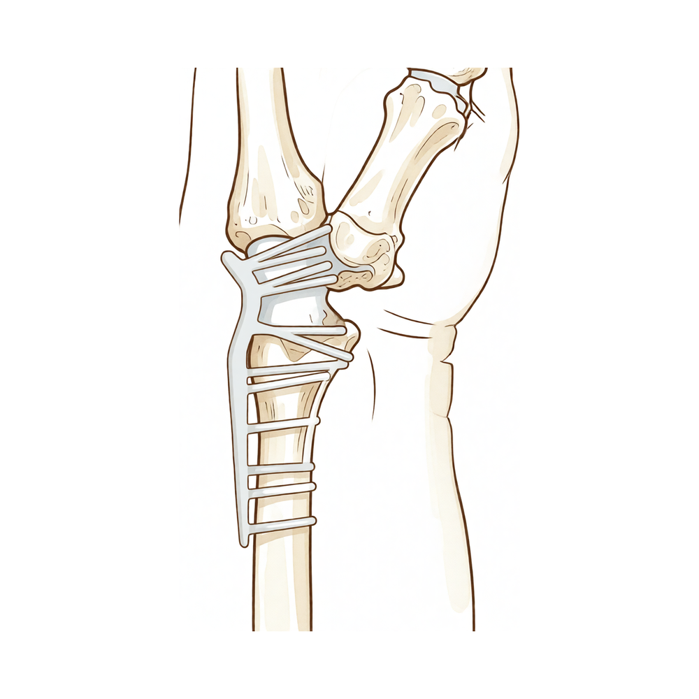
A continuación, el cirujano alinea los huesos en una posición funcional y los mantiene inmóviles durante el proceso de curación. Se coloca una placa metálica moldeada sobre la parte posterior de la muñeca y se fija a los huesos mediante tornillos. La placa mantiene todo en su sitio, evitando que los huesos se desplacen mientras se unen. En algunos casos, el cirujano podría añadir una pequeña cantidad de injerto óseo para favorecer la cicatrización.
Una vez que los huesos quedan bien fijados, el cirujano cierra la incisión con puntos de sutura y aplica un vendaje.
Después de la operación
Despertará en la sala de recuperación, donde las enfermeras lo vigilarán mientras el efecto de la anestesia desaparece. Su muñeca quedará sujeta mediante una férula o escayola, y la herida estará cubierta con un vendaje. Dejamos el vendaje puesto durante unos 10 días; por favor, no lo retire antes de ese plazo a menos que se lo indiquemos. Lo cambiamos o lo retiramos cuando lo veamos en consulta. Se le administrará analgésico antes de darle el alta; además, las enfermeras pueden ajustar la dosis si siente dolor. El mismo día de la cirugía podrá levantarse y caminar, apoyándose en la otra mano. La mayoría de los pacientes permanecen una noche en el hospital tras esta operación, aunque algunos pueden volver a casa ese mismo día. Por favor, asegúrese de que alguien lo acompañe durante las primeras 24 horas después de regresar a casa.
Recuperación
Durante los primeros días, su muñeca estará adolorida e hinchada. Esto mejora gradualmente. Mantener la mano elevada sobre almohadas, incluso por la noche, ayuda a reducir la hinchazón y el malestar. Tome los analgésicos que se le hayan recetado en lugar de esperar a que el dolor empeore.
Mientras los huesos se unen, su muñeca quedará inmovilizada mediante una férula o un yeso. Para las tareas cotidianas como vestirse, comer y asearse, deberá usar la otra mano. Puede moverse por la casa y realizar actividades ligeras, pero no debe conducir mientras la muñeca permanezca inmovilizada. Una vez que se retire el yeso o la férula y su cirujano lo autorice, podrá volver a conducir; nuestra página sobre conducción tras una cirugía de miembro superior explica esto con más detalle.
La terapia de la mano comienza poco después de la cirugía y ayuda a prevenir la rigidez en los dedos, la muñeca y el antebrazo. Su terapeuta de la mano, Ruby Doolan de Extend Rehabilitation, le guiará en los ejercicios y le confeccionará cualquier férula que necesite. Iniciar movimientos suaves desde el principio le permite recuperar la movilidad más rápidamente y, con frecuencia, reduce el número total de sesiones de terapia.
Dado que los huesos de la muñeca quedan unidos, esa zona ya no podrá doblarse. La mayoría de las personas se adaptan bien y logran realizar sus actividades diarias con algunos cambios en sus hábitos. El antebrazo, en cambio, sigue pudiendo girar, por lo que muchas tareas siguen siendo cómodas de realizar. Una vez que los huesos hayan sanado y su agarre sea fuerte, podrá retomar sus labores y otras actividades.
Cada persona sana a su propio ritmo, por lo que su cronograma puede variar. Su cirujano y su terapeuta de la mano lo guiarán en cada etapa del proceso.
Qué puede salir mal
La mayoría de los pacientes evolucionan bien, pero en ocasiones pueden presentarse problemas. Su cirujano y el equipo lo vigilarán de cerca para detectar cualquier incidencia a tiempo.
La placa metálica y los tornillos mantienen los huesos inmóviles mientras sanan. En algunas ocasiones, este material provoca molestias o se desplaza ligeramente. Puede sentir un borde afilado bajo la piel, o un dolor persistente sobre la placa que los analgésicos comunes no alivian. Si esto le molesta, hágalo saber en su próxima revisión. Una vez que los huesos hayan sanado, se puede retirar el material.
En raras ocasiones, los huesos de la muñeca no se unen como estaba previsto; esto se denomina no-unión. Muchas personas con este problema no sienten nada y no presentan síntomas. No obstante, si dicha separación provoca un dolor persistente, su cirujano conversará con usted sobre las opciones disponibles en la revisión.
El nervio que atraviesa el centro de la muñeca puede quedar comprimido por la inflamación postoperatoria; esto se conoce como síndrome del túnel carpiano. Notará hormigueo, sensación de pinchazos o entumecimiento en el pulgar, el índice y el dedo medio, a menudo más intenso por la noche. Informe a su cirujano si esto ocurre; normalmente se trata mediante una pequeña intervención para liberar la presión sobre el nervio.
También pueden surgir problemas en la cicatrización de la herida. Observe la piel alrededor del vendaje: si aparece enrojecimiento que se extiende, aumento de temperatura, secreción o un olor desagradable, podría tratarse de una complicación. Una herida que no evoluciona favorablemente puede requerir tratamiento; si observa estos signos, comuníquese con la clínica de inmediato en lugar de esperar a su próxima cita.
Los tendones, esos cordones que conectan los músculos con los huesos, pueden irritarse o desgarrarse cerca de la muñeca. Podría notar debilidad repentina, por ejemplo que un dedo no se estire correctamente, además de hinchazón o sensibilidad en el dorso de la mano. Comuníquelo a su cirujano lo antes posible.
Si previamente se le realizó un reemplazo de muñeca que fracasó, la artrodesis posterior puede solucionar el problema, aunque la recuperación suele ser un poco más lenta que tras una artrodesis por primera vez.
En la tabla de complicaciones de esta página se detallan las tasas habituales; si desea información específica, puede consultarla.
¿Cuándo debemos ser contactados?
La mayoría de los problemas aparecen durante las primeras semanas. Llámenos si tiene fiebre, o si la piel alrededor de la herida se vuelve más roja, caliente o segrega líquido. Llámenos si el dolor empeora repentinamente, o si nota hormigueo o entumecimiento que antes no tenía. Acuda a urgencias si la pantorrilla está hinchada y le duele, o si le cuesta respirar. Acuda a urgencias si no siente ni puede mover la mano o los dedos en absoluto.
¿Dónde leer más sobre esta enfermedad?
Esta página trata sobre la intervención quirúrgica en sí. La enfermedad que se trata, así como las evidencias sobre cuándo la cirugía resulta beneficiosa y cuándo no, se explican con mayor detalle en la página de Osteoartritis de la muñeca.
Evidence & references
This is the clinical evidence summary written for health professionals. It is technical, and it lists the research this page was built from. You do not need to read it to understand your treatment or to make a decision about it.
Anatomy & Pathophysiology
Bony Anatomy
- The wrist is the anatomic region between the forearm and the hand, including the distal radioulnar, radiocarpal, and ulnocarpal joints and the eight carpal bones [2].
- The proximal row of carpal bones consists of the scaphoid, lunate, triquetrum, and pisiform [2].
- The distal row of carpal bones consists of the trapezium, trapezoid, capitate, and hamate [2].
- The pisiform and trapezoid are the smallest carpal bones, while the capitate is the largest [2].
- The capitate articulates with seven other carpal bones, whereas the pisiform articulates with only one (the triquetrum) [2].
- The radiocarpal joint is formed by the articulation of the distal radius with the scaphoid and lunate, and the triquetrum on the triangular fibrocartilage [2].
- The distal concave articular surfaces of the proximal carpal row form the midcarpal articulations with the distal row [2].
- The distal ulnar convexity articulates at the lesser sigmoid notch of the distal radius [2].
- The sigmoid notch articular surface accommodates the ulnar head through two thirds of its arc [2].
- There is approximately a 20-degree inclination of the distal ulna at its articulation with the radius [2].
- The ulnar styloid lies dorsal to the ulnar head and extends distally [2].
- The distal radius has three articular components: the scaphoid fossa, the lunate fossa, and the sigmoid notch [6].
- A ridge between the scaphoid and lunate fossae corresponds with the scapholunate interval [6].
- The radial styloid allows attachment of the brachioradialis tendon and is the origin of the radial scapholunate and radial lunocapitate ligaments [6].
- The concave elliptical distal radius is oriented in the sagittal plane with an average of 11 degrees of volar tilt [6].
- In the frontal plane, the average radial inclination of the distal radius is 23 degrees [6].
- Radial length, measured from the tip of the radial styloid to the ulnar articular surface, averages 13 mm [6].
- The interosseous membrane connects the shafts of the radius and ulna, with a thickened central portion important for force transmission [6].
Ligamentous Anatomy
- The triangular fibrocartilage complex (TFCC) attaches to the ulnar margin of the lunate fossa of the radius [2].
- The TFCC includes the ulnar collateral ligament, dorsal and volar radioulnar ligaments, articular disc, meniscal homologue, extensor carpi ulnaris sheath, and ulnolunate and ulnotriquetral ligaments [2].
- Extrinsic carpal ligaments connect the radius or the ulna to the carpus [7].
- Volar extrinsic ligaments are generally stronger than dorsal ligaments [7].
- The radioscaphocapitate ligament connects to the waist of the scaphoid and limits ulnar translation of the carpus [7].
- The long radiolunate ligament helps limit ulnar translocation of the carpus [7].
- The short radiolunate ligament helps control lunate position [7].
- The radioscapholunate ligament is a vascular conduit (ligament of Testut) rather than a true ligament [7].
- The ulnolunate ligament attaches to the palmar radioulnar ligament and the lunate [7].
- The ulnocapitate ligament attaches to the ulnar head and is the most superficial or palmar ulnocarpal ligament [7].
- The ulnotriquetral ligament attaches to the palmar radioulnar ligament and the triquetrum [7].
- The dorsal radiocarpal ligament has a trapezoidal shape, passing from the dorsal rim of the distal radius to the lunate and triquetrum [7].
- The dorsal radiocarpal ligament is associated with dorsal and volar intercalated segmental stabilities [7].
- Intrinsic carpal ligaments originate and insert within the carpus [7].
- The scapholunate interosseous ligament (SLIL) is a major stabilizer of the wrist and the most commonly injured wrist ligament [7].
- The SLIL is C-shaped, consisting of dorsal, palmar, and interosseous portions, with the dorsal portion being the strongest and thickest [7].
- The SLIL provides a flexion force on the lunate due to its attachment to the scaphoid [7].
- The lunotriquetral interosseous ligament (LTIL) is C-shaped, with the volar portion being the thickest and strongest [7].
- The LTIL provides an extension moment on the lunate due to its attachment to the triquetrum [7].
- The capitohamate ligament is a thick ligament measuring 5 × 5 mm in cross section, with extensions to the third or fourth metacarpals [7].
- The dorsal intercarpal ligament passes from the dorsal tubercle of the triquetrum to the distal pole of the scaphoid [7].
- The dorsal intercarpal ligament reinforces the elastic dorsal wrist capsule and stabilizes the scapholunate articulation [7].
- The space of Poirier is an area adjacent to the proximal capitate without ligamentous attachment, situated ulnar to the radioscaphocapitate ligament and radial to the long radiolunate ligament [7].
- The space of Poirier is a weak area vulnerable to instability, through which the distal carpal row separates from the lunate during a perilunate dislocation [7].
- The dorsal wrist ganglion is typically located directly over the scapholunate ligament [16].
Biomechanics and Kinematics
- The wrist joint’s motion planes include flexion, extension, radial deviation, ulnar deviation, and circumduction [7].
- There is minimal carpal motion with pronosupination [7].
- Approximately 62° of wrist extension occurs through the radiocarpal joint [7].
- 62% of wrist flexion occurs through the midcarpal joint [7].
- The midcarpal joint is mostly responsible for 20° of radial deviation and 40° of ulnar deviation [7].
- The midcarpal joint is responsible for the “dart thrower’s motion,” which involves moving from radial extension into ulnar flexion positioning of the wrist [7].
- The radius bears 80% of the axial load transmitted through the radiocarpal joint in neutral ulnar variance [7].
- The ulna bears 20% of the axial load transmitted through the radiocarpal joint in neutral ulnar variance [7].
- The proximal row of carpal bones forms an intercalated segment between the distal carpal row and the distal radius, bound into a functional unit by the SLIL and LTIL [7].
- The distal row of carpal bones is rigid with little motion between its bones due to stout intercarpal ligaments, acting as a functional unit with the scaphoid bridging both rows [7].
- During wrist flexion from neutral, the distal row flexes and ulnarly deviates slightly while the scaphoid pronates [7].
- During wrist flexion from neutral, the proximal row flexes differentially, with more rotation through the scaphoid, followed by the triquetrum and the lunate [7].
- During wrist flexion from neutral, the proximal row translates dorsally [7].
- During wrist extension from neutral, the distal row extends and radially deviates slightly while the scaphoid supinates [7].
- During wrist extension from neutral, the proximal row extends differentially, with more motion in the scaphoid, followed by the triquetrum and then the lunate [7].
- During wrist extension from neutral, the proximal row translates palmarly [7].
- The eight carpal bones represent the most complex articular system in the human body [3].
- Injuries to the wrist mechanism can lead to instability, resulting in a painful lack of motion, strength, and function [3].
Investigations
Magnetic Resonance Imaging
- MRI was first reported for hand and wrist imaging in 1986 [8].
- The primary advantages of MRI compared with CT and radiography are improved tissue characterization, especially of soft tissues such as ligamentous structures in the wrist and synovium in the hand, and the lack of ionizing radiation [8].
- MRI is the modality of choice for imaging radiographically occult fractures of the hand and wrist [8].
- Early MRI was limited by low magnetic field strength (0.15 T) and limited image options and processing [8].
- Modern MRI is generally at 1.5T or 3T with a wide variety of imaging options and powerful image processing [8].
- 3T is much preferred for hand and wrist imaging, especially for imaging small fields of view [8].
- Dedicated extremity magnets have been marketed, but image quality is poor compared with conventional MR imaging [8].
- 7T MRI has recently become approved for clinical use [8].
- 7T MRI has more than double the magnetic field strength of 3T MRI and has the potential to become a powerful tool for hand and wrist imaging as applications are developed [8].
- MRI with contrast enhancement is most commonly used to determine whether soft-tissue lesions are solid or cystic [8].
- In rheumatologic imaging, MRI with contrast enhancement is used to better visualize erosions and synovial burden [8].
- Dynamic contrast enhancement has been used with inconsistent results to assess for the presence of avascular necrosis in the lunate or scaphoid after injury [8].
- MR angiography of the hand and wrist can be helpful in situations such as diagnosis of the hypothenar hammer syndrome [8].
- MR arthrography can be performed for evaluation of the triangular fibrocartilage and intercarpal ligament tears, but this is generally unnecessary with the increasing availability of high field MRI [8].
Computed Tomography and Other Modalities
- Hybrid SPECT/CT has been used for the diagnosis of radiographic occult fractures of the wrist [4].
- Cone-beam CT has been used in the diagnosis of scaphoid fractures [4].
- CT and MRI have a diagnostic impact on wrist injuries in young adults [4].
- Cine MRI is a new approach to the diagnosis of scapholunate dissociation [4].
- Cone-beam computed tomography arthrography is an innovative modality for the evaluation of wrist ligament and cartilage injuries [4].
- 3D analysis of the wrist has been described [4].
- Normal and variant anatomy of the wrist and hand can be evaluated on MR imaging [4].
- Low-field MRI has been used for scaphoid fracture evaluation [5].
- Occult wrist fractures can be detected by magnetic resonance imaging [5].
- MR imaging has been used to evaluate the triangular fibrocartilage complex [5].
- MR imaging and computed tomography arthrography have been used for preoperative evaluation of the ulnar collateral ligament [5].
- Histologic and magnetic resonance imaging correlations have been established in Kienbock’s disease [5].
- MRI diagnosis of occult dorsal wrist ganglion has been evaluated [5].
Radiography and Stress Views
- Distal radioulnar joint stress radiography has been used for detecting radioulnar ligament injury [4].
- Radiographic stress views have been compared for scapholunate dynamic instability in a cadaver model [4].
- Simple plain radiographic signs and measures have been evaluated for the accuracy of diagnosing acute scapholunate ligament injuries of the wrist [9].
- Radiographic clues have been described for determining carpal instability and treatment protocol for scaphoid fractures [9].
References
[2] Campbell S Operative Orthopaedics 4 Volume Set. NERVE INJURIES AT THE LEVEL OF THE HAND AND WRIST > ANATOMY.
[3] Green S Operative Hand Surgery. INTRODUCTION.
[4] Campbell S Operative Orthopaedics 4 Volume Set. ANATOMIC RECONSTRUCTION OF THE DISTAL RADIOULNAR LIGAMENTS > RADIOGRAPHIC TECHNIQUES.
[5] Campbell S Operative Orthopaedics 4 Volume Set. ELBOW, WRIST, AND HAND.
[6] A Lange Medical Book Current Diagnosis Treatment In Orthopedics Fifth Edition. 2Musculoskeletal Trauma Surgery > FRACTURES AND DISLOCATIONS OF THE DISTAL AND MID-FOREARM.
[7] Aaos Comprehensive Orthopaedic Review 3. Carpal Instability* > II. Anatomy and Biomechanics (See Chapter 92).
[8] Orthopaedic Knowledge Update 13 Ebook Without Multimedia. Anatomy, Evaluation, Clinical Examination, and Imaging > Imaging: Advances in Imaging of the Hand and Upper Extremity > Magnetic Resonance Imaging.
[9] Campbell S Operative Orthopaedics 4 Volume Set. ANATOMIC RECONSTRUCTION OF THE DISTAL RADIOULNAR LIGAMENTS > DIAGNOSIS AND EVALUATION.
[16] Green S Operative Hand Surgery. BOX 59.1 Ganglions of the Hand and Wrist > Operative Treatment > Dorsal Wrist Ganglion.