Patients › Shoulder
Descompresión subacromial
Arthroscopic subacromial decompression for impingement and bursitis — operation and recovery.
¿Por qué se ha recomendado esta operación?
El Dr. Kieran Hirpara, cirujano de extremidad superior en el Mater Private Hospital Rockhampton, comienza por proponer las opciones menos invasivas que se adapten a su condición. Por lo general, los pacientes son derivados a nuestra clínica por su médico de cabecera; si un fisioterapeuta le ha sugerido que nos consulte, igualmente necesitará una derivación de su médico de cabecera para poder acceder al reembolso de Medicare. En la consulta, tomamos su historia clínica, examinamos su hombro y solicitamos estudios de imagen si es necesario. Así se descartan otras causas de dolor de hombro, como el hombro congelado o la rotura de un tendón.
La descompresión subacromial es una operación para crear más espacio en la zona situada por encima del manguito rotador, el grupo de tendones que mueven el hombro. Se ofrece cuando su dolor coincide con el patrón del pinzamiento, en el que los tendones quedan comprimidos al levantar el brazo, y cuando el tratamiento no quirúrgico no le ha proporcionado una mejora suficiente. Ese tratamiento suele incluir cambios en la actividad, fisioterapia e inyecciones. Solo le propondremos la cirugía después de revisar con usted su exploración y sus estudios de imagen. Cuando se utiliza por los motivos adecuados, a la mayoría de las personas, entre el 70% y el 75%, la operación les ayuda. El objetivo es tener menos dolor al levantar el brazo y poder usar mejor el hombro.
Antes de la operación
Una vez que se ha programado la cirugía, unos sencillos preparativos ayudan a que el día transcurra sin contratiempos. Su cirujano ya dispondrá de los estudios de imagen necesarios para planificar la operación, como radiografías, una resonancia magnética o una ecografía. No coma ni beba durante las siete horas previas a la operación. Pedimos siete horas en lugar de seis para poder adelantar su intervención si la lista de quirófano avanza antes de lo previsto. Si toma medicamentos de forma habitual, lleve una lista escrita de ellos y pregunte a su cirujano si debe suspender alguno antes de la operación. Organice que alguien lo lleve a casa después, ya que no podrá conducir usted mismo. Use ropa holgada y cómoda que sea fácil de quitarse. Si padece otras enfermedades, es posible que necesite análisis de sangre o una evaluación con el anestesista antes del día de la operación.
El día de la intervención
Llegará a la unidad de admisiones quirúrgicas del hospital, donde se le registrará y preparará para la cirugía. Después conocerá al anestesista, el médico que se encarga de su anestesia y del control del dolor. Esta operación se realiza bajo anestesia general combinada con un bloqueo nervioso regional. El anestesista se reunirá con usted antes de la intervención y le explicará ambos procedimientos.
Cuando todo esté listo, será llevado al quirófano y se realizará la operación. Después, despertará en la sala de recuperación, donde las enfermeras lo vigilarán mientras la anestesia va desapareciendo. Comprobarán su hombro, cómo se encuentra y cómo se recupera de la anestesia. Una vez que su estado sea estable, será trasladado a la planta de hospitalización o podrá volver a casa, según la operación que se le haya realizado y cómo evolucione su recuperación. El tiempo habitual de estancia se explica en «Después de la operación».
Descripción del procedimiento quirúrgico
La descompresión subacromial es una operación mínimamente invasiva. El cirujano realiza algunas incisiones pequeñas alrededor del hombro, incluida una en la parte posterior, y trabaja a través de ellas con una pequeña cámara e instrumentos finos. La cámara permite al cirujano ver en una pantalla el espacio situado por encima del manguito rotador, el grupo de tendones que mueven el hombro.
Una vez dentro, el cirujano retira parte de un tejido inflamado llamado bursa y rebaja una pequeña porción de hueso de la cara inferior del acromion, la repisa ósea situada por encima de los tendones. Al retirar este hueso y este tejido se crea más espacio, de modo que los tendones ya no quedan comprimidos al levantar el brazo. Esta parte de la operación se llama acromioplastia. Si el cirujano encuentra algo más que necesite atención, como la rotura de un tendón, lo hablará con usted de antemano.
Cuando termina el trabajo, el cirujano lava el interior del hombro y cierra cada pequeña incisión con puntos de sutura. Se coloca un vendaje sobre las heridas, que se mantiene durante unos 10 días.
Después de la operación
La mayoría de los pacientes permanecen una noche en el hospital tras esta operación, aunque algunos pueden volver a casa el mismo día. Despertará en la sala de recuperación y luego será trasladado a la planta para pasar la noche. Su brazo descansará en un cabestrillo sencillo para mayor comodidad, y este se retira para los ejercicios y para lavarse. Las enfermeras le ayudarán a controlar el dolor con medicamentos. Alguien debe quedarse con usted durante las primeras 24 horas después de llegar a casa. Si le aplicaron un bloqueo nervioso, al principio es posible que sienta el brazo pesado o entumecido; esto es esperable y desaparece en unas 24 horas. Llame a la clínica si el entumecimiento o la debilidad persisten una vez que haya pasado el efecto del bloqueo. Dejamos el vendaje puesto durante unos 10 días; por favor, no lo retire antes de ese plazo a menos que se lo indiquemos. Lo cambiaremos o lo quitaremos cuando venga a la consulta.
Recuperación
Durante los primeros días, sentirá dolor e hinchazón en el hombro. La hinchazón disminuye gradualmente a lo largo de las semanas siguientes, y el dolor se alivia a medida que lo hace. Los medicamentos que le receta su equipo médico, las bolsas de hielo y el reposo ayudan en los primeros días. Su brazo descansa en un cabestrillo sencillo para mayor comodidad, y este se retira para los ejercicios y para lavarse.
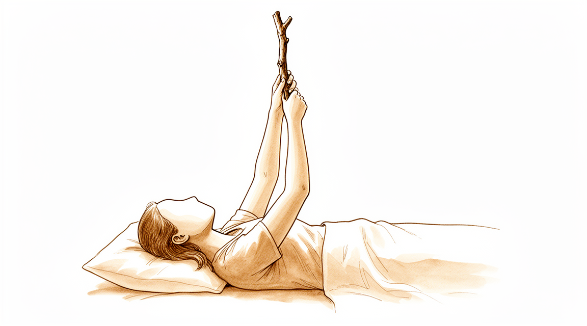
Comenzará con ejercicios suaves poco después de la operación, guiado por su fisioterapeuta. Estos ejercicios evitan que el hombro se ponga rígido mientras los tejidos se curan. Las tareas cotidianas, como vestirse, comer o escribir con el teclado en un escritorio, suelen ser posibles desde el principio. Levantar algo más pesado que una taza de té, o estirar el brazo por encima de la altura del hombro, debe esperar hasta que su hombro esté preparado. Al principio puede resultar incómodo dormir; a muchas personas les resulta más fácil recostadas en un sillón o con almohadas detrás.
Una vez que la hinchazón disminuya y recupere el movimiento, las actividades diarias le resultarán más naturales. Podrá conducir cuando su cirujano se lo autorice, generalmente en la revisión de las seis semanas; consulte nuestra guía Conducción después de cirugía de miembro superior. La vuelta al trabajo, al deporte y a las actividades más exigentes es gradual, y su cirujano y su fisioterapeuta le indicarán cuándo es seguro dar cada paso.
La recuperación varía de una persona a otra. Su cronograma puede ser diferente de los plazos habituales que se muestran en la tabla anterior, y su cirujano y su fisioterapeuta le guiarán durante todo el proceso.
Qué puede salir mal
La mayoría de los pacientes evolucionan bien, pero en ocasiones pueden surgir problemas. Su cirujano y el equipo lo vigilarán de cerca para detectar cualquier anomalía a tiempo.
A veces se forma hueso nuevo donde se rebajó la pequeña porción de hueso. Puede notarse como si volviera el antiguo dolor de pinzamiento, con una sensación de enganche o de roce al levantar el brazo. Si esto ocurre, coméntelo en su próxima revisión.
El hombro también puede volverse rígido y doloroso, con un movimiento que no se suelta ni siquiera con ejercicios suaves. Coménteselo a su fisioterapeuta o llame a la clínica si no mejora.
En ocasiones, el dolor resulta deberse a otra causa, como la rotura de un tendón o la artritis en una articulación cercana. Si, una vez superada la curación inicial, el hombro le sigue doliendo de forma muy parecida a antes, volveremos a revisar con usted el diagnóstico original en su revisión.
En raras ocasiones puede formarse un coágulo de sangre. Acuda al servicio de urgencias si tiene hinchazón o dolor en una pantorrilla, o una falta de aire repentina o dolor en el pecho. Pueden ser signos de un coágulo de sangre.
Llame a la clínica ese mismo día si tiene fiebre, un enrojecimiento que se extiende desde una herida, o salida de líquido o pus por ella. Llame también a la clínica ese mismo día si el dolor sigue empeorando a pesar de los analgésicos.
Llame a la clínica si el entumecimiento o la debilidad en el brazo, la mano o los dedos persisten una vez que haya pasado el efecto del bloqueo nervioso, a las 24 horas aproximadamente. El entumecimiento y la debilidad durante las primeras 24 horas son esperables.
En la tabla de complicaciones de esta página se detallan las tasas típicas, por si desea conocer los datos específicos.
¿Cuándo deben llamarnos?
La mayoría de los problemas aparecen pronto, por lo que conviene saber a qué estar atento. Acuda al servicio de urgencias si tiene hinchazón o dolor en una pantorrilla, o una falta de aire repentina o dolor en el pecho. Pueden ser signos de un coágulo de sangre. Llame a la clínica ese mismo día si tiene fiebre, un enrojecimiento que se extiende desde una herida, o salida de líquido o pus por ella. Llame también a la clínica ese mismo día si el dolor sigue empeorando a pesar de los analgésicos. Llame a la clínica si el entumecimiento o la debilidad en el brazo, la mano o los dedos persisten una vez que haya pasado el efecto del bloqueo nervioso, a las 24 horas aproximadamente. El entumecimiento y la debilidad durante las primeras 24 horas son esperables. Si no consigue contactar con la clínica, acuda al servicio de urgencias más cercano.
¿Dónde leer más sobre esta afección?
Esta página trata sobre la operación en sí. La afección que se trata con ella, incluyendo lo que demuestran las evidencias sobre cuándo la cirugía es útil y cuándo no, se explica con mayor detalle en la página Pinzamiento subacromial y bursitis.
Evidence & references
This is the clinical evidence summary written for health professionals. It is technical, and it lists the research this page was built from. You do not need to read it to understand your treatment or to make a decision about it.
Overview
- Functional outcome 6 years after arthroscopic subacromial decompression is not obviously related to the preoperative degree of cuff pathology, even if a total rupture of small size is present [1].
- The impingement test can be used as a predictor of outcome for patients with impingement syndrome treated by arthroscopic subacromial decompression [2].
- In appropriately selected patients, in-office needle arthroscopy of the shoulder with subacromial decompression can be performed by a simple technique [3].
- Arthroscopic subacromial decompression and limited open repair of small full thickness rotator cuff tears demonstrate an improved outcome when compared to patients with similar pathology for which a formal open repair has been performed [4].
- A clinical randomized study was unable to show any beneficial effect of physiotherapist-supervised rehabilitation after arthroscopic subacromial decompression of the shoulder [5].
- A case report describes a particularly impressive recurrence of a subacromial bone spur 5.2 years after open acromioplasty [6].
- The presence of calcifications within the subacromial space was more common after arthroscopic acromioplasty and correlated with worse results [7].
- The role of acromioplasty for treatment of shoulder pathology remains controversial, with recent level I data questioning its efficacy in isolation or in conjunction with rotator cuff repair [8].
- The coracoacromial ligament appears to have the ability to re-form relatively quickly after subacromial decompression or acromioplasty but takes time to regain strength [9].
- Subacromial decompression is an adequate treatment of impingement syndromes when conservative treatment has failed, with more than 90% excellent and good results after four years and 78% after ten years [10].
- There was no demonstrable benefit to laser assistance in arthroscopic subacromial decompression procedures [11].
- Arthroscopic subacromial decompression is an excellent tool for cuff impingement [12].
- Considering the lack of measurable benefit in conjunction with the significantly higher costs identified, laser assistance cannot be recommended in arthroscopic subacromial decompression procedures [13].
- Most patients returned to their former sports activities after arthroscopic subacromial decompression, including 90% of those in overhead sports and 100% of those in shoulder-sparing and contact sports [15].
- Benefits of tenotomy of the long head of the biceps (LHB) are evident on follow up and do not modify the subacromial space [19].
- Both Neer acromioplasty and modified acromioplasty provide adequate decompression [20].
- The indication for an acromioplasty must be done with precise and indisputable clinical and radiographic arguments [31].
- A meta-analysis found no benefits in measured outcomes at up to 2 years after surgery, but the long-term effects of performing or not performing an acromioplasty at the time of rotator cuff repair are not known [32].
- Despite Clinical Practice Guidelines recommending the nonroutine use of acromioplasty, surgeons continue to perform acromioplasty with rotator cuff repair in most of the cases throughout all subcategorizations analyzed [33].
Anatomy & Pathophysiology
Bony Anatomy
- The acromion has three ossification centers: the metacromion (base), the mesoacromion (middle), and the preacromion (tip) [51].
- Failure of fusion of the acromial ossification centers results in os acromiale [51].
- The scapula is attached to the axial skeleton by the clavicle, specifically via the acromioclavicular and sternoclavicular joints [50].
- The scapular spine is an osseous ridge that separates the supraspinatus and infraspinatus fossae [51].
- The acromion is a flattened bony process that curves forwards from the scapular spine [50].
- In humans, the acromion is a massive structure overlying the humeral head, reflecting the increasing role of the deltoid muscle in shoulder function [58].
- The glenoid is a convex structure of shallow depth shaped like an inverted pear [48].
- The glenoid averages 5° of retroversion in relation to the axis of the scapular body [51].
- The humeral head averages 19° of retroversion and 41° of inclination (neck-shaft angle) [51].
- The articular head of the humerus is spherical and has a diameter of 37 to 57 mm [48].
- The most superior portion of the articular surface of the humeral head averages 8 mm above the greater tuberosity [48].
- The humeral version averages 29.8 degrees (range, 10 to 55 degrees) [48].
- The head is inclined approximately 130 degrees with respect to the humeral shaft [48].
- The neck-shaft angle measures an average of 135 degrees [49].
- The humeral head is retroverted an average of 30 degrees [49].
- The glenoid cavity is a shallow socket, approximately one third the size of the humeral head [49].
- The acromion, the coracoacromial ligament, and the coracoid process form the coracoacromial arch [48].
- The coracoacromial arch is a rigid bony-ligamentous structure that imparts stability to the shoulder girdle [48].
- The rotator cuff, subacromial bursa, and subdeltoid bursa pass underneath the coracoacromial arch [48].
- The acromion process is located posterosuperiorly, the coracoid process anteriorly, and the coracoacromial ligament joins them to form a fibro-osseous canopy over the rotator cuff [56].
- The acromial branch of the thoracoacromial artery runs on the medial aspect of the coracoacromial ligament [62].
- The coracoacromial ligament contributes to anterosuperior stability in rotator cuff deficiency [62].
- The coracoacromial ligament should be preserved with irreparable cuff tears to prevent anterosuperior escape [62].
- The coracoacromial ligament is the arthroscopic landmark for a complete release of the rotator interval for adhesive capsulitis [62].
- The superior shoulder suspensory complex (SSSC) is composed of the glenoid, the coracoid process, the coracoclavicular ligaments, the distal clavicle, the AC joint, and the acromion [51].
- The SSSC provides a stable connection between the scapula and the axial skeleton [51].
- The SSSC is a bone–soft-tissue ring that provides a stable connection of the glenoid and scapula to the clavicle [57].
- The SSSC is composed of four bony landmarks—distal clavicle, acromion, coracoid process, and glenoid neck—and the supporting ligamentous complexes of the AC joint and the CC ligaments [57].
- The superior strut of the SSSC comprises the middle clavicle [51].
- The inferior strut of the SSSC comprises the lateral scapular border/spine of the scapula [51].
- The coracoid process curves forwards from the superior surface of the scapular neck [50].
- The coracoid process has undergone an increase in size over time [58].
- With the shoulder in 90 degrees of abduction, the coracoid extension over the glenohumeral joint can mechanically limit anterior translation of the humerus relative to the glenoid [58].
- The coracobrachialis muscle and the short head of the biceps tendon originate from the coracoid process [51].
- The pectoralis minor muscle inserts onto the medial coracoid process [51].
- The relationship between coracoid morphology and subscapularis tears is controversial [51].
- The scapula is anteverted on the chest wall approximately 30 degrees relative to the body [62].
- The scapula spans second through seventh ribs and serves as an attachment for 17 muscles [62].
- The scapula is suspended by muscles alone and has shifted caudally from the cervical position in lower animals [58].
- The scapular index is extremely high in pronograde animals with a long, narrow scapula [58].
- The scapula is broader in humans and other primates, with the most pronounced differences observed in the infraspinatus fossa [58].
- Broadening of the infraspinatus fossa has resulted in a change in the vector of muscle pull from the axillary border of the scapula to the glenoid fossa [58].
- This adaptation allows the infraspinatus and teres minor muscles to be more effective in their roles as depressors and external rotators of the humeral head [58].
- The supraspinatus fossa and muscle have changed little in size or shape over time [58].
- The distribution of the bony mass of the scapula is highly uneven, with areas of thick bone contrasting with other areas that are almost translucent [50].
- The highest concentration of bony mass in the scapula is in the glenoid, the scapular neck (including the base of the coracoid process), and the lateral border of the scapular body [50].
- The lateral pillar connects the inferior border of the glenoid with the inferior angle [50].
- The spinal pillar arises from the central part of the glenoid and continues medially to become part of the base of the scapular spine [50].
- The two pillars, connected by a markedly thinner medial border of the scapular body, form the basic load-bearing structure of the scapular body [50].
- The weakest bone in the scapula is located primarily in the central part of the biomechanical body, i.e., in the infraspinous fossa [50].
- The weakest area of the circumference of the biomechanical body of the scapula is the connection of the scapular spine and the medial border of the scapula, the spinomedial angle [50].
- Os acromiale is incomplete fusion of secondary ossification centers, most commonly between mesoacromion and meta-acromion [62].
- The clavicle is the first bone to ossify (fifth week of gestation) and is the only long bone to ossify by intramembranous ossification [51].
- The medial (sternal) epiphysis of the clavicle is the last ossification center to fuse, at age 20 to 25 years [51].
- The primary blood supply to the clavicle is periosteal; no nutrient artery is present [51].
- The clavicle serves as the primary stabilizer between the axial skeleton (via the sternoclavicular joint) and the appendicular skeleton (via the acromioclavicular joint) [57].
- The distal clavicle is flat in the AP plane [57].
- The proximal humerus has three centers of ossification: the humeral head (4 to 6 months), the greater tuberosity (1 to 3 years), and the lesser tuberosity (3 to 5 years) [51].
- The proximal humeral ossification centers fuse to the shaft at age 17 to 20 years [51].
- The proximal humerus is primarily cartilaginous at birth; however, the ossification centers can be detected with ultrasonography as early as the 38th week of gestation [55].
- The ossification center for the humeral head is usually present at birth [55].
- The greater tuberosity ossification center appears by 1 to 3 years of age [55].
- The lesser tuberosity ossification center appears by 5 years of age [55].
- These ossification centers fuse by 5 to 7 years of age to form the humeral head [55].
- The proximal humeral physis closes by 14 to 17 years of age in girls and by 16 to 18 years in boys [55].
- Humeral retroversion averages 65 degrees in infants and young children and gradually decreases, approaching adult values by 11 years of age [55].
- Mean values of humeral retroversion are around 26 degrees in healthy adults [55].
- Eighty percent of subsequent growth of the humerus comes from the proximal humeral physis [55].
- The proximal humeral physis accounts for approximately 40% of the growth of the entire upper extremity [55].
- Less than 75% of growth from the proximal humerus occurs before 2 years of age [55].
- More than 85% of growth from the proximal humerus occurs by 8 years of age [55].
- The anatomic neck of the proximal humerus is located at the junction of the articular surface and the tuberosities [48].
- The surgical neck represents an indistinct region (i.e., metadiaphyseal junction) below the tuberosities but above the humeral shaft [48].
- The greater tuberosity is located in a posterior-superior location with respect to the humeral shaft [48].
- The greater tuberosity serves as the attachment site for the supraspinatus, infraspinatus, and teres minor tendons of the rotator cuff [48].
- The lesser tuberosity is located on the anterior aspect of the proximal humerus [48].
- The lesser tuberosity serves as the attachment site for the subscapularis tendon [48].
- The bicipital groove lies between the greater and lesser tuberosities and serves as a pathway for the long head of the biceps [48].
- The distal aspect of the bicipital groove is internally rotated with respect to the proximal portion [48].
- The transverse humeral ligament is an important stabilizer of the biceps tendon [62].
- The anatomic neck is at the junction of the head and tuberosities [49].
- The surgical neck lies below the greater and lesser tuberosities [49].
- The major blood supply to the humeral head is through the ascending branch of the anterior humeral circumflex artery, which penetrates the head at the bicipital groove and becomes the arcuate artery [49].
- The anterolateral ascending branch of the anterior humeral circumflex artery provides the primary blood supply to the humeral head [51].
- The anterolateral ascending branch of the anterior humeral circumflex artery travels proximally in the lateral aspect of the intertubercular groove [51].
- The terminal intraosseous portion of the artery enters at the proximal aspect of the intertubercular groove as the arcuate artery [51].
- The proximal humerus receives its blood supply from the anterior and posterior humeral circumflex branches from the third division of the axillary artery [48].
- The posterior humeral circumflex artery travels with the axillary nerve, enters the quadrilateral space posteriorly, and anastomoses with a branch of the anterior circumflex to supply the posterior cuff [48].
- The anterior humeral circumflex artery arises from the axillary artery at the inferior border of the subscapularis [48].
- The anterior humeral circumflex artery provides vascular inflow to the humeral head by way of its terminal anterolateral branch known as the artery of Laing (also known as the arcuate artery) [48].
- The ascending branch of the anterior humeral circumflex artery courses parallel to the lateral aspect of the long head biceps tendon and enters the humeral head at the interface of the bicipital groove and greater tuberosity [48].
- Injury to the arcuate artery may result in osteonecrosis of the humeral head [48].
- Additional extraosseous collateral branches can permit humeral head perfusion despite complete ligation of the arcuate artery [48].
- Recent quantitative assessment has shown that 64% of the humeral head blood supply arises from the posterior humeral circumflex artery [55].
- The brachial plexus and axillary artery are anterior to the coracoid process of the scapula and humeral head [49].
- Nerves innervating muscles around the shoulder include the axillary, suprascapular, subscapular, and musculocutaneous nerves [49].
- The axillary nerve circles the humeral neck just inferior to the glenohumeral joint as it courses posteriorly [55].
- The axillary nerve is a terminal branch coming off the posterior cord of the brachial plexus just proximal to the coracoid process [54].
- The axillary nerve passes beneath the conjoined tendon anterior to the subscapularis 3 to 5 mm medial to the musculotendinous junction [54].
- The axillary nerve is adjacent to the inferior capsule before entering the quadrilateral space posteriorly [54].
- The axillary nerve splits into the anterior and posterior branches within the quadrangular space [54].
- The anterior and middle deltoid muscle receives sole innervation from the anterior branch of the axillary nerve [54].
- The posterior deltoid muscle innervation varies, with one study showing supply only from the anterior branch in 2.3% of cases, from the posterior branch in 8.5%, and from both branches in 89.1% [54].
- The posterior branch of the axillary nerve branches to supply the teres minor muscle and then terminates as the superior lateral brachial cutaneous nerve [54].
- In the anterior deltopectoral approach, the axillary nerve can be palpated by sweeping a finger inferiorly across the subscapularis muscle tendon interface [54].
- In the anterolateral deltoid splitting approach, the axillary nerve crosses approximately 5 cm inferior to the anterolateral acromial corner [54].
- In the posterior deltoid splitting approach, the axillary nerve is approximately 7 cm from the posterior acromial corner [54].
- The deltoid has anatomic subdivisions (acromial, clavicular, and scapular) [53].
- The deltoid forward flexes and abducts the shoulder and courses from the clavicle and acromion superiorly, coalescing into a common tendinous insertion onto the lateral upper third of the humeral shaft [55].
- The pectoralis major powers adduction and internal rotation due to its tendinous insertion anteriorly onto the lateral wall of the bicipital groove [55].
- The pectoralis major forms the roof of the distal continuation of the bicipital tunnel, which is a closed space that extends proximally to the glenohumeral joint [55].
- The subscapularis originates from the anterior scapula and inserts anteriorly onto the lesser tuberosity [55].
- The greater tuberosity provides attachment superiorly and posteriorly for the supraspinatus, infraspinatus, and teres minor, all of which originate from the posterior scapula [55].
- The rotator cuff consists of four muscles: the subscapularis, supraspinatus, infraspinatus, and teres minor muscles [49].
- The teres major is not a rotator cuff muscle [49].
- The cuff muscles serve as depressors of the humeral head to allow the deltoid to efficiently abduct the humerus [49].
- The infraspinatus and teres minor are external rotators, while the subscapularis is an internal rotator of the humerus [49].
- The rotator cuff is a sheet of conjoined tendons closely applied over the shoulder capsule and inserting mainly into the greater tuberosity of the humerus [56].
- The subscapularis is inserted into the lesser tuberosity [56].
- The cuff is made up of subscapularis in front, supraspinatus above and infraspinatus and teres minor behind [56].
- The rotator cuff has an important function in stabilizing the head of the humerus by pulling it firmly into the glenoid whenever the deltoid lifts the arm forwards or sideways [56].
- The tendinous insertions of the rotator cuff muscles, the articular capsule, the coracohumeral ligament, and the glenohumeral ligament complex blend into a confluent sheet before insertion into the humeral tuberosities [61].
- The tendons of the infraspinatus and supraspinatus muscles join approximately
Classification
Diagnostic Criteria and Predictors
- Diagnosis of impingement syndrome was based on a history of pain in the anterior aspect of the shoulder, a “painful arc“ during elevation, and the presence of tenderness over the anterior cuff associated with a positive impingement sign [27].
- The Neer impingement test was performed if there was any doubt about the diagnosis [27].
- Complete subsidence of pain after injection of local anaesthetic into the subacromial bursa was deemed an excellent response to the diagnostic test [27].
- Marked diminution of pain after injection of local anaesthetic into the subacromial bursa was deemed a good response to the diagnostic test [27].
- Little or no relief of pain after injection of local anaesthetic into the subacromial bursa was deemed a poor response to the diagnostic test [27].
Pathological Associations
- There is an association between the shape of the acromion and severity of rotator cuff pathology, with greater prevalence of the type-3 hooked acromion in individuals with impingement and rotator cuff tears [37].
- A flat, horizontal acromial orientation is associated with subacromial impingement, degenerative changes to the rotator cuff, and subacromial spur formation [37].
- Low degree chronic inflammation of the bursa is a common sign in impingement syndrome, but not always detectable [17].
- There is no correlation between clinical and pathohistological findings regarding the degree of inflammation in the subacromial bursa [17].
- Calcification on x-ray evaluation is a condition that is found in both painful and painfree shoulders [35].
Staging and Etiology
- The staging of cases in one series was not based on Neer's classification, but on the fact whether they had a rotator cuff tear or not, without considering age intervals [46].
- The presence of a rotator cuff tear was the most important factor that affected the surgical treatment results in a series of patients with subacromial impingement syndrome [46].
Clinical Presentation
Diagnostic Criteria and Signs
- The diagnosis of impingement syndrome is based on a history of pain in the anterior aspect of the shoulder, a “painful arc“ during elevation, and the presence of tenderness over the anterior cuff associated with a positive impingement sign [27].
- The Neer impingement test is performed if there is any doubt about the diagnosis [27].
- The ideal patient for arthroscopic subacromial decompression presents with subacromial stenosis, tenderness at the insertion of the supraspinatus tendon, a positive subacromial painful arc, and a positive Jobe test [39].
Pathology and Histology
- Low degree chronic inflammation of the subacromial bursa is a common sign in impingement syndrome, but it is not always detectable [17].
- There is no correlation between the degree of bursal inflammation and the diagnosis, duration or severity of pain, or Constant score [17].
- There is no correlation between clinical and pathohistological findings in impingement syndrome [17].
- The functional outcome 6 years after arthroscopic subacromial decompression is not obviously related to the preoperative degree of cuff pathology, even if a total rupture of small size is present [1].
Prognostic Factors
- Workers’ Compensation patients had a satisfactory rate of 32% after arthroscopic subacromial decompression, whereas non–Workers’ Compensation patients had a satisfactory rate of 59% [18].
- Inadequate decompression was noted in 14 of 20 failed patients who underwent open acromioplasty after arthroscopic subacromial decompression [18].
Investigations
Radiographic Evaluation
- Standardized plain films are almost always sufficient to garner the information needed for shoulder imaging, and there is information that can be gathered from properly taken plain films that cannot be obtained from CT scans [41].
- The first key radiographic view is the anteroposterior (AP) view in the plane of the scapula, taken so that the x-ray beam passes through the glenohumeral joint [41].
- The AP view in the plane of the scapula shows the superoinferior position of the humeral head relative to the glenoid, the presence of osteophytes on the humeral head and glenoid, narrowing of the joint space, and the degree of medial displacement of the humerus in relation to the lateral acromial line [41].
- The second key radiographic view is the axillary view taken with the arm in the functional position of elevation in the plane of the scapula [41].
- The axillary view is oriented so that both the spinoglenoid notch and the scapular neck are visible [41].
- The axillary view demonstrates the glenohumeral relationships in the functional position of elevation and is referred to as the "truth view" [41].
- CT scans have the disadvantage of being taken with the arm in the adducted position, unlike the axillary truth view [41].
- The standardized anteroposterior and axillary views indicate the thickness of the cartilage space between the humerus and the glenoid, relative positions of the humeral head and the glenoid, presence of osteophytes, degree of osteopenia, and extent of bony deformity and erosion [41].
- The axillary truth view shows posterior subluxation or "functional decentering" that is not evident in images taken with the arm at the side [41].
- At least two X-ray views should be obtained for shoulder imaging: an anteroposterior in the plane of the glenoid and an axillary projection with the arm in abduction to show the relationship of the humeral head to the glenoid [65].
- The standard shoulder series should include orthogonal views of the shoulder, including a true AP view in the scapular plane, an AP view, an axillary view, and a scapular Y view [73].
- The true AP view in the scapular plane visualizes the anterior greater tuberosity in profile and can reveal proximal humeral migration when the arm is held in neutral rotation with the shoulder in slight abduction [73].
- The AP view with the arm held in internal rotation visualizes the posterior aspect of the greater tuberosity and the lesser tuberosity in profile [73].
- The axillary view enables determination of the humeral head position in the glenoid fossa and may detect occult, locked posterior shoulder dislocation in a patient who exhibits a lack of passive external rotation [73].
- The axillary view provides good visualization of the coracoid process, acromion, and distal clavicle [73].
- The scapular Y view provides visualization of the coracoacromial arch and can reveal coracoacromial spurs, which have been closely associated with the presence of rotator cuff pathology [73].
- The scapular Y view is a reliable alternative for evaluation of glenohumeral subluxation and dislocation and can show scapular body abnormalities and acromial shape [73].
- The acromiohumeral distance is normally 7 to 14 mm [73].
- The width of the glenohumeral joint space should be symmetric superiorly and inferiorly [73].
- Neer classified acromial morphology as type I (flat), type II (curved), and type III (hooked) [73].
- Type III acromial morphology has been shown to have a correlation with the presence of rotator cuff disease, although no direct causal relationship has been demonstrated [73].
- The Neer classification of acromial morphology has shown relatively poor interobserver reliability [73].
- Initially, all patients are usually asked to have AP and lateral plain radiographs of the shoulder related to their chief report [72].
- Plain radiographs are often the only required studies needed for assessing acute shoulder trauma, including fractures or dislocations [72].
- Arthritis, calcific tendinitis, and osteolysis of the distal clavicle can be observed on plain radiograph [72].
Magnetic Resonance Imaging
- MRI is the modality of choice for evaluating the rotator cuff, biceps, and subacromial/subdeltoid bursa [72].
- T1-weighted MRI can reveal Hill-Sachs lesions and is often used with magnetic resonance (MR) arthrograms to provide a more detailed picture of the joint surfaces [72].
- T2-weighted MRI provides better visualization of full thickness rotator cuff tears [72].
- Magnetic resonance imaging (MRI) is useful to identify osteonecrosis of the humeral head, or a bone tumour [65].
- MRI can identify labral tears and rotator cuff tears, although the accuracy for these is enhanced by combining the scan with arthrography [65].
- MR accuracy in identifying labral and rotator cuff tears in the literature ranges from 70% to 100% [70].
- The acquired multi-planar imaging of MRI allows for the detailed evaluation of the glenoid, labrum, joint capsule, and rotator cuff in different planes [70].
- MR arthrography (MRA) refers to MRI of a joint that has been injected with an intra-articular contrast agent such as diluted gadolinium or saline solution [70].
- MRA increases the sensitivity for detecting tears and other lesions by distending the joint capsule and outlining the cartilage, ligaments, and labrum with contrast [70].
- MRA has proven utility by increasing both sensitivity and specificity in detecting injuries to the capsulolabral–ligamentous complex as compared to traditional MRI [70].
- In a meta-analysis of 6 studies including 4,667 shoulders, MRA had greater diagnostic test accuracy for the detection of glenoid labral lesions than MRI, with MRA sensitivity of 88% and specificity of 93% versus MRI sensitivity of 76% and specificity of 87% [70].
- Abduction and external rotation (ABER) of the arm is an alternative position utilized to increase the sensitivity and specificity for detecting anteroinferior labroligamentous injury [70].
- Limited range of motion or pain may prohibit patients from performing the ABER provocative maneuver [70].
- MRAs can demonstrate a patulous capsule on the coronal, sagittal, and axial imaging in patients with multidirectional instability [70].
- MRAs can be helpful in evaluating lesions of the rotator interval and other associated findings that may ultimately affect the eventual surgical plan [70].
- The presence of glenoid dysplasia, increased capsular cross-sectional area, and increased glenoid retroversion have all been found to be associated with increased posterior labral tears and symptomatic instability [70].
- Glenoid retroversion was significantly increased in patients with symptomatic posterior labral tears, but there was no significant association between instability and increased humeral head subluxation [70].
- The diagnosis of multidirectional instability is a clinical one, and the need for expensive and/or invasive imaging should be weighed against the information that will be gained from these studies [70].
- MR arthrography is considered the benchmark for evaluation for labral tears and rarely is indicated for evaluation of rotator cuff pathology [72].
- When MRI or MR arthrography is contraindicated, such as in patients with a pacemaker or vascular clips, CT arthrography is indicated [72].
Ultrasonography
- Ultrasonography is a low-cost alternative to MRI and arthrography for evaluating both skeletal and soft-tissue structures of the shoulder [72].
- Ultrasonography can provide immediate, real-time visualization of the rotator cuff, biceps tendon, and calcific deposits [72].
- Ultrasonography can be used to measure the subacromial space and detect atrophy of rotator cuff muscles [72].
- As a result of providing images in real-time, ultrasonography can evaluate impingement in various positions and motions [72].
- Ultrasonography is highly operator dependent and is not as useful for evaluating labral tears or rotator cuff tears that are very small or larger than 3 cm [72].
- Ultrasonography is a simple and accurate test for identifying rotator cuff tears and calcific tendinitis [65].
- Ultrasonography can be useful in guiding injections or barbotage, which involves aspirating calcific deposits in the rotator cuff [65].
- The sensitivity of ultrasonography for the detection of full-thickness rotator cuff tears is 98% [73].
- The specificity of ultrasonography for the detection of full-thickness rotator cuff tears is 80% [73].
- The positive predictive value of ultrasonography for the detection of full-thickness rotator cuff tears is 90% [73].
- The negative predictive value of ultrasonography for the detection of full-thickness rotator cuff tears is 95% [73].
- The accuracy of ultrasonography for the detection of full-thickness rotator cuff tears is 94% [73].
- The sensitivity of MRI for the detection of full-thickness rotator cuff tears is 100% [73].
- The specificity of MRI for the detection of full-thickness rotator cuff tears is 68% [73].
- The positive predictive value of MRI for the detection of full-thickness rotator cuff tears is 85% [73].
- The negative predictive value of MRI for the detection of full-thickness rotator cuff tears is 100% [73].
- The accuracy of MRI for the detection of full-thickness rotator cuff tears is 89% [73].
Computed Tomography
- CT imaging is frequently used to evaluate fractures of the shoulder, to assess for bony lesions in recurrent instability cases, or for preoperative templating for shoulder arthritis [72].
- Computed tomography (CT) is helpful for planning fracture surgery and shoulder joint replacement [65].
- CT with three-dimensional reconstructions is the advanced imaging study of choice for determining the extent of glenoid bone loss in the setting of shoulder instability [73].
- Although CT scans may offer a few degrees of increased precision in the measurement of glenoid version, this precision does not improve the quality of the surgery or the clinical outcome [41].
- Three-dimensional reconstructions can reveal fine details of the shoulder anatomy, but this additional information rarely changes the planning or conduct of the arthroplasty [41].
Arthroscopy
- Arthroscopy is useful for diagnosing and treating subacromial impingement, intra-articular lesions, detachment of the glenoid labrum and rotator cuff tears [65].
- This approach allows for visualization, decompression, and curettage of the lesion, as well as identification and treatment of additional glenohumeral pathology [24].
Treatment
Outcomes and Efficacy
- Subacromial decompression with more than 90% excellent and good results after four years and 78% after ten years is an adequate treatment of impingement syndromes when conservative treatment has failed [10].
- In a study of two series of patients treated for impingement syndrome by undergoing arthroscopic subacromial decompression, 48% of patients in Group 1 were graded as satisfactory and 52% unsatisfactory according to Neer criteria [18].
- In a study of two series of patients treated for impingement syndrome by undergoing arthroscopic subacromial decompression, 86% of patients in Group 2 were graded as satisfactory according to Neer criteria [18].
- The average American Shoulder and Elbow Society score at follow-up was 90.4 in a study of patients treated with arthroscopic subacromial decompression [18].
- Arthroscopic subacromial decompression has been shown to be a reliable method of treating subacromial impingement in the medium- and long-term [21].
- Medium- and long-term results of arthroscopic subacromial decompression are comparable to open decompression [21].
- Satisfactory pain relief and return to preinjury work activities can be achieved by acromioplasty in patients who have the potential for secondary gain [23].
Rehabilitation and Return to Work
- Many departments now carry out arthroscopic subacromial decompression as a day-case procedure if a patient’s health allows [21].
- It is believed that recovery is quicker for an arthroscopic subacromial decompression than for an open decompression as there is less disturbance of the surrounding soft tissues [21].
- In a study of 68 patients, the average return to work for manual workers was 64 days (range 1–320) and for non-manual workers was 27 days (range 4–90) following arthroscopic subacromial decompression [21].
- Ellman stated that patients in office-based occupations returned to work in 2–3 days and those with manual jobs returned within 6 weeks of surgery, although actual figures were not given [21].
- Altchek et al. reported 40 patients returning to work an average of 9 days [21].
- In a study of 75 consecutive anterior acromioplasties, group 1 patients (workers' compensation) required a significantly longer time to return to work (average 14.2 weeks) compared to group 2 (4.7 weeks) and group 3 (2.5 weeks) [23].
- Ninety-one percent of employed patients were able to return to full employment in a study of 75 consecutive anterior acromioplasties [23].
Surgical Techniques and Adjuncts
- Benefits of tenotomy of the long head of biceps are evident on follow up and do not modify the subacromial space [19].
- Both Neer acromioplasty and modified acromioplasty methods provide adequate decompression [20].
- At a mean follow-up of 18 months, no differences related to shoulder function as evaluated by the Constant score were found between groups in a study of open anterior acromioplasty with preservation of the coracoacromial ligament [14].
- The resection of the inferior medial acromion and distal clavicle spurs does not cause late clinical problems and achieves the primary goal of fully decompressing the supraspinatus outlet [38].
- This technique is very effective for symptomatic acromioclavicular arthritis of the shoulder [29].
- The authors introduce a computer image-guided precise acromioplasty (CIG-PAP) technique, an individualized treatment based on three-dimensional planning, to reduce a large critical shoulder angle (CSA) to the desired range of 30-33 degrees [44].
- Patient-controlled lidocaine analgesia in the subacromial space appears to be a safe method for achieving high levels of pain control in patients undergoing an acromioplasty [76].
Complications and Anatomical Considerations
- The authors report a case of a particularly impressive recurrence of a subacromial bone spur 5.2 years after open acromioplasty [6].
- It appears that the coracoacromial ligament has the ability to re-form relatively quickly after subacromial decompression or acromioplasty but takes time to regain strength [9].
- Simulating a longer acromion did not result in any new impingement in acromions that were free of impingement after acromioplasty [16].
- Risk factors for experiencing an acromial fracture include osteopenia and overzealous bone resection [28].
- The patient continued to have moderate pain with use of the arm at 18 months post-surgery and had not been able to return to employment in a case report of acromion nonunion after anterior acromioplasty [36].
- Physical examination at 1 year revealed no pain with shoulder motion and the ability to forward flex the arm to 90° and internally rotate to place the hand in the front pocket in a case report of severe heterotopic ossification after arthroscopic acromioplasty [26].
- Understanding the acromial malunion and ultimately restoring anatomy were essential to the success of a case involving deltoid dysfunction [25].
- This approach allows for visualization, decompression, and curettage of the lesion, as well as identification and treatment of additional glenohumeral pathology in arthroscopic decompression and bone grafting of proximal humeral intraosseous ganglion [24].
Complications
Bony Complications
- Acromial fracture is a complication of arthroscopic subacromial decompression [28].
- Acromion nonunion after anterior acromioplasty has been reported as a complication [36].
- A patient with acromion nonunion after anterior acromioplasty continued to have moderate pain with use of the arm at 18 months post-surgery and had not been able to return to employment [36].
- Recurrent acromial bone spur after open subacromial decompression has been reported [6].
- A recurrence of a subacromial bone spur was observed 5.2 years after open acromioplasty [6].
- Excessive bone removal tends to jeopardize the superior acromion and deltoid origin [34].
- Excessive bone removal removes the passive stabilizing effect of the inferior acromion against superior humeral subluxation [34].
Heterotopic Ossification and Calcification
- Heterotopic ossification is rarely described as a complication of the Neer anterolateral acromioplasty [75].
- Heterotopic ossification is rarely described as a complication of distal clavicle resection [75].
- Postoperative heterotopic ossification occurred with an incidence of 3.2% (16 of 497) after primary Neer acromioplasties and distal clavicle resections [75].
- Severe heterotopic ossification after arthroscopic acromioplasty has been reported as a complication [26].
- Physical examination at 1 year after severe heterotopic ossification revealed no pain with shoulder motion and the ability to forward flex the arm to 90° and internally rotate to place the hand in the front pocket [26].
- The presence of calcifications within the subacromial space was more common after arthroscopic acromioplasty [7].
- The presence of calcifications within the subacromial space after arthroscopic acromioplasty correlated with worse results [7].
- Calcifications are resorbed spontaneously [35].
Soft Tissue and Neurological Complications
- Deltoid pull-off is a complication identified in failed arthroscopic subacromial decompression cases [77].
- Suprascapular nerve compression is a complication identified in failed arthroscopic subacromial decompression cases [77].
- Painful neuroma is a complication identified in failed arthroscopic subacromial decompression cases [77].
- Adhesive capsulitis or adhesions are complications identified in failed arthroscopic subacromial decompression cases [77].
- Twenty-one shoulders had continued impingement from inadequate decompression in a series of failed arthroscopic subacromial decompression cases [77].
Ligamentous Regeneration
- The coracoacromial ligament has the ability to re-form relatively quickly after subacromial decompression or acromioplasty [9].
- The coracoacromial ligament takes time to regain strength after subacromial decompression or acromioplasty [9].
- A macroscopically normal coracoacromial ligament was found at revision arthroscopy after failure of arthroscopic subacromial decompression [47].
Intraoperative and Postoperative Physiological Effects
- A considerable swelling of the shoulder developed in all patients in the later part of the operation during arthroscopic acromioplasty [43].
- The swelling developed during arthroscopic acromioplasty always disappeared by the time the patients were evaluated the day after surgery [43].
- No complications were recorded in the study of intramuscular pressure and fluid absorption during arthroscopic acromioplasty [43].
Recovery
Return to Work and Activities
- In a cohort of 68 patients, the average return to work for manual laborers was 64 days (range 1–320) and for non-manual workers was 27 days (range 4–90) following arthroscopic subacromial decompression [21].
- In a series of 75 anterior acromioplasties, 91% of employed patients were able to return to full employment [23].
- Patients with workers' compensation claims required an average of 14.2 weeks to return to work, compared to 4.7 weeks for those in accident litigation and 2.5 weeks for those with no financial gain associated with their shoulder pain [23].
- A patient with acromion nonunion after anterior acromioplasty had not been able to return to employment at 18 months post-surgery [36].
Rehabilitation and Functional Outcomes
- A clinical randomized study was unable to show any beneficial effect of physiotherapist-supervised rehabilitation compared to self-training after arthroscopic subacromial decompression [5].
- Subacromial decompression yielded more than 90% excellent and good results after four years and 78% after ten years in patients where conservative treatment had failed [10].
- In a study comparing open anterior acromioplasty with and without preservation of the coracoacromial ligament, no differences related to shoulder function as evaluated by the Constant score were found at a mean follow-up of 18 months [14].
Tissue Regeneration and Healing
- The coracoacromial ligament has the ability to re-form relatively quickly after subacromial decompression or acromioplasty but takes time to regain strength [9].
- Macroscopically normal coracoacromial ligaments have been observed at revision arthroscopy after failure of arthroscopic subacromial decompression, indicating apparent reformation of the ligament [47].
Complications and Adverse Events
- A case of recurrence of a subacromial bone spur was reported 5.2 years after open acromioplasty [6].
- Excessive bone removal during anterior acromioplasty tends to jeopardize the superior acromion and deltoid origin and removes the passive stabilizing effect of the inferior acromion against superior humeral subluxation [34].
- Acromial malunion after prior acromioplasty has been associated with deltoid dysfunction [25].
Key Evidence
- [L4] We conclude that the functional outcome 6 years after arthroscopic subacromial decompression is not obviously related to the preoperative degree of cuff pathology, even if a total rupture of small size is present. [1] (10.1016/s1058-2746(99)90094-0)
- [L3] Our evidence indicates that the impingement test can be used as a predictor of outcome for patients with impingement syndrome treated by arthroscopic subacromial decompression. [2] (10.1016/j.jse.2003.12.006)
- [L5] In appropriately selected patients, in-office needle arthroscopy of the shoulder with subacromial decompression can be performed by this simple technique. [3] (10.1016/j.eats.2023.04.012)
- [L4] These findings are consistent with results in a similar series of patients undergoing subacromial decompression for incomplete rotator cuff tears and demonstrate an improved outcome when compared to patients with similar pathology for which a formal open repair has been performed by the authors. [4] (10.1016/s1058-2746(96)80165-0)
- [L1] This study was unable to show any beneficial effect of physiotherapist-supervised rehabilitation after arthroscopic subacromial decompression of the shoulder. [5] (10.1016/s1058-2746(99)90000-9)
- [L5] The authors report a case of a particularly impressive recurrence of a subacromial bone spur 5.2 years after open acromioplasty. [6] (10.1016/s1058-2746(05)80040-0)
- [L3] The presence of calcifications within the subacromial space was more common after arthroscopic acromioplasty and correlated with worse results. [7] (10.1016/s1058-2746(09)80001-3)
- [L5] The role of acromioplasty for treatment of shoulder pathology remains controversial, with recent level I data questioning its efficacy in isolation or in conjunction with rotator cuff repair. [8] (10.1097/bte.0000000000000045)
- [L4] It appears that the ligament does have the ability to re-form relatively quickly after subacromial decompression or acromioplasty but takes time to regain strength. [9] (10.1016/j.jse.2003.09.016)
- [L4] The subacromial decompression with more than 90 % excellent and good resuits after four and 78 % after ten years is an adequate treatment of impingement syndromes when conservative treatment has failed. [10] (10.1016/s1058-2746(96)80232-1)
- [L1] There was no demonstrable benefit to laser assistance in arthroscopic subacromial decompression procedures. [11] (10.1016/s1058-2746(96)80492-7)
- [L4] Arthroscopic subacromial decompression is an excellent tool for cuff impingement. [12] (10.1016/s1058-2746(95)80147-2)
- [L1] Considering the lack of measurable benefit in conjunction with the significantly higher costs identified, we cannot recommend laser assistance in arthroscopic subacromial decompression procedures. [13] (10.1016/s1058-2746(99)90142-8)
- [L3] At a mean follow-up of 18 months, no differences related to shoulder function as evaluated by the Constant score were found between the two groups. [14] (10.1067/mse.2003.128198)
- [L4] Most patients returned to their former sports activities, including 90% of those in overhead sports and 100% of those in shoulder-sparing and contact sports. [15] (10.1016/s1058-2746(95)80073-5)
- [Paper] Simulating a longer acromion did not result in any new impingement in acromions that were free of impingement after acromioplasty. [16] (10.1016/s1058-2746(95)80193-6)
- [L4] [17] (10.1016/s1058-2746(95)80145-6)
- [L4] [18] (10.1067/mse.2001.114679)
- [L4] Benefits of tenotomy of LHB are evident on follow up and does not modify the subacromial space. [19] (10.1016/s1058-2746(95)80109-x)
- [L4] Both methods provide adequate decompression. [20] (10.1016/s1058-2746(96)80234-5)
- [L3] [21] (10.1111/j.1445-2197.2005.03529.x)
- [L4] [23] (10.1016/s1058-2746(05)80048-5)
- [L5] This approach allows for visualization, decompression, and curettage of the lesion, as well as identification and treatment of additional glenohumeral pathology. [24] (10.1097/bte.0000000000000046)
- [L5] Understanding the acromial malunion and ultimately restoring her anatomy were essential to the success of this case. [25] (10.1097/bte.0000000000000063)
- [L5] Physical examination at 1 year revealed no pain with shoulder motion and the ability to forward flex the arm to 90° and internally rotate to place the hand in the front pocket. [26] (10.1016/s1058-2746(99)90083-6)
- [L3] [27] (10.1016/s1058-2746(99)90134-9)
- [L4] These cases suggest that risk factors for experiencing an acromial fracture include osteopenia and overzealous bone resection. [28] (10.1016/s1058-2746(09)80044-x)
- [L4] This technique is very effective for symptomatic acromioclavicular arthritis of the shoulder. [29] (10.1016/s1058-2746(98)90091-x)
- [L4] The indication for an acromioplasty must be done with precise and indisputable clinical and radiographic arguments. [31] (10.1097/00132589-200209000-00008)
- [L4] Despite Clinical Practice Guidelines recommending the nonroutine use of acromioplasty, surgeons continue to perform acromioplasty with rotator cuff repair in most of the cases throughout all subcategorizations analyzed. [33] (10.5435/jaaosglobal-d-22-00075)
- [L5] Furthermore, excessive bone removal tends to jeopardize the superior acromion and deltoid origin, and remove the passive stabilizing effect of the inferior acromion against superior humeral subluxation. [34] (10.1016/s1058-2746(96)80096-6)
- [L4] Calcification on x-ray evaluation is a condition that is found in both painful and painfree shoulders, and the calcifications are resorbed spontaneously. [35] (10.1016/s1058-2746(98)90047-7)
- [L5] The patient continued to have moderate pain with use of the arm at 18 months post-surgery and had not been able to return to employment. [36] (10.1016/s1058-2746(09)80059-1)
- [L1] [37] (10.1097/bte.0000000000000047)
- [Paper] The resection of the inferior medial acromion and distal clavicle spurs, although theoretically causing an increase in clavicular mobility, does not cause late clinical problems and does achieve the primary goal of fully decompressing the supraspinatus outlet. [38] (10.1097/bte.0b013e31802ca571)
- [L4] The ideal patient for ASD is the patient with subacromial stenosis, who presents with the typical clinical signs of tenderness at the insertion of the supraspinatus tendon, a positive subacromial painful arc, and a positive Jobe test. [39] (10.1016/s1058-2746(95)80072-7)
- [L4] [43] (10.1016/s1058-2746(99)90069-1)
- [L5] The authors introduce a computer image-guided precise acromioplasty (CIG-PAP) technique, an individualized treatment based on three-dimensional planning, to reduce a large critical shoulder angle (CSA) to the desired range of 30-33 degrees. [44] (10.1016/j.eats.2022.06.026)
- [L4] [46] (10.1016/s1058-2746(96)80126-1)
- [L4] [47] (10.1067/mse.2001.116519)
- [L4] [75] (10.1016/s1058-2746(05)80050-3)
- [L3] Patient-controlled lidocaine analgesia in the subacromial space appears to be a safe method for achieving high levels of pain control in patients undergoing an acromioplasty. [76] (10.1067/mse.2000.104092)
- [L4] [77] (10.1016/s1058-2746(98)90099-4)
References
[1] The influence of cuff pathology on shoulder function after arthroscopic subacromial decompression: A 3- and 6-year follow-up study. Journal of Shoulder and Elbow Surgery. 1999. DOI: 10.1016/s1058-2746(99)90094-0
[2] Can the impingement test predict outcome after arthroscopic subacromial decompression?. Journal of Shoulder and Elbow Surgery. 2004. DOI: 10.1016/j.jse.2003.12.006
[3] In‐Office Nano‐Arthroscopy of the Shoulder with Acromioplasty. Arthroscopy Techniques. 2023. DOI: 10.1016/j.eats.2023.04.012
[4] Arthroscopic subacromial decompression and limited open repair of small full thickness rotator cuff tears. Journal of Shoulder and Elbow Surgery. 1996. DOI: 10.1016/s1058-2746(96)80165-0
[5] Self-training versus physiotherapist-supervised rehabilitation of the shoulder in patients treated with arthroscopic subacromial decompression: A clinical randomized study. Journal of Shoulder and Elbow Surgery. 1999. DOI: 10.1016/s1058-2746(99)90000-9
[6] Recurrent acromial bone spur after open subacromial decompression. Journal of Shoulder and Elbow Surgery. 1995. DOI: 10.1016/s1058-2746(05)80040-0
[7] Comparison of open and arthroscopic subacromial decompression. Journal of Shoulder and Elbow Surgery. 1994. DOI: 10.1016/s1058-2746(09)80001-3
[8] Tips and Technical Pearls for Performing an Arthroscopic Acromioplasty in a Reproducible and Accurate Manner. Techniques in Shoulder & Elbow Surgery. 2015. DOI: 10.1097/bte.0000000000000045
[9] Mechanical properties of regenerated coracoacromial ligament after subacromial decompression. Journal of Shoulder and Elbow Surgery. 2004. DOI: 10.1016/j.jse.2003.09.016
[10] Subacromial decompression for impingement syndrom of the shoulder: 4- and 10-year-follow-up in comparison. Journal of Shoulder and Elbow Surgery. 1996. DOI: 10.1016/s1058-2746(96)80232-1
[11] Arthroscopic subacromial decompression: A randomized, prospective study comparing a holmium laser vs. An electrocautery. Journal of Shoulder and Elbow Surgery. 1996. DOI: 10.1016/s1058-2746(96)80492-7
[12] Three year follow up after arthroscopic subacromial decompression in partial thickness rotator cuff tears. Journal of Shoulder and Elbow Surgery. 1995. DOI: 10.1016/s1058-2746(95)80147-2
[13] Cost-benefit comparison: Holmium laser versus electrocautery in arthroscopic acromioplasty. Journal of Shoulder and Elbow Surgery. 1999. DOI: 10.1016/s1058-2746(99)90142-8
[14] Open anterior acromioplasty with preservation of the coracoacromial ligament: A modified surgical technique. Journal of Shoulder and Elbow Surgery. 2003. DOI: 10.1067/mse.2003.128198
[15] Does arthroscopic subacromial decompression allow return to shoulder demanding activities?. Journal of Shoulder and Elbow Surgery. 1995. DOI: 10.1016/s1058-2746(95)80073-5
[16] The effect of anterior acromioplasty on rotator cuff contact: An experimental and computer simulation. Journal of Shoulder and Elbow Surgery. 1995. DOI: 10.1016/s1058-2746(95)80193-6
[17] Anterior acromionectomy with acromioplasty for the treatment of shoulder impingement syndrome. Journal of Shoulder and Elbow Surgery. 1995. DOI: 10.1016/s1058-2746(95)80145-6
[18] Arthroscopic subacromial decompression. Journal of Shoulder and Elbow Surgery. 2001. DOI: 10.1067/mse.2001.114679
[19] Arthroscopic acromioplasty for rotator cuff tears: A French multicentric study of 210 cases. Journal of Shoulder and Elbow Surgery. 1995. DOI: 10.1016/s1058-2746(95)80109-x
[20] Comparison of the resuts of the neer acromioplasty and modified acromioplasty. Journal of Shoulder and Elbow Surgery. 1996. DOI: 10.1016/s1058-2746(96)80234-5
[21] Rate of return to work and driving following arthroscopic subacromial decompression. ANZ Journal of Surgery. 2005. DOI: 10.1111/j.1445-2197.2005.03529.x
[23] Anterior acromioplasty: Effect of litigation and workers' compensation. Journal of Shoulder and Elbow Surgery. 1995. DOI: 10.1016/s1058-2746(05)80048-5
[24] Arthroscopic Decompression and Bone Grafting of Proximal Humeral Intraosseous Ganglion. Techniques in Shoulder & Elbow Surgery. 2015. DOI: 10.1097/bte.0000000000000046
[25] Acromial Malunion After Prior Acromioplasty Associated With Deltoid Dysfunction. Techniques in Shoulder & Elbow Surgery. 2016. DOI: 10.1097/bte.0000000000000063
[26] Severe heterotopic ossification after arthroscopic acromioplasty: A case report. Journal of Shoulder and Elbow Surgery. 1999. DOI: 10.1016/s1058-2746(99)90083-6
[27] Arthroscopic subacromial decompression: Results and factors affecting outcome. Journal of Shoulder and Elbow Surgery. 1999. DOI: 10.1016/s1058-2746(99)90134-9
[28] Acromial fracture: A complication of arthroscopic subacromial decompression. Journal of Shoulder and Elbow Surgery. 1994. DOI: 10.1016/s1058-2746(09)80044-x
[29] 31 A modified arthroscopic acromioplasty for acromioclavicular arthritis. Journal of Shoulder and Elbow Surgery. 1998. DOI: 10.1016/s1058-2746(98)90091-x
[31] Arthroscopic Conventional Acromioplasty. Techniques in Shoulder and Elbow Surgery. 2002. DOI: 10.1097/00132589-200209000-00008
[32] The_Changing_Role_of_Acromioplasty_S0749806312014181. 2014.
[33] Trends in Acromioplasty Utilization During Arthroscopic Rotator Cuff Repair: An Epidemiological Study of 139,586 Patients. JAAOS: Global Research and Reviews. 2022. DOI: 10.5435/jaaosglobal-d-22-00075
[34] The effect of anterior acromioplasty on rotator cuff contact: An experimental and computer simulation. Journal of Shoulder and Elbow Surgery. 1996. DOI: 10.1016/s1058-2746(96)80096-6
[35] Change of calcifications after arthroscopic subacromial decompression. Journal of Shoulder and Elbow Surgery. 1998. DOI: 10.1016/s1058-2746(98)90047-7
[36] Acromion nonunion after anterior acromioplasty: A case report. Journal of Shoulder and Elbow Surgery. 1992. DOI: 10.1016/s1058-2746(09)80059-1
[37] Is Acromioplasty of Benefit for Rotator Cuff Repair?. Techniques in Shoulder & Elbow Surgery. 2015. DOI: 10.1097/bte.0000000000000047
[38] The Evaluation and Surgical Treatment of the Acromioclavicular Joint in Conjunction With Subacromial Decompression (Coplaning). Techniques in Shoulder & Elbow Surgery. 2007. DOI: 10.1097/bte.0b013e31802ca571
[39] Reasons for failures in arthroscopic subacromial decompression (ASD). Journal of Shoulder and Elbow Surgery. 1995. DOI: 10.1016/s1058-2746(95)80072-7
[41] Rockwood And Matsen S The Shoulder. Arthroscopic Management of Prearthritic and Arthritic Conditions of the Shoulder and the Postarthroplasty Shoulder > Radiographic Evaluation.
[43] Intramuscular pressure and fluid absorption during arthroscopic acromioplasty. Journal of Shoulder and Elbow Surgery. 1999. DOI: 10.1016/s1058-2746(99)90069-1
[44] Computer Image‐Guided Precise Acromioplasty for Reducing the Critical Shoulder Angle. Arthroscopy Techniques. 2022. DOI: 10.1016/j.eats.2022.06.026
[46] Heterotopic ossification after acromioplasty and distal clavicle resection. Journal of Shoulder and Elbow Surgery. 1996. DOI: 10.1016/s1058-2746(96)80126-1
[47] Regeneration of the coracoacromial ligament after acromioplasty and arthroscopic subacromial decompression. Journal of Shoulder and Elbow Surgery. 2001. DOI: 10.1067/mse.2001.116519
[48] Rockwood And Matsen S The Shoulder. Shoulder and Elbow Specialty Clinic Workers’ Survey > ANATOMY.
[49] A Lange Medical Book Current Diagnosis Treatment In Orthopedics Fifth Edition. 2Musculoskeletal Trauma Surgery > SHOULDER AND ARM INJURIES.
[50] Rockwood And Green S Fractures In Adults. 29: Principles of Nonunion and Bone Defect Treatment > Applied Anatomy Related to Scapular Fractures.
[51] Aaos Comprehensive Orthopaedic Review 3. Anatomy of the Shoulder, Arm, and Elbow > I. Shoulder.
[53] Rockwood And Matsen S The Shoulder. Risk Factors for Failure of Arthroscopic Stabilization > ANATOMY.
[54] Rockwood And Matsen S The Shoulder. Developmental Anatomy of the Shoulder and Anatomy of the Glenohumeral Joint > EDITOR COMMENTARY.
[55] Rockwood And Matsen S The Shoulder. Fractures, Dislocations, and Acquired Problems of the Shoulder in Children > FRACTURES OF THE PROXIMAL HUMERUS.
[56] Apley And Solomon S Concise System Of Orthopaedics And Trauma. DISORDERS OF THE ROTATOR CUFF.
[57] Aaos Comprehensive Orthopaedic Review 3. Fractures of the Clavicle, Scapula, and Glenoid > I. Clavicular Fractures.
[58] Rockwood And Matsen S The Shoulder. Developmental Anatomy of the Shoulder and Anatomy of the Glenohumeral Joint > Development of Individual Regions.
[61] Campbell S Operative Orthopaedics 4 Volume Set. ANTERIOR CRUCIATE LIGAMENT RECONSTRUCTION WITH BONE-PATELLAR TENDON-BONE GRAFT > SHOULDER INJURIES > ANATOMY AND BIOMECHANICS.
[62] Miller S Review Of Orthopaedics. Genetics of musculoskeletal conditions and abnormalities are summarized in Table 1.27 > UPPER EXTREMITY > SHOULDER.
[65] Apley And Solomon S Concise System Of Orthopaedics And Trauma. INVESTIGATION.
[70] Rockwood And Green S Fractures In Adults. 29: Principles of Nonunion and Bone Defect Treatment > Magnetic Resonance Imaging and Arthrography.
[72] Orthopaedic Knowledge Update 13 Ebook Without Multimedia. Shoulder Anatomy and Biomechanics, Clinical Evaluation, Imaging > Clinical Evaluation > Imaging.
[73] Aaos Comprehensive Orthopaedic Review 3. Imaging of the Shoulder and Elbow > I. Shoulder.
[75] Heterotopic ossification after acromioplasty and distal clavicle resection. Journal of Shoulder and Elbow Surgery. 1995. DOI: 10.1016/s1058-2746(05)80050-3
[76] Patient-controlled lidocaine analgesia for acromioplasty surgery. Journal of Shoulder and Elbow Surgery. 2000. DOI: 10.1067/mse.2000.104092
[77] 39 The surgical treatment of failed arthroscopic subacromial decompression. Journal of Shoulder and Elbow Surgery. 1998. DOI: 10.1016/s1058-2746(98)90099-4