Patients › General-Health
Bultos y protuberancias en la mano, la muñeca y los dedos
Found a lump on your hand, wrist or finger? The common causes and what they mean: ganglion cysts, fatty lumps (lipomas), giant cell tumours, mucous cysts and carpal bossing, and when to see someone.
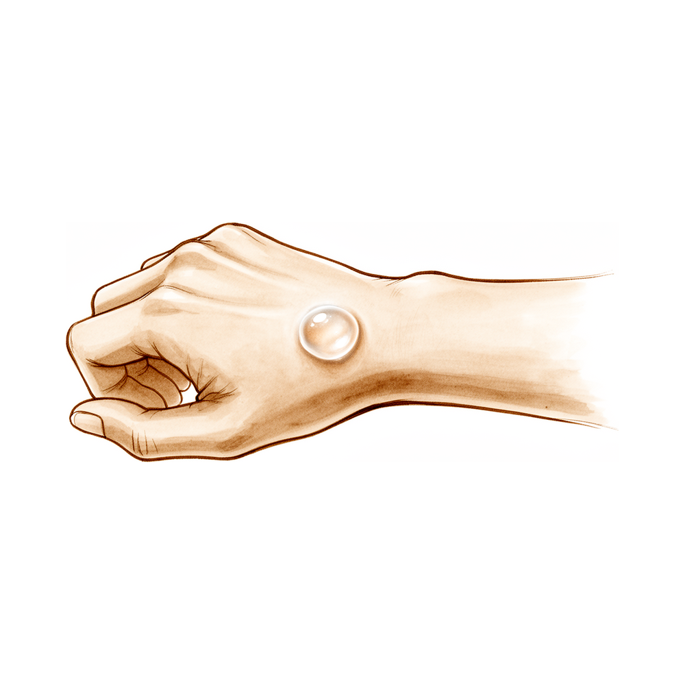
Qué está sintiendo
Un bulto en el dorso de la muñeca suele ser un quiste ganglionar, una hinchazón llena de líquido que sale de una articulación de la muñeca o del revestimiento de un tendón. La mayoría de los bultos del dorso de la muñeca se originan cerca de una de las pequeñas articulaciones de la muñeca. Algunas personas tienen un dolor sordo en la muñeca junto con el bulto. Otras no tienen ningún dolor, solo la hinchazón.
El dolor a menudo empeora después de haber usado mucho la mano, por ejemplo al agarrar herramientas, escribir o llevar la compra. El reposo suele calmarlo. Los quistes ganglionares también pueden aparecer en la palma o a lo largo del tendón de un dedo. Un bulto en el centro de la palma puede no doler en absoluto. Un bulto cerca del tendón de un dedo puede hacer que doblar o estirar ese dedo resulte incómodo, de modo que escribir en el teclado, abrocharse una camisa o sujetar un bolígrafo pueden resultar torpes.
A veces el bulto presiona un nervio cercano. Eso puede causar hormigueo, entumecimiento o debilidad en los dedos, o un dolor que le despierta por la noche. Si el entumecimiento o el dolor en los dedos anular y meñique aparece de forma repentina, debe ser evaluado. Si sus síntomas no se calman, empeoran a lo largo de las semanas, le despiertan por la noche o le impiden trabajar o usar la mano, consulte a su médico de cabecera o solicite una evaluación especializada.
No todos los bultos de la mano son quistes ganglionares. Algunos bultos son puntos de calcio endurecido, y otros se originan en el propio hueso. Un único dedo hinchado también puede estar relacionado con una forma de artritis infantil, y los niños también pueden tener quistes ganglionares en la muñeca. En los niños de 10 años o menos, el 64 % de los quistes ganglionares de la muñeca desaparecen por sí solos, y en los menores de 10 años, entre el 69 % y el 79 % se reducen hasta desaparecer en un plazo de 12 a 18 meses.
Hay una señal de alarma que requiere atención ese mismo día. Si el dedo, la mano o el brazo se ponen calientes, rojos, hinchados y dolorosos, sobre todo si tiene fiebre, acuda al servicio de urgencias ese mismo día. No necesita derivación de su médico de cabecera.
¿Qué está ocurriendo realmente?
Un quiste ganglionar es una pequeña bolsa de líquido espeso y gelatinoso. Crece a partir de una articulación o del revestimiento de un tendón, del mismo modo que un punto débil de la cámara de una bicicleta se abomba a través del neumático. En el dorso de la muñeca, la mayoría de los quistes ganglionares se originan en una pequeña articulación situada en el centro de la muñeca, donde dos de los pequeños huesos de la muñeca se mantienen unidos mediante una correa de ligamento. El líquido de esa articulación sale a través de un punto débil de la correa y se acumula bajo la piel formando un bulto.
Nadie sabe por qué se forma ese punto débil. Los huesos de la muñeca y los ligamentos que lo rodean pueden tener un aspecto completamente normal. Algunos quistes ganglionares parecen estar relacionados con un pequeño desgarro o una holgura de esa correa de ligamento, lo que también podría explicar el dolor sordo que aparece al usar la mano.
El bulto en sí es inofensivo, pero ocupa espacio. Si presiona uno de los nervios que pasan por la muñeca o la palma, aparecen hormigueo, entumecimiento o debilidad en los dedos. A veces, ese dolor en el dorso de la muñeca se debe a que el bulto presiona un pequeño nervio cercano, más que a la propia articulación. Un quiste ganglionar puede incluso presionar un nervio sin que usted note ningún bulto.
Los quistes ganglionares de otras zonas funcionan de la misma manera. Uno en el revestimiento del tendón de un dedo se sitúa justo al lado del cordón de fibras que dobla el dedo, por lo que doblarlo puede resultar incómodo. Uno en el centro de la palma no suele causar ningún problema. Algunos bultos se originan dentro del hueso en lugar de a su lado; allí, el líquido o la sangre que se acumulan en el interior del hueso aumentan la presión, lo que explica un dolor profundo y persistente.
En los niños, estos bultos a menudo simplemente desaparecen por sí solos a medida que crece la muñeca.
Qué podemos hacer al respecto
Muchos quistes ganglionares de la muñeca no necesitan ningún tratamiento. Algunos simplemente se calman por sí solos, y el dolor que empeora después de un uso intenso a menudo se alivia con el reposo y con un cambio en la forma de usar la mano. Si el bulto resulta molesto, una muñequera de soporte o una férula pueden calmar las molestias mientras usted sigue con su día a día.
Si desea que el bulto desaparezca sin una operación, se puede drenar. Con una aguja fina se extrae el líquido gelatinoso del quiste. El drenaje da resultado completo en aproximadamente la mitad de los casos, y hacer hasta tres intentos elimina aproximadamente el 85 % de ellos. Llevar una férula después no cambia el resultado, por lo que no la recomendamos. Las inyecciones de corticoides también pueden calmar la hinchazón y el dolor en algunas de las afecciones que causan bultos en la mano, y los comprimidos o geles antiinflamatorios pueden aliviar el dolor. En algunos bultos óseos, los medicamentos antiinflamatorios a largo plazo pueden ser una alternativa a la cirugía cuando una operación causaría más problemas de los que resuelve.
Si el bulto le sigue causando molestias a pesar de estas medidas, su médico de cabecera o su médico pueden evaluarlo y hablar con usted sobre si un procedimiento le ayudaría. En algunas afecciones, la cirugía extirpa el bulto o el tejido que lo alimenta, lo que impide que vuelva a llenarse. Drenarlo primero, antes de cualquier operación, es un paso razonable en los bultos del dorso de la muñeca.
Qué esperar
Para muchas personas, un quiste ganglionar de la muñeca es un bulto que va y viene, más que un problema duradero. El dolor a menudo empeora después de un uso intenso y se calma con el reposo. Algunos bultos se reducen o desaparecen por sí solos, y otros permanecen igual durante años sin causar muchas molestias. Otros vuelven a llenarse después del drenaje, por lo que a veces el drenaje necesita más de un intento.
Si no se trata el bulto, el pronóstico a menudo no es peor que si se trata. En los bultos de la cara palmar de la muñeca, los síntomas a los 2 y a los 5 años son prácticamente iguales tanto si el bulto se extirpa como si se drena o simplemente se vigila. Eso puede hacer que esperar sea una opción razonable si el bulto no le molesta mucho. El drenaje da resultado completo en aproximadamente la mitad de los casos, como se explicó antes en esta página.
Cuando un bulto sigue causando molestias, el tratamiento suele resolver bien el problema. Las personas a las que se les extirpa un bulto suelen comprobar que el dolor se alivia y que la mano vuelve a funcionar con normalidad. A algunas les queda una pequeña zona de rigidez o una cicatriz que tarda en asentarse. Para los bultos que reaparecen después del tratamiento sigue habiendo opciones, y la mayoría de las personas acaban recuperando un uso cómodo de la mano.
Los bultos relacionados con la artritis de las articulaciones de los dedos tienden a comportarse de forma distinta. El bulto puede ir y venir, pero el desgaste de fondo de la articulación no, por lo que el dolor puede persistir y empeorar a lo largo de los meses. Las férulas, los medicamentos antiinflamatorios y las inyecciones pueden calmar el dolor y, si eso no es suficiente, un procedimiento sobre la propia articulación suele aliviarlo.
Los síntomas nerviosos merecen un poco más de atención. El hormigueo, el entumecimiento o la debilidad causados por un bulto que presiona un nervio a menudo se calman una vez que se alivia la presión, a veces sin ninguna operación. Si esos síntomas no se calman, empeoran a lo largo de las semanas, le despiertan por la noche o le impiden trabajar o usar la mano, consulte a su médico de cabecera o solicite una evaluación especializada.
Cuándo consultar a un especialista
La mayoría de los bultos de este tipo no son urgentes, pero algunas señales requieren atención rápida. Si el dedo, la mano o el brazo se ponen calientes, rojos, hinchados y dolorosos, sobre todo si tiene fiebre, acuda al servicio de urgencias ese mismo día. No necesita derivación de su médico de cabecera. Si no puede contactar con la clínica fuera del horario de atención o durante un fin de semana, acuda al servicio de urgencias más cercano.
Consulte a su médico de cabecera o solicite una evaluación especializada si sus síntomas no se calman, empeoran a lo largo de las semanas, le despiertan por la noche o le impiden trabajar o usar la mano. Pida consulta antes si un bulto presiona un nervio y causa hormigueo, entumecimiento o debilidad en los dedos. El entumecimiento o el dolor en los dedos anular y meñique que aparece de forma repentina también debe ser evaluado. También conviene que se evalúe un bulto que se mueve bajo la piel cuando usted estira el pulgar.
Evidence & references
This is the clinical evidence summary written for health professionals. It is technical, and it lists the research this page was built from. You do not need to read it to understand your treatment or to make a decision about it.
Overview
- The ultimate etiology of a dorsal wrist ganglion remains unknown, and no underlying abnormality in the structural components of the wrist joint has been documented [1].
- Fibrous flexor sheath ganglions are more common in the third decade of life and in the middle finger [4].
- In a series of children 10 years of age and younger with ganglia of the wrist joint, nine of 14 (64%) disappeared spontaneously [13].
- Occult dorsal wrist ganglions had a high prevalence (11 of 14) in a selected patient group with recalcitrant dorsal wrist pain in the area of the scapholunate interval [25].
- Ganglions associated with radial nerve palsy were identified at the elbow in 14 patients [33].
- An intraneural ganglion cyst originating from the radio-carpal joint can cause isolated sensory impairment of the thumb due to median nerve involvement [21].
- Lipomas of the hand are rare, representing only 1 to 3.8% of benign hand tumors [10].
- Lipoma in the deep palmar space of the hand is an unusual tumor that can be unusually large at initial examination and can first be seen with paresthesias in branches of the median nerve [20].
- A tumourous deposition of calcium pyrophosphate dihydrate crystals in the palmar aspect of the wrist has been reported in a 63-year-old man [3].
- Tumoral calcinosis can present with bilateral hand involvement, including local calcinosis cutis of the skin and bone changes [5].
- The radiographic appearance of aneurysmal bone cysts in the hand differs from that in other skeletal locations and must be differentiated from other common lesions [7].
- Pachydermodactyly usually is not associated with any other disease and requires treatment only for cosmetic reasons or when it interferes with the patient's hand function [9].
- Focal myositis involving muscle of the hand has been reported as a first incidence in the literature [50].
- Juvenile aponeurotic fibromata of the hand are predominantly palmar, with only two cases occurring on the dorsum out of forty-three documented in the literature [99].
- Fibro-osseous pseudotumor should be considered as a differential diagnosis for a mass in the hand with a clinical picture of malignancy to avoid unnecessary radical surgery [101].
- The incidence of bone islands in the hands of asymptomatic children between the ages of 5 and 13 years is 3.8% [94].
- The incidence of juxta-articular bone cysts in the hands of asymptomatic children between the ages of 5 and 13 years is 2.8% [94].
- Trigger finger occurs in 2% to 3% of the general population, with women more commonly affected than men [73].
- Trigger finger is more common in patients with systemic diseases such as diabetes mellitus, hypothyroidism, sarcoidosis, rheumatoid arthritis, and septic tenosynovitis [73].
- In cases of dorsal carpometacarpal cyst, associated carpal boss should be systematically screened for to ensure the true cause is treated [51].
Background & Causes
Ganglions and Mucous Cysts
- Digital mucous cysts and wrist ganglia show no anatomic difference at the light and scanning electron microscopic levels, supporting the concept of a similar cause for both [65].
- In children 10 years of age and younger with ganglia of the wrist joint, 9 of 14 (64%) disappeared spontaneously [13].
- Ganglions have been described as arising from most joints of the hand and wrist, including a reported case arising from a metacarpophalangeal joint [16].
- An intratendinous ganglion can present as a mass on the dorsum of the wrist that moves with the extensor pollicis longus tendon [17].
- Ganglions arising from wrist joints can present as painless masses in the center of the palm without signs or symptoms of median or ulnar nerve compression [18].
- In patients with chronic wrist pain, normal radiographic findings, and relief of discomfort upon dorsal capsule injection, dorsal capsulectomy with partial scapho-lunate ligament resection produced pathological findings consistent with incipient ganglion formation [15].
- In a selected patient group with recalcitrant dorsal wrist pain in the area of the scapholunate interval, occult dorsal wrist ganglions had a high prevalence (11 of 14) [25].
- Partial ruptures of the dorsal scapho-lunate ligament will cause dorsal wrist pain [95].
- A midpalm ganglion can cause neuropathy of the isolated motor branch of the median or ulnar nerve [68].
- Ganglion-related compression neuropathy of the palmar cutaneous branch of the median nerve should be a diagnostic consideration even in the absence of a palpable mass [12].
- An intraneural ganglion cyst originating from the radio-carpal joint can cause isolated sensory impairment of the thumb due to median mononeuropathy [21].
- Ganglions associated with radial nerve palsy have been identified at two different locations at the elbow in 14 patients [33].
- Cases strongly suggest a causal relationship between ganglion cysts and carpal tunnel syndrome in some patients [92].
- The potentially serious complication of septic arthritis of the distal interphalangeal joint of the finger secondary to an infected mucous cyst is documented with a report of four cases seen within the past 6 years [97].
- Pseudoaneurysm formation of the radial artery following wrist ganglion surgery has not been previously reported even though the radial artery is often closely related and may be adherent to the wall of anterior wrist ganglia [98].
- The cause of an intraosseous ganglion of the distal ulna is not clear, but perhaps a torn triangular fibrocartilage disk produced a local area of increased stress resulting in the lesion [96].
Lipomas
Calcific and Crystal Deposition
- A tumourous deposition of calcium pyrophosphate dihydrate crystals can occur in the palmar aspect of the wrist [3].
- Tumoral calcinosis can present with bilateral hand involvement, including local calcinosis cutis of the skin and bone changes not previously reported in the literature [5].
- Symmetrical calcium deposits being present in both hands at birth has been reported as a congenital condition [39].
Other Soft Tissue and Bony Lesions
- Soft tissue masses of the hand pose a challenging diagnostic and therapeutic issue [11].
- The radiographic appearance of an aneurysmal bone cyst in the hand differs from that in other skeletal locations and must be differentiated from other common lesions [7].
- Focal myositis involving muscle of the hand has been reported as a first incidence [50].
- Synovial chondrometaplasia of the hand has been described as a case report [45].
- Single-digit swelling may be a presenting feature of juvenile rheumatoid arthritis in 18% of cases [34].
Diagnostic Considerations
- A careful history suggests the correct diagnosis in approximately 90% of patients with hand problems [29].
- Magnetic resonance imaging is a useful preoperative method to identify the cause and location of nerve compression at the wrist when the symptoms and physical findings are inconclusive [23].
- Distinguishing between a flexor sheath ganglion, trigger digit, and Dupuytren's may be accomplished with a detailed physical examination of the MCP region of the affected digit [30].
Symptoms & Presentation
Ganglions
- Fibrous flexor sheath ganglions are more common in the third decade and in the middle finger [4].
- A ganglion-related compression neuropathy of the palmar cutaneous branch of the median nerve should be a diagnostic consideration even in the absence of a palpable mass [12].
- A ganglion arising from a metacarpophalangeal joint has been reported [16].
- An intratendinous ganglion on the dorsum of the wrist may present as a mass that moves with the extensor pollicis longus tendon, posing a differential diagnostic challenge [17].
- At 2 and 5 year follow-up, no difference in symptoms was found for palmar wrist ganglions regardless of whether they were excised, aspirated, or left alone [19].
- A dorsal wrist ganglion can present as an anterior wrist ganglion [22].
- Ultrasonography revealed a small hypoechogenic area on the dorsal aspect between the scaphoid and the lunate or just dorsal to the lunate in all five cases of occult dorsal wrist ganglion examined [79, 80].
Lipomas
- A lipoma in the deep palmar space of the hand is an unusual tumor that can be unusually large at initial examination and can first be seen with paresthesias in branches of the median nerve [20].
Calcific and Crystal Deposition
- Tumoral calcinosis can present with an unusual distribution of lesions restricted to multiple digits [43].
- Erosive tumoral calcinosis localized at the P.I.P. joint of a ring finger can completely erode the distal epiphysis of the middle phalanx [76].
- Symmetrical calcium deposits in both hands at birth have been reported as a case of congenital bilateral calcinosis cutis [39].
Bone Cysts and Tumors
- An aneurysmal bone cyst of the hamate bone can present with grip strength of 45 kg in the affected right hand compared with 47 kg in the left hand [14].
- An aneurysmal bone cyst of the trapezium has been reported as a new location for this lesion [26, 27].
- An aneurysmal bone cyst of the distal phalanx has been reported [36].
- Soft tissue masses of the hand pose a challenging diagnostic and therapeutic issue, as illustrated by a case of fibro-osseous pseudotumor [11].
Neuropathies and Nerve Compression
- Ulnar neuropathy at the wrist can be associated with a recurrent branch through the flexor carpi ulnaris tendon [2].
- The acute onset of numbness with or without intolerable pain in the ring and little fingers after a long-term remission period following initial surgery for cubital tunnel syndrome in patients with elbow osteoarthritis appears to be the characteristic clinical profile of recurrent cubital tunnel syndrome caused by ganglion [42].
Other Soft Tissue Lesions
- A subungual dermatofibroma of the thumb has been reported [40].
Management
General Assessment and History
- The natural inclination to study radiographs or special imaging studies prior to a thorough history and physical examination should be avoided, as this introduces cognitive bias which can affect thinking and decision making [63].
- Physical examination always needs to be preceded by a thorough investigation of the patient’s medical history, with special emphasis on the mechanism of injury and acuity [63].
- Palpation for areas of maximal tenderness is one of the most useful tools in the diagnosis of wrist pathology, especially in patients with chronic dysfunctions [63].
- A careful assessment of neural and vascular status is imperative, with particular attention being paid to the median and ulnar nerves, which may be injured by direct contusion, compression from displaced bones, or swelling within the carpal canal [63].
- Bilateral grip and pinch strength are useful to uncover underlying pathology in chronic cases [63].
- Sensory testing should always accompany an examination of suspected nerve compression, using threshold or density testing [63].
Ganglions
- Even in the absence of a palpable mass, a ganglion-related compression neuropathy of the palmar cutaneous branch of the median nerve should be a diagnostic consideration [12].
- Magnetic resonance imaging is a useful preoperative method to identify the cause and location of nerve compression at the wrist when symptoms and physical findings are inconclusive [23].
- Dorsal wrist ganglions can be confused with carpal bosses, which are osteoarthritic spurs or prominences that develop at the base of the second or third carpometacarpal joints [62].
- A small ganglion is associated with a carpal boss in 30% of cases [62].
- Every effort should be made to treat the carpal boss nonoperatively using splinting, nonsteroidal antiinflammatory medications, and ultrasound-guided cortisone injections prior to proceeding with surgery [62].
- The potential for persistent symptoms following surgery for a carpal boss must be emphasized [62].
- In children aged <10 years, ganglions mainly occur on the volar wrist and can be treated expectantly, with 69% to 79% displaying spontaneous regression within a span of 12-18 months [48].
- The outcome of immobilization following aspiration of carpal and digital ganglions was successful in 52% of the wrists and digits that were immobilized and in 50% of those that were not [8].
- Aspiration treatment of ganglion cysts of the wrist and hand can safely remove 85% of these tumors if one, two or three separate treatments are administered [82].
- Patients with carpal ganglions who wish to avoid or delay operative treatment should be advised that aspiration-puncture followed by 3 weeks of immobilization will be successful in approximately 50% of cases [83].
- Performing at least 1 aspiration before surgical excision improves the cost-effectiveness of dorsal wrist ganglions treatment [35].
- The practice of treating wrist ganglions with a sclerosant must be abandoned due to the risk of catastrophic complications such as radial artery injury [31].
- Improved functional activity and decreased pain were noted in all patients following dorsal carpal ganglion excision [46].
- Worse hand function following dorsal wrist ganglion excision was associated with recurrence following prior surgery, worse baseline hand function and lower treatment credibility [47].
- Full recovery of wrist function occurred in all cases of recurrent ganglion treated with a fascial flap except one who had slight restriction of flexion [24].
- At a follow-up examination 4 years after operation for a dorsal wrist ganglion presenting as an anterior wrist ganglion, the ganglion had not recurred, and motion and strength were equal to the contralateral wrist and hand [22].
- Manipulation of the wrist for the management of postoperative stiffness is rarely, if ever, indicated [41].
- To assure relief of symptoms for a dorsal ganglion with anomalous muscles, treatment must include division of the fourth dorsal extensor compartment in the case of the former muscle, and excision in the case of the latter [77].
Bone Lesions
- For stable aneurysmal bone cysts in the carpus, where excision could lead to carpal instability, conservative management is recommended [72].
- In the 11th month after surgery for an aneurysmal bone cyst of the trapezium, the patient had a full range of wrist motion and was free of pain [26].
- Grip strength was 45 kg in the affected right hand compared with 47 kg in the left hand following treatment for an aneurysmal bone cyst of the hamate bone [14].
- Osteoid osteoma is a benign bone lesion that has been well described in the hand and distal end of the radius [66].
- Five percent to 15% of osteoid osteomas occur in the hand and wrist, most commonly in the neck of the proximal phalanx and carpus [66].
- Patients with osteoid osteoma are typically seen initially in the late second or early third decade of life with a deep, dull ache that is constant and frequently relieved with nonsteroidal antiinflammatory drugs (NSAIDs) [66].
- Diagnosis of hand lesions for osteoid osteoma is frequently delayed, with a mean time to diagnosis of longer than 15 months in two large case series [66].
- Treatment of osteoid osteoma in the hand or carpus has consisted of surgical excision with either curettage of the nidus or en bloc excision [66].
- Persistence of the lesion has been reported if the nidus is not completely excised [66].
- Successful use of percutaneous CT-guided laser photoagulation has been reported for osteoid osteoma [66].
- Radiofrequency ablation may be considered for carefully selected osteoid osteoma lesions that are not subcutaneous or near major nerves [66].
- Prolonged use of NSAIDs may be an alternative to surgical treatment if the clinical and radiographic findings are strongly supportive of a diagnosis of osteoid osteoma and excision or biopsy of the lesion might produce excessive morbidity [66].
- Wrist scores improved 34 points, from 51 to 85 points, by 1 year after arthroscopically assisted treatment of intraosseous ganglions of the lunate with trabeculation being noted within the grafting lunate [84].
- The patient achieved complete relief from pain and returned to normal employment following treatment for an intraosseous ganglion of the trapezoid [81].
Soft Tissue Masses and Inflammatory Conditions
- Tendon xanthoma is a physical manifestation of hyperlipidemia that may respond favorably to dietary modification and appropriate pharmacotherapy [87].
- In the three reported cases of acute calcific tendinitis in the hand and wrist, symptoms resolved in an average of seven or eight days with the use of Aspirin 300 mg q.i.d. and a resting splint [75].
- The patient had tumoral calcinosis with an unusual distribution of lesions restricted to multiple digits [43].
- The patient had bilateral hand involvement, including local calcinosis cutis of the skin and bone changes not previously reported in the literature, in a case of tumoral calcinosis [5].
- A 63-year-old man had a tumourous deposition of calcium pyrophosphate dihydrate crystals in the palmar aspect of the wrist [3].
- Local amputation provided complete relief of symptoms and there has been no recurrence in two years for a pseudo-malignant osseous tumour of soft tissue [89].
Rheumatoid Arthritis and Tenosynovitis
- The advent of disease-modifying antirheumatic drugs (DMARDs) has led to clinical control of joint disease in systemic rheumatoid arthritis [53].
- Dorsal extensor tenosynovitis most commonly affects the fourth dorsal compartment tenosynovium [53].
- The enlarged tenosynovium in rheumatoid arthritis feels irregular and diffuse, which differentiates the tenosynovitis from the well-defined ganglia or other wrist masses [53].
- Conservative management in rheumatoid tenosynovitis of the hand and wrist requires rheumatologic support for systemic pharmacologic management [53].
- Splinting has an adjunctive role to relieve pain in rheumatoid tenosynovitis [53].
- Targeted injection into the tenosynovial inflammation is more effective than the administration of systemic corticosteroids for isolated tenosynovitis [53].
- Surgical treatment for tenosynovitis is to remove the inflamed tenosynovium while preserving normal structures [53].
- Tenosynovectomy has been shown to decrease pain and improve disease outcomes [53].
- Painful dorsal wrist swelling may be the presenting symptom in rheumatoid arthritis [55].
- The tenosynovial swelling may contribute to de Quervain disease, trigger finger, or carpal tunnel syndrome, whereas rheumatoid arthritis as the underlying cause may not be suspected [55].
- If the synovitis is only moderate, and if changes in the bones are absent, but pain is significant, dorsal synovectomy of the wrist may be of lasting benefit [55].
- Persistent swelling at the dorsum of the wrist that continues for 6 weeks or longer despite adequate medical treatment may be an indication for a dorsal synovectomy [55].
- Dorsal synovectomy may be considered a prophylactic measure to avoid extensor tendon rupture [55].
- Any tendons ruptured at the wrist level can be repaired or reconstructed at the time of synovectomy [55].
- If synovitis involves the wrist and the metacarpophalangeal joints, synovectomy often can be done at both levels during the same operation, usually only on one limb at a time [55].
- Hypertrophy of the volar wrist synovium even though undetectable clinically can cause median nerve compression and symptoms of carpal tunnel syndrome [55].
- Compression of the nerve in rheumatoid arthritis should be relieved surgically if conservative treatment with splinting and corticosteroid injections has been unsuccessful [55].
- If hypertrophy of the tenosynovium on the volar aspect of the wrist is obvious clinically with or without symptoms of compression of the median nerve, a palmar (flexor) tenosynovectomy may be useful in relieving pain and in preventing rupture of tendons [55].
- The indications for surgical treatment of the rheumatoid wrist are based on pain, functional disability, deformation including its esthetic aspect, and possible future development of the disease [56].
- The two main concerns associated with the rheumatoid wrist are tendon ruptures and irreversible joint damage [56].
- Key risk factors in determining the possible occurrence of a relevant complication of the inflammation process include long-standing disease (>5 years), persistent synovitis (>1 year), and a LDE stage 4 or higher wrist [56].
- If the carpus is subluxated with an ensuing prominent ulna, and this is associated with local synovitis of the ulnar tendons and/or distal radioulnar joint, surgical intervention should be considered [56].
- Three dimensional-CT scans can help improve the ability to predict the risk of extensor tendon rupture around the ulnar side of the wrist [56].
- The classic interventions used to stabilize rheumatoid wrists are partial wrist fusions [56].
- When relevant risk factors for future deterioration are present, the indication for wrist stabilization should be given precedence even for oligosymptomatic patients [56].
- Prophylactic surgery may need to be considered even for patients with minor pain and disturbances to prevent ongoing damage to joints and tendons [56].
Malignant Tumors
- Malignancies of the proximal palmar surface of the hand and volar aspect of the wrist often require amputation [67].
- A dissection that attempts to “salvage” the median nerve or one or two flexors in the middle of an expanding tumor or reactive zone is only likely to spread the disease [67].
- Tumors that arise on the dorsum may allow preservation of the hand if staging studies show that the lesion has not penetrated into the palm and the excision margin verifies a safe plane of normal tissue [67].
- Isolated intraosseous carpal lesions that have not invaded soft tissue are rare but can be excised locally [67].
- The carpal bones are intraarticular, so the onset of symptoms is generally associated with synovitis and joint invasion; therefore below-elbow amputation or complete en bloc excision of the entire radiocarpal articulation and carpus is likely to be necessary [67].
- Growths on the volar aspect of the distal part of the forearm must be widely excised with negative margins [67].
- Most lesions on the volar aspect of the distal forearm can be treated with wide excision [67].
- At times below-elbow amputation is required for tumors on the volar aspect of the distal forearm [67].
- If a tumor arises in a location that does not specifically involve the ulnar nerve and artery, it may be possible to save a portion of the hand and wrist along with the neurovascular bundle and to consider later reconstruction after longitudinal hemiamputation [67].
- Tumors on the extreme ulnar side of the wrist can be handled in a way that mirrors those on the radial aspect, although lesions in the extreme end of the ulna may be amenable to wide excision of the distal ulna [67].
- Intracompartmental lesions within the distal radius or ulna can be treated by wide excision of the bone and arthrodesis or autograft replacement [67].
- When tumors have invaded tissues or crossed compartments extensively, above- or below-elbow amputation may be required [67].
- Wherever a tumor is located, treatment must be individualized to achieve the goal of functional restoration without risking local recurrence and later distant spread [67].
Nerve Compression and Neuropathy
- Postoperatively the patient's symptoms were relieved and after 3 months he had normal and full use of his hand and wrist following treatment for ulnar neuropathy at the wrist associated with a recurrent branch through the flexor carpi ulnaris tendon [2].
- At 11 months, a full range of extension had been regained in the fingers with the wrist in neutral position, though strength of extension was still not quite full and thumb elevation to the plane of the palm lacked 15°, following treatment for posterior interosseous nerve axonotmesis from compression by a ganglion [85].
- Because of the nature of the pathology, carpal tunnel syndrome caused by an occult ganglion would not respond to conservative treatment [88].
Key Considerations
Ganglions
- In children aged 10 years or younger with ganglia of the wrist joint, 9 of 14 (64%) disappeared spontaneously [13].
- A ganglion arising from a metacarpophalangeal joint has been reported as the first such case [16].
- An intratendinous ganglion on the dorsum of the wrist that moves with the extensor pollicis longus tendon presents a clinical problem regarding differential diagnosis [17].
- Four years after operation for a dorsal wrist ganglion presenting as an anterior wrist ganglion, the ganglion had not recurred and motion and strength were equal to the contralateral wrist and hand [22].
- Full recovery of wrist function occurred in all cases treated with a fascial flap for recurrent ganglion except one who had slight restriction of flexion [24].
- Ultrasound of the wrist can be used as a first-line imaging procedure in clinically inconclusive situations, and ultrasound evidence of an occult dorsal ganglion is a reliable indicator for surgery [70].
- Performing at least 1 aspiration before surgical excision improves the cost-effectiveness of dorsal wrist ganglions treatment, as patient preferences may preclude routinely performing 2 aspirations [35].
- Recurrent cubital tunnel syndrome caused by ganglion is characterized by acute onset of numbness with or without intolerable pain in the ring and little fingers after a long-term remission period following initial surgery in patients with elbow osteoarthritis [42].
- Long-term results after treatment of mucous cysts with simple aspiration, decapping, excision with skin grafts and flaps, or observation only were similar [91].
Other Soft Tissue Masses
- The fundamental consideration in the management of hand tumours is to reconcile adequate tumour clearance with the maintenance of function and appearance [69].
- Recurrence of symptoms for glomus tumours occurred in only two cases after a pain-free interval of 2 years [90].
- Marginal excision appears to result in a good outcome with no recurrence at short- to medium-term follow-up for parosteal lipomas of the phalanges [86].
- Fifteen months after operation for a subungual dermatofibroma of the thumb, the nail has returned to an almost normal shape and appearance with no signs of recurrence, although the nail plate shows longitudinal furrows [40].
Bony Lesions
- Aneurysmal bone cysts can involve the trapezium [26, 27].
- An aneurysmal bone cyst of the hamate bone resulted in a grip strength of 45 kg in the affected right hand compared with 47 kg in the left hand [14].
- Four years after operation for an aneurysmal bone cyst in the metacarpal of a child, the fibular graft was well taken up and the child had good function of the hand with full range of movements in the metacarpo-phalangeal and proximal interphalangeal joints [93].
- At the 4-year follow-up for an aneurysmal bone cyst of the distal phalanx, the patient exhibited a full range of motion, no pain, and no signs of recurrence [36].
- Tumoral calcinosis can involve the hands bilaterally, including local calcinosis cutis of the skin and bone changes not previously reported in the literature [5].
Surgical Approaches and Outcomes
- Although the dorsal approach to the wrist disturbs the extensor pollicis longus tendon and exposes the extensor carpi radialis brevis tendon, patients have not had problems with thumb extension [6].
- Postoperatively, symptoms of ulnar neuropathy at the wrist associated with a recurrent branch through the flexor carpi ulnaris tendon were relieved, and after 3 months the patient had normal and full use of his hand and wrist [2].
Key Evidence
- [L5] The ultimate etiology of a dorsal wrist ganglion remains unknown, and the authors have been unable to document any underlying abnormality in the structural components of the wrist joint. [1] (10.1097/00130911-200209000-00008)
- [L5] Postoperatively the patient's symptoms were relieved and after 3 months he had normal and full use of his hand and wrist. [2] (10.1016/s0266-7681(05)80199-8)
- [L5] A 63-year-old man had a tumourous deposition of calcium pyrophosphate dihydrate crystals in the palmar aspect of the wrist. [3] (10.1016/0266-7681(91)90183-o)
- [L4] They were found to be more common in the third decade and in the middle finger. [4] (10.1016/s0266-7681(97)80068-x)
- [L5] The patient had bilateral hand involvement, including local calcinosis cutis of the skin and bone changes not previously reported in the literature. [5] (10.1016/s0363-5023(85)80225-2)
- [L5] Although the approach disturbs the extensor pollicis longus tendon and exposes the extensor carpi radialis brevis tendon, the authors' patients have not had problems with thumb extension. [6] (10.1016/s0363-5023(86)80251-9)
- [L4] The radiographic appearance in the hand differs from that in other skeletal locations and must be differentiated from other common lesions. [7] (10.1016/s0363-5023(79)80132-x)
- [L1] The outcome was successful in 52% of the wrists and digits that were immobilized and in 50% of those that were not. [8] (10.1016/s0363-5023(09)91073-5)
- [L5] When present, it usually is not associated with any other disease and requires treatment only for cosmetic reasons or when it interferes with the patient's hand function. [9] (10.1016/s0363-5023(84)80033-7)
- [L4] Lipomas of the hand are rare, representing only 1 to 3.8% of benign hand tumors. [10] (10.1016/j.hansur.2017.10.135)
- [L5] Soft tissue masses of the hand pose a challenging diagnostic and therapeutic issue. [11] (10.1142/s2424835516720127)
- [L4] These two cases suggest that even in the absence of a palpable mass, a ganglion-related compression neuropathy of the palmar cutaneous branch of the median nerve should be a diagnostic consideration. [12] (10.1016/0363-5023(94)90195-3)
- [L4] In a small series of children 10 years of age and younger with ganglia of the wrist joint, nine of 14 (64%) disappeared spontaneously. [13] (10.1016/s0363-5023(77)80137-8)
- [L5] Grip strength was 45 kg in the affected right hand compared with 47 kg in the left hand. [14] (10.1016/s0363-5023(84)80064-7)
- [L4] In patients with chronic wrist pain, normal radiographic findings, and relief of discomfort upon dorsal capsule injection, dorsal capsulectomy with partial scapho-lunate ligament resection produced pathological findings consistent with incipient ganglion formation. [15] (10.1016/0266-7681(85)90034-8)
- [L5] Although ganglia have been described as arising from most joints of the hand and wrist, to our knowledge this is the first report of a ganglion arising from a metacarpophalangeal joint. [16] (10.1016/s0266-7681(98)80076-4)
- [L5] The clinical problem was the inability to make a differential diagnosis of a mass on the dorsum of the wrist that moved with the extensor pollicis longus tendon. [17] (10.1016/s0363-5023(85)80218-5)
- [L4] We report 2 cases of ganglions arising from wrist joints that presented as painless masses in the center of the palm without signs or symptoms of median or ulnar nerve compression. [18] (10.1016/j.jhsa.2003.11.006)
- [L3] At 2 and 5 year follow-up, regardless of treatment, no difference in symptoms was found, regardless of whether the palmar wrist ganglion was excised, aspirated or left alone. [19] (10.1016/s0266-7681(02)00365-0)
- [L4] Lipoma in the deep palmar space of the hand is an unusual tumor that can be unusually large at initial examination and can first be seen with paresthesias in branches of the median nerve. [20] (10.1016/0363-5023(89)90193-7)
- [L5] The case illustrates an unusual aetiology of thumb involvement in a median mononeuropathy caused by an intraneural ganglion cyst originating from the radio-carpal joint. [21] (10.1016/s0266-7681(05)80156-1)
- [L5] At a follow-up examination 4 years after operation, the ganglion had not recurred, and motion and strength were equal to the contralateral wrist and hand. [22] (10.1016/0363-5023(94)90096-5)
- [L5] Magnetic resonance imaging is a useful preoperative method to identify the cause and location of nerve compression at the wrist when the symptoms and physical findings are inconclusive. [23] (10.1016/0363-5023(89)90182-2)
- [L4] Full recovery of wrist function occurred in all cases except one who had slight restriction of flexion. [24] (10.1016/j.main.2003.12.007)
- [L4] In our selected patient group (recalcitrant dorsal wrist pain in the area of the scapholunate interval), occult dorsal wrist ganglions had a high prevalence (11 of 14). [25] (10.1016/s0363-5023(05)80288-6)
- [L5] In the 11th month after surgery, he had a full range of wrist motion and was free of pain. [26] (10.1016/j.jhsb.2006.02.011)
- [L5] In the 11th month after surgery, he had a full range of wrist motion and was free of pain. [27] (10.1016/j.jhsb.2005.12.007)
- [L4] Distinguishing between a flexor sheath ganglion, trigger digit, and Dupuytren's may be accomplished with a detailed physical examination of the MCP region of the affected digit. [30] (10.1177/15589447221109644)
- [L4] After this catastrophic complication of the treatment of a benign condition, the practice of treating wrist ganglions with a sclerosant must be abandoned. [31] (10.1177/1753193409105561)
- [L4] Ganglions associated with radial nerve palsy at two different locations were identified at the elbow in 14 patients. [33] (10.1016/j.jhsb.2006.09.014)
- [L4] Single-digit swelling may be a presenting feature of JRA in 18% of cases. [34] (10.1016/s0363-5023(97)80124-4)
- [L2] As patient preferences may preclude routinely performing 2 aspirations, performing at least 1 aspiration before surgical excision improves the cost-effectiveness of dorsal wrist ganglions treatment. [35] (10.1016/j.jhsa.2022.09.002)
- [L5] At the 4-year follow-up, the patient exhibited a full range of motion, no pain, and no signs of recurrence. [36] (10.1016/j.jhsg.2026.101052)
- [L5] We present the first reported case of symmetrical calcium deposits being present in both hands at birth. [39] (10.1016/j.jhsb.2006.04.023)
- [L5] Fifteen months after operation, the thumb nail has returned to an almost normal shape and appearance with no signs of recurrence, although the nail plate shows longitudinal furrows. [40] (10.1016/s0266-7681(05)80218-9)
- [L5] Manipulation of the wrist for the management of postoperative stiffness is rarely, if ever, indicated. [41] (10.1016/s0363-5023(83)80096-3)
- [L4] The acute onset of numbness with or without intolerable pain in the ring and little fingers after a long-term remission period following initial surgery for CuTS in patients with elbow osteoarthritis appears to be the characteristic clinical profile of recurrent CuTS caused by ganglion. [42] (10.1142/s2424835518500224)
- [L5] The patient has tumoral calcinosis with an unusual distribution of lesions restricted to multiple digits. [43] (10.1016/s0363-5023(85)80223-9)
- [L5] A case of synovial chondrometaplasia of the hand is described. [45] (10.1016/s0363-5023(84)80158-6)
- [L4] Improved functional activity and decreased pain were noted in all patients. [46] (10.1016/s0266-7681(99)90053-0)
- [L2] Worse hand function was associated with recurrence following prior surgery, worse baseline hand function and lower treatment credibility. [47] (10.1177/17531934231153029)
- [L4] In children aged <10 years, ganglions mainly occur on the volar wrist and can be treated expectantly, with 69% to 79% displaying spontaneous regression within a span of 12-18 months. [48] (10.1016/j.jhsa.2021.12.015)
- [L5] This case represents the first reported incidence of focal myositis that involves muscle of the hand. [50] (10.1016/0363-5023(88)90218-3)
- [L4] In cases of dorsal carpometacarpal cyst, associated carpal boss should be systematically screened for to ensure the true cause is treated. [51] (10.1016/j.hansur.2016.12.004)
- [L5] Digital mucous cysts and wrist ganglia show no anatomic difference at the light and scanning electron microscopic levels, supporting the concept of a similar cause for both. [65] (10.1016/s0363-5023(88)80143-6)
- [L4] A midpalm ganglion can cause neuropathy of the isolated motor branch of the median or ulnar nerve. [68] (10.1053/jhsu.2001.24146)
- [L5] The fundamental consideration in the management of hand tumours is to reconcile adequate tumour clearance with the maintenance of function and appearance. [69] (10.1016/0266-7681(91)90159-l)
- [L3] It was concluded that ultrasound of the wrist can be used as a first-line imaging procedure in clinically inconclusive situations and that ultrasound evidence of an occult dorsal ganglion is a reliable indicator for surgery. [70] (10.1016/s0363-5023(97)80044-5)
- [L5] For stable lesions in the carpus, where excision could lead to carpal instability, conservative management is recommended. [72] (10.1016/s0266-7681(05)80005-1)
- [L4] In the three reported cases, symptoms resolved in an average of seven or eight days with the use of Aspirin 300 mg q.i.d. and a resting splint. [75] (10.1016/0266-7681(91)90181-m)
- [L5] The authors report an exceptional case of erosive tumoral calcinosis localized at the P.I.P. joint of a ring finger, which had completely eroded the distal epiphysis of the middle phalanx. [76] (10.1016/j.main.2004.09.001)
- [L5] To assure relief of symptoms, treatment must include division of the fourth dorsal extensor compartment in the case of the former muscle, and excision in the case of the latter. [77] (10.1016/s0363-5023(79)80111-2)
- [L4] Ultrasonography revealed a small hypoechogenic area on the dorsal aspect between the scaphoid and the lunate or just dorsal to the lunate in all five cases examined. [79] (10.1016/0266-7681_88_90133-7)
- [L4] Ultrasonography revealed a small hypoechogenic area on the dorsal aspect between the scaphoid and the lunate or just dorsal to the lunate in all five cases examined. [80] (10.1016/0266-7681(88)90133-7)
- [L5] The patient achieved complete relief from his pain and returned to his normal employment. [81] (10.1016/0363-5023(94)90266-6)
- [L2] Aspiration treatment of ganglion cysts of the wrist and hand can safely remove 85% of these tumors if one, two or three separate treatments are administered. [82] (10.1016/s0363-5023(87)80221-6)
- [L2] Patients with carpal ganglions who wish to avoid or delay operative treatment should be advised that aspiration-puncture followed by 3 weeks of immobilization will be successful in approximately 50% of cases. [83] (10.1016/s0363-5023(87)80108-9)
- [L4] Wrist scores improved 34 points, from 51 to 85 points, by 1 year after surgery with trabeculation being noted within the grafting lunate. [84] (10.1053/jhsu.2003.50027)
- [L5] At 11 months, a full range of extension had been regained in the fingers with the wrist in neutral position, though strength of extension was still not quite full and thumb elevation to the plane of the palm lacked 15°. [85] (10.1016/0266-7681(90)90100-i)
- [L4] Marginal excision appears to result in a good outcome with no recurrence at short- to medium-term follow-up for parosteal lipomas of the phalanges. [86] (10.1016/j.jhsa.2020.10.029)
- [L4] The importance of this lesion to the hand surgeon is to recognize it as a physical manifestation of a potentially life-threatening disorder (hyperlipidemia) that may respond favorably to dietary modification and appropriate pharmacotherapy. [87] (10.1016/s0363-5023(88)80057-1)
- [L5] Because of the nature of the pathology the condition would not respond to conservative treatment. [88] (10.1016/0266-7681(93)90099-2)
- [L5] Local amputation provided complete relief of symptoms and there has been no recurrence in two years. [89] (10.1016/0266-7681(84)90061-5)
- [L4] Recurrence of symptoms occurred in only two cases after a pain-free interval of 2 years. [90] (10.1016/s0266-7681(96)80110-0)
- [L4] The long-term results after treatment with simple aspiration or decapping, or by excision with skin grafts and flaps as needed or with observation only were similar. [91] (10.1016/s0363-5023(84)80076-3)
- [L4] These cases strongly suggest a causal relationship between ganglion cysts and carpal tunnel syndrome in some patients. [92] (10.1016/s0363-5023(88)80144-8)
- [L5] Four years after operation, the fibular graft is well taken up and the child has a good function of the hand with full range of movements in the metacarpo-phalangeal and proximal interphalangeal joints. [93] (10.1016/0266-7681(88)90186-6)
- [L3] The incidence of bone islands in the hands of asymptomatic children between the ages of 5 and 13 years is 3.8% and the incidence of cysts in the same population is 2.8%. [94] (10.1016/j.jhsb.2006.03.158)
- [L4] Partial ruptures of the dorsal scapho-lunate ligament will cause dorsal wrist pain. [95] (10.1016/s0363-5023(03)80341-6)
- [L5] The cause of the patient's pain is not clear, but perhaps the torn triangular fibrocartilage disk produced a local area of increased stress resulting in the intraosseous ganglion of the distal ulna. [96] (10.1016/s0363-5023(87)80124-7)
- [L4] The potentially serious complication of septic arthritis of the distal interphalangeal joint of the finger secondary to an infected mucous cyst is documented with a report of four cases seen within the past 6 years. [97] (10.1016/s0363-5023(84)80152-5)
- [L5] Pseudoaneurysm formation of the radial artery following wrist ganglion surgery has not been previously reported even though the radial artery is often closely related and may be adherent to the wall of anterior wrist ganglia. [98] (10.1016/s0266-7681(96)80190-2)
- [L5] Out of forty-three juvenile aponeurotic fibromata of the hand documented in the literature, only two cases occurred on the dorsum. [99] (10.1016/s0266-7681(85)80040-1)
- [L5] For a mass in the hand with a clinical picture of malignancy, hand surgeons should consider fibro-osseous pseudotumor as a differential diagnosis to avoid unnecessary radical surgery. [101] (10.1016/s0363-5023(96)80036-0)
References
[1] Internal Decompression of Dorsal Wrist Ganglions. Techniques in Hand and Upper Extremity Surgery. 2002. DOI: 10.1097/00130911-200209000-00008
[2] Ulnar Neuropathy at the Wrist Associated with a Recurrent Branch through the Flexor Carpi Ulnaris Tendon. Journal of Hand Surgery. 1996. DOI: 10.1016/s0266-7681(05)80199-8
[3] Tumourous Deposition of Calcium Pyrophosphate Dihydrate Crystals in the Wrist. Journal of Hand Surgery. 1991. DOI: 10.1016/0266-7681(91)90183-o
[4] The Distribution of Fibrous Flexor Sheath Ganglions. Journal of Hand Surgery. 1997. DOI: 10.1016/s0266-7681(97)80068-x
[5] Tumoral calcinosis: A case report and review of the literature. The Journal of Hand Surgery. 1985. DOI: 10.1016/s0363-5023(85)80225-2
[6] The dorsal approach to the wrist revisited. The Journal of Hand Surgery. 1986. DOI: 10.1016/s0363-5023(86)80251-9
[7] Aneurysmal bone cyst involving the hand: A review and report of two cases. The Journal of Hand Surgery. 1979. DOI: 10.1016/s0363-5023(79)80132-x
[8] Efficacy of immobilization following aspiration of carpal and digital ganglions. The Journal of Hand Surgery. 1992. DOI: 10.1016/s0363-5023(09)91073-5
[9] Pachydermodactyly: A case report and discussion of the pathologic entity. The Journal of Hand Surgery. 1984. DOI: 10.1016/s0363-5023(84)80033-7
[10] Les lipomes de la main, à propos de 4 cas et revue de la littérature. Hand Surgery and Rehabilitation. 2017. DOI: 10.1016/j.hansur.2017.10.135
[11] Fibro-Osseous Pseudotumor of the Hand. The Journal of Hand Surgery (Asian-Pacific Volume). 2016. DOI: 10.1142/s2424835516720127
[12] Ganglion-related compression neuropathy of the palmar cutaneous branch of the median nerve: A report of two cases. The Journal of Hand Surgery. 1994. DOI: 10.1016/0363-5023(94)90195-3
[13] Dorsal wrist ganglions in children. The Journal of Hand Surgery. 1977. DOI: 10.1016/s0363-5023(77)80137-8
[14] Aneurysmal bone cyst of the hamate bone. The Journal of Hand Surgery. 1984. DOI: 10.1016/s0363-5023(84)80064-7
[15] The occult dorsal carpal ganglion. The Journal of Hand Surgery: Journal of the British Society for Surgery of the Hand. 1985. DOI: 10.1016/0266-7681(85)90034-8
[16] A Palmar Ganglion Arising from the Metacarpophalangeal Joint. Journal of Hand Surgery. 1998. DOI: 10.1016/s0266-7681(98)80076-4
[17] A case of an intratendinous ganglion. The Journal of Hand Surgery. 1985. DOI: 10.1016/s0363-5023(85)80218-5
[18] Wrist joint ganglion presenting as a painless mass in the palm: report of 2 cases. The Journal of Hand Surgery. 2004. DOI: 10.1016/j.jhsa.2003.11.006
[19] Palmar Wrist Ganglion: Does Intervention Improve Outcome A Prospective Study of the Natural History and Patient-Reported Treatment Outcomes. Journal of Hand Surgery. 2003. DOI: 10.1016/s0266-7681(02)00365-0
[20] Large lipomas in the deep palmar space. The Journal of Hand Surgery. 1989. DOI: 10.1016/0363-5023(89)90193-7
[21] Isolated Sensory Impairment of the Thumb Due to an Intraneural Ganglion Cyst in the Median Nerve. Journal of Hand Surgery. 1995. DOI: 10.1016/s0266-7681(05)80156-1
[22] Dorsal wrist ganglion presenting as anterior wrist ganglion. The Journal of Hand Surgery. 1994. DOI: 10.1016/0363-5023(94)90096-5
[23] Diagnosis of ganglion in Guyon's canal by magnetic resonance imaging. The Journal of Hand Surgery. 1989. DOI: 10.1016/0363-5023(89)90182-2
[24] Fascial flap for recurrent ganglion. Chirurgie de la Main. 2004. DOI: 10.1016/j.main.2003.12.007
[25] Evaluating dorsal wrist pain: MRI diagnosis of occult dorsal wrist ganglion. The Journal of Hand Surgery. 1995. DOI: 10.1016/s0363-5023(05)80288-6
[26] The Trapezium: a New Location for an Aneurysmal Bone Cyst. Journal of Hand Surgery. 2006. DOI: 10.1016/j.jhsb.2006.02.011
[27] The Trapezium: a New Location for an Aneurysmal Bone Cyst. Journal of Hand Surgery. 2006. DOI: 10.1016/j.jhsb.2005.12.007
[29] A Lange Medical Book Current Diagnosis Treatment In Orthopedics Fifth Edition. 9Hand Surgery > DIAGNOSIS OF DISORDERS OF THE HAND.
[30] A Simple Physical Exam Maneuver to Distinguish Trigger Digit, Dupuytren’s Nodule, and Flexor Sheath Ganglion. HAND. 2022. DOI: 10.1177/15589447221109644
[31] Radial artery injury caused by a sclerosant injected into a palmar wrist ganglion. Journal of Hand Surgery (European Volume). 2009. DOI: 10.1177/1753193409105561
[33] THE TWO LOCATIONS OF GANGLIONS CAUSING RADIAL NERVE PALSY. Journal of Hand Surgery (European Volume). 2007. DOI: 10.1016/j.jhsb.2006.09.014
[34] Isolated digital swelling as the initial presentation of juvenile rheumatoid arthritis. The Journal of Hand Surgery. 1997. DOI: 10.1016/s0363-5023(97)80124-4
[35] Minimizing Costs for Dorsal Wrist Ganglion Treatment: A Cost-Minimization Analysis. The Journal of Hand Surgery. 2023. DOI: 10.1016/j.jhsa.2022.09.002
[36] Aneurysmal Bone Cyst of the Distal Phalanx: A Case Report. Journal of Hand Surgery Global Online. 2027. DOI: 10.1016/j.jhsg.2026.101052
[39] Congenital Bilateral Calcinosis Cutis of the Hands. Journal of Hand Surgery. 2006. DOI: 10.1016/j.jhsb.2006.04.023
[40] Subungual Dermatofibroma of the Thumb. Journal of Hand Surgery. 1996. DOI: 10.1016/s0266-7681(05)80218-9
[41] Rotatory subluxation of the scaphoid after excision of dorsal carpal ganglion and wrist manipulation—A case report. The Journal of Hand Surgery. 1983. DOI: 10.1016/s0363-5023(83)80096-3
[42] Recurrent Cubital Tunnel Syndrome Caused by Ganglion: A Report of Nine Cases. The Journal of Hand Surgery (Asian-Pacific Volume). 2018. DOI: 10.1142/s2424835518500224
[43] Tumoral calcinosis in multiple digits: A case report. The Journal of Hand Surgery. 1985. DOI: 10.1016/s0363-5023(85)80223-9
[45] Synovial chondrometaplasia of the hand: Case report and review of the literature. The Journal of Hand Surgery. 1984. DOI: 10.1016/s0363-5023(84)80158-6
[46] The Effect of Dorsal Carpal Ganglion Excision on the Scaphoid Shift Test. Journal of Hand Surgery. 1999. DOI: 10.1016/s0266-7681(99)90053-0
[47] Factors associated with self-reported pain and hand function following dorsal wrist ganglion excision. Journal of Hand Surgery (European Volume). 2023. DOI: 10.1177/17531934231153029
[48] Pediatric Ganglions of the Hand and Wrist: A Review of Current Literature. The Journal of Hand Surgery. 2022. DOI: 10.1016/j.jhsa.2021.12.015
[50] Focal myositis in the hand. The Journal of Hand Surgery. 1988. DOI: 10.1016/0363-5023(88)90218-3
[51] Surgical treatment of carpal boss by simple resection: Results in 25 cases at a mean of 8 years’ follow-up. Hand Surgery and Rehabilitation. 2017. DOI: 10.1016/j.hansur.2016.12.004
[53] Green S Operative Hand Surgery. PROLIFERATIVE TENOSYNOVITIS > Rheumatoid Arthritis.
[55] Campbell S Operative Orthopaedics 4 Volume Set. THUMB CARPODNETACARPAL ARTHRODESIS WITH KIRSCHNER WIRE OR BLADE-PLATE FIXATION > SYNOVITIS OF THE WRIST.
[56] Green S Operative Hand Surgery. Indication for Wrist Surgery.
[62] Green S Operative Hand Surgery. BOX 59.1 Ganglions of the Hand and Wrist > Carpmetacarpal Boss.
[63] Green S Operative Hand Surgery. Diagnosis and Treatment > Assessment of the Symptomatic Wrist.
[65] A surface ultrastructure study of ganglia and digital mucous cysts. The Journal of Hand Surgery. 1988. DOI: 10.1016/s0363-5023(88)80143-6
[66] Green S Operative Hand Surgery. BOX 59.1 Ganglions of the Hand and Wrist > Osteoid Osteoma.
[67] Green S Operative Hand Surgery. BOX 59.1 Ganglions of the Hand and Wrist > Wrist and Distal Forearm.
[68] Neuropathy of motor branch of median or ulnar nerve induced by midpalm ganglion. The Journal of Hand Surgery. 2001. DOI: 10.1053/jhsu.2001.24146
[69] Soft Tissue Tumours of the Hand. Journal of Hand Surgery. 1991. DOI: 10.1016/0266-7681(91)90159-l
[70] Diagnostic validity of ultrasound in patients with persistent wirst pain and suspected occult ganglion. The Journal of Hand Surgery. 1997. DOI: 10.1016/s0363-5023(97)80044-5
[72] Aneurysmal Bone Cyst of the Capitate. Journal of Hand Surgery. 1995. DOI: 10.1016/s0266-7681(05)80005-1
[73] Aaos Comprehensive Orthopaedic Review 3. Tendinopathy of the Hand and Wrist* > II. Trigger Finger.
[75] Acute Calcific Tendinitis in the Hand and Wrist. Journal of Hand Surgery. 1991. DOI: 10.1016/0266-7681(91)90181-m
[76] Calcinose circonscrite érosive d'un doigt long : à propos d'une observation. Chirurgie de la Main. 2004. DOI: 10.1016/j.main.2004.09.001
[77] The dorsal ganglion with anomalous muscles. The Journal of Hand Surgery. 1979. DOI: 10.1016/s0363-5023(79)80111-2
[79] The Dorsal Occult Ganglion of the Wrist and Ultrasonography. Journal of Hand Surgery. 1988. DOI: 10.1016/0266-7681_88_90133-7
[80] The dorsal occult ganglion of the wrist and ultrasonography. The Journal of Hand Surgery: Journal of the British Society for Surgery of the Hand. 1988. DOI: 10.1016/0266-7681(88)90133-7
[81] Intraosseous ganglion of the trapezoid. The Journal of Hand Surgery. 1994. DOI: 10.1016/0363-5023(94)90266-6
[82] Management of ganglion cysts of the hand by simple aspiration. The Journal of Hand Surgery. 1987. DOI: 10.1016/s0363-5023(87)80221-6
[83] Ganglions of the wrist and digits: Results of treatment by aspiration and cyst wall puncture. The Journal of Hand Surgery. 1987. DOI: 10.1016/s0363-5023(87)80108-9
[84] Arthroscopically assisted treatment of intraosseous ganglions of the lunate: A new technique. The Journal of Hand Surgery. 2003. DOI: 10.1053/jhsu.2003.50027
[85] Posterior Interosseous Nerve Axonotmesis from Compression by a Ganglion. Journal of Hand Surgery. 1990. DOI: 10.1016/0266-7681(90)90100-i
[86] Parosteal Lipoma of the Proximal Phalanx of Hand. The Journal of Hand Surgery. 2021. DOI: 10.1016/j.jhsa.2020.10.029
[87] Tendon xanthoma: A physical manifestation of hyperlipidemia. The Journal of Hand Surgery. 1988. DOI: 10.1016/s0363-5023(88)80057-1
[88] Ultrasonography in the Diagnosis of Carpal Tunnel Syndrome Caused by an Occult Ganglion. Journal of Hand Surgery. 1993. DOI: 10.1016/0266-7681(93)90099-2
[89] Pseudo-Malignant Osseous Tumour of Soft Tissue. Journal of Hand Surgery. 1984. DOI: 10.1016/0266-7681(84)90061-5
[90] Glomus Tumours of the Hand. Journal of Hand Surgery. 1996. DOI: 10.1016/s0266-7681(96)80110-0
[91] The treatment of mucous cysts: Long-term follow-up in sixty-two cases. The Journal of Hand Surgery. 1984. DOI: 10.1016/s0363-5023(84)80076-3
[92] Ganglion cysts and carpal tunnel syndrome. The Journal of Hand Surgery. 1988. DOI: 10.1016/s0363-5023(88)80144-8
[93] Aneurysmal bone cyst in the metacarpal of a child: A case report. The Journal of Hand Surgery: Journal of the British Society for Surgery of the Hand. 1988. DOI: 10.1016/0266-7681(88)90186-6
[94] The Distribution of Bone Islands and Juxta-Articular Bone Cysts in the Growing Hand. Journal of Hand Surgery. 2006. DOI: 10.1016/j.jhsb.2006.03.158
[95] Occult ganglions of the dorsal scapho-lunate ligament. Histological and long term review of 65 operated cases. The Journal of Hand Surgery. 2003. DOI: 10.1016/s0363-5023(03)80341-6
[96] Intraosseous ganglion of the distal ulna. The Journal of Hand Surgery. 1987. DOI: 10.1016/s0363-5023(87)80124-7
[97] Infected mucous cyst of the finger. The Journal of Hand Surgery. 1984. DOI: 10.1016/s0363-5023(84)80152-5
[98] Pseudoaneurysm of the Radial Artery Complicating Excision of a Wrist Ganglion. Journal of Hand Surgery. 1996. DOI: 10.1016/s0266-7681(96)80190-2
[99] Juvenile Aponeurotic Fibroma. Journal of Hand Surgery. 1985. DOI: 10.1016/s0266-7681(85)80040-1
[101] Fibro-osseous pseudotumor that may be mistaken for a malignant tumor in the hand: A case report and review of the literature. The Journal of Hand Surgery. 1996. DOI: 10.1016/s0363-5023(96)80036-0