Patients › Elbow
Reparación del bíceps distal
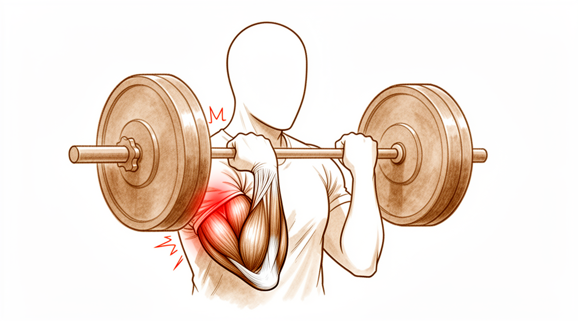
Distal biceps rupture repair — restoring elbow flexion strength and addressing the “Popeye” deformity.
¿Por qué se ha recomendado esta operación?
El Dr. Kieran Hirpara, cirujano de extremidades superiores en el Mater Private Hospital Rockhampton, adapta el tratamiento a su lesión específica. Por lo general, los pacientes son derivados a nuestra clínica por su médico de cabecera; si un fisioterapeuta le ha sugerido que nos consulte, igualmente necesitará una derivación de su médico de cabecera para poder acceder al reembolso de Medicare. En su consulta, tomamos su historia clínica, examinamos su brazo y, si es necesario, solicitamos estudios por imagen. Estos estudios pueden confirmar si el tendón que conecta el músculo bíceps braquial con el hueso del antebrazo se ha roto por completo o solo parcialmente.
Esta operación consiste en volver a unir ese tendón roto al hueso del que proviene. Generalmente se indica en casos de rotura completa, especialmente si usted es una persona activa, trabaja con las manos o practica deportes. En caso de rotura parcial, normalmente intentamos primero un tratamiento no quirúrgico: modificación de actividades, fisioterapia o terapia de la mano, uso de férulas o inyecciones. La cirugía se considera cuando estos tratamientos no logran la mejora deseada. El objetivo es recuperar la fuerza necesaria para flexionar el codo y girar la palma de la mano hacia arriba, de modo que pueda volver a utilizar su brazo con normalidad.
Antes de la operación
Su cirujano le indicará qué medicamentos debe suspender antes de la cirugía y cuándo hacerlo. Deberá dejar de comer y beber siete horas antes, para que podamos adelantar su turno si el programa quirúrgico lo permite. Organice que alguien lo lleve a casa después de la intervención. Lleve consigo una lista de los medicamentos que toma actualmente y vístase con ropa cómoda y holgada. Las pruebas de imagen como radiografías, ecografías o resonancias magnéticas ayudan a planificar la operación. Si padece otras enfermedades, es posible que necesite análisis de sangre o una evaluación con el anestesista.
El día de la intervención
Llega usted a la unidad de admisiones quirúrgicas del hospital, donde se le registrará y preparará para el quirófano. Allí conocerá al anestesista. Esta operación se realiza bajo anestesia general; usted permanecerá completamente dormido durante todo el procedimiento. En algunos pacientes también se aplica un bloqueo nervioso regional para aliviar el dolor postoperatorio; el anestesista decide al respecto ese mismo día según sus circunstancias individuales.
A continuación, se le lleva al quirófano, donde se realiza la operación. Una vez finalizada, despierta usted en la sala de recuperación. Las enfermeras le vigilan mientras la anestesia va perdiendo efecto. Cuando su estado sea estable, será trasladado a la planta de hospitalización o podrá volver a casa, según el tipo de intervención y su recuperación.
Descripción del procedimiento quirúrgico
La operación se realiza mediante una sola incisión en la parte frontal del codo, justo sobre el lugar donde se desgarró el tendón. El cirujano separa los tejidos para acceder al hueso del antebrazo, el cual presenta una pequeña protuberancia donde antes se fijaba el tendón.
El extremo desgarrado del tendón se limpia y se sutura con hilos resistentes. A continuación, el cirujano perfora un pequeño túnel en el hueso y pasa los hilos a través de él. Un pequeño botón metálico acompaña dichos hilos y queda apoyado plano sobre la cara opuesta del hueso, funcionando como anclaje. Los hilos tiran del tendón hacia el interior del túnel, contra la superficie ósea. Allí es donde el tendón vuelve a cicatrizar, reuniéndose con el punto del hueso del cual se había desprendido.
Finalmente, la herida se cierra con puntos de sutura y se cubre con un apósito. Este apósito se deja puesto durante unos 10 días; la sección “Después de la operación” describe lo que sucede entonces.
Después de la operación
Despertará en la sala de recuperación y luego será trasladado a la planta de hospitalización. La mayoría de los pacientes permanecen una noche en el hospital tras esta operación, aunque algunos pueden volver a casa el mismo día. Para mayor comodidad, su brazo quedará sostenido en un cabestrillo, y se iniciarán movimientos suaves desde el principio. Las enfermeras le administrarán analgésicos y se asegurarán de que se sienta cómodo. Alguien debe acompañarle durante las primeras 24 horas después de llegar a casa. Dejamos el vendaje puesto durante unos 10 días; por favor, no lo retire antes de ese plazo a menos que se lo indiquemos. Lo cambiaremos o lo quitaremos cuando vengamos a verle. Puede moverse por la casa y realizar tareas ligeras con la otra mano.
Recuperación
Durante los primeros días, el codo le dolerá y se hinchará. Esto mejora gradualmente. El alivio del dolor y el descanso son de gran ayuda; además, mantener la mano elevada sobre una almohada reduce el dolor pulsátil. Es normal que aparezcan hematomas en el antebrazo.
Para mayor comodidad, el brazo se sostiene en un cabestrillo; sin embargo, se inician movimientos suaves desde el principio, en lugar de mantener el codo inmóvil. La terapia de la mano postoperatoria corre a cargo de Ruby Doolan en Extend Rehabilitation. Ruby le indicará los ejercicios a realizar y confeccionará cualquier férula que necesite. Al inicio, se practican movimientos sencillos para evitar la rigidez del codo y la muñeca. Puede realizar tareas ligeras en casa usando la otra mano. No debe levantar nada con el brazo operado hasta que su terapeuta se lo autorice.
Con el paso de las semanas, la hinchazón disminuye y recupera la movilidad. Su terapeuta añadirá ejercicios de fortalecimiento suaves a medida que el tendón cicatriza. Una vez que se retire el cabestrillo y su cirujano le dé el visto bueno, podrá volver a conducir. Consulte nuestra página sobre conducción tras una cirugía de miembro superior. El regreso al trabajo dependerá de la naturaleza de sus funciones: el trabajo de oficina se reanuda antes que las actividades que requieran levantar pesas. Las actividades deportivas podrán retomarse más adelante, una vez recuperada la fuerza y la capacidad de agarre.
La mayoría de las personas vuelven a trabajar y a realizar sus actividades favoritas. La recuperación varía según cada individuo, por lo que su cronograma puede ser distinto. Su cirujano y su terapeuta de mano le guiarán en cada etapa del proceso.
Qué puede salir mal
La mayoría de los pacientes evolucionan bien, pero en ocasiones pueden presentarse problemas. Su cirujano y el equipo lo vigilarán de cerca para detectar cualquier incidencia a tiempo.
En algunos casos, el tendón reparado no se mantiene en su lugar. Si siente un chasquido o estallido repentino en el codo, o si la fuerza que había ido recuperando desaparece de golpe, comuníquese con la clínica de inmediato. Esto es poco frecuente y, cuando ocurre, suele suceder en las primeras semanas tras la cirugía. Seguir las indicaciones de su terapeuta respecto a levantar objetos y realizar actividades reduce la probabilidad de que esto le suceda.
Los nervios que pasan cerca del codo pueden irritarse durante la intervención. Si esto ocurre, es posible que note hormigueo, entumecimiento o una zona de la piel que se siente insensible o “adormecida”. Algunas personas notan debilidad al estirar los dedos o el pulgar. Con frecuencia esto es temporal; sin embargo, informe a su cirujano o terapeuta en la siguiente consulta si observa cualquiera de estos cambios. Si el entumecimiento o la debilidad son intensos o empeoran, llame a la clínica en lugar de esperar.
El codo también puede volverse rígido si no se mueve según lo previsto. Es posible que le resulte difícil estirar el brazo por completo o doblarlo tanto como antes; además, girar la palma de la mano hacia arriba podría sentirse bloqueado. Continúe realizando los movimientos suaves y comunique a su terapeuta de inmediato si el progreso se estanca. Si la rigidez se convierte en un problema real, su cirujano hablará con usted sobre los pasos siguientes.
Comente cualquier anomalía en su próxima revisión, aunque parezca insignificante. Incluso aquellos síntomas que desaparecen por sí solos merecen ser mencionados, pues una atención temprana facilita la resolución de cualquier problema.
En la tabla de complicaciones de esta página se detallan las tasas típicas, por si desea conocer los datos específicos.
¿Cuándo deben llamarnos?
Llámenos si tiene fiebre, o si la herida se vuelve más roja, hinchada o comienza a supurar. Llámenos si el dolor empeora repentinamente. Si siente un “chasquido” o “estallido” en el codo, llámenos de inmediato o, si es fuera del horario de atención, acuda al servicio de urgencias. Acuda a urgencias si presenta hinchazón o dolor en la pantorrilla, o dificultad para respirar, ya que estos pueden ser signos de un coágulo sanguíneo. Acuda a urgencias si pierde la sensibilidad en el brazo o la mano, o si no puede moverlos. Si no está seguro, llámenos. Preferimos atender cualquier preocupación, por pequeña que sea, antes que pasar por alto algo que requiera atención inmediata.
¿Dónde leer más sobre esta afección?
Esta página trata sobre la intervención quirúrgica en sí. La afección que se trata, así como las evidencias sobre cuándo la cirugía resulta útil y cuándo no, se explican con mayor detalle en la página Rotura del bíceps distal.
Evidence & references
This is the clinical evidence summary written for health professionals. It is technical, and it lists the research this page was built from. You do not need to read it to understand your treatment or to make a decision about it.
Anatomy & Pathophysiology
Bony Anatomy
- The elbow is a trocho-ginglymoid joint consisting of medial and lateral articulations that provide bony stability [3].
- The medial ulnohumeral articulation is formed by the trochlea articulating with the ulna within the greater sigmoid notch [3].
- The ulnohumeral joint has highly congruent anatomy through almost 180° of articular contact, with the exception of a bare area in the greater sigmoid notch devoid of cartilage [3].
- The coronoid process has medial and lateral facets that buttress the trochlea anteriorly [3].
- The sublime tubercle is located just distal and medial to the coronoid and serves as the attachment site for the anterior bundle of the medial ulnar collateral ligament [3].
- The medial epicondyle is larger and more posteriorly oriented than the lateral epicondyle and serves as the attachment site for the origins of the flexor pronator mass [3].
- The lateral radiocapitellar joint is formed by the capitellum and radial head [3].
- The radius is held in close approximation to the ulna at the proximal radioulnar joint by the annular ligament [3].
- The area of the ulna that articulates with the margin of the radial head at the proximal radioulnar joint is known as the lesser sigmoid notch [3].
- The radial head is a concave elliptical structure covered with articular cartilage along the radiocapitellar joint and approximately 270° of the articular margin [3].
- The radial head articulates with both the capitellum and the lesser sigmoid notch [3].
- The lateral epicondyle is the origin of the lateral extensor musculature [3].
- The origin of the lateral ulnar collateral ligamentous complex is located just distal to the lateral epicondyle at the geometric center of the radiocapitellar articulation [3].
- The distal humeral articulation is angled 30° from the longitudinal axis [3].
- The anterior humeral line should pass through the center of the axis of rotation [3].
- The axis of rotation is 5° to 7° angulated in the coronal plane to the epicondylar axis, with the medial side more distal than the lateral side [3].
- This coronal angulation accounts for the change from a valgus carrying angle to a more varus position as the elbow is flexed [3].
- There is a high correlation between the size of the radial head and capitellum on the left and right sides of the same individual [2, 3].
- The olecranon provides a broad attachment site for the triceps [3].
- The ulna bends approximately 8° medially at 8 cm from the tip of the olecranon [3].
- The articulation to the tip of the coronoid is approximately 30° from the long axis of the ulna in the sagittal plane [3].
Ligaments & Soft Tissue
- Elbow stability is determined by primary stabilizers, which include the ulnohumeral articulation, the medial ulnar collateral ligament, and the lateral ulnar collateral ligament complex [1].
- Secondary stabilizers of the elbow include the radiocapitellar articulation, the common flexor tendon, the common extensor tendon, and the joint capsule [1].
- The medial ulnar collateral ligament is comprised of the anterior bundle, posterior bundle, and transverse ligament [2].
- The anterior bundle of the medial ulnar collateral ligament is the strongest component and the primary restraint to valgus stress [2].
- The anterior bundle of the medial ulnar collateral ligament is subdivided into anterior and posterior bands that provide reciprocal function, with the anterior band tight in extension and the posterior band tight in flexion [2].
- The lateral ulnar collateral ligament origin center is 10.7 mm from the lateral epicondyle [2].
- The lateral ulnar collateral ligament insertion is 3.3 mm from the apex of the supinator crest [2].
- Tensile forces are present at the medial elbow, while compressive forces are present at the lateral elbow [4, 5].
- The joint capsule allows maximum distension at approximately 70 to 80 degrees of flexion [4, 5].
- The anterior capsule attaches at a point approximately 6 mm distal to the tip of the coronoid [4, 5].
- The coronoid tip is an intraarticular structure that is visualized during elbow arthroscopy [4, 5].
- The medial or ulnar collateral ligament is the primary valgus stabilizer [4, 5].
- The anterior bundle of the medial ulnar collateral ligament is the most important component for stability [4, 5].
- The posterior bundle of the medial ulnar collateral ligament has the greatest change in length and becomes taut at flexion beyond 120 degrees [4, 5].
- The lateral ulnar collateral ligament is the posterolateral stabilizer [4, 5].
- Osborne’s ligament stabilizes the ulnar nerve in the cubital tunnel [4, 5].
- The ligament of Struthers is a variant anatomy arising from the supracondylar process to attach to the medial epicondyle and is a potential site of median nerve compression [4, 5].
Muscles & Insertions
- The brachialis is the strongest elbow flexor and attaches to the coronoid 11 mm distal to the tip [4, 5].
- The biceps brachii inserts at the ulnar margin of the radial tuberosity, with the long head inserting proximal and the short head inserting distal [4, 5].
- The biceps brachii is a powerful supinator of the forearm [4, 5].
- The triceps is the primary elbow extensor and inserts on the olecranon process [4, 5].
- The mobile wad consists of the brachioradialis, extensor carpi radialis longus, and extensor carpi radialis brevis [4, 5].
- The flexor-pronator mass consists of the pronator teres, flexor carpi radialis, palmaris longus, flexor carpi ulnaris, and flexor digitorum superficialis [4, 5].
- The triceps has three distinct insertional areas to the olecranon corresponding to the posterior capsular insertion, the deep muscular portion, and the superficial tendinous portion [2].
- The deep muscular head of the triceps corresponds to the medial head, while the tendinous portion corresponds to the long and lateral heads [2].
- The width of the triceps insertion is 2.6 cm and is located 1.1 cm from the tip of the olecranon [2].
Biomechanics & Motion
- The normal elbow has a range of motion from 0° to 140° from extension to flexion [1].
- The normal elbow has a range of motion of 75° in pronation and 85° in supination [1].
- A functional arc for the elbow is 100° for flexion and extension and forearm rotation [1].
Investigations
Physical Examination
- The physical examination of the elbow is directed by the history and the location of the patient's pain in the anterior, posterior, medial, or lateral aspect [1].
- Active and passive flexion, extension, supination, and pronation should be evaluated using a goniometer for accurate measurement [9].
- The contralateral elbow should be examined for comparison during range of motion assessment [9].
- Pain should be assessed during the mid-arc or at the terminal ends of motion [9].
- Mid-arc range of motion pain is more common with intrinsic disease and may not improve with contracture release alone [9].
- The ulnar nerve is of utmost importance in the physical examination due to its anatomic proximity to the elbow [9].
- An assessment for ulnar nerve subluxation should be performed [9].
- Subluxation of the ulnar nerve is a relative contraindication for an arthroscopic procedure secondary to possible iatrogenic nerve injury [9].
- Electromyography and nerve conduction velocity studies should be performed if there is any question about neurologic dysfunction [9].
Imaging
- Plain radiographs remain the hallmark and the best screening test for elbow evaluation [1].
- Standard radiographic views include AP, lateral, and oblique views [9].
- Serial radiography is used as follow-up when heterotopic ossification is present [9].
- Primary bony landmarks identified on radiographs include the ulnohumeral joint, coronoid process, radial head, capitellum, radiocapitellar joint, olecranon tip, coronoid/olecranon fossae, and trochlear ridge [9].
- CT is helpful when assessing for malunion architecture and the location and pattern of osteophytes and/or loose bodies [9].
- Three-dimensional CT is used to check for heterotopic ossification [9].
- CT is not necessary when the stiffness is entirely soft-tissue related [9].
- CT is beneficial if any joint incongruity or abnormal bony anatomy is present [9].
- MRI can be used to evaluate ligaments and tendons, but it is rarely indicated for elbow stiffness [9].
- MRI may be most helpful in evaluating associated injuries including partial or complete tears of the medial collateral ligament in the context of valgus extension overload syndrome [11].
- CT with two-dimensional reconstruction and three-dimensional surface rendering best visualizes the pathology of valgus extension overload syndrome [11].
- AP, lateral, oblique, and axillary views of the elbow may reveal posteromedial olecranon osteophytes and/or loose bodies in valgus extension overload syndrome [11].
- Radiographic evaluations are essential when diagnosing an osteochondritis dissecans lesion of the elbow [12].
- Important aspects of osteochondritis dissecans lesions may be better seen with MRI [12].
- Standard AP and lateral radiographs should be obtained for the evaluation of elbow osteoarthritis [13].
- Radiographs typically show osteophyte formation at the coronoid process, coronoid fossa, radial fossa, radial head, olecranon tip, and olecranon fossa in elbow osteoarthritis [13].
- Joint spaces at the ulnohumeral joint are usually preserved in elbow osteoarthritis [13].
- Joint spaces at the radiocapitellar joint are mildly narrowed in elbow osteoarthritis [13].
- Loose bodies may be evident on radiographs, which typically underestimate the number present [13].
- CT may be useful for surgical planning in elbow osteoarthritis, allowing a detailed assessment of osteophytes and the presence of loose bodies [13].
References
[1] Orthopaedic Knowledge Update 13 Ebook Without Multimedia. Anatomy, Biomechanics, Physical Examination, and Imaging of the Elbow > Summary and Conclusions.
[2] Orthopaedic Knowledge Update 13 Ebook Without Multimedia. Anatomy, Biomechanics, Physical Examination, and Imaging of the Elbow > Annotated References.
[3] Orthopaedic Knowledge Update 13 Ebook Without Multimedia. Anatomy, Biomechanics, Physical Examination, and Imaging of the Elbow > Anatomy > Bony Anatomy.
[4] Miller S Review Of Orthopaedics. SECTION 16 PATELLAR TRACKING IN TOTAL KNEE ARTHROPLASTY > TABLE 2.3 Shoulder Spaces.
[5] Miller S Review Of Orthopaedics. Genetics of musculoskeletal conditions and abnormalities are summarized in Table 1.27 > TABLE 2.3 Shoulder Spaces.
[9] Aaos Comprehensive Orthopaedic Review 3. Elbow Stiffness* > IV. Evaluation.
[11] Aaos Comprehensive Orthopaedic Review 3. Elbow Injuries in the Athlete* > III. Valgus Extension Overload Syndrome and Posterior Impingement.
[12] Orthopaedic Knowledge Update. Osteochondritis Dissecans of the Knee and Elbow* > Summary.
[13] Aaos Comprehensive Orthopaedic Review 3. Arthritis and Arthroplasty of the Elbow > I. Osteoarthritis.