Patients › Rehabilitation
Fijación del húmero proximal (reducción abierta y fijación interna)
Rehabilitation after locking-plate fixation of a proximal humerus fracture, gated on radiographic healing at review.
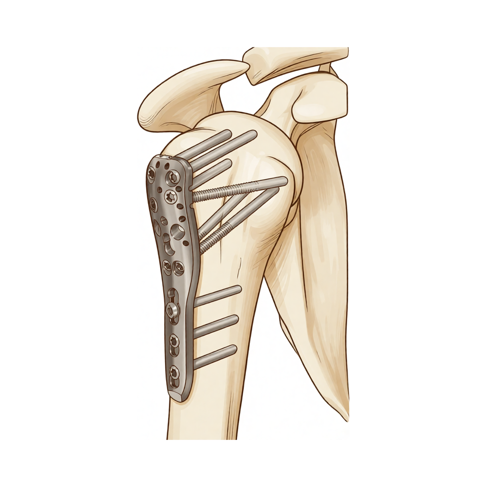
Este protocolo describe la rehabilitación posterior a la fijación quirúrgica de una fractura del húmero proximal (una rotura del hueso del brazo cerca del hombro, reparada mediante una placa bloqueante y tornillos, mediante reducción abierta y fijación interna, ORIF) a cargo del Dr. Kieran Hirpara en el Mater Private Hospital Rockhampton. Lleve esta página o su versión en PDF a su primera sesión de fisioterapia para garantizar una rehabilitación coordinada. Su fisioterapeuta guiará su rehabilitación individualmente a través de las siguientes fases, según el proceso de curación de su fractura.
Si tiene alguna duda respecto a la herida quirúrgica, no dude en contactar con el consultorio. A menudo resulta útil tomar una fotografía de la herida y enviarla por correo electrónico para su evaluación.
Qué esperar
La rehabilitación tras la fijación de una fractura difiere de la rehabilitación posterior a la mayoría de las cirugías programadas del hombro. La placa y los tornillos mantienen el hueso roto en su posición, pero el hueso en sí aún debe sanar; la velocidad de esa curación varía considerablemente de una persona a otra. Por ese motivo, el siguiente cronograma constituye una guía típica y no un plan fijo: cada avance en su programa depende tanto del número de semanas transcurridas desde la cirugía como de cómo se está soldando la fractura según sus radiografías, tal como lo confirme el Dr. Hirpara en sus revisiones. No pase a la siguiente fase únicamente basándose en el calendario; espere a su revisión médica.
Hay otro factor que diferencia esta intervención. Las protuberancias óseas en la parte superior del húmero (las tuberosidades) son los puntos de inserción de los tendones del manguito rotador; en muchas fracturas proximales del húmero, estos fragmentos forman parte de la fractura y se fijan nuevamente con la placa o con puntos de sutura. Ejercitar los músculos del manguito rotador de forma excesiva o demasiado pronto podría tirar de esos fragmentos antes de que hayan sanado. Gracias a las placas bloqueantes modernas, la fijación es sólida desde el primer día, por lo que se recomienda un movimiento suave desde el inicio: podrá realizar movimientos activos asistidos y movimientos activos suaves dentro de los límites del dolor, levantando el brazo dentro de un rango indoloro. Lo que requiere precaución es la carga sobre el manguito rotador: si las tuberosidades no forman parte de la fractura o están bien fijadas, su cirujano podría autorizarle a iniciar la rotación activa y el ejercicio del manguito rotador antes; si las tuberosidades están implicadas y aún sanan, se retrasará un poco más el ejercicio activo del manguito rotador y la rotación externa forzada. El Dr. Hirpara le indicará cuál de estos casos se aplica a usted.
Su programa de ejercicios incluye tres tipos de movimientos; su equipo le indicará cuáles son aplicables para usted:
- Amplitud de movimiento activa: se permite el movimiento sin ayuda ni asistencia.
- Amplitud de movimiento activa asistida: se utiliza el otro brazo o algún objeto para ayudar a mover el brazo.
- Amplitud de movimiento pasiva: el brazo permanece totalmente relajado; el otro brazo o una fuerza externa realiza el 100 % del movimiento.
Al despertar de la cirugía, tendrá el brazo en un cabestrillo. Úselo todo el tiempo durante las primeras tres semanas aproximadamente, y luego vaya retirándolo progresivamente en las semanas siguientes según le resulte cómodo; los protocolos publicados indican un promedio de tres semanas de inmovilización, y la evidencia científica respalda el movimiento temprano frente a un período prolongado con el cabestrillo. No es necesario dormir con él puesto. Quíteselo varias veces al día para hacer los ejercicios, para lavarse y vestirse; cuando esté sentado tranquilamente en casa, puede dejar el brazo fuera del cabestrillo. No debe conducir durante al menos seis semanas después de cualquier cirugía del hombro; su cirujano le autorizará a conducir, generalmente en la revisión de la sexta semana.
Resumen del proceso:
- Fase I — Protección y movimiento temprano: semanas 0–6
- Fase II — Recuperación de la movilidad activa: semanas 6–12
- Fase III — Fortalecimiento: aproximadamente entre los 3 y 4½ meses
- Fase IV — Retorno a la actividad plena: a partir de los 4½–6 meses
Fase I — Protección y movimiento temprano (Semanas 0–6)
Kieran Hirpara 4.0
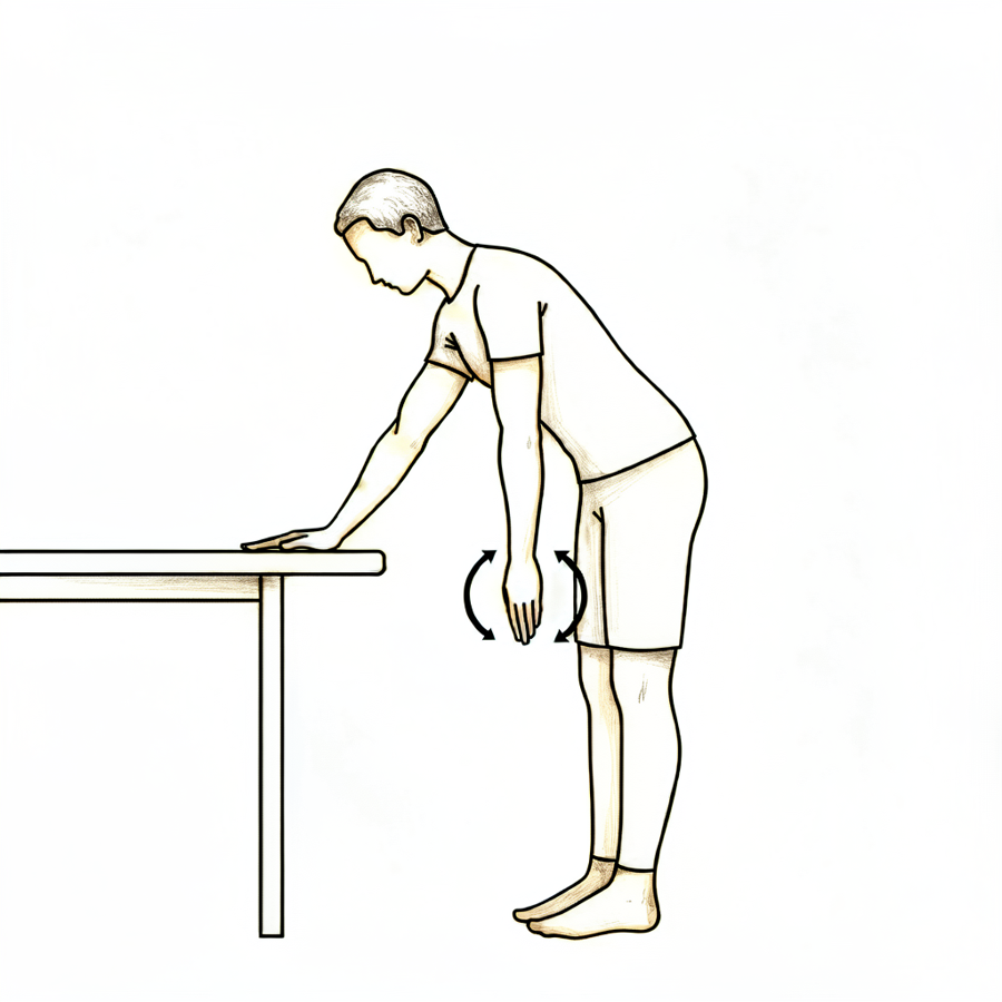
Ejercicios de péndulo
Incline el cuerpo hacia adelante, apoyándose con la otra mano en una mesa, y deje que el brazo operado cuelgue relajado. Realice pequeños círculos: primero en sentido horario y luego en sentido antihorario, balanceando su cuerpo suavemente. El brazo se mueve pasivamente; el movimiento proviene de su cuerpo, no de los músculos del hombro.
Varias veces al día
Kieran Hirpara 4.0
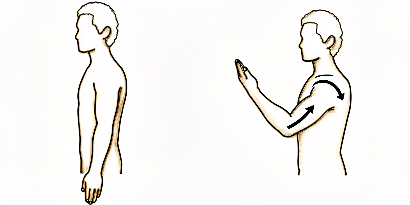
Flexiones activas de codo
Con el brazo fuera del cabestrillo y el brazo superior apoyado a un costado, doble el codo tanto como le resulte cómodo y luego estírelo por completo. Solo debe moverse el codo; mantenga el hombro inmóvil y el brazo junto al cuerpo.
Varias veces al día
Kieran Hirpara 4.0
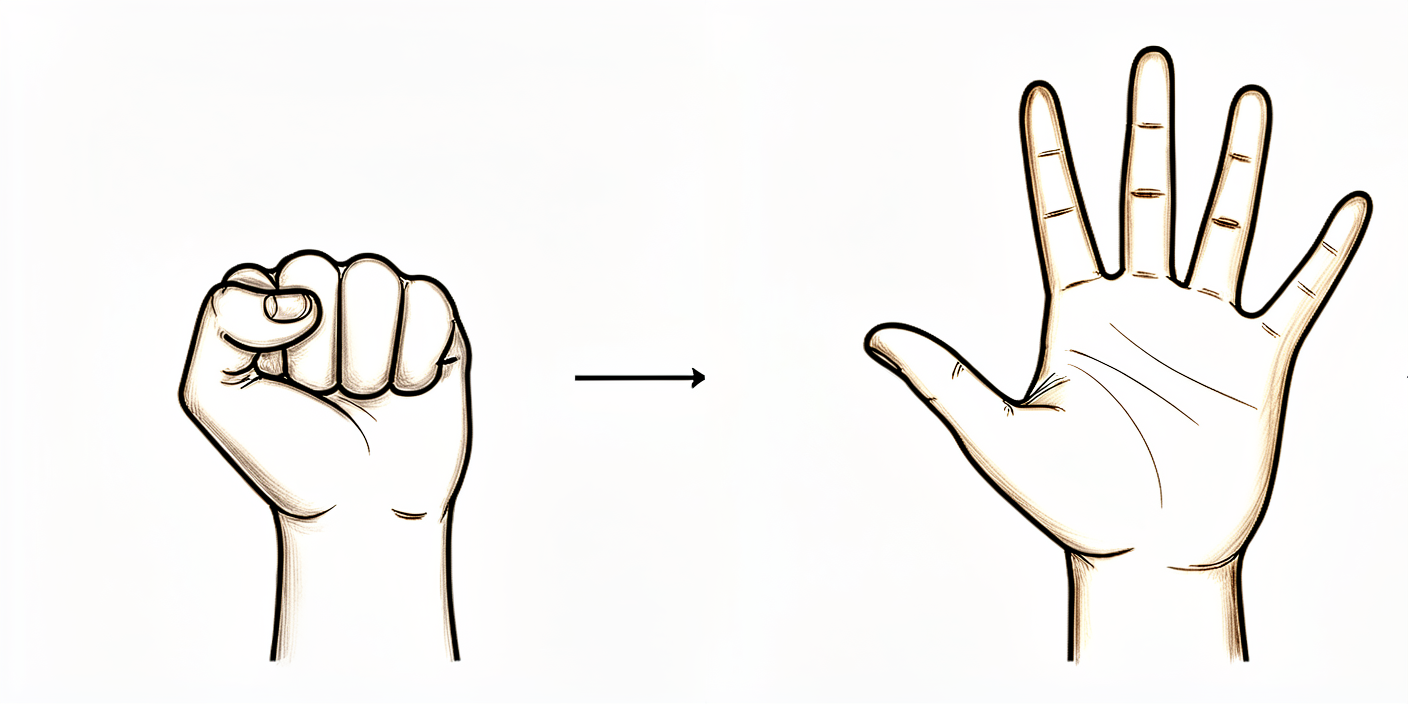
Aprietes de pelota
Con el brazo apoyado, apriete una pelota blanda o haga un puño cerrado, y luego abra la mano por completo. Cada vez que el brazo quede fuera del cabestrillo, mueva los dedos, la muñeca y la mano.
Varias veces al día
Kieran Hirpara 4.0
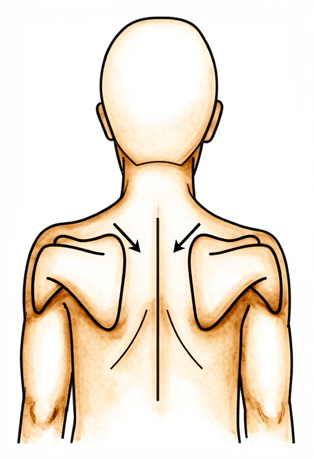
Posicionamiento escapular
Ya sea sentado o de pie, lleva suavemente los omóplatos hacia abajo y hacia atrás sin mover los brazos; mantén esa posición unos segundos y luego relájate. Procura que las orejas, los hombros y las caderas queden alineados.
Según las indicaciones de su fisioterapeuta.
Kieran Hirpara 4.0
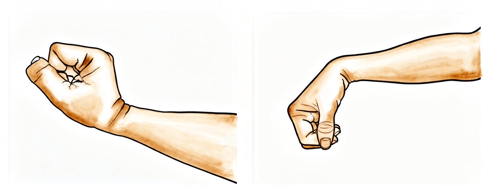
Movimientos de la muñeca
Con el brazo fuera del cabestrillo, mantenga el movimiento de la muñeca flexionándola hacia adelante, hacia atrás y suavemente de lado a lado. Esto permite que todo el brazo siga funcionando mientras se protege el hombro.
Varias veces al día
Kieran Hirpara 4.0
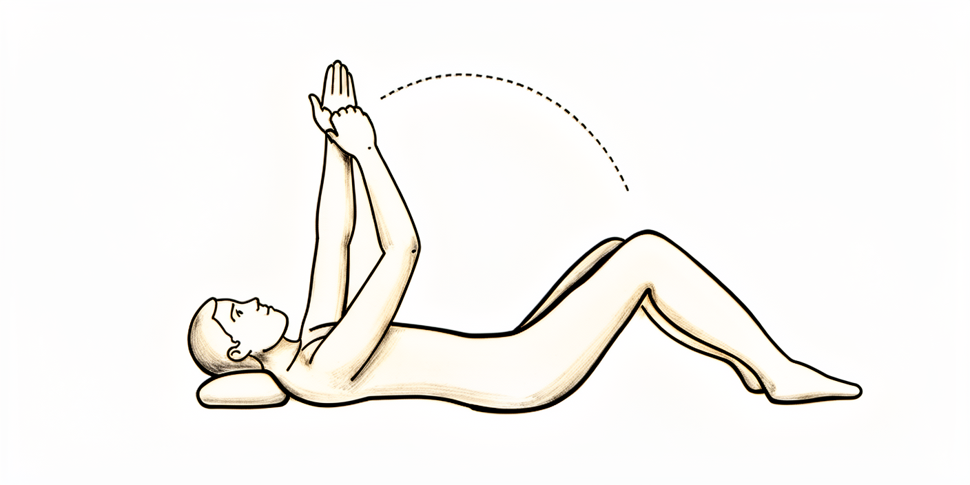
Elevación frontal asistida (en decúbito)
Acuéstese boca arriba y utilice el brazo sano para ayudar a levantar el brazo operado hacia adelante; luego, bájelo lentamente mientras el brazo sano sigue ayudando. Empezar acostado aprovecha la gravedad y facilita los primeros levantamientos. Mantenga el movimiento dentro de un rango cómodo y sin dolor; deténgase en el punto en que sienta alguna tensión, sin forzarlo.
Varias veces al día, dentro de un rango sin dolor.
Kieran Hirpara 4.0
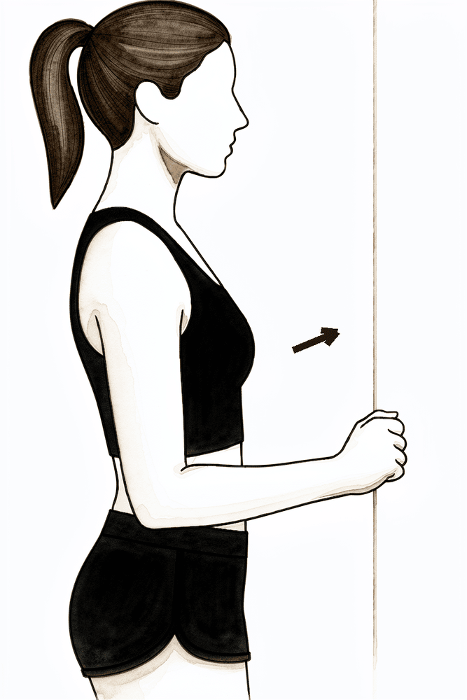
Isométrico de elevación frontal suave
Párese de cara a una pared, con el brazo operado a un lado del cuerpo y el puño suave apoyado contra la pared delante de usted. Presione suavemente la mano contra la pared sin mover el brazo, mantenga la posición unos segundos y luego relájese. Esto activa los músculos responsables de elevar el hombro sin ejercer tensión sobre la fractura en proceso de curación. Solo debe hacerlo si su fisioterapeuta ya le ha indicado que lo practique.
Según las indicaciones de su fisioterapeuta.
Durante las primeras seis semanas, el objetivo es proteger la fractura fijada y evitar que el resto del brazo (así como la articulación del hombro) se vuelva rígido. Gracias a que la placa de fijación mantiene el hueso estable desde el inicio, se recomienda realizar movimientos suaves desde el principio, en lugar de esperar seis semanas. Un fisioterapeuta le guiará en ejercicios suaves antes de que regrese a casa: ejercicios de péndulo (dejar el brazo colgar y balancearlo suavemente mediante el movimiento del cuerpo), y mover el codo, la muñeca y la mano fuera del cabestrillo varias veces al día. Desde el inicio, y dentro de un rango cómodo y sin dolor, puede comenzar a mover el brazo con ayuda de la otra mano (movimiento activo asistido) y también de forma autónoma (movimiento activo), elevándolo frente a usted según le resulte cómodo. Lo que requiere precaución es la carga sobre el manguito rotador: evite forzar el brazo hacia afuera (rotación externa) y no realice ejercicios de resistencia para el manguito rotador hasta que sus tuberosidades hayan sanado, tal como confirma el Dr. Hirpara. Utilice hielo para aliviar el dolor y tome sus analgésicos antes de realizar los ejercicios y las sesiones de fisioterapia.
Para su fisioterapeuta:
Objetivos
- Proteger la fijación ósea y favorecer su sanación
- Controlar el dolor y la hinchazón
- Iniciar movimientos activos asistidos, progresando hacia la elevación activa dentro de un rango sin dolor
- Mantener la movilidad completa del cuello, codo, muñeca y mano
Manejo
- Usar el cabestrillo todo el tiempo durante unas 3 semanas; luego retirarlo gradualmente según la comodidad del paciente (según la literatura médica); retirarlo varias veces al día para ejercicios e higiene; no es necesario usarlo mientras esté acostado
- Ejercicios de péndulo/Codman varias veces al día
- Desde el inicio, si la fijación es estable, realizar movimientos activos asistidos y elevaciones suaves hacia adelante dentro de un rango cómodo y sin dolor; comenzar en posición supina y avanzar hacia la posición vertical según el control del paciente; ampliar el rango de movimiento según le resulte cómodo
- Movimientos pasivos de amplitud articular cuando el movimiento activo aún no sea tolerado: elevación frontal en el plano escapular hasta unos 90°, rotación interna hasta el abdomen (nunca detrás de la espalda)
- La rotación externa se mantendrá inicialmente en unos 30–40° con el brazo pegado al cuerpo; el cirujano podrá aumentar este valor si las tuberosidades no están afectadas o están bien fijadas
- Movilidad activa del cuello, codo, muñeca y mano fuera del cabestrillo; ejercicios de apretar pelotas para fortalecer la prensión
- Ejercicios para la movilidad escapular (elevación, depresión, retracción y protracción)
- Isométricos suaves del deltoides y músculos periescapulares pueden introducirse según la comodidad del paciente
- Aplicar crioterapia y analgésicos antes de las sesiones; realizar movilización y desensibilización de la cicatriz una vez que la herida haya sanado
Precauciones
- Evitar rotaciones externas forzadas o con resistencia, así como cargas intensas sobre el manguito rotador hasta confirmar la sanación de las tuberosidades (bajo supervisión del cirujano); la elevación activa dentro de un rango sin dolor sí está permitida
- No realizar rotación interna detrás de la espalda; evitar la abducción en el plano coronal
- Los movimientos iniciales deben ser suaves y sin dolor; nunca forzados
- No levantar cargas superiores a unos 0.5–1 kg con el brazo operado
- No soportar peso con el brazo operado (no empujarse hacia arriba desde una silla o cama)
- No conducir durante seis semanas (esto aplica a cualquier cirugía de hombro)
- Evitar movimientos forzados o que provoquen dolor en los extremos del rango articular
Criterios para avanzar
- Evidencia radiográfica de progreso en la sanación, confirmada en la revisión con el Dr. Hirpara alrededor de la semana 6
- Control adecuado del dolor
- Mantenimiento de la movilidad completa del codo, muñeca y mano
- Capacidad de realizar elevaciones activas asistidas y luego activas dentro de un rango sin dolor
Fase II — Recuperación del movimiento activo (Semanas 6–12)
Kieran Hirpara 4.0
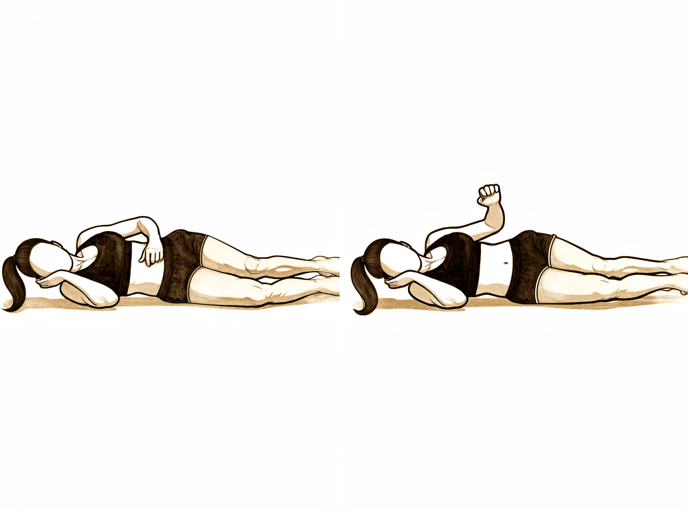
Rotación externa en decúbito lateral
Acuéstese sobre el lado no operado, con el brazo operado encima; doble el codo a 90 grados y acérquelo al cuerpo. Manteniendo el codo en esa posición, gire el antebrazo hacia arriba, alejándolo del abdomen, hasta donde le resulte cómodo; luego bájelo de forma controlada. No añada ningún peso: el brazo debe moverse por su propia fuerza.
Según las indicaciones de su fisioterapeuta.
Kieran Hirpara 4.0
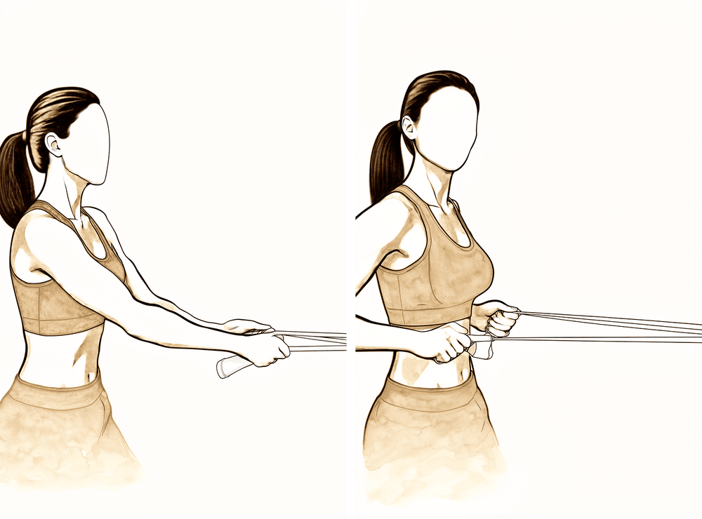
Ejercicio de remo con banda elástica
Fije una banda elástica delante de usted a la altura de la cintura. De pie, tire de la banda hacia sí, llevando suavemente el omóplato hacia abajo y hacia atrás; luego suelte la banda de forma controlada. Mantenga la resistencia en un nivel bajo.
Según las indicaciones de su fisioterapeuta.
Kieran Hirpara 4.0
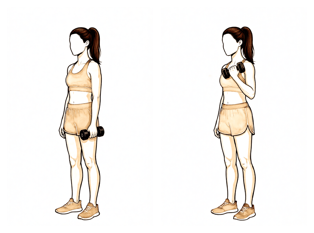
Flexiones de bíceps
Sostenga un peso ligero con la palma de la mano hacia arriba; luego flexione el codo para acercar el peso a su hombro y, a continuación, bájelo lentamente. Mantenga el brazo superior inmóvil a lo largo del costado, y asegúrese de que el peso sea ligero: en esta fase no debe superar los 1–2 kg aproximadamente.
Según las indicaciones de su fisioterapeuta.
Kieran Hirpara 4.0
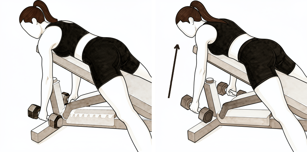
Remo con apoyo
Incline el cuerpo hacia adelante apoyando el brazo sano sobre una mesa, de modo que el brazo operado quede colgando con un peso ligero. Retraiga el omóplato y eleve el peso hacia la cadera, luego bájelo de forma controlada. Mantenga el tronco estable: el movimiento debe provenir del omóplato y el brazo, no de la espalda.
Según las indicaciones de su fisioterapeuta.
Kieran Hirpara 4.0
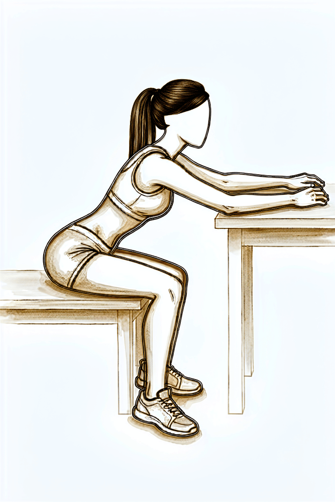
Deslizamientos sobre la mesa
Siéntese a una mesa con la mano apoyada sobre una toalla o un paño. Incline el cuerpo hacia adelante y deslice la mano lejos de usted a lo largo de la mesa, permitiendo que el brazo se extienda más hacia adelante; luego deslícela de vuelta. La mesa soporta el peso del brazo, por lo que esta es una forma suave de recuperar el movimiento hacia adelante.
Según las indicaciones de su fisioterapeuta.
Kieran Hirpara 4.0
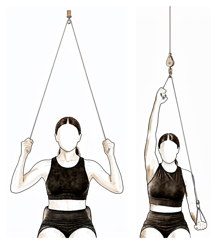
Polea de puerta
Siéntese debajo de una polea de puerta, sosteniendo un mango en cada mano. Tire hacia abajo con el brazo sano para elevar el brazo operado hasta donde le resulte cómodo; luego, bájelo lentamente. Deje que sea el brazo sano el que realice el esfuerzo y manténgase siempre dentro de un rango de movimiento cómodo.
Según las indicaciones de su fisioterapeuta.
Kieran Hirpara 4.0
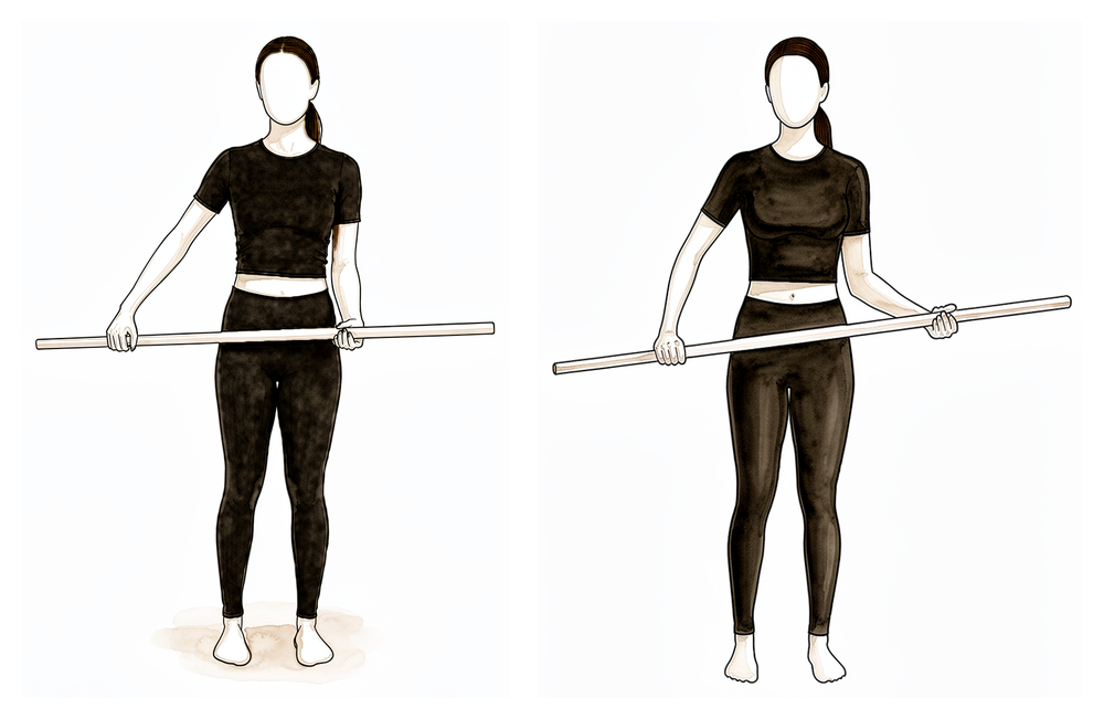
Rotación externa asistida mediante un palo
Acuéstese boca arriba, con el codo pegado al costado y doblado en ángulo recto. Sostenga un palo con ambas manos y utilice el brazo sano para girar la mano operada hacia afuera. Una vez que se confirme la cicatrización, intente aumentar ligeramente ese giro externo; sin embargo, debe mantenerse dentro del rango indicado por su cirujano y fisioterapeuta.
Según las indicaciones de su fisioterapeuta.
Kieran Hirpara 4.0
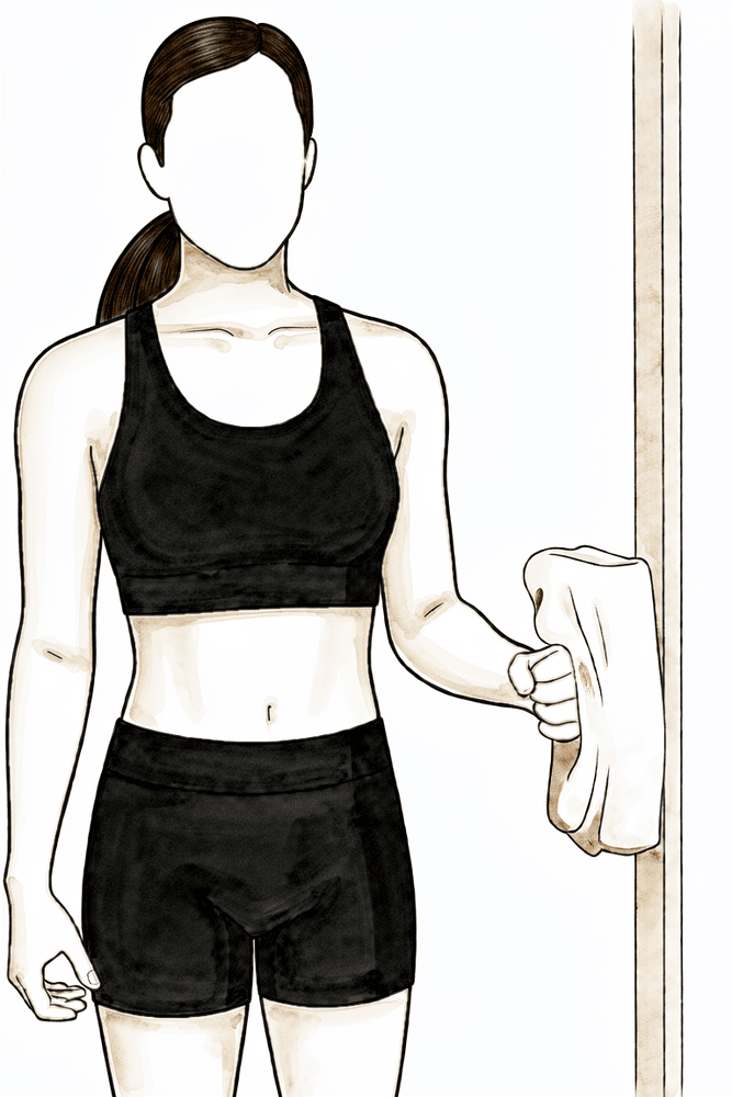
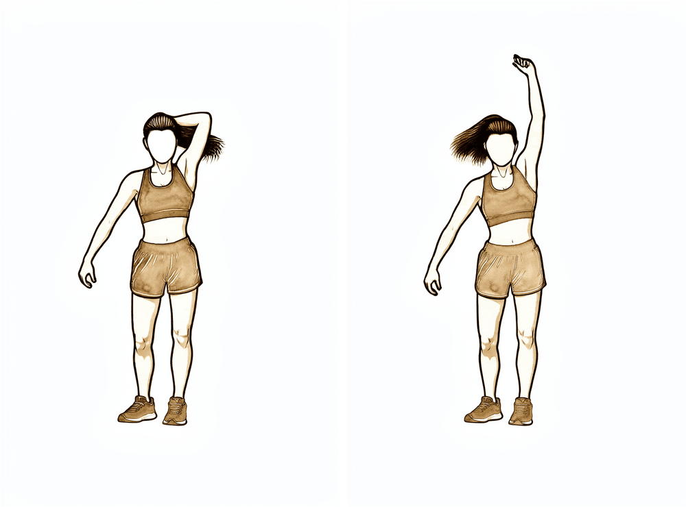
Rotación externa isométrica
Párese con el codo pegado al costado y doblado en ángulo recto, con el dorso de la mano apoyado contra una pared o marco de puerta. Presione suavemente hacia afuera sin mover el brazo, mantenga la posición unos segundos y luego relájese. Esto comienza a activar el manguito rotador sin necesidad de movimiento; úselo únicamente después de que el Dr. Hirpara haya confirmado que su recuperación es adecuada.
Según las indicaciones de su fisioterapeuta.
Kieran Hirpara 4.0
Extensiones de tríceps
Sosteniendo un peso ligero, doble y luego estire el codo para ejercitar el músculo situado en la parte posterior del brazo. Mantenga el brazo firme y el peso ligero: en esta fase no debe superar los 1–2 kg aproximadamente.
Según las indicaciones de su fisioterapeuta.
Kieran Hirpara 4.0
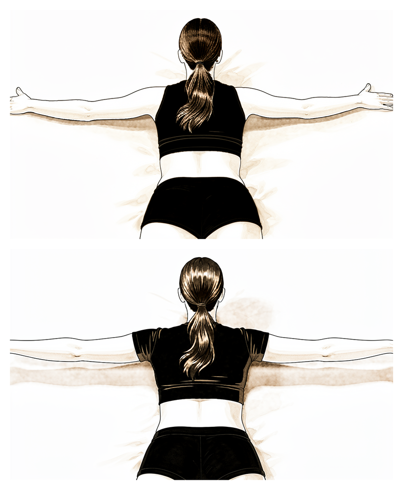
Remo escapular en posición prona
Acuéstese boca abajo con el brazo colgando por el borde de una cama o banco. Retraiga el omóplato y eleve el brazo ligeramente hacia el costado, apretando los omóplatos entre sí; luego bájelo de forma controlada. Esto fortalece los músculos que estabilizan el omóplato.
Según las indicaciones de su fisioterapeuta.
En la revisión a las seis semanas, el Dr. Hirpara examinará sus radiografías. Si la cicatrización avanza según lo esperado, se dejará de usar el cabestrillo y usted comenzará a mover el brazo por sí mismo, primero con ayuda (utilizando el otro brazo, un palo o un sistema de poleas) y luego de forma activa. Por lo general, es más fácil iniciar los movimientos activos del brazo estando acostado boca arriba, donde la gravedad no supone un obstáculo tan grande; posteriormente se pasa a realizarlos sentado o de pie, a medida que gana control. En esta fase, una vez que el Dr. Hirpara confirma la cicatrización, se inician ejercicios isométricos suaves para el manguito rotador; los ejercicios con resistencia se añadirán más adelante. Podrá volver a conducir cuando ya no use el cabestrillo, tenga suficiente movilidad y control del brazo para conducir de forma segura, y haya dejado de tomar analgésicos fuertes; en caso de duda, consulte al médico en su próxima revisión.
Para su fisioterapeuta:
Objetivos
- Restaurar el rango de movimiento pasivo por completo
- Pasar de movimientos activos asistidos a movimientos activos en todos los planos
- Recuperar el ritmo escapulohumeral normal y minimizar patrones compensatorios
- Volver a realizar actividades cotidianas ligeras con normalidad
Manejo
- Se debe abandonar por completo el uso del cabestrillo a más tardar en la revisión de la semana 6
- Movilidad activa asistida: ejercicios progresivos en posición de silla de jardín, deslizamientos contra mesas o paredes, ejercicios con poleas y palos; se amplían los límites de movimiento respecto a la Fase I según la tolerancia del paciente
- Movilidad activa a partir de la semana 6–8: flexión del brazo acostado, progresando hacia elevaciones en posición vertical; rotación externa y flexión en decúbito lateral; ejercicios de remo y puñetazos suaves
- Ejercicios isométricos submáximos para el manguito rotador y el deltoides a partir de la semana 6–8, con el brazo pegado al cuerpo, una vez confirmada la cicatrización
- Ejercicios isométricos ligeros para el codo (flexiones de bíceps, extensiones de tríceps) y fortalecimiento escapular (retracción, remos en decúbito prono)
- Movilizaciones glenohumerales y escapulotorácicas según sea necesario; se aumentan los grados de movilización conforme avanza la cicatrización
- Corrección postural; se pueden seguir aplicando calor o hielo y analgésicos antes y después de las sesiones, según preferencia del paciente
Precauciones
- No realizar ejercicios isotónicos de fortalecimiento del manguito rotador hasta confirmar la consolidación ósea, normalmente no antes de la semana 8–12
- Evitar presiones excesivas en los extremos de rango de movimiento ni estiramientos pasivos agresivos
- El peso que se puede levantar con el brazo operado está limitado a unos 1–2 kg
- Vigilar y corregir cualquier tendencia a levantar el hombro o inclinar el tronco al realizar movimientos de elevación
Criterios para avanzar a la siguiente fase
- Rango de movimiento pasivo completo o casi completo
- Capacidad de elevar el brazo de forma activa con buena mecánica, al menos hasta la altura del hombro
- Tolerancia adecuada a los ejercicios isométricos sin aumento de dolor
- Consolidación ósea progresiva según se observa en las radiografías, confirmada en la revisión con el Dr. Hirpara
Fase III — Fortalecimiento (aproximadamente entre los meses 3 y 4½)
Kieran Hirpara 4.0
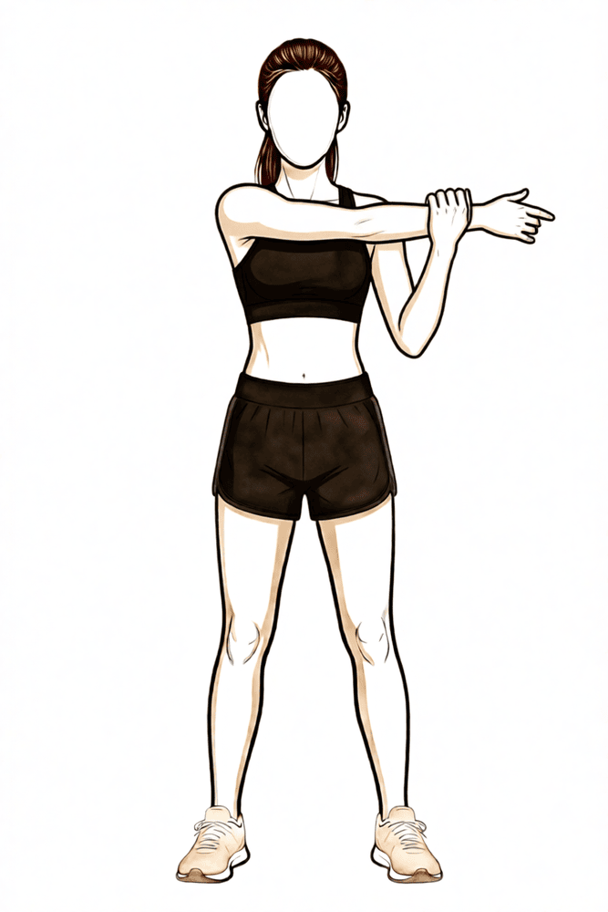
Estiramiento cruzado del cuerpo
Utilice la otra mano para llevar el brazo operado hacia el pecho hasta que sienta un estiramiento en la parte posterior del hombro. Mantenga esa posición brevemente y luego relájese. El estiramiento debe generar solo una molestia moderada; nunca debe provocar dolor intenso.
Según las indicaciones de su fisioterapeuta.
Kieran Hirpara 4.0
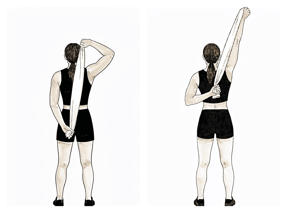
Estiramiento detrás de la espalda
Sujete una toalla detrás de la espalda, con el brazo sano arriba y el brazo operado abajo. Utilice la mano superior para tirar suavemente de la mano inferior hacia arriba, por la espalda, hasta sentir un estiramiento cómodo; mantenga esa posición y luego suelte. Aumente gradualmente el rango de movimiento; nunca fuerce el estiramiento ni vaya más allá del punto en el que sienta dolor intenso.
Según las indicaciones de su fisioterapeuta.
Kieran Hirpara 4.0
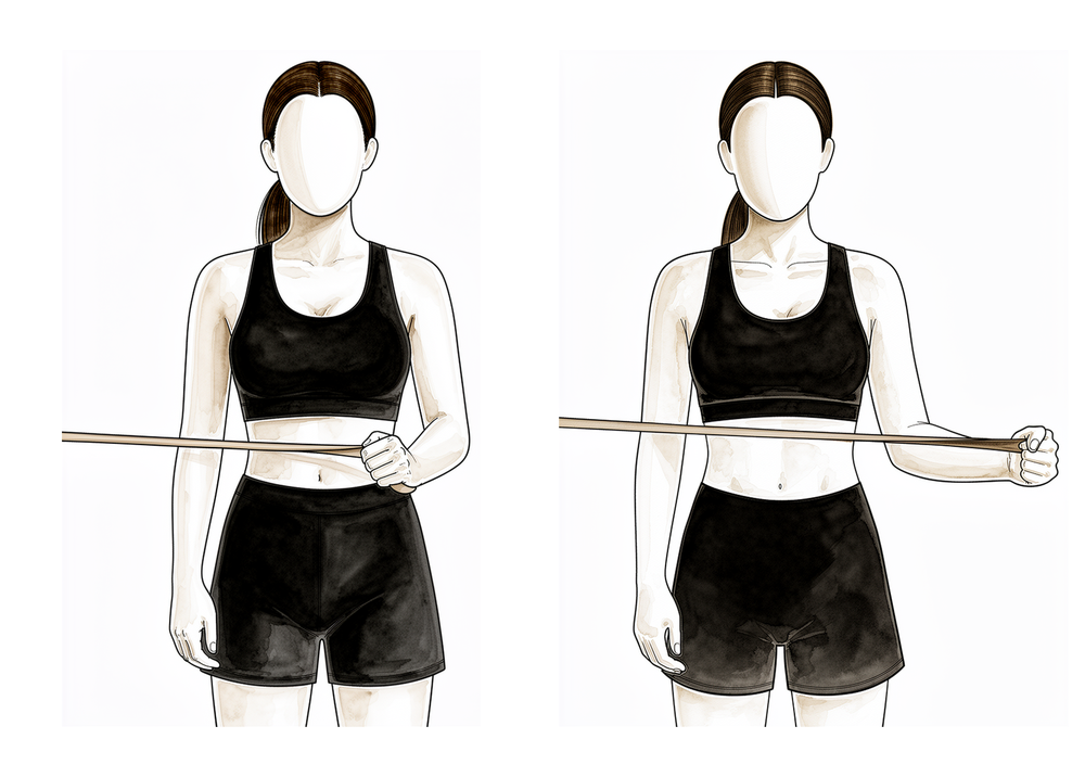
Rotación externa con banda elástica
Párese con el codo pegado al costado y doblado en ángulo recto, sosteniendo una banda elástica fijada a la altura de la cintura. Manteniendo el codo junto al cuerpo, gire el antebrazo hacia afuera contra la resistencia de la banda y luego regrese lentamente a la posición inicial. Este ejercicio fortalece el manguito rotador con el brazo junto al cuerpo, que es el punto de partida para el fortalecimiento de la rotación.
2 a 3 series de 8 a 12 repeticiones, con baja resistencia.
Kieran Hirpara 4.0
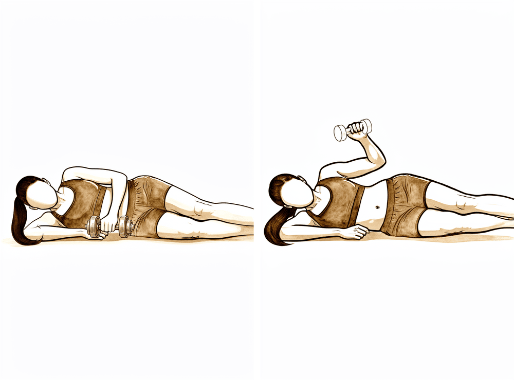
Rotación externa en decúbito lateral con peso
Acuéstese sobre el lado no operado, con el codo superior doblado en ángulo recto y pegado al cuerpo. Sosteniendo un peso ligero, gire el antebrazo hacia el techo y luego bájelo lentamente. Mantenga el peso bajo y realice más repeticiones a medida que recupere la fuerza del manguito rotador.
2 a 3 series de 8 a 12 repeticiones, con carga baja.
Kieran Hirpara 4.0
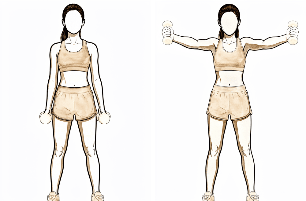
Elevación en «lata llena» (plano escapular)
Sostenga un peso ligero con el pulgar apuntando hacia arriba; luego eleve el brazo en una dirección diagonal hacia adelante y hacia afuera —no directamente hacia el costado— hasta aproximadamente la altura del hombro, y bájelo lentamente. Este ejercicio trabaja tanto la parte frontal del hombro como el manguito rotador. Utilice siempre un peso ligero y deténgase si siente algún dolor persistente.
2 a 3 series de 8 a 12 repeticiones, con carga baja.
Kieran Hirpara 4.0
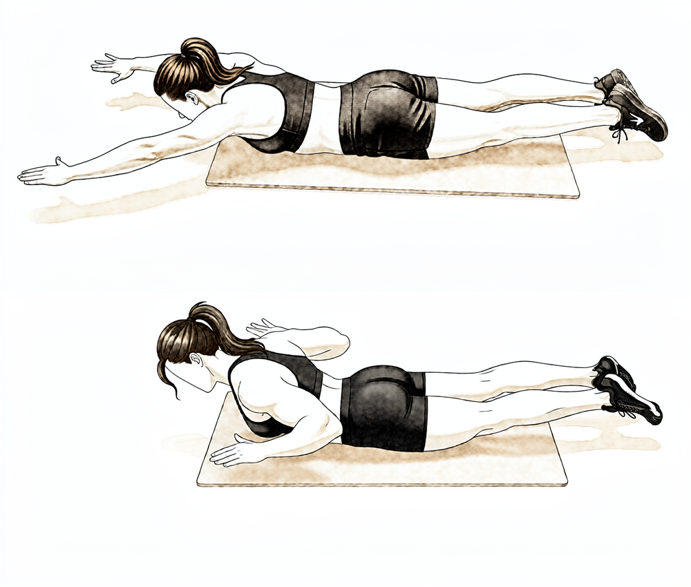
Remo en posición prona
Acuéstese boca abajo con el brazo operado colgando hacia el suelo. Levante el codo hacia el techo, acercando la escápula a la columna vertebral, y luego bájelo lentamente. Comience sin ningún peso y mantenga el movimiento bajo control.
Según las indicaciones de su fisioterapeuta.
Kieran Hirpara 4.0
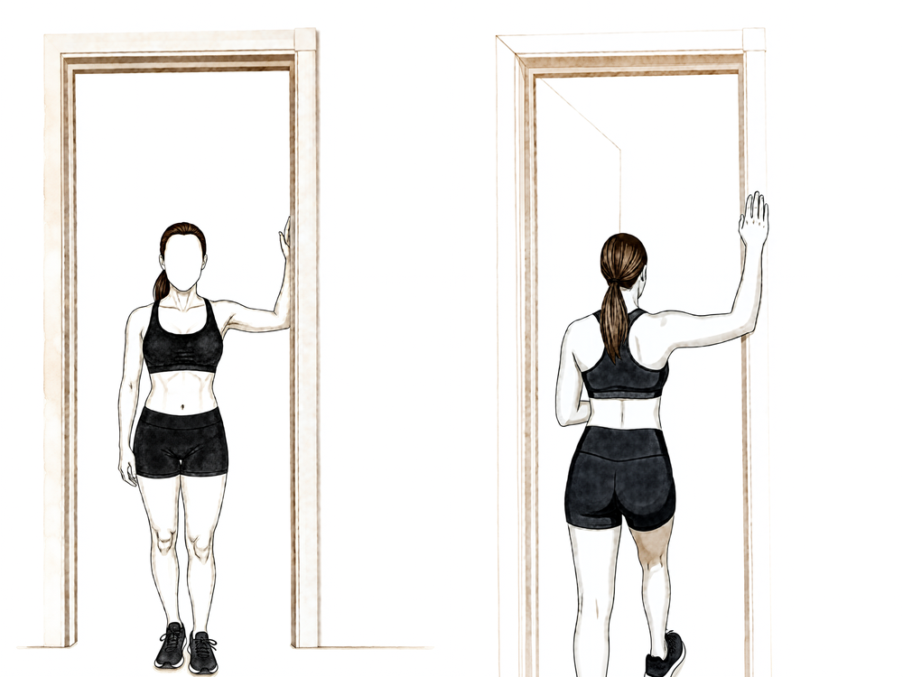
Estiramiento torácico en el umbral de la puerta
Párese en el umbral de una puerta, con los antebrazos apoyados en el marco y los codos a la altura de los hombros. Pase un pie al otro lado y incline su cuerpo suavemente hacia adelante hasta sentir un estiramiento en la parte frontal del pecho; luego mantenga esa posición. Haga el movimiento con suavidad y nunca fuerce.
Mantenga la posición durante ~20–30 segundos, según las indicaciones de su fisioterapeuta.
Kieran Hirpara 4.0
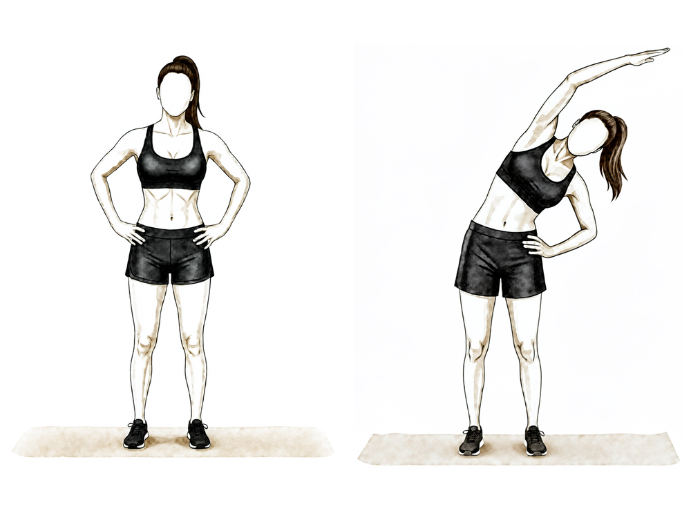
Estiramiento del dorsal ancho (lado de la espalda)
Levante el brazo operado por encima de la cabeza; si es necesario, use la mano sana para guiarlo. Incline su cuerpo suavemente hacia el lado opuesto hasta sentir un estiramiento a lo largo del costado de la espalda, y luego mantenga esa posición. Asegúrese de que sea una sensación cómoda y que se mantenga dentro de su rango de movilidad.
Mantenga la posición durante ~20–30 segundos, según las indicaciones de su fisioterapeuta.
Una vez consolidada la fractura y recuperado el movimiento activo, se centra la atención en recuperar la fuerza muscular. El entrenamiento de resistencia comienza de forma suave (con bandas elásticas y pesos ligeros para el manguito rotador, el deltoides y los músculos de la escápula) y avanza gradualmente. Se continúa con los estiramientos, buscando alcanzar un rango de movimiento completo en todas las direcciones, incluido el movimiento hacia atrás de la espalda. En esta fase, las actividades cotidianas normales suelen volver a la normalidad, y las actividades recreativas más ligeras también se reanudan, siempre bajo la orientación de su fisioterapeuta.
Para su fisioterapeuta:
Objetivos
- Rango de movimiento activo completo en todos los planos, con mecánica normal
- Recuperación progresiva de la fuerza y resistencia del manguito rotador, deltoides y músculos estabilizadores de la escápula
Manejo
- Pasar de ejercicios isométricos a ejercicios con bandas elásticas y, posteriormente, con pesos libres ligeros (aproximadamente 0.5–2 kg) para el manguito rotador, deltoides y músculos estabilizadores de la escápula: carga baja y mayor número de repeticiones (por ejemplo, 2–3 series de 8–12 repeticiones); sesiones de resistencia unas 3 veces por semana para evitar sobrecargas.
- El fortalecimiento de la rotación se inicia con el brazo a un lado, por debajo de la altura del hombro.
- Enfatizar el trabajo del deltoides anterior y la acción conjunta del trapecio y el serrato anterior para lograr una base escapular estable.
- Programa de flexibilidad dirigido a alcanzar el límite del rango de movimiento en todos los planos: estiramiento de la cápsula posterior (cruzando el cuerpo), rotación interna detrás de la espalda, estiramientos de la pared torácica anterior y del pectoral menor, así como estiramientos en el marco de una puerta.
- Iniciar la rotación interna detrás de la espalda y realizar movilizaciones de grado III–IV según lo permita el rango de movimiento.
- Uso de ergómetro de parte superior del cuerpo con baja resistencia; entrenamiento aeróbico general.
Precauciones
- El fortalecimiento debe realizarse dentro de un rango cómodo; no debe provocar dolor persistente.
- Durante esta fase, no se deben levantar cargas superiores a 4–5 kg con el brazo operado.
- Evitar soportar demasiado peso a través del brazo (flexiones y ejercicios similares se harán más adelante).
Criterios para avanzar
- Rango de movimiento activo completo, sin necesidad de estrategias compensatorias.
- Tolerancia al programa de fortalecimiento sin reaparición de dolor ni pérdida del rango de movimiento.
Fase IV — Retorno a la actividad plena (a partir de los 4½–6 meses)
La fase final consiste en un retorno gradual a actividades que requieren levantar pesos, trabajo manual, tareas por encima de la cabeza y práctica deportiva. El entrenamiento de fuerza avanza hacia resistencias mayores y ejercicios compuestos; además, cuando sea relevante para su trabajo o deporte, se incorporan ejercicios más rápidos y dinámicos. La mayoría de las personas vuelven a sus actividades habituales alrededor de los seis meses, aunque la fuerza y la confianza siguen mejorando hasta un año después. El momento adecuado para finalizar el proceso depende de las funciones que deba desempeñar el brazo; por ello, el retorno a trabajos manuales pesados o a deportes de contacto y actividades por encima de la cabeza se acuerda con el Dr. Hirpara y su fisioterapeuta, en lugar de determinarse únicamente por el calendario.
Para su fisioterapeuta:
Objetivos
- Retorno a todas las actividades laborales, recreativas y deportivas
- Que la fuerza del brazo operado se acerque a la del otro brazo
Manejo
- Aumento progresivo de la resistencia mediante bandas elásticas, pesas libres y ejercicios compuestos en el gimnasio
- Progresión en flexiones (desde la pared hasta el suelo, pasando por el banco y las rodillas) y ejercicios de estabilidad en cadena cerrada según tolerancia
- A partir de los 4½ meses: carga excéntrica, ejercicios pliométicos (con pelotas pesadas), así como ejercicios propioceptivos y de estabilización rítmica, según sea pertinente
- Rotación resistida a 90° de elevación, además de programas deportivos o laborales específicos, según corresponda
Precauciones
- La progresión debe guiarse por la ausencia de síntomas: si aparece dolor o disminución del rango de movimiento, se debe reducir la intensidad y recuperar primero un movimiento cómodo
Criterios para dar por finalizado el tratamiento
- La fuerza del brazo operado alcanza al menos el 80 % de la del otro brazo, según medición
- No hay dolor durante el fortalecimiento progresivo
- El paciente puede realizar de forma independiente el programa de mantenimiento en casa
Después de seguir el protocolo
Las fases descritas anteriormente se han adaptado de protocolos de rehabilitación publicados para la fijación de fracturas del húmero proximal: Massachusetts General Brigham Sports Medicine, Twin Cities Orthopedics, el UConn Musculoskeletal Institute, el NYU Langone Orthopedic Center y South Bend Orthopaedics, además de una revisión sistemática sobre la rehabilitación tras este tipo de fracturas. Los protocolos publicados para esta intervención varían más que para la mayoría de las cirugías de hombro, ya que el ritmo adecuado depende de cómo se haya fijado la fractura y de su proceso de curación; por ello, su avance a través de las fases será guiado por el Dr. Hirpara en sus revisiones y ajustado por su fisioterapeuta entre dichas revisiones. Esta página complementa las indicaciones generales de recuperación de la clínica; consulte control del dolor postoperatorio y cuidado de la herida. Para información sobre la cirugía en sí, consulte fijación del húmero proximal.
Si desea conocer la evidencia científica que sustenta este protocolo (los estudios sobre el movimiento temprano frente al retrasado tras la fijación con placa, el debate entre cirugía y uso de cabestrillo, así como las complicaciones que el avance gradual busca prevenir), dispondrá de un resumen documentado con todas las referencias, disponible en formato PDF junto a esta página.
Evidence & references
This is the clinical evidence summary written for health professionals. It is technical, and it lists the research this page was built from. You do not need to read it to understand your treatment or to make a decision about it.
Proximal Humerus Fracture Fixation (Locking-Plate ORIF) — Post-operative Rehabilitation: Evidence
Topic scope: Post-operative rehabilitation after open reduction and internal fixation (ORIF) of a proximal humerus fracture with a locking (angular-stable) plate and screws. This page covers the evidence behind the phased rehabilitation program — early protected/passive motion, deferred active and resisted motion, and graded strengthening — and the surgical-outcome facts that shape it. It does not cover the separate pathways of non-operative fracture management, intramedullary nailing, hemiarthroplasty or reverse total shoulder arthroplasty, although the operative-versus-non-operative debate is summarised because it frames who is offered this operation at all.
Defining principle of the rehab here (mobilise early, protect the tuberosities): A locking plate is an angular-stable construct — the screws lock into the plate, so the fixation holds even in osteoporotic bone without relying on bone-to-screw friction. Because that stability is present from day one, this protocol now permits early active (and active-assisted) shoulder elevation in a pain-free range for stable fixation, consistent with the early-active-motion RCT evidence (Loew 2025), rather than holding the shoulder passive-only for six weeks. The element still keyed to biological healing is rotation and rotator-cuff loading: where the tuberosity fragments to which the rotator cuff attaches are part of the fracture, active/resisted rotation and cuff work are held back until those fragments unite, whereas where the tuberosities are uninvolved or solidly fixed the surgeon may clear cuff loading sooner. The brake is therefore biological, not mechanical — the plate is strong immediately — and it is now selective (rotation/cuff), not a blanket movement ban. Progression of cuff loading remains governed by fracture stability and radiographic healing, not the calendar. This places the protocol close to the early-active-movement end of the spectrum while still being more tuberosity-aware than a pure debridement/decompression, and far less tuberosity-dependent than a fracture arthroplasty, where healing of the tuberosities to the prosthesis dictates a slower, stricter cuff-loading timetable.
The operation
A proximal humerus fracture is a break of the upper end of the arm bone, near the shoulder. In ORIF the fragments are realigned (reduced) and held with a pre-contoured locking plate on the outer surface of the bone, fixed with multiple locking screws into the humeral head. Where the tuberosities (the bony knobs carrying the rotator-cuff attachments) are part of the fracture, they are reduced and secured to the plate, often reinforced with heavy sutures through the cuff. The plate provides immediate mechanical stability; the rehabilitation then protects the biological healing of the fracture and the tuberosity fragments.
Evidence by theme
1. Modern locking plates permit early active motion — supported by RCT evidence and adopted here for stable fixation
The historical "restrictive" protocol kept the arm immobilised with no active movement for ~6 weeks, with passive limits of flexion to ~90°, external rotation to ~20° and internal rotation to the belly. The rationale for early motion is to prevent the shoulder stiffening (adhesive capsulitis is a recognised complication of these fractures) while still protecting the bone. Because an angular-stable locking plate is mechanically strong from day one, early active elevation can be permitted for stable fixation; the element kept keyed to biological healing is rotation and rotator-cuff loading, because that is what pulls on the tuberosity fragments.
The more aggressive question — can patients move actively from the start? — has now been tested. A prospective randomised controlled trial (Loew et al., J Orthop, 2025) compared a conventional 4-week sling-immobilisation group against an early functional group with no movement or force restrictions after locking-plate ORIF (both groups avoided heavy lifting and impact for 3 months). At 24 months there was no significant difference in DASH or Constant score: Constant score averaged 81.3 (conventional) vs 78.4 (early functional), with relative Constant score 89.8% of the uninjured side in both groups — i.e. early active motion was non-inferior. Moderate (single RCT). This is consistent with the broader signal that early intensive mobilisation yields similar outcomes to conventional later mobilisation after operative treatment. In line with this evidence, Dr Hirpara's protocol now permits early active elevation in a pain-free range for stable fixation; the literature still has no consensus on the optimal regimen, so the one element kept deliberately cautious and keyed to radiographic healing is active/resisted rotation and rotator-cuff loading, because that is the movement that stresses the healing tuberosity fragments.
2. Post-operative protocols are highly heterogeneous, but converge on short immobilisation and early passive ROM
The best summary of practice is a systematic review of 45 cohorts (40 articles, 3,507 patients, 3,519 fractures) (Budharaju et al., Shoulder Elbow, 2024). Across studies:
- Sling immobilisation averaged 3.1 weeks (most commonly 3 weeks; range 0–6).
- Passive ROM began at ~0.9 weeks on average (most commonly at 2 days).
- Active ROM began at ~2.5 weeks on average (most commonly at 3 weeks).
- Strengthening began at ~5.5 weeks on average (most commonly at 6 weeks).
The authors emphasised substantial variability regardless of management, concluded that this heterogeneity limits cross-study comparison, and noted that early mobilisation may produce superior function — supporting consideration of shorter immobilisation. The synthesis page's "~3 weeks in the sling, then wean" reflects this averaged convention, not a trial-proven optimum. Moderate for the descriptive pattern; weak/consensus for any specific timetable.
3. Progression is governed by fracture stability and healing, not the calendar — because of the tuberosities and biological complications
The locking plate is strong immediately; what limits the rehab is the bone. Two facts anchor the "wait-for-healing" rule:
- Tuberosity / cuff loading. When the tuberosities are part of the fracture, loading the rotator cuff (forced/resisted external rotation and resisted cuff work) too early risks displacing fragments before they unite. This is the explicit reason the early phases keep rotation cautious and defer cuff loading until healing, even though early active elevation in a pain-free range is permitted; where the tuberosities are uninvolved or solidly fixed, the surgeon may clear cuff loading sooner.
- The major complications of locking-plate ORIF are largely biological and mechanical, and several are loading- and reduction-sensitive. A systematic review (Thanasas / Brorson-class series) reports the commonest complications as intra-articular screw perforation (~9–12%), varus collapse (~6.8%), loss of reduction, avascular necrosis of the humeral head (~4.6%, reported range 0–15%), subacromial impingement (~5%), adhesive capsulitis (~4%), nonunion (~1.5%) and deep infection (~1.4%). Screw perforation and avascular necrosis frequently coincide, because a head that collapses or undergoes AVN lets fixed-length locked screws migrate into the joint. Moderate (pooled observational series).
This complication profile is why progression waits on radiographs: premature loading risks tipping a borderline reduction into varus collapse or screw cut-out. Some surgeons even advocate early planned plate removal to avoid secondary screw penetration once the head shows AVN/collapse (Dimitriou et al., J Orthop, 2019) — a salvage strategy, not part of routine rehab, but it illustrates how mechanical and biological failure interact.
4. Who is offered ORIF at all — the operative-versus-non-operative debate (PROFHER and after)
The single most influential trial is PROFHER (Rangan et al., JAMA, 2015; 250 patients, 32 UK centres, displaced fractures involving the surgical neck): surgery showed no important difference in Oxford Shoulder Score versus sling-based non-operative care over 2 years, and was more expensive. The 5-year follow-up (Handoll/Keding et al., Bone Joint J, 2017) confirmed no significant difference in shoulder function or quality of life persisting to 5 years. A smaller RCT in displaced 3-part fractures in the elderly (Fjalestad et al., J Shoulder Elbow Surg, 2012) similarly found no functional advantage to internal fixation over non-operative care in that group, and a systematic review and meta-analysis (Beks et al., J Shoulder Elbow Surg, 2018) found no clear superiority of operative treatment across observational and randomised data combined. Strong (multiple RCTs + SR-MA).
The clinical upshot — and the reason this matters to a rehab page — is that ORIF is selectively indicated, typically in younger patients, in fractures where reduction and stable fixation are achievable and worthwhile, and where the alternative (non-operative care or arthroplasty) is judged less favourable. The decision is individualised; "difficulty in decision-making" for displaced fractures is itself documented as affecting outcomes (Okike et al., J Shoulder Elbow Surg, 2018). Patients should understand that being offered ORIF is a considered judgement, not an automatic consequence of the fracture.
5. Adjacent rehabilitation evidence (non-operative immobilisation duration)
Although it concerns non-operatively treated fractures, a relevant randomised controlled trial (Tanji et al., J Bone Joint Surg Am, 2021) compared 1 versus 3 weeks of immobilisation and supports the broad theme that earlier movement is at least as good as longer immobilisation for many proximal humerus fractures. It does not directly govern the post-ORIF protocol but reinforces the same direction-of-travel away from prolonged slings. Moderate (RCT, non-operative population).
Phased post-operative timeline (consistent with the synthesis page)
| Phase | Window | Sling | Shoulder motion | Strengthening | Governing rule |
|---|---|---|---|---|---|
| I — Protection & early passive motion | Weeks 0–6 | Full-time ~3 weeks, then weaned; off for exercises/hygiene; not in bed | Early active-assisted/active elevation permitted in a pain-free range from the outset (supine-start → upright); passive as needed; pendulums; elbow/wrist/hand active. ER kept to a gentle ~30–40° default; rotation cautious — defer active/resisted cuff work until tuberosities heal (surgeon-guided) | None at the shoulder (deltoid/periscapular isometrics as comfort allows) | Protect fixation + tuberosities; settle pain/swelling |
| II — Regaining active movement | Weeks 6–12 | Discarded by the 6-week review at the latest | Active-assisted → active in all planes (supine-start elevation); submaximal cuff/deltoid isometrics once healing confirmed | Light elbow/scapular work; no resisted cuff work until union (typically not before 8–12 weeks) | Radiographic healing at the ~6-week review gates active motion |
| III — Strengthening | ~Months 3–4½ | Off | Full active ROM in all planes, including behind-the-back | Graded bands → light free weights (~0.5–2 kg) for cuff, deltoid, scapula | Union confirmed; symptom-guided load progression |
| IV — Return to full activity | ~4½–6 months | Off | Full, with dynamic/overhead drills as relevant | Heavier resistance, compound + sport/work-specific | Return-to-task agreed with surgeon/physio, not the calendar |
The phase boundaries are individualised at Dr Hirpara's reviews on the basis of X-ray healing — this is the operative consequence of the "stability- and healing-governed, not calendar-governed" principle.
Key controversies / evidence quality
-
Early active versus delayed (protected) mobilisation after ORIF. A single RCT (Loew 2025) found unrestricted early active motion non-inferior to 4-week immobilisation at 24 months, and the broader literature leans toward shorter immobilisation. The protocol here aligns with that early-active-motion evidence: early active and active-assisted elevation in a pain-free range is permitted for stable fixation, rather than holding the shoulder passive-only for six weeks. Because there is still no consensus and no large confirmatory trial (protocols remain heterogeneous — Budharaju 2024), the protocol keeps one deliberate, evidence-aware caution: active/resisted rotation and cuff loading is keyed to radiographic tuberosity healing rather than released wholesale on day one. Moderate evidence, unsettled.
-
Operative versus non-operative treatment of displaced fractures. PROFHER (2015) and its 5-year follow-up (2017), plus an elderly-3-part RCT (Fjalestad 2012) and a meta-analysis (Beks 2018), found no clear functional benefit of surgery on average — which is why ORIF is selectively, not routinely, offered. The trials enrolled broad/older populations; the subgroup most likely to benefit from fixation (younger patients, good bone, reconstructable head-preserving fractures) is exactly where this operation is concentrated. Strong evidence overall; subgroup benefit remains debated.
-
The post-operative rehab protocol itself is consensus/expert. No high-level RCT defines the optimal phase structure, ROM limits or strengthening onset after ORIF. The timings here are drawn from published surgeon protocols and the systematic-review averages, individualised at review. Weak/consensus.
Evidence-strength flags (summary)
- STRONG (RCT / SR-MA): operative versus non-operative equivalence on average for displaced fractures (PROFHER 2-yr JAMA 2015 + 5-yr Bone Joint J 2017; Fjalestad 2012 RCT; Beks 2018 SR-MA).
- MODERATE (single RCT / pooled series): early active motion non-inferior to immobilisation after ORIF at 24 months (Loew 2025 RCT); 1-vs-3-week immobilisation non-operatively (Tanji 2021 RCT); locking-plate complication profile — screw perforation, varus collapse, AVN, etc. (pooled observational series); descriptive practice pattern of short sling + early passive ROM (Budharaju 2024 SR of 45 cohorts).
- WEAK / CONSENSUS: the specific phased rehabilitation timetable after ORIF (no defining rehab RCT; published surgeon protocols + systematic-review averages; progression individualised by radiographic healing).
Citations
RAG corpus (180,000+ Orthopaedic articles) — real DOIs
- Five-year follow-up results of the PROFHER trial comparing operative and non-operative treatment of adults with a displaced fracture of the proximal humerus. Bone Joint J. 2017. DOI: 10.1302/0301-620x.99b3.bjj-2016-1028
- Operative versus nonoperative treatment of proximal humeral fractures: a systematic review, meta-analysis, and comparison of observational studies and randomized controlled trials. J Shoulder Elbow Surg. 2018. DOI: 10.1016/j.jse.2018.03.009
- Internal fixation versus nonoperative treatment of displaced 3-part proximal humeral fractures in elderly patients: a randomized controlled trial. J Shoulder Elbow Surg. 2012. DOI: 10.1016/j.jse.2010.12.018
- One Versus 3-Week Immobilization Period for Nonoperatively Treated Proximal Humeral Fractures: a randomized controlled trial. J Bone Joint Surg Am. 2021. DOI: 10.2106/jbjs.20.02137
- Contemporary Management of Proximal Humeral Fractures. J Am Acad Orthop Surg. 2024. DOI: 10.5435/jaaos-d-24-01073
- The Use of Precontoured Humeral Locking Plates in the Management of Displaced Proximal Humerus Fracture. J Am Acad Orthop Surg. 2009. DOI: 10.5435/00124635-200909000-00005
- Use of locking plates in the treatment of proximal humerus fractures. J Shoulder Elbow Surg. 2010. DOI: 10.1016/j.jse.2010.01.001
- Functional results and unfavorable events after treatment of proximal humerus fractures using a new locking plate system. BMC Musculoskelet Disord. 2023. DOI: 10.1186/s12891-023-06176-5
- Fracture site augmentation with calcium phosphate cement reduces screw penetration after open reduction–internal fixation of proximal humeral fractures. J Shoulder Elbow Surg. 2012. DOI: 10.1016/j.jse.2011.09.017
- Difficulty in decision making in the treatment of displaced proximal humerus fractures: the effect of uncertainty on surgical outcomes. J Shoulder Elbow Surg. 2018. DOI: 10.1016/j.jse.2017.09.033
Literature (URLs)
- Loew M, et al. Postoperative treatment of proximal humerus fractures with an early active motion protocol: a prospective randomized controlled trial. J Orthop. 2025. https://www.sciencedirect.com/science/article/pii/S1058274625001867 (Constant 81.3 conventional vs 78.4 early-functional at 24 months; early active motion non-inferior)
- Budharaju A, Hones KM, Hao KA, et al. Rehabilitation protocols in proximal humerus fracture management: a systematic review. Shoulder Elbow. 2024;16(4):449–458. https://pmc.ncbi.nlm.nih.gov/articles/PMC11437559/ (45 cohorts; sling 3.1 wk, passive 0.9 wk, active 2.5 wk, strengthening 5.5 wk; early mobilisation may improve function)
- Rangan A, et al. (PROFHER). Surgical vs nonsurgical treatment of adults with displaced fractures of the proximal humerus: the PROFHER randomized clinical trial. JAMA. 2015;313(10):1037–1047. https://pubmed.ncbi.nlm.nih.gov/25756440/ (250 patients; no important difference in Oxford Shoulder Score at 2 years)
- Complications associated with locking plate of proximal humerus fractures (systematic review of complication rates). PMC. https://pmc.ncbi.nlm.nih.gov/articles/PMC5858203/ (screw perforation ~9–12%, varus collapse ~6.8%, AVN ~4.6%)
- Avascular necrosis and posttraumatic arthritis after proximal humerus fracture internal fixation: evaluation and management. PMC. https://pmc.ncbi.nlm.nih.gov/articles/PMC9889581/ (AVN reported range 0–15%)
- Late screw-related complications in locking plating of proximal humerus fractures: a systematic review. Injury. https://www.sciencedirect.com/science/article/abs/pii/S0020138319306989
- Dimitriou D, et al. Early locking plate removal following ORIF of proximal humeral fractures could prevent secondary implant-related complications. J Orthop. 2019;17:106–109. https://pmc.ncbi.nlm.nih.gov/articles/PMC6919395/
Published rehabilitation protocols (patient-guidance — basis for the phase structure)
- Massachusetts General Brigham Sports Medicine. Rehabilitation Protocol for Proximal Humeral Fracture Open Reduction Internal Fixation (ORIF). https://www.massgeneral.org/assets/MGH/pdf/orthopaedics/sports-medicine/physical-therapy/rehabilitation-protocol-for-proximal-humeral-fracture-with-ORIF.pdf
- LaPrade CM. Post-Surgical Physical Therapy Protocol: Proximal Humerus Fracture ORIF. Twin Cities Orthopedics. https://tcomn.com/wp-content/uploads/2024/08/CML_Proximal-Humerus-ORIF-PT_10-2024.pdf
- Coyner KJ. ORIF Proximal Humerus Fractures Protocol. UConn Musculoskeletal Institute. https://www.drcoyner.com/pdf/orif-proximal-humerus-fractures-protocol.pdf
- Jazrawi LM. Rehabilitation Protocol: Proximal Humerus Open Reduction & Internal Fixation (ORIF). NYU Langone Orthopedic Center. https://www.newyorkortho.com/pdf/proximal-humerus-fracture-orif-post-op-instructions-and-rehab.pdf
- South Bend Orthopaedics. ORIF Proximal Humerus Fracture Rehab Protocol. https://www.sbortho.com/wp-content/uploads/2023/09/br-pt-fracture-orif-proximal-humerus.pdf