Patients › Rehabilitation
Liberación del túnel radial
Post-operative exercises and precautions after radial tunnel release, including radial nerve glides.
Este protocolo le servirá de guía durante su recuperación tras la liberación del túnel radial realizada por el Dr. Kieran Hirpara en el Mater Private Hospital Rockhampton. Explica qué puede esperar durante las semanas posteriores a la cirugía y detalla el programa de ejercicios que figura en el folleto postoperatorio. Lleve esta página o su versión en PDF a su primera sesión de fisioterapia o terapia de la mano para garantizar una rehabilitación coordinada. Su terapeuta podrá modificar el plan según el progreso de su recuperación.
Si tiene alguna duda respecto a la herida quirúrgica, no dude en contactar con el consultorio. A menudo resulta útil tomar una fotografía de la herida y enviarla por correo electrónico para su evaluación.
Qué esperar
El cuidado de la herida se explica en las indicaciones de cuidado de heridas del consultorio. A medida que la herida cicatriza, el nervio liberado puede adherirse al tejido circundante; los ejercicios de deslizamiento que se describen a continuación son muy importantes para mantenerlo en movimiento y evitar que quede fijado.
En ocasiones, la herida puede volverse sensible. Esto es normal, y se puede prevenir o minimizar iniciando una desensibilización diaria: tocando y frotando suavemente la herida (o el vendaje), comenzando inmediatamente después de la cirugía. Este tipo de “retroalimentación sensorial” ayuda al nervio a normalizar su respuesta al tacto y a las texturas.
Una vez que la herida haya cicatrizado por completo, comience a masajear la cicatriz: realice círculos firmes sobre la incisión. Consulte las indicaciones de cuidado de heridas para obtener más información sobre el manejo de cicatrices.
Es importante tener expectativas realistas respecto a la recuperación. El nervio radial debe desplazarse y estirarse una distancia considerable durante los movimientos habituales del brazo; por eso, mantenerlo en movimiento desde el principio evita que se adhiera a los tejidos en proceso de cicatrización [1]. Aun así, el alivio del dolor tras la liberación del túnel radial suele ser gradual y no inmediato; en algunas personas, el alivio es solo parcial. En realidad, esta intervención es menos predecible que otros procedimientos de liberación nerviosa: el síndrome del túnel radial es difícil de diagnosticar con certeza y a menudo se superpone al codo de tenista, lo cual explica las variaciones en los resultados. Los estudios a largo plazo publicados indican buenos resultados en aproximadamente dos tercios de los pacientes en general, siendo los mejores resultados aquellos con síntomas exclusivos del túnel radial [2][3]. La recuperación suele ser más lenta y menos completa cuando también existe codo de tenista (epicondilitis lateral), más de una compresión nerviosa en el mismo brazo o cuando se trata de un caso cubierto por seguros de accidentes laborales [2][4]. Su programa de deslizamiento nervioso y la desensibilización gradual son los aspectos de la rehabilitación que usted puede controlar mejor; la práctica diaria constante brinda al nervio la mejor oportunidad de recuperarse.
Precauciones y limitaciones
Se recomienda un uso funcional moderado de la mano para tareas cotidianas como el aseo personal, la alimentación, vestirse, escribir y teclear. Más allá de eso, las limitaciones son sencillas: no se debe levantar peso, agarrar objetos, soportar carga ni utilizar maquinaria vibratoria (por ejemplo, herramientas eléctricas o cortadoras de césped) durante las primeras 6 semanas tras la cirugía; además, la conducción queda restringida durante las primeras 2–3 semanas.
Para su fisioterapeuta:
Objetivos
- Evitar que el nervio liberado se adhiera a la herida en proceso de cicatrización (programa de deslizamiento nervioso)
- Reducir la sensibilidad de la herida mediante una desensibilización gradual
- Mantener el rango de movimiento de la muñeca, el antebrazo y el codo
- Facilitar el uso funcional moderado de la mano en actividades de la vida diaria
Manejo
- Desensibilización diaria: golpecitos o fricción suaves sobre la herida (vendaje), comenzando inmediatamente después de la cirugía
- Masaje de la cicatriz (círculos firmes sobre la incisión) una vez que la herida haya cicatrizado por completo
- Programa de ejercicios en casa según las fichas siguientes: estiramiento de flexión/extensión de muñeca; estiramiento de supinación/pronación de muñeca; flexión/extensión de codo; deslizamientos del nervio radial
- Se prefieren técnicas de deslizamiento nervioso suaves (“slider”) en lugar de tensiones bruscas en los límites de movimiento: estas técnicas logran un mayor desplazamiento nervioso con una tensión mucho menor, lo cual es más tolerable para un nervio recientemente descomprimido [1][5]
- La movilización nerviosa puede considerarse como complemento al programa; la evidencia científica sobre la movilización neural en afecciones nerviosas es favorable aunque con grados variables de certeza; por ello, la progresión debe guiarse por los síntomas [6]
Precauciones
- Solo se permite un uso funcional moderado de la mano (aseo personal, alimentación, vestirse, escribir, teclear)
- No se debe levantar peso, agarrar objetos, soportar carga ni utilizar maquinaria vibratoria (p. ej., herramientas eléctricas, cortadoras de césped) durante las primeras 6 semanas tras la cirugía
- La conducción queda restringida durante las primeras 2–3 semanas
- Los deslizamientos y estiramientos nerviosos deben ser suaves y prácticamente indoloros; evite forzar el movimiento hasta rangos que reproduzcan el dolor nervioso previo a la operación
Estos son los ejercicios que figuran en su folleto postoperatorio; deben realizarse tras la cirugía y continuarse en casa según las indicaciones de su fisioterapeuta o terapeuta de mano. El número de repeticiones, el tiempo de mantenimiento y la frecuencia se indican en cada ficha.
Sus ejercicios
Kieran Hirpara 4.0
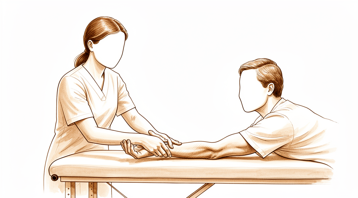
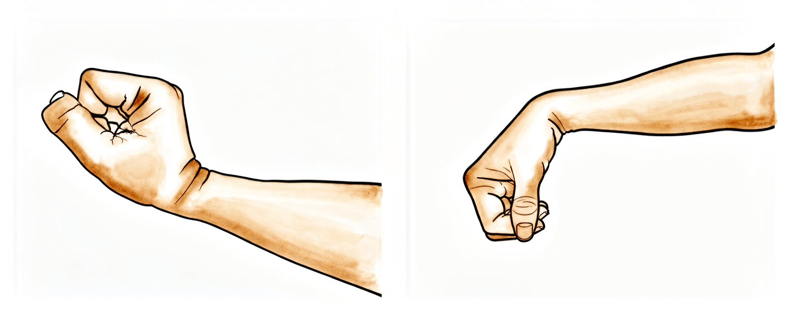
Estiramiento para flexión/extensión de la muñeca
Apoye el codo sobre una mesa y mueva su muñeca suavemente hacia atrás y hacia adelante (o apóyela en el borde de una mesa o sillón, tal como se muestra en la imagen). Cuando se sienta más cómodo, use la otra mano para presionar la palma y mover la muñeca hacia atrás y luego hacia adelante, manteniendo los dedos relajados. Mantenga cada estiramiento durante 15 segundos; repita el ejercicio 5 veces en cada dirección.
10 repeticiones, 4–5 veces al día.
Kieran Hirpara 4.0
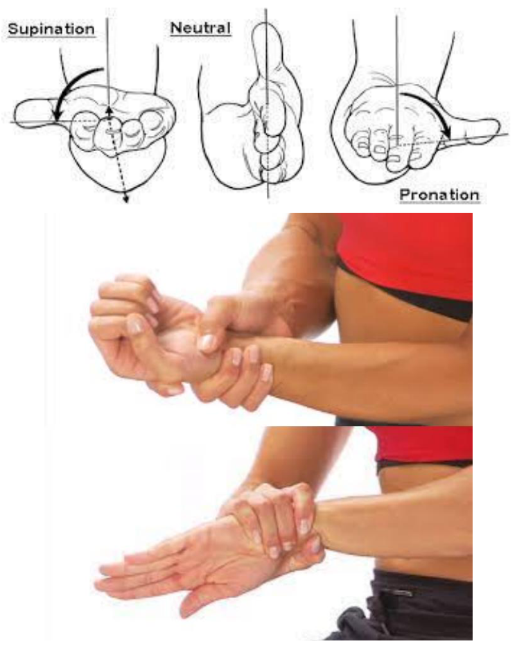
Estiramiento de supinación/pronación de la muñeca
Flexione el codo junto al cuerpo, con la palma orientada hacia el techo. Gire la mano de modo que la palma quede hacia arriba y mantenga esa posición durante 15 segundos; luego gire la palma hacia abajo y manténgala otros 15 segundos. Repita este proceso 5 veces. A medida que se sienta más cómodo, utilice la mano opuesta — sujetando por la MUÑECA, no por la mano — para empujar suavemente la rotación un poco más en cada posición hasta sentir un estiramiento.
5 repeticiones, 4 veces al día, 5–7 días por semana.
Kieran Hirpara 4.0
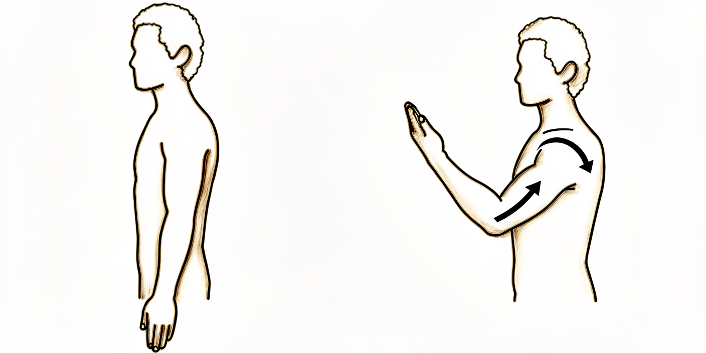
Flexión/extensión del codo
Con el brazo a lo largo del cuerpo, estire suavemente el antebrazo y el codo tanto como pueda. Mantenga esta posición de estiramiento máximo durante 3–5 segundos. Repita el ejercicio en la dirección opuesta: doble el antebrazo hacia arriba e intente tocar su hombro con la mano.
10 repeticiones, 2 veces al día, a diario.
Kieran Hirpara 4.0
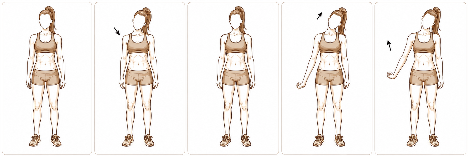
Deslizamientos del nervio radial
Párese con los brazos relajados a los costados. Baje el hombro y estire los dedos hacia el suelo. Realice una rotación interna del brazo (con el pulgar orientado hacia el cuerpo) y flexione la muñeca, con la palma hacia arriba. Incline suavemente la cabeza en dirección opuesta al lado que está estirando, luego eleve el brazo hacia arriba y lejos del cuerpo. Mantenga cada posición durante 3 a 5 segundos.
5–8 repeticiones, 2–4 veces al día, 6–7 días a la semana
Este programa de ejercicios fue elaborado en colaboración con Sarah Farrell, Licenciada en Terapia Ocupacional (BOccThy) y Terapeuta de Mano Acreditada (AHT).
Después de seguir este protocolo
Este protocolo se complementa con las recomendaciones generales de recuperación del consultorio; consulte control del dolor postoperatorio, cuidado de la herida y conceptos básicos de la terapia de la mano. En cuanto a la intervención quirúrgica en sí y la afección que trata, consulte liberación del túnel radial y síndrome del túnel radial.
Referencias
[1] Wright TW, Glowczewskie F, Cowin D, Wheeler DL. Desplazamiento y tensión del nervio radial en el codo y la muñeca durante el movimiento de las extremidades superiores. J Hand Surg Am. 2005;30(5):990–996. https://pubmed.ncbi.nlm.nih.gov/16182056/ [2] Lee JT, Azari K, Jones NF. Resultados a largo plazo de la liberación del túnel radial: efecto de la existencia simultánea de codo de tenista, múltiples síndromes de compresión y seguros de accidentes laborales. J Plast Reconstr Aesthet Surg. 2008;61(9):1095–1099. https://www.sciencedirect.com/science/article/abs/pii/S1748681507004044 [3] Sotereanos DG, Varitimidis SE, Giannakopoulos PN, Westkaemper JG. Resultados del tratamiento quirúrgico del síndrome del túnel radial. J Hand Surg Am. 1999;24(3):566–570. https://pubmed.ncbi.nlm.nih.gov/10357537/ [4] Naam NH, Nemani S. Síndrome del túnel radial. Orthop Clin North Am. 2012;43(4):529–536. (Síndrome del túnel radial, StatPearls.) https://www.ncbi.nlm.nih.gov/books/NBK555937/ [5] Coppieters MW, Butler DS. ¿Se deslizan realmente los “deslizadores” y se tensan los “tensadores”? Análisis de las técnicas neurodinámicas y consideraciones sobre su aplicación. Man Ther. 2008;13(3):213–221. https://pubmed.ncbi.nlm.nih.gov/17398140/ [6] Basson A, Olivier B, Ellis R, Coppieters M, Stewart A, Mudzi W. Eficacia de la movilización neural en afecciones neuromusculoesqueléticas: revisión sistemática y metaanálisis. J Orthop Sports Phys Ther. 2017;47(9):593–615. https://pubmed.ncbi.nlm.nih.gov/28704626/
Evidence & references
This is the clinical evidence summary written for health professionals. It is technical, and it lists the research this page was built from. You do not need to read it to understand your treatment or to make a decision about it.
Radial Tunnel Release — Evidence Brief & Post-operative Rehabilitation
Topic scope: post-operative rehabilitation after surgical decompression / neurolysis of the posterior interosseous nerve (deep branch of the radial nerve) in the radial tunnel of the proximal forearm, performed for radial tunnel syndrome (RTS). This is an elbow / proximal-forearm topic — anatomically and clinically distinct from carpal-tunnel and cubital-tunnel decompression. Like other nerve decompressions it is an early-motion pathway (early elbow/forearm/wrist motion, radial-nerve glides, oedema and scar care). The scope deliberately foregrounds the diagnostic controversy and the more variable, lower success rates that distinguish RTS release from the better-validated carpal-tunnel and cubital-tunnel operations.
Defining principle of the rehab here: a decompressed nerve does not create a healing construct that needs months of protection — it needs early, gentle movement to stop it adhering to the operative bed and to restore its glide. So the rehab is an early-motion programme: light functional hand use from day 1, radial-nerve sliders, graded desensitisation and (once healed) scar massage; heavier loading deferred to ~6 weeks. But two honesty caveats sit over the whole topic. First, RTS is a contested diagnosis — there is no confirmatory imaging or electrodiagnostic test, it is a diagnosis of exclusion, and a substantial body of opinion regards it as a variant of recalcitrant lateral epicondylitis. Second, outcomes after release are more variable and on average lower than carpal- or cubital-tunnel release — good results cluster around two-thirds overall, and fall further with co-existing tennis elbow, multiple compression sites, or a workers'-compensation context. Patient expectations should be set accordingly.
A. THE DIAGNOSTIC CONTROVERSY (read first — it frames everything)
RTS is among the most contested entities in upper-limb surgery, and the rehab brief is incomplete without it:
- No confirmatory test. Electromyography and nerve-conduction studies are characteristically normal in RTS (compression is intermittent/dynamic and predominantly of a motor nerve carrying few pain fibres), and MRI is frequently negative — denervation oedema in supinator/extensors is suggestive but inconsistent, and a normal scan does not exclude the diagnosis. RTS is therefore a clinical diagnosis of exclusion, resting on point tenderness ~4 cm distal to the lateral epicondyle (over the radial tunnel rather than the epicondyle), pain on resisted supination / resisted long-finger extension, and — for some surgeons — temporary relief from a diagnostic local-anaesthetic block at the radial tunnel.
- Overlap with lateral epicondylitis (tennis elbow). The two coexist frequently and share the lateral-elbow pain territory. A recognised school of thought holds that "RTS" is often severe, recalcitrant lateral epicondylitis rather than a discrete compression neuropathy. Importantly, routine PIN release added to lateral-epicondylitis surgery has not been shown to improve outcomes, so the diagnosis should be secure before a decompression is planned.
- Practical consequence. Surgery is a last resort after prolonged failed conservative care (activity modification, splinting, anti-inflammatories, sometimes a steroid injection), and is best reserved for patients with proximal-forearm pain and no better explanation. This uncertainty is the single most important reason post-operative expectations must be framed honestly.
B. RELEASE OUTCOMES (variable — and why)
- Headline success ~two-thirds. Across the older long-term series, roughly 67% good, 15% fair, 18% poor after radial tunnel decompression — markedly more variable than carpal- or cubital-tunnel release. A 2008 long-term series (Lee, Azari, Jones) and a 1999 series (Sotereanos et al.) both document this spread; the Sotereanos cohort reported good/excellent results in only ~39% by objective assessment (though ~64% by patient self-rating), underscoring how outcome depends on the metric used.
- Co-existing lateral epicondylitis lowers success. Success falls to roughly 40% when tennis elbow coexists, versus far higher with isolated RTS.
- Multiple compression sites and workers'-compensation context lower success — reported ~58% success in compensation cases vs ~73% without. These are the same modifiers named in the patient protocol.
- 2025 systematic review (Raymond et al., HAND). 11 studies, 401 limbs (381 patients). Outcomes were heterogeneous; a dorsal approach between ECRB and EDC was associated with the most favourable Roles-and-Maudsley scores and satisfaction. The review's central message is that the overall evidence is low-grade (observational), the diagnosis non-standardised, and the effectiveness of conservative treatment essentially untested — a "tendency" toward benefit rather than proof.
- Resorption-style "spontaneous improvement" does not apply here — unlike calcific tendinitis, RTS does not self-resolve through a biological cycle; conservative care manages symptoms rather than curing a deposit.
C. SURGICAL APPROACH (shapes the early rehab)
- What is done. Complete neurolysis of the radial nerve at its bifurcation, decompressing the deep branch (PIN) and superficial sensory branch, releasing the arcade of Frohse (the proximal supinator edge), the leash of Henry (radial recurrent vessels), the ECRB fascial edge, and the distal supinator border. Any constrictive bands or vessels are divided.
- Approaches. Dorsal (Thompson, between ECRB/EDC or the brachioradialis–ECRL interval), volar/anterior (Henry), or transmuscular. Anatomical studies map the trade-offs; the dorsal ECRB–EDC interval performed best in the 2025 review. The superficial radial branch matters — it is a recognised source of post-operative dysaesthesia if irritated.
- Rehab implication. A muscle-splitting/dorsal exposure through the extensor mass means early gentle forearm rotation and wrist motion are encouraged but heavy resisted supination/extension is deferred; the incision sits over a mobile, frequently sensitive area, so desensitisation and scar care carry real weight here.
D. POST-OP THERAPY ROLE (nerve/tendon glides, oedema, scar)
The decompressed nerve must glide, not adhere. The mechanical rationale is well quantified: the radial nerve translates and stretches a measurable amount across the elbow and wrist during ordinary arm motion (Wright et al. 2005), so early motion is what keeps it free of the healing bed.
- Early motion, immediately. Early active elbow, forearm and wrist movement within pain limits from the first post-op days; most protocols use no rigid splinting (or a removable splint for comfort/night only).
- Radial-nerve glides — favour "sliders" over "tensioners". Sliding (slider) neurodynamic techniques achieve substantially greater nerve excursion at much lower nerve strain than end-range tensioners — preferable around a freshly decompressed nerve. Neural-mobilisation evidence across neuromusculoskeletal conditions is supportive but of variable certainty, so progression is symptom-guided and essentially pain-free; mechanism work (e.g., the MONET protocol) is still maturing.
- Oedema and desensitisation. Graded desensitisation (tapping/rubbing over the dressing) from day 1 normalises touch and pre-empts a sensitive scar — particularly relevant given superficial- radial-branch proximity.
- Scar management once healed. Massage, pressure, and silicone are advocated to loosen skin–tissue adhesions and aid remodelling, started once the wound is closed/sutures out.
- Strengthening deferred. Light functional ADL use throughout; resisted strengthening of wrist/ elbow and fine-motor work introduced from ~6 weeks. Heavy work and vibration tools avoided to ~6–8 weeks.
Phased post-op timeline (maps to the patient protocol phases)
| Phase | Window | Splint | Motion / nerve work | Load / strengthening | Notes |
|---|---|---|---|---|---|
| I — Protect & glide | Day 0–2 wk | None, or removable for comfort/night | Early pain-free active elbow/forearm/wrist ROM; radial-nerve sliders; desensitisation from day 1 | Light functional ADL use only (self-care, feeding, dressing, writing, typing) | Stop the nerve adhering; settle the wound. No lifting/gripping/weight-bearing/vibration tools. Driving limited first 1–2 wk |
| II — Restore motion | 2–6 wk | Off | Progress full active + gentle assisted ROM; continue sliders; scar massage once healed | Still no resisted loading; ADL use continues | Sensitivity/dysaesthesia common and usually settles; keep glides gentle |
| III — Strengthen & return | ~6 wk onward | Off | Full ROM goal; sliders as needed | Begin graded wrist/elbow strengthening + fine-motor work from ~6 wk; advance work/heavy tasks thereafter | Vibration tools/heavy work resume ~6–8 wk. Pain relief is often gradual and may be partial — counsel accordingly |
E. COMPLICATIONS / DOWNSIDES
- Incomplete or no pain relief — the dominant "complication," tied directly to diagnostic uncertainty; relief is frequently gradual and sometimes partial.
- Superficial-radial-branch dysaesthesia / scar sensitivity — recognised; desensitisation and careful technique mitigate it.
- Transient PIN weakness (finger/thumb extension) from retraction — usually recovers.
- Adhesion/recurrence of symptoms if early glide is neglected.
- Standard wound risks (infection, haematoma) — uncommon.
F. KEY CONTROVERSIES / EVIDENCE QUALITY
- Does RTS exist as a discrete entity? Genuinely contested. No confirmatory test; substantial opinion equates much of it with recalcitrant lateral epicondylitis. This is the defining controversy and must shape consent and expectation-setting. Unresolved — expert opinion divided.
- Patient selection drives outcome more than technique. Isolated RTS does best; coexisting tennis elbow, multiple compressions, and compensation context predict worse results. Moderate (consistent across cohorts).
- Approach choice. A dorsal ECRB–EDC interval was favoured in the 2025 SR, but the evidence is observational and confounded by diagnostic heterogeneity. Weak–moderate.
- The rehab protocol itself is consensus/expert — drawn from surgeon and hand-therapy guidance (early motion, sliders, desensitisation, scar care), not from a rehab RCT. Phase timings are typical, not trial-derived. Weak / consensus.
- Conservative-treatment efficacy is essentially untested — the 2025 SR notes no usable trials of non-operative care, so "failed conservative management" before surgery rests on practice convention. Weak.
G. EVIDENCE STRENGTH FLAGS (summary)
- STRONG: the mechanical rationale for early nerve glide — quantified radial-nerve excursion/ strain across elbow and wrist (Wright et al. 2005); slider-vs-tensioner excursion/strain physiology.
- MODERATE: patient-selection modifiers of outcome (lateral epicondylitis, multiple compressions, workers' compensation lower success); ~two-thirds overall good-result rate from long-term cohorts; dorsal-approach signal from the 2025 systematic review (low-grade studies).
- WEAK / CONSENSUS: the existence and diagnostic criteria of RTS (no confirmatory test; overlap with lateral epicondylitis); the post-operative rehabilitation protocol (surgeon/ hand-therapy guidance, no rehab RCT); neural-mobilisation certainty (supportive but variable); efficacy of conservative care (essentially untested).
CITATIONS
RAG corpus (180,000+ Orthopaedic articles)
- Posterior Interosseous Nerve Compression in the Forearm, AKA Radial Tunnel Syndrome. HAND. 2022. DOI: 10.1177/15589447221122822
- Radial Tunnel Syndrome: Emphasis on the Superficial Branch of the Radial Nerve. J Hand Surg Eur. 2009. DOI: 10.1177/1753193408099832
- Anatomical Study of the Surgical Approaches to the Radial Tunnel. J Hand Surg Am. 2015. DOI: 10.1016/j.jhsa.2015.03.009
- MR Imaging Features of Radial Tunnel Syndrome: Initial Experience. Radiology. 2006. DOI: 10.1148/radiol.2401050028
- Management of Lateral Epicondylitis: Current Concepts. J Am Acad Orthop Surg (JAAOS). 2008. DOI: 10.5435/00124635-200801000-00004
- Uncommon Nerve Compression Syndromes of the Upper Extremity. J Am Acad Orthop Surg (JAAOS). 1998. DOI: 10.5435/00124635-199811000-00006
- Radial Nerve Excursion and Strain at the Elbow and Wrist Associated With Upper-Extremity Motion. J Hand Surg Am. 2005. DOI: 10.1016/j.jhsa.2005.06.008
- Evidence and Techniques in Rehabilitation Following Nerve Injuries. Hand Clin. 2013. DOI: 10.1016/j.hcl.2013.04.012
- Preventive Strategies, Exercises and Rehabilitation of Hand Compression Neuropathies. J Hand Ther. 2022. DOI: 10.1016/j.jht.2021.11.003
- Mechanisms of Neurodynamic Treatments (MONET): a protocol for a mechanistic study. BMC Musculoskelet Disord. 2024. DOI: 10.1186/s12891-024-07713-6
Radial-tunnel literature (URLs)
- Clinical Outcomes of Operative Management for Radial Tunnel Syndrome According to Surgical Approach: a Systematic Review. HAND. 2025. https://journals.sagepub.com/doi/10.1177/15589447251315761
- The Epidemiology of Radial Tunnel Syndrome and Its Overlap With Lateral Epicondylitis. J Hand Surg Am. 2023. https://www.jhandsurg.org/article/S0363-5023(23)00138-7/abstract
- Lee JT, Azari K, Jones NF. Long-term results of radial tunnel release — the effect of co-existing tennis elbow, multiple compression syndromes and workers' compensation. J Plast Reconstr Aesthet Surg. 2008. https://www.sciencedirect.com/science/article/abs/pii/S1748681507004044
- Sotereanos DG, et al. Results of surgical treatment for radial tunnel syndrome. J Hand Surg Am. 1999. https://pubmed.ncbi.nlm.nih.gov/10357537/
- Interventions for treating the radial tunnel syndrome: a systematic review of observational studies (DARE). https://www.ncbi.nlm.nih.gov/books/NBK75403/
- Radial Tunnel Syndrome (StatPearls). https://www.ncbi.nlm.nih.gov/books/NBK555937/
- Orthopedic Management of Radial Tunnel Syndrome: A Diagnostic and Treatment Dilemma. PMC. https://pmc.ncbi.nlm.nih.gov/articles/PMC10081130/
- Radial Tunnel Syndrome: Case Report and Comprehensive Critical Review of a Compression Neuropathy Surrounded by Controversy. PMC. https://pmc.ncbi.nlm.nih.gov/articles/PMC9896270/
Published rehab protocols (patient-guidance — basis for the phase structure)
- Radial Tunnel Release post-op protocol (Santa Barbara Orthopedic / Mencias). https://www.sbortho.com/wp-content/uploads/2023/09/radial-tunnel-release-new.pdf
- Radial Tunnel Syndrome — conservative and post-operative rehabilitation. Physiopedia. https://www.physio-pedia.com/Radial_Tunnel_Syndrome
- Basson A, et al. The effectiveness of neural mobilization for neuromusculoskeletal conditions: a systematic review and meta-analysis. J Orthop Sports Phys Ther. 2017. https://pubmed.ncbi.nlm.nih.gov/28704626/
- Coppieters MW, Butler DS. Do "sliders" slide and "tensioners" tension? Man Ther. 2008. https://pubmed.ncbi.nlm.nih.gov/17398140/