Patients › Elbow
Codo de tenista
Tennis elbow (lateral epicondylitis) — causes, symptoms, and conservative treatment options for pain relief.
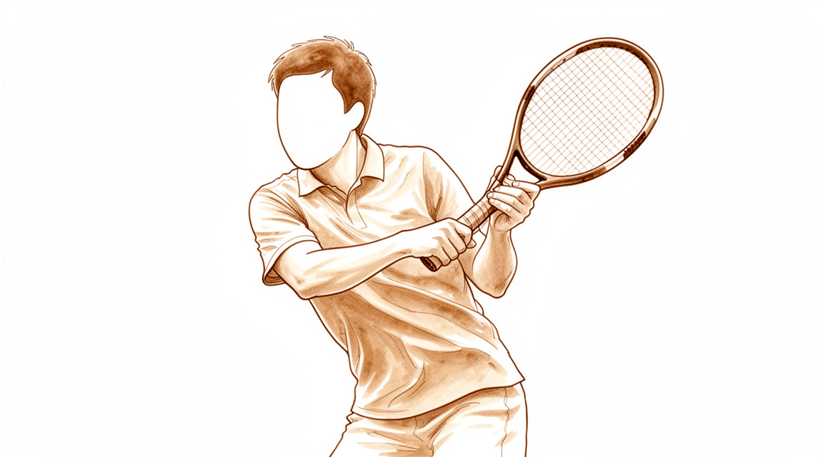
Qué está sintiendo
El codo de tenista provoca dolor en la parte externa del codo, en una protuberancia ósea llamada epicóndilo lateral o a su alrededor. El dolor suele extenderse hacia abajo por el antebrazo y, a veces, hacia arriba, a la parte superior del brazo. Normalmente lo desencadenan las actividades que hacen trabajar los músculos de la parte posterior del antebrazo, porque estos músculos comparten un único punto de inserción en esa zona ósea.
El dolor puede ir desde leve y ocasional hasta constante e intenso. Cuando es intenso, puede afectar a todas sus actividades diarias. También puede alterar su sueño. Muchas personas notan que empeora después de una actividad o al despertarse.
Las tareas cotidianas que implican girar o agarrar se vuelven difíciles. Girar un picaporte, sostener una taza de café, dar la mano o levantar una tetera pueden desencadenar el dolor. Por lo general, el codo sigue moviéndose en todo su rango, así que el problema no es la rigidez, sino el dolor.
En la mayoría de las personas, el codo de tenista mejora por sí solo. Alrededor del 90 % de las personas ven que ha mejorado en el plazo de un año sin cirugía. No necesita precipitarse con ningún tratamiento.
Algunos signos requieren atención más pronto que tarde. Consulte a su médico de cabecera o solicite una valoración por un especialista si sus síntomas no mejoran, empeoran a lo largo de las semanas, le despiertan por la noche o le impiden trabajar o usar la mano o el brazo.
¿Qué está ocurriendo realmente?
El punto doloroso es donde los tendones se insertan en el hueso de la parte externa del codo. Los tendones son cordones resistentes que unen el músculo al hueso. Los afectados en este caso recorren la parte posterior del antebrazo y comparten un único punto de anclaje en esa zona ósea.
A pesar de su nombre, en realidad no es un problema de inflamación. Cuando se examina el tendón al microscopio, el tejido muestra desgaste y deshilachado en lugar de hinchazón y enrojecimiento. Imagine una cuerda muy usada: algunas fibras se han roto, y la cuerda ha intentado repararse pero no ha terminado el trabajo correctamente. El tejido de reparación que se forma está desorganizado y es más débil que el original. Por eso la zona sigue sensible, y por eso duele agarrar o girar: esas acciones tiran directamente del cordón dañado.
El nombre médico es epicondilitis lateral, y también puede verlo como tendinopatía lateral del codo. Ambos nombres describen lo mismo: un desgaste de esa inserción del tendón por el uso repetido. Es mucho más frecuente en la parte externa del codo que en la interna, por eso la denominación codo de tenista suele referirse a esta zona.
El daño se localiza en un tendón concreto de ese grupo, el que estabiliza la muñeca cuando usted agarra algo. Como todos los músculos del antebrazo que extienden la muñeca comparten ese único punto de anclaje, el dolor en ese punto se extiende hacia abajo por el antebrazo cuando usa la mano.
Otros problemas pueden causar dolor en la misma zona, y no siempre es fácil distinguirlos. Por eso es importante una exploración física cuidadosa del codo, y por eso a veces se utilizan estudios de imagen para comprobar cuánto desgaste hay. Esta afección también puede alterar ligeramente su percepción de la posición de la articulación, algo que la fisioterapia puede ayudar a corregir.
Lo fundamental es esto: el tendón está desgastado, no destruido, y el tejido sigue teniendo la capacidad de repararse por sí mismo con tiempo y la carga adecuada.
Qué podemos hacer al respecto
El Dr. Kieran Hirpara, cirujano de extremidades superiores en el Mater Private Hospital Rockhampton, comienza con las opciones menos invasivas que se adapten a su condición. Por lo general, los pacientes son derivados a nuestra clínica por su médico de cabecera; si un fisioterapeuta le ha sugerido que nos consulte, igualmente necesitará una derivación de su médico de cabecera para poder acceder al reembolso de Medicare. En su consulta, tomamos su historia clínica, examinamos su codo y solicitamos estudios de imagen solo si van a cambiar lo que hacemos. Para un problema de larga evolución como este, normalmente probamos primero el tratamiento no quirúrgico y solo consideramos la cirugía cuando no ha proporcionado suficiente mejoría.
El primer paso es cambiar la forma en que somete el tendón a carga. Eso significa reducir las tareas de agarrar y girar que desencadenan el dolor y, después, fortalecer de nuevo el tendón con un programa de carga progresiva. La fisioterapia busca ayudarle a superar más rápido los meses dolorosos, más que cambiar el resultado final, porque la mayoría de los codos mejoran con el tiempo de una forma u otra. Dele una oportunidad genuina y prolongada, medida en meses. Como leyó antes en esta página, alrededor de nueve de cada diez codos mejoran en el plazo de un año sin cirugía, y por eso ponemos el listón alto para operar.
Aquí no utilizamos las inyecciones de cortisona como opción por defecto. La razón probable es que el dolor se calma antes de que el tendón haya cicatrizado, de modo que vuelve a someter a carga un tendón que no está preparado. Si alguna vez se plantea una inyección, es una decisión meditada orientada a la función a corto plazo, no un paso rutinario. Otras inyecciones, como el plasma rico en plaquetas o la sangre extraída de su propio brazo, no se recomiendan para esta afección.
La cirugía solo entra en juego para el pequeño grupo de personas que siguen incapacitadas tras un intento real de tratamiento no quirúrgico. La intervención limpia la parte desgastada y deshilachada del tendón en su punto de inserción al hueso de la parte externa del codo. Puede realizarse mediante pequeñas incisiones de cirugía mínimamente invasiva o mediante una pequeña incisión abierta, y ningún método ha demostrado ser mejor que los demás. La decisión de operar es compartida y la tomamos juntos, una vez que usted sepa en qué consiste la operación y lo que exige la recuperación.
Qué esperar
Para la mayoría de las personas, el codo de tenista sigue una evolución lenta pero constante. Los síntomas suelen aliviarse a lo largo de meses, no de días, y el patrón suele ser de mejoría gradual con algunas recaídas por el camino. El tiempo que tarda no depende de cuánto tiempo lleve con el problema: un tendón que lleva un año doliendo tiene la misma probabilidad de mejorar que uno que lleva unos pocos meses.
Ya ha leído la cifra principal: alrededor de nueve de cada diez codos mejoran en el plazo de un año sin cirugía. Ese es el punto de partida honesto, y se aplica tanto si no hace nada como si sigue un programa de carga o prueba otro tratamiento no quirúrgico. Lo que el tratamiento puede cambiar es el camino, no el destino. Un programa bien llevado busca que supere los meses dolorosos con más comodidad, mientras el tendón realiza en segundo plano su lento trabajo de reparación.
Si su codo es uno de los pocos que siguen doliendo a pesar de un intento genuino de tratamiento no quirúrgico, la cirugía todavía puede ofrecerle un alivio real.
La recuperación tras la cirugía es gradual. Primero mejora el dolor; después, a lo largo de semanas o meses, vuelven la fuerza de agarre y la confianza para usar la mano en las tareas diarias. La terapia de mano posterior a la cirugía la realizará Ruby Doolan en Extend Rehabilitation, quien guiará sus ejercicios y le confeccionará cualquier férula que necesite.
Un pequeño número de personas sigue teniendo dolor después de la cirugía, y alrededor del 1,5 % necesita otra operación. Haberse puesto tres o más inyecciones antes de la cirugía aumenta esa probabilidad. Si sus síntomas no mejoran, empeoran a lo largo de las semanas, le despiertan por la noche o le impiden trabajar o usar la mano o el brazo, consulte a su médico de cabecera o solicite una valoración por un especialista.
Cuándo consultar a un especialista
La mayoría de los casos de codo de tenista mejoran por sí solos, y alrededor de nueve de cada diez codos están mejor en el plazo de un año sin cirugía. Pero algunos signos indican que es momento de buscar ayuda. Consulte a su médico de cabecera o solicite una valoración por un especialista si su dolor no mejora después de varios meses, si está empeorando, si le despierta por la noche o si le impide trabajar o usar la mano y el brazo con normalidad. Alrededor de una de cada cinco personas con esta afección tiene un dolor lo bastante intenso como para limitar su trabajo y su vida diaria, así que necesitar ayuda no es algo inusual. Si ya ha recibido tres o más inyecciones en el tendón y el dolor sigue limitándole, solicite una valoración por un especialista, porque ese historial cambia lo que un cirujano querrá comentar con usted.
En mayor profundidad
Advanced reading: the deeper science (optional)
Esta sección profundiza más de lo necesario para que usted tome sus propias decisiones de tratamiento. Vale la pena leer más sobre el codo de tenista, ya que se trata de una de esas afecciones en las que el tratamiento que parece más eficaz durante el primer mes es, en realidad, el que peor resultado arroja al cabo de un año.
La afección suele curarse por sí sola, aunque lentamente
Si se deja sin intervención, el codo de tenista mejora con el tiempo. En el estudio que se describe a continuación, el grupo al que se le indicó simplemente esperar y observar, sin inyecciones ni fisioterapia, fue reevaluado a las 52 semanas; 56 de 62 (90%) de los pacientes afirmaron haber mejorado mucho o haberse recuperado por completo [1].
Esa cifra constituye el punto de referencia con el que se debe comparar cualquier tratamiento. Una intervención solo merece la pena si logra mejores resultados que la recuperación espontánea, y lo cierto es que, por lo general, la recuperación ocurre por sí sola.
La paradoja de los corticoides
Un ensayo aleatorizado pionero comparó la inyección de corticoides, la fisioterapia y la simple espera, siguiendo a los pacientes durante un año completo [1].
A las seis semanas, la inyección arrojó resultados excelentes: 51 de 65 (78%) de los pacientes del grupo de inyección mostraron éxito, frente a 16 de 60 (27%) de quienes simplemente esperaron; el número necesario a tratar fue de 2 [1].
Posteriormente, la situación se invirtió. De esos pacientes que inicialmente respondieron bien, 47 de 65 (72%) experimentaron posteriormente una recaída. Al cabo de 52 semanas, el grupo de inyección presentó resultados significativamente peores que el grupo de fisioterapia en todos los parámetros evaluados; además, fue peor que quienes no recibieron ningún tratamiento en dos de las tres medidas principales [1].
La explicación ofrecida por los propios autores merece ser citada: la inyección alivia el dolor de forma tan rápida que los pacientes vuelven a someter al tendón a cargas, aun cuando este aún no se ha recuperado del todo. La conclusión del ensayo fue que los corticoides “deben emplearse con precaución” en el codo de tenista; una afirmación sorprendente respecto a un tratamiento que la mayoría de pacientes esperan recibir.
Por ello, la aplicación de una inyección en estos casos debe ser una decisión meditada, orientada a mejorar la función a corto plazo, y no una opción automática; además, no sustituye en modo alguno al programa de carga progresiva.
Entonces, ¿qué merece la pena hacer?
La fisioterapia superó a la simple espera a las seis semanas en todos los aspectos; a las 52 semanas, ambos grupos resultaron indistinguibles, ya que casi todos los pacientes de ambos grupos se habían recuperado [1]. Por tanto, su utilidad radica en ayudar a superar más rápidamente los meses dolorosos, no en cambiar el desenlace final. Cabe destacar que el grupo que recibió fisioterapia también requirió menos tratamientos adicionales durante el proceso [1]. Teniendo esto en cuenta, el plan razonable consiste en controlar la carga de trabajo y seguir un programa progresivo para los tendones, asumiendo que el tiempo de recuperación se mide en meses.
La cirugía se reserva para esa minoría de pacientes que siguen discapacitados tras un intento genuino y prolongado de tratamiento no quirúrgico. El motivo por el cual ese umbral es tan elevado es precisamente la evolución natural de la enfermedad mencionada: si se opera prematuramente, se está compitiendo contra una afección que, en nueve de cada diez casos, habría resuelto por sí sola.
Inyección de células tendinosas cultivadas
Se le podría ofrecer, o podría leer al respecto, el implante de tenocitos autólogos (ATI; comercializado en Australia como OrthoATI), que consiste en una inyección de células tendinosas suyas cultivadas en el laboratorio. Este procedimiento es seguro y biológicamente razonable; los resultados obtenidos en casos de codo de tenista refractario al tratamiento se han mantenido durante más de cuatro años. No obstante, en toda la literatura médica mundial solo existen cinco estudios que incluyen un total de 50 pacientes, y en ninguno de ellos hubo grupo de comparación. Si se compara esto con la evolución natural de la enfermedad descrita anteriormente, resulta evidente la necesidad de ser cautelosos. En la página sobre implante de tenocitos autólogos se detalla todo lo que se sabe y lo que no se sabe al respecto, junto con las referencias correspondientes.
Referencias
[1] Bisset L, Beller E, Jull G, Brooks P, Darnell R, Vicenzino B. Movilización con movimiento y ejercicios, inyección de corticoides o esperar para ver qué sucede en el codo de tenista: ensayo aleatorizado. BMJ. 2006;333(7575):939. https://doi.org/10.1136/bmj.38961.584653.ae
Evidence & references
This is the clinical evidence summary written for health professionals. It is technical, and it lists the research this page was built from. You do not need to read it to understand your treatment or to make a decision about it.
Overview
- A 2018 review provides an overview of current concepts regarding the diagnosis and treatment of tennis elbow and its impact on work participation [1].
- The term "Tennis Elbow" is considered inaccurate by some authors, and the diagnosis is often made too quickly, which may explain high rates of recalcitrant complaints due to insufficient consideration of differential diagnoses [3].
- Most published literature on the nonoperative treatment of lateral tennis elbow consists of poorly designed trials with nebulous selection criteria and low patient numbers [2].
- Based on placebo or no-treatment control arms of randomized trials, approximately 90% of people with untreated tennis elbow achieve symptom resolution at 1 year [5].
- Pooled data from randomized controlled trials indicate a lack of intermediate- to long-term clinical benefit after nonsurgical treatment of lateral epicondylitis compared with observation only or placebo [12].
- Systematic reviews have been unable to reach conclusions regarding the benefit of forearm bracing in tennis elbow [41].
- Most patients with lateral epicondylitis resolve spontaneously or with standard conservative management, but refractory cases may benefit from interventional therapies or surgical approaches [25].
- While numerous treatment options exist for epicondylitis, no single universally accepted protocol has emerged [15].
- There is wide variability in the treatments offered when physiotherapy fails patients with tennis elbow [30].
- Controversy remains regarding the optimal modality for quickest recovery and the role of surgical intervention for refractory cases of lateral epicondylosis [31].
- Due to a small number of studies, large heterogeneity in interventions, small sample sizes, and poor reporting of outcomes, there was insufficient evidence to support or refute the effectiveness of surgery for lateral elbow pain [20].
- Current research evidence suggests that surgery for tennis elbow is no more effective than nonsurgical treatment, based on evidence with significant methodological limitations [53].
- It is recommended to consider non-invasive techniques such as extracorporeal shock wave therapy prior to surgery in tennis elbow [54].
- If a good hand therapy program and other nonoperative measures are ineffective, percutaneous tenotomy is a viable surgical option for tennis elbow [14].
- After surgical treatment for lateral elbow tendonopathy, pain relief and restoration of elbow function can be achieved [8].
- Denervation of the elbow for the management of tennis elbow is a simple and safe procedure [16].
- The effectiveness of ultrasound percutaneous tenotomy appears to improve up to 1 year after surgery, making it an emerging viable alternative for the treatment of medial or lateral epicondylitis [51].
- Recovery from debridement of extensors and drilling of the lateral epicondyle was slow and never immediate, unlike in series where extensors were simply released without decortication of the lateral epicondyle [28].
- In experienced hands, elbow arthroscopy is a safe modality of treatment for a variety of pathologies [10].
Anatomy & Pathophysiology
Anatomy
- The pathology of lateral epicondylitis is primarily localized to the origin of the extensor carpi radialis brevis (ECRB) [83].
- The ECRB and extensor digitorum communis (EDC) have tendinous origins and lie deep to the extensor carpi radialis longus (ECRL), which has a muscular origin [83].
- The common extensors lie superficial to the lateral collateral ligament complex proximally and to the supinator distally [83].
- The ECRB tendon lies superficial to the joint capsule and is therefore accessible arthroscopically [83].
- The posterior interosseous nerve enters the supinator distal to the radial head [83].
- Compression of the posterior interosseous nerve at the radial tunnel can coexist with lateral epicondylitis [83].
- The pathologic process of lateral epicondylitis mainly involves the origin of the extensor carpi radialis brevis but can involve the tendons of the extensor carpi radialis longus and the extensor digitorum communis [84].
- The anatomic basis of the injury to the extensor carpi radialis brevis origin involves hypovascular zones, eccentric tendon stresses, and a microscopic degenerative response [40].
- The superficial head of supinator has a biomechanical basis in the aetiology of both lateral epicondylitis and radial tunnel syndrome [101].
- The common extensor tendon is thicker in men and in the dominant elbow compared to non-dominant elbows [122].
Pathophysiology
- Lateral epicondylitis is initiated as a microtear, most often within the origin of the extensor carpi radialis brevis [84].
- The current consensus is that lateral epicondylitis is a degenerative disease rather than primarily an inflammatory condition [18].
- Histologic studies of lateral epicondylitis show angiofibroblastic hyperplasia [83].
- Histologic findings in lateral epicondylitis include neovascularization, infiltration by mucopolysaccharide, a disordered collagen scaffold, bone formation, and angiofibroblastic proliferation [83].
- Inflammation is not usually seen in lateral epicondylitis but is likely present in the early stages [83].
- The lesion in lateral epicondylitis occurs in a vascular watershed area that is relatively avascular, limiting healing potential [83].
- Tennis elbow may start as an inflammatory condition but progresses to a degenerative state [4].
- Tendinosis at initial onset is similar to a stagnant state of fibroplasia [4].
- The primary lesion of lateral epicondylitis is classically found in the origin of the ECRB, but it can also be seen in the EDC [83].
- Microscopic findings in lateral epicondylitis show immature reparative tissue that resembles angiofibroblastic hyperplasia [84].
- Lateral epicondylitis is typically caused by eccentric contractions of the extensor carpi radialis brevis muscle during the backhand swing [83].
- Repetitive exposure to bending and straightening the elbow is a significant risk factor for lateral epicondylitis [121].
- Self-reported physical exposures involving repetitive and extensive or prolonged wrist bend, twisting, and forearm movements are associated with incident cases of lateral epicondylitis [52].
- Combined physical exertion and elbow movements are strongly associated with lateral epicondylitis [9].
- Biomechanical exposure involving the wrist and/or elbow at work is associated with the incidence of lateral epicondylitis [87].
- Some individuals may have a genetic predisposition to develop tennis elbow [84].
- Increased MRI signal in the ECRB origin is common in both symptomatic and asymptomatic elbows [66].
- The presence of hypoechogenicity and bone changes on ultrasound indicates a stressed common extensor origin-lateral epicondyle complex in elbows with lateral epicondylalgia [13].
- In tennis elbow patients, hand/wrist and shoulder strength and extensor carpi radialis activity are reduced compared to controls [42].
- Grip force is markedly reduced at the pathological side in tennis elbow patients, with a striking reduction when measured with a straight elbow compared to 90° flexion [105].
Classification
- The term "Tennis Elbow" is considered inaccurate, and the diagnosis is often made too quickly [3].
- Insufficient consideration of differential diagnoses may explain high rates of recalcitrant complaints in patients diagnosed with tennis elbow [3].
- Almost half of patients (46.5%) presenting with lateral sided elbow pain are diagnosed with a condition other than lateral epicondylitis [29].
- Differential diagnosis of lateral elbow instability should be considered in patients presenting with tennis elbow [11].
- Accurate diagnosis of medial epicondylitis requires distinguishing it from other elbow conditions [19].
- There is considerable terminological heterogeneity in the description of lateral elbow pain [50].
- The lack of clear and recognized diagnostic criteria is associated with the terminological heterogeneity in evaluating and treating patients with lateral elbow pain [50].
- The presence of hypoechogenicity and bone changes on musculoskeletal ultrasound indicates a stressed common extensor origin-lateral epicondyle complex in elbows with lateral epicondylalgia [13].
- Tennis elbow is reflective of tendon overuse and failed tendon healing [68].
- The primary site of the basic underlying lesion in lateral tennis elbow is in the origin of the extensor carpi radialis brevis (ECRB) [70].
- Overuse in the ECRB origin results in microscopic rupture and subsequent tendinous nonrepair with immature reparative tissue [70].
- The findings of microscopic rupture and nonrepair in the ECRB origin have been termed angiofibroblastic hyperplasia [70].
- The onset of tennis elbow is hastened by overuse of the arm and elbow, consistent with it being a degenerative disease [18].
- There is evidence for a possible neurogenic involvement in the pathophysiology of tennis elbow [33].
- In tennis elbow patients, hand/wrist and shoulder strength are reduced compared to controls [42].
- In tennis elbow patients, extensor carpi radialis (ECR) activity is reduced compared to controls [42].
- The Patient Rated Tennis Elbow Evaluation (PRTEE) is the core outcome for capturing the disability domain in clinical settings and research for lateral elbow tendinopathy [124].
- The PRTEE and its subscales offer insights into the domains of pain and function in addition to disability [124].
- Time off work is recommended as an interim measure for measuring participation in lateral elbow tendinopathy [124].
- Pain-free grip strength is recommended as an interim measure for measuring physical function capacity in lateral elbow tendinopathy [124].
- A numerical rating scale for pain on gripping is recommended as an interim measure for measuring pain on loading in lateral elbow tendinopathy [124].
Clinical Presentation
Epidemiology and Demographics
- Lateral epicondylitis is a common, generally self-limiting tendinosis affecting patients aged 35 to 55 years [45].
- Lateral elbow tendinosis is relatively common, particularly among individuals aged 40 to 49 years [58].
Pathophysiology and Terminology
- The term 'Tennis Elbow' is considered inaccurate by some authors, and the diagnosis is often made too quickly, potentially explaining high rates of recalcitrant complaints due to insufficient consideration of differential diagnoses [3].
- Findings are consistent with the hypothesis that tennis elbow is a degenerative disease, the onset of which is hastened by overuse of the arm and elbow [18].
- There is considerable terminological heterogeneity in the description of lateral elbow pain, associated with the lack of clear and recognised diagnostic criteria [50].
- The results give further evidence for a possible neurogenic involvement in the pathophysiology of tennis elbow [33].
Risk Factors
- Self-reported physical exposures that implicate repetitive and extensive/prolonged wrist bend/twisting and forearm movements were associated with incident cases of lateral epicondylitis [52].
Clinical Diagnosis and Examination
- The diagnosis of lateral epicondylitis was based on self-reported symptoms and clinical signs according to the Japanese Orthopaedic Association guidelines, which include pain in the elbow joint within 2 weeks, pain in the lateral epicondyle region on resisted extension of the wrist with the elbow extended, and tenderness in the lateral epicondyle [79].
- Physical examination of the elbow should focus on functional anatomy and includes inspection, palpation, range of motion, strength, stability, and special tests [86].
- Characteristic elements of the history for lateral elbow tendinopathy include pain lifting things from a bag with a pronated hand, turning doorknobs, taking milk from the fridge, shaking hands, taking a laptop out of a bag, and bumping the lateral elbow [86].
- Physical examination maneuvers to elicit lateral elbow tendinopathy include direct palpation of the ECRB origin, the tennis elbow shear test, pain along with resisted wrist or long finger extension, and the laptop test [86].
- Almost half of the patients (46.5%) were diagnosed with a diagnosis other than lateral epicondylitis [29].
- Differential diagnosis of lateral elbow instability in patients presenting with tennis elbow should be considered [11].
Imaging
- The presence of hypoechogenicity and bone changes on musculoskeletal ultrasound indicates the presence of a stressed common extensor origin-lateral epicondyle complex in elbows with lateral epicondylalgia [13].
- MRI is used to evaluate lateral elbow tendinopathy if the lateral ulnar collateral ligament (LUCL) is suspected as part of the pathology [86].
- Ultrasonography is used to evaluate lateral elbow tendinopathy [86].
Investigations
Clinical Diagnosis and Differential Diagnosis
- The diagnosis of tennis elbow is often made too quickly, which may explain high rates of recalcitrant complaints due to insufficient consideration of differential diagnoses [3].
- Almost half of the patients (46.5%) presenting with lateral sided elbow pain were diagnosed with a condition other than lateral epicondylitis [29].
- The physical examination for the elbow is directed by the history and the location of the patient's pain in the anterior, posterior, medial, or lateral aspect of the joint [81].
- In tennis elbow patients compared to controls, hand/wrist and shoulder strength and extensor carpi radialis (ECR) activity were reduced [42].
Imaging: Ultrasound
- The presence of hypoechogenicity and bone changes on ultrasound indicates the presence of a stressed common extensor origin-lateral epicondyle complex in elbows with lateral epicondylalgia [13].
- A systematic review and meta-analysis evaluated the diagnostic test accuracy of ultrasound for the detection of lateral epicondylitis [46].
Imaging: Magnetic Resonance Imaging (MRI)
- Increased MRI signal in the extensor carpi radialis brevis (ECRB) origin is common in both symptomatic and asymptomatic elbows [66].
- The routine use of MRI for the diagnosis of lateral epicondylitis is low, although its use is associated with downstream effects [107].
- MRI can be used to evaluate ligaments and tendons of the elbow, but it is rarely indicated [78].
- Magnetic resonance imaging findings in refractory tennis elbows have a relationship to surgical treatment [48].
- The reliability and validity of magnetic resonance imaging in the assessment of chronic lateral epicondylitis has been evaluated [80].
Imaging: Radiography
- Lateral epicondyle calcifications are much more common in lateral epicondylitis than previously reported, possibly owing to modern digital radiography and magnification [129].
- Lateral epicondyle calcifications do not appear to be related to clinical factors including patient-reported measures [129].
- Bony changes at the lateral epicondyle, such as a gunsight type spur, have been described in tennis elbow syndrome [6].
Pathophysiology and Prognosis
- Tennis elbow is a degenerative disease, the onset of which is hastened by overuse of the arm and elbow [18].
- Although tennis elbow may start out as an inflammatory condition, it progresses to a degenerative state [4].
- Based on placebo or no-treatment control arms of randomized trials, about 90% of people with untreated tennis elbow achieve symptom resolution at 1 year [5].
- The results provide evidence for a possible neurogenic involvement in the pathophysiology of tennis elbow [33].
- The strength of associations between combined physical exertion and elbow movements and lateral epicondylitis is emphasized in working populations [9].
Treatment
Non-Operative Management
- Approximately 90% of people with untreated tennis elbow achieve symptom resolution at 1 year based on placebo or no-treatment control arms of randomized trials [5].
- Lateral epicondylitis is usually self-limited, resolving over a 12- to 18-month period without treatment [55].
- The available data suggest that lateral epicondylitis often resolves spontaneously, warranting considerable circumspection before embarking on treatment [27].
- Most patients with lateral epicondylitis resolve spontaneously or with standard conservative management [25].
- Pooled data from RCTs indicate a lack of intermediate- to long-term clinical benefit after nonsurgical treatment of lateral epicondylitis compared with observation only or placebo [12].
- Patient education regarding activity modification to reduce exposure to aggravating activity appears to play a crucial role in resolving the pain associated with tennis elbow [4].
- There is general agreement that exercise is beneficial to treatment outcome for tennis elbow, although there is a lack of evidence to support a particular exercise prescription to increase tolerance for loading the common extensor tendon [4].
- Painful eccentric exercise has been shown to be effective in the management of chronic mid-portion Achilles tendinopathy, but not insertional Achilles tendinopathy [4].
- The significant short term benefits of corticosteroid injection are paradoxically reversed after six weeks, with high recurrence rates, implying that this treatment should be used with caution in the management of tennis elbow [37].
- Corticosteroid injection has little, if any, benefit over placebo and usually does not change the natural history of the disease [75].
- Recent data suggest that poorer results are seen in the intermediate and long term following corticosteroid injections relative to placebo, and patients who undergo corticosteroid injection may actually be worse at the end of a year [75].
- Local PRP injections were associated with superior outcomes for reducing pain and improving elbow joint function compared with local corticosteroids treatment for lateral epicondylitis at a follow-up of 6 months [59].
- Normal saline injections yielded a statistically significant and clinically meaningful improvement in pain and functional outcomes in patients with lateral epicondylitis [97].
- Peri-articular hyaluronic acid treatment for tennis elbow was significantly better than control in improving pain at rest and after maximal grip testing [56].
- Iontophoresis treatment was well tolerated by most patients and was effective in reducing symptoms of epicondylitis at short-term follow-up [61].
- Systematic reviews have been unable to come to any conclusions with regard to the benefit of forearm bracing in tennis elbow [41].
- Evidence was found for effectiveness of shock wave treatment for tennis elbow under well-defined, restrictive conditions only [60].
- Non-invasive techniques such as extracorporeal shock wave therapy are recommended to be considered prior to surgery in tennis elbow [54].
- Nonsurgical treatment is the mainstay of management for lateral epicondylitis, involving options such as rest, physical therapy, and injections [95].
- The 6 most frequently prescribed nonoperative treatments for lateral epicondylitis by fellowship-trained upper extremity surgeons were home exercise program/stretching (81%), nonsteroidal anti-inflammatory drugs (75%), steroid injection (71%), counterforce bracing (68%), formal physical therapy (65%), and wrist brace (47%) [104].
- There is wide variability of treatments offered when physiotherapy fails patients with tennis elbow [30].
- A high-repetition, low-resistance home exercise program may be useful for the management of lateral epicondylitis [75].
- Poorer outcomes are seen in patients who complain of severe pain, have concomitant neck pain, are involved in workers’ compensation claims, have concomitant depression, or have poor coping skills [75].
Operative Management
- There was insufficient evidence to support or refute the effectiveness of surgery for lateral elbow pain due to a small number of studies, large heterogeneity in interventions across trials, small sample sizes and poor reporting of outcomes [20].
- The literature and clinical experience confirm greater than 90% of people with lateral epicondylitis are successfully treated nonoperatively [36].
- Individuals with symptomatic tennis elbow who do not respond to rehabilitation and injections may be candidates for surgical intervention [36].
- Most surgical patients for lateral epicondylitis are in their fifth decade of life (range, 30–63 years), have had symptoms in their dominant arm for an average of 19 months (range, 6–132 months), and have had failure of nonoperative treatment including an average of 3.1 corticosteroid injections with little to no improvement [36].
- The majority of patients undergoing surgery for tennis elbow have surgery on their dominant elbow, with an average incidence of dominant arm involvement of 74% [36].
- Percutaneous tenotomy is a viable surgical option if a good hand therapy program and other nonoperative measures are ineffective in treating the patient with tennis elbow [14].
- For isolated lateral epicondylitis, a percutaneous release performed in the office is an effective treatment for recalcitrant lateral epicondylitis that has failed nonoperative management [63].
- The effectiveness of ultrasound percutaneous tenotomy for epicondylitis appears to improve up to 1 year after the surgery, making it an emerging viable alternative for the treatment of medial or lateral epicondylitis [51].
- Recovery after debridement of extensors and drilling of the lateral epicondyle was slow and was never found to be immediate as described in other series of tennis elbow procedures where the extensors were simply released and the lateral epicondyle was not decorticated [28].
- Good to excellent results have been achieved in 83% to 96% of patients using variations in technique for epicondylitis, with poorer outcomes reported in patients with ulnar nerve symptoms [75].
- The use of suture anchors to reattach the extensor carpi radialis brevis tendon after debridement and decortication provided significantly better short-term and long-term clinical results compared to traditional elbow arthroscopy and extensor carpi radialis brevis tendon debridement and decortication without anchors [103].
- Denervation of the elbow for management of tennis elbow is a simple safe procedure [16].
- Outcomes between open and arthroscopic procedures for lateral epicondylitis are comparable [75].
- Arthroscopic treatment of lateral epicondylitis is accompanied by an increased risk of neurovascular injury and instability compared with open release of the elbow [75].
- Failed surgical treatment of lateral epicondylitis is multifactorial, and distinguishing the cause is critical for appropriate management [21].
- The median time of minimum conservative management before surgery for lateral epicondylitis was six months (IQR 6 months) [98].
- Surgical intervention for refractory medial epicondylitis often has a high success rate with patients generally demonstrating an improvement in patient-reported outcomes and an encouraging number returning to work with limited complications [23].
- Chronic elbow dislocation is a rare complication of tennis elbow surgery that can be treated by open reduction and external articular distrator [11].
Complications
Surgical Complications
- Heterotopic ossification has been reported as a complication following arthroscopic treatment of lateral epicondylitis [35].
- Recovery after debridement of extensors and drilling of the lateral epicondyle is slow and not immediate, unlike procedures where extensors are simply released without decortication [28].
- Short-term complication rates appear comparable between open and arthroscopic treatment for lateral epicondylitis [110].
- In a review of revision surgery for recalcitrant lateral epicondylitis, pathologic changes in the extensor carpi radialis brevis were not addressed in 27 of 35 elbows, and damaged tissue was not completely excised in 7 elbows [102].
- Three or more preoperative injections is the most significant risk factor for revision surgery after operative treatment of lateral epicondylitis [108].
Diagnostic and Management Complications
- The diagnosis of tennis elbow is often made too quickly, potentially explaining high rates of recalcitrant complaints due to insufficient consideration of differential diagnoses [3].
- The term 'Tennis Elbow' is considered inaccurate by some authors [3].
Recovery
Prognosis and Natural History
- Lateral epicondylitis often resolves spontaneously, warranting considerable circumspection before embarking on treatment [27].
- Enthesopathy of the extensor carpi radialis brevis origin is a benign, self-limiting disorder with a natural history of spontaneous resolution [72].
- No treatments have been proven to alter the course of enthesopathy of the extensor carpi radialis brevis origin [72].
- Poor prognosis at 1 year of follow-up for lateral epicondylitis was related to manual work and high baseline pain [113].
- No relation was found between the type of medical treatment given or chosen and prognosis for lateral epicondylitis at 1 year [113].
- The annual incidence of lateral epicondylitis per 10,000 patients and the proportion of cases treated surgically remained constant from 2007 to 2014 [69].
Non-Operative Management
- The significant short-term benefits of corticosteroid injection are paradoxically reversed after six weeks, with high recurrence rates [37].
- Corticosteroid injection should be used with caution in the management of tennis elbow due to high recurrence rates after six weeks [37].
- There is general agreement that exercise is beneficial to treatment outcome for tennis elbow [4].
- There is a lack of evidence to support a particular exercise prescription to increase tolerance for loading the common extensor tendon [4].
- It is unclear which interventions may be helpful in pain modulation in chronic tennis elbow [4].
- It is unknown if commonly used physical agents may resolve the neurochemical response and its associated pain mediation in either acute or chronic tennis elbow [4].
- Ultrasound and electrotherapy may be used to facilitate tissue healing in tennis elbow [4].
- Theoretically, ultrasound and electrotherapy may be able to stimulate cellular responses to promote tissue healing in tendinosis [4].
- Evidence was found for the effectiveness of shock wave treatment for tennis elbow under well-defined, restrictive conditions only [60].
- Radial extracorporeal shock wave therapy, symptom duration of longer than 6 months, and short follow-up duration (less than 24 weeks) were related to better effects in lateral epicondylitis [65].
Operative Management
- Recovery following debridement of extensors and drilling of the lateral epicondyle was slow and was never found to be immediate [28].
- Immediate recovery has been described in other series of tennis elbow procedures where the extensors were simply released and the lateral epicondyle was not decorticated [28].
- No patient exhibited reduced wrist or elbow ranges of motion at follow-up compared with those ranges on the uninvolved opposite side following elongation of the extensor carpi radialis brevis tendon for refractory tennis elbow [43].
- Due to a small number of studies, large heterogeneity in interventions across trials, small sample sizes and poor reporting of outcomes, there was insufficient evidence to support or refute the effectiveness of surgery for lateral elbow pain [20].
Key Evidence
- [L4] This review gives an overview of the current concepts of diagnosis and treatment of tennis elbow and the impact on work participation. [1] (10.1177/1758573218797973)
- [L5] Most of the published literature on the nonoperative treatment of patients with lateral tennis elbow consists of poorly designed trials with nebulous selection criteria and low patient numbers. [2] (10.1016/s1058-2746(99)90081-2)
- [Letter] The authors agree that the term 'Tennis Elbow' is inaccurate and that the diagnosis is often made too quickly, potentially explaining high rates of recalcitrant complaints due to insufficient consideration of differential diagnoses. [3] (10.1177/1758573218816086)
- [Paper] [4] (10.1197/j.jht.2006.02.016)
- [L1] Based on the placebo or no-treatment control arms of randomized trials, about 90% of people with untreated tennis elbow achieve symptom resolution at 1 year. [5] (10.1097/corr.0000000000002058)
- [L4] [6] (10.1067/mse.2001.112020)
- [L4] After surgical treatment for lateral elbow tendonopathy, pain relief and restoration of elbow function can be achieved. [8] (10.1053/jhsu.2001.28432)
- [L4] This study emphasizes the strength of the associations between combined physical exertion and elbow movements and lateral epicondylitis. [9] (10.1002/ajim.22140)
- [L4] In experienced hands, elbow arthroscopy is a safe modality of treatment for a variety of pathologies. [10] (10.1016/j.arthro.2007.03.080)
- [L5] Differential diagnosis of lateral elbow instability in patients presenting with tennis elbow should be considered. [11] (10.1016/j.main.2007.05.002)
- [L1] Pooled data from RCTs indicate a lack of intermediate- to long-term clinical benefit after nonsurgical treatment of lateral epicondylitis compared with observation only or placebo. [12] (10.1007/s11999-014-4022-y)
- [L4] The presence of hypoechogenicity and bone changes indicates presence of a stressed common extensor origin-lateral epicondyle complex in elbows with lateral epicondylalgia. [13] (10.1186/1471-2342-14-10)
- [L4] If a good hand therapy program and other nonoperative measures are ineffective in treating the patient with tennis elbow, percutaneous tenotomy is a viable surgical option. [14] (10.1016/s0894-1130(12)80105-0)
- [L5] This article is a review of recently published information on elbow tendinopathy and tendon ruptures intended to assist clinicians in diagnosis and management, noting that while numerous treatment options exist for epicondylitis, no single universally accepted protocol has emerged. [15] (10.1016/j.jhsa.2009.01.022)
- [L2] Denervation of the elbow for management of tennis elbow is a simple safe procedure. [16] (10.5435/jaaosglobal-d-24-00352)
- [L4] The findings are consistent with the hypothesis that tennis elbow is a degenerative disease, the onset of which is hastened by overuse of the arm and elbow. [18] (10.1177/036354657900700405)
- [L5] Accurate diagnosis requires distinguishing it from other elbow conditions, and treatment is guided by the specific pathologic stage of the tendon. [19] (10.1016/j.csm.2004.04.011)
- [L1] Due to a small number of studies, large heterogeneity in interventions across trials, small sample sizes and poor reporting of outcomes, there was insufficient evidence to support or refute the effectiveness of surgery for lateral elbow pain. [20] (10.1002/14651858.cd003525.pub2)
- [L5] Failed surgical treatment of lateral epicondylitis is multifactorial, and distinguishing the cause is critical for appropriate management. [21] (10.1016/j.xrrt.2023.07.006)
- [L4] Surgical intervention for refractory medial epicondylitis often has a high success rate with patients generally demonstrating an improvement in patient-reported outcomes and an encouraging number returning to work with limited complications. [23] (10.1177/03635465221095565)
- [L4] Most patients with lateral epicondylitis resolve spontaneously or with standard conservative management, but refractory cases may benefit from interventional therapies or surgical approaches. [25] (10.5397/cise.2019.22.4.227)
- [L4] Overall, the available data suggest that lateral epicondylitis often resolves spontaneously, warranting considerable circumspection before embarking on treatment. [27] (10.1016/j.otsr.2019.09.004)
- [L4] However, recovery was slow and was never found to be immediate as described in other series of tennis elbow procedures where the extensors were simply released and the lateral epicondyle was not decorticated. [28] (10.1016/j.jse.2005.07.002)
- [L3] Almost half of the patients (46.5%) were diagnosed with a diagnosis other than lateral epicondylitis. [29] (10.1016/j.jseint.2024.08.047)
- [L4] There is wide variability of treatments offered when physiotherapy fails patients with tennis elbow. [30] (10.1177/1758573217738199)
- [L5] This article serves to provide an updated review of the various treatment options and management for lateral epicondylosis, noting that while most patients experience relief with non-operative management, controversy remains regarding the optimal modality for quickest recovery and the role of surgical intervention for refractory cases. [31] (10.1016/j.jhsa.2024.07.003)
- [L4] The results give further evidence for a possible neurogenic involvement in the pathophysiology of tennis elbow and in medial epicondylalgia. [33] (10.1016/s0736-0266(03)00183-9)
- [L4] To our knowledge, we present the first case of HO development after elbow arthroscopy for lateral epicondylitis. [35] (10.1177/1558944716668844)
- [L4] [36] (10.1097/blo.0b013e3181483dc4)
- [L1] The significant short term benefits of corticosteroid injection are paradoxically reversed after six weeks, with high recurrence rates, implying that this treatment should be used with caution in the management of tennis elbow. [37] (10.1136/bmj.38961.584653.ae)
- [L4] [40] (10.1016/j.jhsa.2007.07.019)
- [L1] Systematic reviews have been unable to come to any conclusions with regard to the benefit of forearm bracing in tennis elbow. [41] (10.1097/bte.0b013e318047c176)
- [L4] [42] (10.1002/jor.20458)
- [L4] No patient exhibited reduced wrist or elbow ranges of motion at follow-up compared with those ranges on the uninvolved opposite side. [43] (10.1177/17531934211042318)
- [L5] Lateral epicondylitis is a common, generally self-limiting tendinosis affecting patients aged 35 to 55 years. [45] (10.1302/0301-620x.95b9.29285)
- [L2] [46] (10.1016/j.otsr.2014.01.006)
- [L1] In this SR, a considerable terminological heterogeneity emerged in the description of LEP, associated with the lack of clear and recognised diagnostic criteria in evaluating and treating patients with lateral elbow pain. [50] (10.3390/healthcare10061095)
- [Paper] The effectiveness of the technique appears to improve up to 1 year after the surgery and hence, is emerging as a viable and attractive alternative for the treatment of medial or lateral epicondylitis. [51] (10.1097/bte.0b013e318291487e)
- [L2] Self-reported physical exposures that implicate repetitive and extensive/prolonged wrist bend/twisting and forearm movements were associated with incident cases of lateral and medial epicondylitis in a large longitudinal study. [52] (10.1136/oemed-2012-101341)
- [L1] Current research evidence suggests that surgery for tennis elbow is no more effective than nonsurgical treatment based on evidence with significant methodological limitations. [53] (10.1177/1758573217745041)
- [L1] We therefore recommend considering non-invasive techniques such as ESWT treatment prior to surgery in tennis elbow. [54] (10.1016/j.hansur.2020.12.008)
- [L2] Lateral epicondylitis is a condition that is usually self-limited, resolving over a 12- to 18-month period without treatment. [55] (10.1007/s11552-014-9642-x)
- [L1] Peri-articular HA treatment for tennis elbow was significantly better than control in improving pain at rest and after maximal grip testing. [56] (10.1186/1758-2555-2-4)
- [L3] The study indicates that lateral elbow tendinosis is relatively common, particularly among individuals aged 40 to 49 years. [58] (10.1177/0363546514568087)
- [L1] Local PRP injections was associated with superior outcomes for reducing pain and improving elbow joint function compared with local corticosteroids treatment for LE at a follow-up of 6 months. [59] (10.1016/j.ijsu.2019.05.003)
- [L1] In a qualitative systematic per-study analysis identifying common and diverging details of 10 randomized-controlled trials, evidence was found for effectiveness of shock wave treatment for tennis elbow under well-defined, restrictive conditions only. [60] (10.1093/bmb/ldm019)
- [L1] Iontophoresis treatment was well tolerated by most patients and was effective in reducing symptoms of epicondylitis at short-term follow-up. [61] (10.1177/03635465030310020601)
- [L4] For isolated lateral epicondylitis, a percutaneous release performed in the office is an effective treatment for recalcitrant lateral epicondylitis that has failed nonoperative management. [63] (10.1097/00132589-200112000-00003)
- [L1] Radial ESWT, symptom duration of longer than 6 months, and short follow-up duration (less than 24 weeks) were related to better effects. [65] (10.1097/corr.0000000000001246)
- [L4] Increased MRI signal in the ECRB origin is common in symptomatic and in asymptomatic elbows. [66] (10.1016/j.jse.2016.01.033)
- [L4] [68] (10.1016/s0278-5919(03)00051-6)
- [L4] The annual incidence of lateral epicondylitis per 10,000 patients and the proportion of cases treated surgically remained constant from 2007 to 2014. [69] (10.1007/s11420-017-9559-3)
- [L4] [70] (10.1097/00132589-200001030-00007)
- [L5] Enthesopathy of the extensor carpi radialis brevis origin is a benign, self-limiting disorder with a natural history of spontaneous resolution, for which no treatments have been proven to alter the course. [72] (10.5435/jaaos-d-15-00233)
- [L3] [79] (10.1016/j.jseint.2024.01.008)
- [L2] The results of this meta-analysis strongly support the hypothesis of an association between biomechanical exposure involving wrist and/or elbow at work and incidence of lateral epicondylitis. [87] (10.1002/acr.22874)
- [L5] Nonsurgical treatment is the mainstay of management for lateral epicondylitis, involving options such as rest, physical therapy, and injections. [95] (10.5435/00124635-200801000-00004)
- [L1] NS injections yielded a statistically significant and clinically meaningful improvement in pain and functional outcomes in patients with lateral epicondylitis. [97] (10.1177/0363546519899644)
- [L4] [98] (10.1016/j.xrrt.2024.08.008)
- [L5] This study demonstrates a biomechanical basis for the superficial head of supinator in the aetiology of both lateral epicondylitis and radial tunnel syndrome. [101] (10.1016/j.jhsb.2004.06.001)
- [L5] [102] (10.1016/j.csm.2004.06.004)
- [L1] [103] (10.1097/bte.0000000000000027)
- [L4] [104] (10.1177/1558944718770212)
- [L4] Grip force was markedly reduced at the pathological side, but there was also a striking reduction of the grip force at the pathological side when the grip force was measured with a straight elbow, compared with the standard position of 90° flexion. [105] (10.1016/s0894-1130(97)80026-9)
- [L3] Although there is variation in the use of MRI for lateral epicondylitis and its use is associated with downstream effects, the routine use of MRI for the diagnosis of lateral epicondylitis is low. [107] (10.1016/j.jhsa.2023.03.025)
- [L4] [108] (10.1016/j.jse.2016.10.022)
- [L3] Surgical management of lateral epicondylitis varies depending on surgeons' fellowship training, and short-term complication rates appear comparable between open and arthroscopic treatment. [110] (10.1016/j.arthro.2017.04.078)
- [L2] Poor prognosis at 1yr of follow-up for lateral epicondylitis was related to manual work and high baseline pain, whilst no relation was found between the type of medical treatment given/chosen and prognosis. [113] (10.1093/rheumatology/keg360)
- [L4] Repetitive exposure to bending/straightening the elbow was a significant risk factor for medial and lateral epicondylitis. [121] (10.1093/rheumatology/ker228)
- [L3] This study presents the US characteristics and normal values of the CET, finding that the tendon was thicker in men and in the dominant elbow with no difference regarding age groups. [122] (10.1177/2325967117704186)
- [Paper] [124] (10.1136/bjsports-2021-105044)
- [L4] Lateral epicondyle calcifications are much more common in lateral epicondylitis than previously reported, possibly owing to modern digital radiography and magnification, although they do not appear to be related to clinical factors including patient-reported measures. [129] (10.1016/j.jhsa.2017.03.016)
References
[1] Tennis elbow. Shoulder & Elbow. 2018. DOI: 10.1177/1758573218797973
[2] Lateral tennis elbow: “Is there any science out there?”. Journal of Shoulder and Elbow Surgery. 1999. DOI: 10.1016/s1058-2746(99)90081-2
[3] In response to the letter to the editor by Dr. Dimitrios Stasinopoulos. Shoulder & Elbow. 2019. DOI: 10.1177/1758573218816086
[4] Tennis Elbow: Blending Basic Science with Clinical Practice. Journal of Hand Therapy. 2006. DOI: 10.1197/j.jht.2006.02.016
[5] Persistent Tennis Elbow Symptoms Have Little Prognostic Value: A Systematic Review and Meta-analysis. Clinical Orthopaedics & Related Research. 2021. DOI: 10.1097/corr.0000000000002058
[6] Bony changes at the lateral epicondyle of possible significance in tennis elbow syndrome. Journal of Shoulder and Elbow Surgery. 2001. DOI: 10.1067/mse.2001.112020
[8] V-Y slide of the common extensor origin for lateral elbow tendonopathy. The Journal of Hand Surgery. 2001. DOI: 10.1053/jhsu.2001.28432
[9] Work‐related risk factors for lateral epicondylitis and other cause of elbow pain in the working population. American Journal of Industrial Medicine. 2012. DOI: 10.1002/ajim.22140
[10] “Humero Radial Plica” Causing Lateral Elbow Pain, an Analysis of 117 Elbow Arthroscopies (SS‐66). Arthroscopy. 2007. DOI: 10.1016/j.arthro.2007.03.080
[11] Chronic elbow dislocation: a rare complication of tennis elbow surgery. Successful treatment by open reduction and external articular distrator. Chirurgie de la Main. 2007. DOI: 10.1016/j.main.2007.05.002
[12] Does Nonsurgical Treatment Improve Longitudinal Outcomes of Lateral Epicondylitis Over No Treatment? A Meta-analysis. Clinical Orthopaedics & Related Research. 2015. DOI: 10.1007/s11999-014-4022-y
[13] The diagnostic validity of musculoskeletal ultrasound in lateral epicondylalgia: a systematic review. BMC Medical Imaging. 2014. DOI: 10.1186/1471-2342-14-10
[14] Surgical and therapeutic management of tennis elbow: An update. Journal of Hand Therapy. 1991. DOI: 10.1016/s0894-1130(12)80105-0
[15] Elbow Tendinopathy and Tendon Ruptures: Epicondylitis, Biceps and Triceps Ruptures. The Journal of Hand Surgery. 2009. DOI: 10.1016/j.jhsa.2009.01.022
[16] A Comparative Study Between Denervation and Extensor Release for Management of Resistant Tennis Elbow. JAAOS: Global Research and Reviews. 2026. DOI: 10.5435/jaaosglobal-d-24-00352
[18] An epidemiologic study of tennis elbow. The American Journal of Sports Medicine. 1979. DOI: 10.1177/036354657900700405
[19] Diagnosis and treatment of medial epicondylitis of the elbow. Clinics in Sports Medicine. 2004. DOI: 10.1016/j.csm.2004.04.011
[20] Surgery for lateral elbow pain. Cochrane Database of Systematic Reviews. 2011. DOI: 10.1002/14651858.cd003525.pub2
[21] Failed surgical treatment for lateral epicondylitis: literature review and treatment considerations for successful outcomes. JSES Reviews, Reports, and Techniques. 2024. DOI: 10.1016/j.xrrt.2023.07.006
[23] Surgical Techniques and Clinical Outcomes for Medial Epicondylitis: A Systematic Review. The American Journal of Sports Medicine. 2022. DOI: 10.1177/03635465221095565
[25] Current Trends for Treating Lateral Epicondylitis. Clinics in Shoulder and Elbow. 2019. DOI: 10.5397/cise.2019.22.4.227
[27] Management of lateral epicondylitis. Orthopaedics & Traumatology: Surgery & Research. 2019. DOI: 10.1016/j.otsr.2019.09.004
[28] Debridement of extensors and drilling of the lateral epicondyle for tennis elbow: A retrospective follow-up study. Journal of Shoulder and Elbow Surgery. 2006. DOI: 10.1016/j.jse.2005.07.002
[29] Clinical Diagnosis Of Lateral Sided Elbow Pain: A Prediction Model To Recognise A Diagnosis Other Than A Tennis Elbow. JSES International. 2024. DOI: 10.1016/j.jseint.2024.08.047
[30] Management of tennis elbow: a survey of UK clinical practice. Shoulder & Elbow. 2017. DOI: 10.1177/1758573217738199
[31] Management of Lateral Epicondylosis. The Journal of Hand Surgery. 2024. DOI: 10.1016/j.jhsa.2024.07.003
[33] Neurokinin 1‐receptors and sensory neuropeptides in tendon insertions at the medial and lateral epicondyles of the humerus Studies on tennis elbow and medial epicondylalgia. Journal of Orthopaedic Research. 2004. DOI: 10.1016/s0736-0266(03)00183-9
[35] Heterotopic Ossification After the Arthroscopic Treatment of Lateral Epicondylitis. HAND. 2017. DOI: 10.1177/1558944716668844
[36] Surgical Treatment of Lateral Epicondylitis. Clinical Orthopaedics & Related Research. 2007. DOI: 10.1097/blo.0b013e3181483dc4
[37] Mobilisation with movement and exercise, corticosteroid injection, or wait and see for tennis elbow: randomised trial. BMJ. 2006. DOI: 10.1136/bmj.38961.584653.ae
[40] Lateral Epicondylitis: Review and Current Concepts. The Journal of Hand Surgery. 2007. DOI: 10.1016/j.jhsa.2007.07.019
[41] Tennis Elbow Counterforce Bracing. Techniques in Shoulder & Elbow Surgery. 2007. DOI: 10.1097/bte.0b013e318047c176
[42] Upper limb muscle imbalance in tennis elbow: A functional and electromyographic assessment. Journal of Orthopaedic Research. 2007. DOI: 10.1002/jor.20458
[43] Functional outcomes following elongation of the extensor carpi radialis brevis tendon for refractory tennis elbow. Journal of Hand Surgery (European Volume). 2021. DOI: 10.1177/17531934211042318
[45] Lateral epicondylitis. The Bone & Joint Journal. 2013. DOI: 10.1302/0301-620x.95b9.29285
[46] The diagnostic test accuracy of ultrasound for the detection of lateral epicondylitis: A systematic review and meta-analysis. Orthopaedics & Traumatology: Surgery & Research. 2014. DOI: 10.1016/j.otsr.2014.01.006
[48] Campbell S Operative Orthopaedics 4 Volume Set. POSTERIOR SURGICAL APPROACH FOR QUADRILATERAL SPACE SYNDROME > ELBOW INJURIES.
[50] Treatment, Diagnostic Criteria and Variability of Terminology for Lateral Elbow Pain: Findings from an Overview of Systematic Reviews. Healthcare. 2022. DOI: 10.3390/healthcare10061095
[51] Ultrasound Percutaneous Tenotomy for Epicondylitis. Techniques in Shoulder & Elbow Surgery. 2013. DOI: 10.1097/bte.0b013e318291487e
[52] Self-reported physical exposure association with medial and lateral epicondylitis incidence in a large longitudinal study: Table 1. Occupational and Environmental Medicine. 2013. DOI: 10.1136/oemed-2012-101341
[53] Surgery for tennis elbow: a systematic review. Shoulder & Elbow. 2017. DOI: 10.1177/1758573217745041
[54] Lateral elbow tendinopathy: surgery versus extracorporeal shock wave therapy. Hand Surgery and Rehabilitation. 2021. DOI: 10.1016/j.hansur.2020.12.008
[55] Non-Surgical Treatment of Lateral Epicondylitis: A Aystematic Review of Randomized Controlled Trials. HAND. 2014. DOI: 10.1007/s11552-014-9642-x
[56] Management of Tennis Elbow with sodium hyaluronate periarticular injections. BMC Sports Science, Medicine and Rehabilitation. 2010. DOI: 10.1186/1758-2555-2-4
[58] The Epidemiology and Health Care Burden of Tennis Elbow. The American Journal of Sports Medicine. 2015. DOI: 10.1177/0363546514568087
[59] Comparison of platelet rich plasma and corticosteroids in the management of lateral epicondylitis: A meta-analysis of randomized controlled trials. International Journal of Surgery. 2019. DOI: 10.1016/j.ijsu.2019.05.003
[60] Repetitive shock wave therapy for lateral elbow tendinopathy (tennis elbow): a systematic and qualitative analysis. British Medical Bulletin. 2007. DOI: 10.1093/bmb/ldm019
[61] Iontophoretic Administration of Dexamethasone Sodium Phosphate for Acute Epicondylitis: A Randomized, Double-Blinded, Placebo-Controlled Study. The American Journal of Sports Medicine. 2003. DOI: 10.1177/03635465030310020601
[63] Management of Lateral Epicondylitis With Percutaneous Release. Techniques in Shoulder & Elbow Surgery. 2001. DOI: 10.1097/00132589-200112000-00003
[65] Does the Type of Extracorporeal Shock Therapy Influence Treatment Effectiveness in Lateral Epicondylitis? A Systematic Review and Meta-analysis. Clinical Orthopaedics & Related Research. 2020. DOI: 10.1097/corr.0000000000001246
[66] Incidental magnetic resonance imaging signal changes in the extensor carpi radialis brevis origin are more common with age. Journal of Shoulder and Elbow Surgery. 2016. DOI: 10.1016/j.jse.2016.01.033
[68] Elbow tendinopathy: tennis elbow. Clinics in Sports Medicine. 2003. DOI: 10.1016/s0278-5919(03)00051-6
[69] Epidemiology and Disease Burden of Lateral Epicondylitis in the USA: Analysis of 85, 318 Patients. HSS Journal®: The Musculoskeletal Journal of Hospital for Special Surgery. 2018. DOI: 10.1007/s11420-017-9559-3
[70] Arthroscopic Versus Open Techniques for Extensor Tendinosis of the Elbow. Techniques in Shoulder and Elbow Surgery. 2000. DOI: 10.1097/00132589-200001030-00007
[72] Enthesopathy of the Extensor Carpi Radialis Brevis Origin. Journal of the American Academy of Orthopaedic Surgeons. 2016. DOI: 10.5435/jaaos-d-15-00233
[75] Green S Operative Hand Surgery. Surgical Treatment > Expected Outcomes.
[78] Aaos Comprehensive Orthopaedic Review 3. Elbow Stiffness* > IV. Evaluation.
[79] Prevalence and factors associated with lateral epicondylitis among hospital healthcare workers. JSES International. 2024. DOI: 10.1016/j.jseint.2024.01.008
[80] Campbell S Operative Orthopaedics 4 Volume Set. ELBOW, WRIST, AND HAND.
[81] Orthopaedic Knowledge Update 13 Ebook Without Multimedia. Anatomy, Biomechanics, Physical Examination, and Imaging of the Elbow > Summary and Conclusions.
[83] Aaos Comprehensive Orthopaedic Review 3. Lateral and Medial Epicondylitis > I. Lateral Epicondylitis.
[84] Campbell S Operative Orthopaedics 4 Volume Set. POSTERIOR SURGICAL APPROACH FOR QUADRILATERAL SPACE SYNDROME > ELBOW INJURIES > ELBOW TENOPATHIES.
[86] Orthopaedic Knowledge Update 13 Ebook Without Multimedia. Anatomy, Biomechanics, Physical Examination, and Imaging of the Elbow > Biomechanics > Clinical Examination.
[87] Lateral Epicondylitis and Physical Exposure at Work? A Review of Prospective Studies and Meta‐Analysis. Arthritis Care & Research. 2016. DOI: 10.1002/acr.22874
[95] Management of Lateral Epicondylitis: Current Concepts. Journal of the American Academy of Orthopaedic Surgeons. 2008. DOI: 10.5435/00124635-200801000-00004
[97] Effect of Normal Saline Injections on Lateral Epicondylitis Symptoms: A Systematic Review and Meta-analysis of Randomized Clinical Trials. The American Journal of Sports Medicine. 2020. DOI: 10.1177/0363546519899644
[98] Surgical management of lateral epicondylitis: a scoping review of published literature. JSES Reviews, Reports, and Techniques. 2025. DOI: 10.1016/j.xrrt.2024.08.008
[101] The Role of Supinator in the Pathogenesis of Chronic Lateral Elbow Pain: A Biomechanical Study. Journal of Hand Surgery. 2004. DOI: 10.1016/j.jhsb.2004.06.001
[102] Lateral epicondylitis. Clinics in Sports Medicine. 2004. DOI: 10.1016/j.csm.2004.06.004
[103] Tennis Elbow Repair With or Without Suture Anchors. Techniques in Shoulder & Elbow Surgery. 2014. DOI: 10.1097/bte.0000000000000027
[104] A Survey of Fellowship-Trained Upper Extremity Surgeons on Treatment of Lateral Epicondylitis. HAND. 2018. DOI: 10.1177/1558944718770212
[105] Grip force reduction in patients with tennis elbow: Influence of elbow position. Journal of Hand Therapy. 1997. DOI: 10.1016/s0894-1130(97)80026-9
[107] The Use and Downstream Associations of Magnetic Resonance Imaging for Lateral Epicondylitis. The Journal of Hand Surgery. 2024. DOI: 10.1016/j.jhsa.2023.03.025
[108] Three or more preoperative injections is the most significant risk factor for revision surgery after operative treatment of lateral epicondylitis: an analysis of 3863 patients. Journal of Shoulder and Elbow Surgery. 2017. DOI: 10.1016/j.jse.2016.10.022
[110] Trends in Surgical Treatment of Lateral Epicondylitis Among Recently Trained Orthopaedic Surgeons. Arthroscopy. 2017. DOI: 10.1016/j.arthro.2017.04.078
[113] Prognostic factors in lateral epicondylitis: a randomized trial with one-year follow-up in 266 new cases treated with minimal occupational intervention or the usual approach in general practice. Rheumatology. 2003. DOI: 10.1093/rheumatology/keg360
[121] Occupation and epicondylitis: a population-based study. Rheumatology. 2011. DOI: 10.1093/rheumatology/ker228
[122] Ultrasonographic Characteristics of the Common Extensor Tendon of the Elbow in Asymptomatic Individuals: Thickness, Color Doppler Activity, and Bony Spurs. Orthopaedic Journal of Sports Medicine. 2017. DOI: 10.1177/2325967117704186
[124] Development of a core outcome set for lateral elbow tendinopathy (COS-LET) using best available evidence and an international consensus process. British Journal of Sports Medicine. 2022. DOI: 10.1136/bjsports-2021-105044
[129] Radiographic and Clinical Analysis of Lateral Epicondylitis. The Journal of Hand Surgery. 2017. DOI: 10.1016/j.jhsa.2017.03.016