Patients › Shoulder
Revisión de reparación del manguito rotador
¿Por qué se ha recomendado esta operación?
El Dr. Kieran Hirpara, cirujano de extremidad superior en el Mater Private Hospital Rockhampton, comienza por proponer las opciones menos invasivas que se adapten a su condición. Por lo general, los pacientes son derivados a nuestra clínica por su médico de cabecera; si un fisioterapeuta le ha sugerido que nos consulte, igualmente necesitará una derivación de su médico de cabecera para poder acceder al reembolso de Medicare. En su consulta, tomamos su historia clínica, examinamos su hombro y solicitamos estudios de imagen si es necesario. En el caso de problemas crónicos, normalmente probamos primero tratamientos no quirúrgicos, como cambios en las actividades, fisioterapia o inyecciones. La cirugía se considera únicamente cuando estos pasos no logran mejorar su situación.
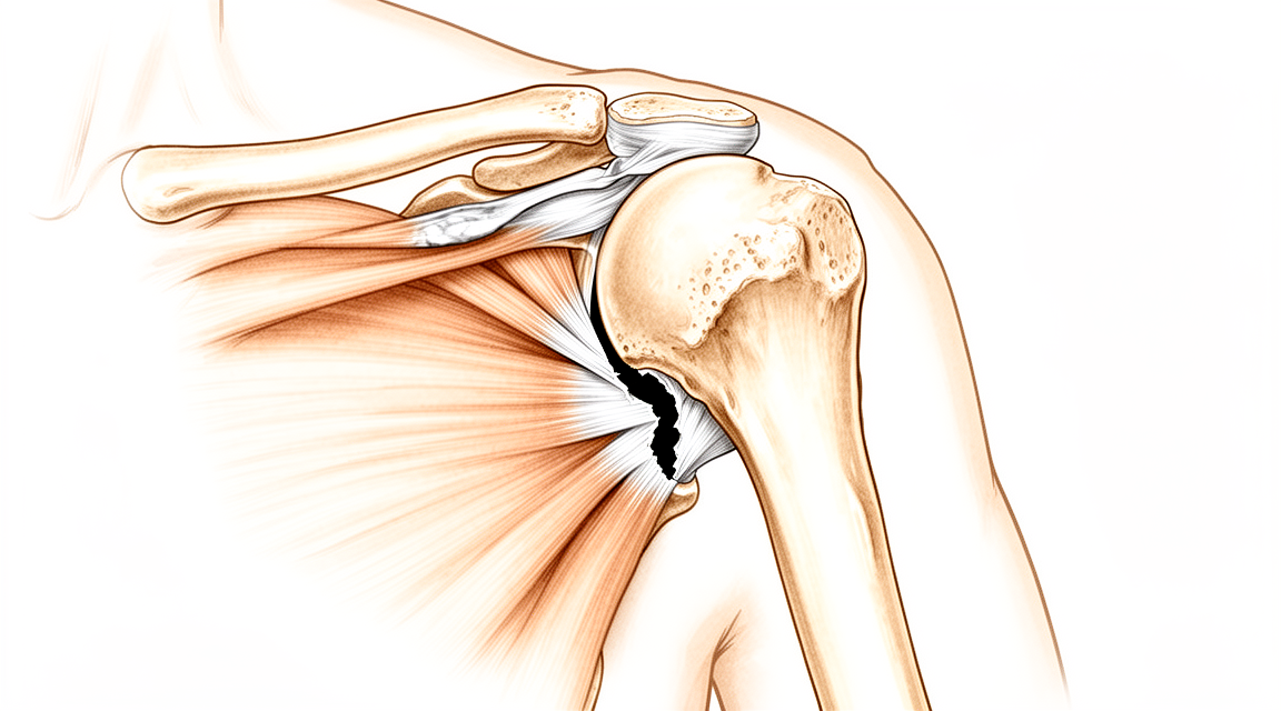
La reparación revisada del manguito rotador es una segunda intervención destinada a volver a reparar un tendón del hombro que se ha vuelto a desgarrar o que no sanó tras una reparación previa. La recomendamos cuando el dolor, el dolor nocturno, la debilidad o la rigidez persisten a pesar de ese tratamiento inicial. Estudios como la ecografía o la resonancia magnética nos ayudan a evaluar el estado del tendón y del músculo circundante antes de tomar una decisión. El objetivo de la operación es aliviar su dolor y mejorar el movimiento y la funcionalidad de su hombro. Muchas personas mantienen esos beneficios hasta 5 años después de la cirugía. Analizaremos con usted los beneficios y los riesgos; la decisión final la tomarán usted y nosotros en conjunto.
Antes de la operación
Antes de la cirugía, solicitamos estudios de imagen para planificar la intervención. Estos pueden incluir radiografías, ecografías o resonancias magnéticas (un estudio que muestra en detalle los tejidos blandos alrededor del hombro). El día de la operación, deberá abstenerse de comer y beber durante siete horas antes del procedimiento. Pedimos que sea durante siete horas para poder adelantar su cirugía si la agenda quirúrgica lo permite; su cirujano le confirmará la hora exacta. Si toma medicamentos de forma regular, traiga una lista de los mismos y le indicaremos cuáles debe suspender. Organice que alguien lo lleve a casa después de la operación, ya que no podrá conducir por sí mismo. Use ropa holgada y cómoda, fácil de poner y quitar. Si padece otras enfermedades, es posible que necesite análisis de sangre o una consulta con el anestesista (el especialista encargado de administrar la anestesia).
El día de la intervención
Llega usted a la unidad de admisiones quirúrgicas del hospital, donde se le registra y prepara para el quirófano. Posteriormente, se reunirá con el anestesista. Esta operación se realiza bajo anestesia general combinada con un bloqueo nervioso regional. Usted permanecerá completamente dormido durante la intervención; el bloqueo nervioso (una inyección que adormece los nervios que inervan el brazo antes de que usted despierte) le proporcionará alivio del dolor durante las primeras 12 a 24 horas posteriores a la cirugía. El anestesista se reunirá con usted antes de la operación y le explicará ambos procedimientos. A continuación, será llevado al quirófano, donde se realiza la operación. Una vez finalizada, despertará en la sala de recuperación, donde las enfermeras lo vigilarán mientras la anestesia va desapareciendo. Cuando su estado sea estable, será trasladado a la planta de hospitalización o podrá volver a casa, según el tipo de intervención y su recuperación.
Descripción del procedimiento quirúrgico
La reparación revisora del manguito rotador se realiza mediante cirugía mínimamente invasiva. El cirujano realiza varias incisiones pequeñas alrededor del hombro, incluida una en la parte posterior, y utiliza una cámara diminuta dentro de la articulación. Esta cámara permite visualizar el tendón desgarrado y el hueso del cual se ha separado.
Por lo general, el plan quirúrgico consiste en volver a fijar el tendón mediante pequeños anclajes colocados en el hueso; dichos anclajes retienen los puntos de sutura que atraviesan el tendón. Los anclajes se colocan en dos filas: una más cercana a la articulación y otra más alejada, que ejerce una fuerza de tracción para mantener firmemente el tendón contra el hueso, facilitando así su cicatrización. Según los hallazgos del cirujano durante la operación, podría optarse por utilizar una sola fila de anclajes o un tipo distinto de anclaje situado más cerca de la articulación.
Con frecuencia, se coloca un parche blando elaborado con tejido biológico debajo del tendón en el lugar de la reparación, con el fin de favorecer su adhesión al hueso. Puede obtener más información al respecto en nuestra página sobre el andamio biológico EnFix.
En caso de que el hueso al que se une el tendón esté desgastado, el cirujano podría añadir una pequeña cantidad de injerto óseo para reconstruirlo, de modo que los anclajes tengan una superficie sólida a la que adherirse. Si el tendón presenta daños excesivos para ser reinsertado, se le explicarán otras alternativas, como el uso de un tendón de donante para cerrar la brecha, tanto antes como durante el tratamiento.
Finalmente, las incisiones se cierran con puntos de sutura y se coloca un vendaje sobre las heridas; dicho vendaje debe permanecer en su lugar durante unos 10 días.
Después de la operación
La mayoría de los pacientes permanecen una noche en el hospital tras esta operación, aunque algunos pueden volver a casa el mismo día. Despertará en la sala de recuperación y luego será trasladado a la planta de hospitalización. Las enfermeras lo revisarán y le administrarán analgésicos según sea necesario. El bloqueo nervioso aplicado durante la cirugía suele mantener el hombro cómodo durante las primeras 12 a 24 horas, por lo que al principio es posible que sienta poco dolor. Para mayor comodidad, su brazo descansará en un cabestrillo sencillo. Puede quitárselo para lavarse y para realizar los ejercicios que le enseñaremos. Dejamos el vendaje puesto durante unos 10 días; por favor, no lo retire antes de ese plazo a menos que se lo indiquemos. Lo cambiaremos o lo retiraremos cuando venga a la consulta. Una enfermera le ayudará a sentarse, ponerse de pie y caminar una corta distancia, generalmente unas horas después de despertar. Por favor, organice que alguien se quede con usted durante las primeras 24 horas después de volver a casa.
Recuperación
Los primeros días después de la cirugía consisten en descanso y comodidad. El bloqueo nervioso desaparece en aproximadamente un día, y entonces el hombro puede doler más que antes. La hinchazón y los hematomas alrededor del hombro y el brazo son normales y desaparecen durante las siguientes semanas. Las compresas de hielo, el descanso y los analgésicos que le recetamos ayudan a aliviar estos síntomas. Muchas personas encuentran más cómodo dormir sentadas en una silla o apoyadas en almohadas al principio, ya que acostarse boca arriba ejerce presión sobre el hombro.
Para mayor comodidad, el brazo se sostiene mediante un cabestrillo sencillo. Debe quitárselo para lavarse y para realizar los ejercicios que le indique su fisioterapeuta. Estos ejercicios comienzan de forma suave, con movimientos pequeños que evitan la rigidez del hombro mientras el tendón cicatriza. Con el tiempo, los movimientos se vuelven más amplios y fuertes, siempre bajo la guía de su fisioterapeuta y según su propia tolerancia. A diario, podrá caminar, moverse por la casa y utilizar libremente el otro brazo. Necesitará ayuda para tareas más pesadas, como levantar objetos, cargar cosas o alcanzar cosas por encima de la cabeza, hasta que su hombro esté listo.
Una vez que la hinchazón disminuye y recupera la movilidad, las actividades cotidianas resultan más fáciles. Primero vuelven a ser posibles vestirse, lavarse y realizar tareas domésticas ligeras. No podrá conducir hasta que su cirujano se lo autorice en la revisión, generalmente alrededor de la sexta semana; consulte nuestra guía sobre Conducción después de cirugía de miembro superior para más detalles. El regreso al trabajo y a la práctica deportiva se produce de forma gradual, una vez que su cirujano y fisioterapeuta consideren que el tendón ha cicatrizado adecuadamente.
La recuperación varía de una persona a otra. Su cronograma personal puede diferir, y su cirujano y fisioterapeuta le guiarán durante todo el proceso.
Qué puede salir mal
La mayoría de los pacientes evolucionan bien, pero en ocasiones pueden surgir problemas. Su cirujano y el equipo lo vigilarán de cerca para detectar cualquier incidencia a tiempo.
Lo que más controlamos es la posibilidad de que el tendón se desgarre nuevamente. Esto puede manifestarse como dolor que reaparece tras haber disminuido, acompañado de debilidad al levantar objetos o al estirar el brazo. La mayoría de los nuevos desgarros ocurren dentro de los primeros seis meses; por eso le haremos un seguimiento riguroso durante ese periodo. Si el dolor o la debilidad regresan, infórmenos de inmediato en lugar de esperar a su próxima consulta.
Las infecciones son poco frecuentes, pero requieren atención urgente. Acuda a su médico de cabecera o al servicio de urgencias si nota un dolor profundo y palpitante que no cede con analgésicos comunes, enrojecimiento que se extiende desde la zona de la incisión, fiebre o si el hombro se siente caliente al tacto. En algunos casos, una reparación fallida puede estar relacionada con una infección de bajo grado difícil de detectar. Ante un dolor o rigidez intensos e inexplicables en el hombro, siempre debemos descartar una infección; por ello, no dude en comentárnoslo.
No todas las cirugías de revisión logran el objetivo de un hombro sin dolor y fuerte. Algunas personas observan que su movilidad no mejora tanto como esperaban, o que persisten la rigidez y la debilidad. Si el tejido tendinoso es delgado, el desgarro es grande o ya existe artritis, el resultado podría no ser el deseado por ninguno de los dos. Antes de tomar una decisión, hablaremos con usted con total transparencia sobre lo que revelan sus estudios de imagen.
Existen también factores que incrementan el riesgo de que la reparación fracase. Ciertos antibióticos administrados poco después de la cirugía se han asociado con fallos en la reparación y la necesidad de una segunda operación. Si le recetan algún medicamento nuevo en las semanas posteriores a la intervención, conviene consultarnos o a su médico de cabecera primero. Asimismo, los problemas de colesterol pueden influir en la cicatrización del tendón; por eso tenemos en cuenta su salud general al explicarle qué esperar.
En la tabla de complicaciones de esta página se detallan las tasas habituales, por si desea conocer los datos concretos.
¿Cuándo deben contactarnos?
Llámenos si nota fiebre, enrojecimiento que se extiende desde la herida, secreción proveniente de la herida o un dolor que sigue empeorando. Infórmenos de inmediato si el dolor o la debilidad regresan después de haber comenzado a mejorar, o si retrocede en su progreso de recuperación. Acuda a urgencias si experimenta dolor intenso y repentino, hinchazón en la pantorrilla, dificultad para respirar, pérdida de sensibilidad en el brazo o si no puede moverlo. La mayoría de las roturas que se repiten ocurren dentro de los primeros seis meses; por eso, durante ese período vigilamos de cerca su hombro.
¿Dónde leer más sobre esta afección?
Esta página trata sobre la intervención quirúrgica en sí. La afección que se trata, así como las evidencias sobre cuándo la cirugía resulta útil y cuándo no, se explican con mayor detalle en la página Trastornos del manguito rotador.
Evidence & references
This is the clinical evidence summary written for health professionals. It is technical, and it lists the research this page was built from. You do not need to read it to understand your treatment or to make a decision about it.
Overview
- Interposition grafting using fascia lata autograft can repair multiple rotator cuff injuries to the greatest extent [1].
- Interposition grafting using fascia lata autograft can restore the function of the shoulder joint [1].
- Interposition grafting using fascia lata autograft can effectively relieve pain [1].
- Biologic augmentation with a bioinductive collagen patch in revision rotator cuff tear repair reduces the retear rate at 12-month follow-up by 25% [2].
- Biologic augmentation with a bioinductive collagen patch in revision rotator cuff tear repair yields similar improvements in clinical outcomes [2].
- Biologic augmentation with a bioinductive collagen patch in revision rotator cuff tear repair is associated with no increased risk of complication [2].
Anatomy & Pathophysiology
Bony Anatomy
- The proximal humerus comprises four main parts: the humeral head, greater tuberosity (GT), lesser tuberosity (LT), and humeral shaft [5].
- The articular head of the proximal humerus is spherical with a diameter of 37 to 57 mm [5].
- The most superior portion of the articular surface of the humeral head averages 8 mm above the greater tuberosity [5].
- Humeral version averages 29.8 degrees, with a range of 10 to 55 degrees [5].
- The humeral head is inclined approximately 130 degrees with respect to the humeral shaft [5].
- The anatomic neck of the proximal humerus is located at the junction of the articular surface and the tuberosities [5].
- The surgical neck represents an indistinct region, or metadiaphyseal junction, below the tuberosities but above the humeral shaft [5].
- The greater tuberosity is located in a posterior-superior location with respect to the humeral shaft and serves as the attachment site for the supraspinatus, infraspinatus, and teres minor tendons [5].
- The lesser tuberosity is located on the anterior aspect of the proximal humerus and serves as the attachment site for the subscapularis tendon [5].
- The bicipital groove lies between the greater and lesser tuberosities and serves as a pathway for the long head of the biceps [5].
- The distal aspect of the bicipital groove is internally rotated with respect to the proximal portion [5].
- The glenoid is a convex structure of shallow depth shaped like an inverted pear [5].
- The glenoid articulates with the humeral head and serves as the attachment for the labrum and joint capsule [5].
- The acromion, coracoacromial ligament, and coracoid process form the coracoacromial arch, a rigid bony-ligamentous structure that imparts stability to the shoulder girdle [5].
- The rotator cuff, subacromial bursa, and subdeltoid bursa pass underneath the coracoacromial arch [5].
- The scapula is attached to the axial skeleton by the clavicle, specifically via the acromioclavicular and sternoclavicular joints [7].
- The scapular body is triangular when viewed anteroposteriorly, with its base situated superiorly and its apex inferiorly [7].
- The glenoid is connected with the flat body of the scapula by the scapular neck [7].
- The hook-shaped coracoid process curves forwards from the superior surface of the scapular neck [7].
- The scapular spine arises from the posterior surface of the scapular body and ends in a flattened bony process, the acromion, which curves forwards [7].
- The distribution of bony mass in the scapula is highly uneven, with the highest concentration in the glenoid, scapular neck, and lateral border of the scapular body [7].
- Two bony pillars extend between the glenoid and the scapular body to transmit compressive forces from the glenoid fossa [7].
- The lateral pillar connects the inferior border of the glenoid with the inferior angle of the scapula [7].
- The spinal pillar arises from the central part of the glenoid and continues medially to become part of the base of the scapular spine [7].
- The weakest bone in the scapula is located primarily in the central part of the biomechanical body, specifically in the infraspinous fossa [7].
- The weakest area of the circumference of the biomechanical body of the scapula is the spinomedial angle, where the scapular spine connects to the medial border [7].
- The humeral head averages 19° of retroversion and 41° of inclination (neck-shaft angle) [8].
- The glenoid averages 5° of retroversion in relation to the axis of the scapular body [8].
- The subchondral bone of the glenoid is relatively flat, with the articular concavity augmented by cartilage and a circumferential labrum [8].
- The neck-shaft angle measures an average of 135 degrees, and the humeral head is retroverted an average of 30 degrees [6].
Vascular Anatomy
- The proximal humerus receives its blood supply from the anterior and posterior humeral circumflex branches from the third division of the axillary artery [5].
- The posterior humeral circumflex artery travels with the axillary nerve, enters the quadrilateral space posteriorly, and anastomoses with a branch of the anterior circumflex to supply the posterior cuff [5].
- The anterior humeral circumflex artery arises from the axillary artery at the inferior border of the subscapularis and provides vascular inflow to the humeral head via its terminal anterolateral branch, known as the artery of Laing or arcuate artery [5].
- The ascending branch of the anterior humeral circumflex artery courses parallel to the lateral aspect of the long head biceps tendon and enters the humeral head at the interface of the bicipital groove and greater tuberosity [5].
- Injury to the arcuate artery may result in osteonecrosis of the humeral head [5].
- Additional extraosseous collateral branches can permit humeral head perfusion despite complete ligation of the arcuate artery [5].
- The major blood supply to the humeral head is through the ascending branch of the anterior humeral circumflex artery, which penetrates the head at the bicipital groove and becomes the arcuate artery [6].
- The anterolateral ascending branch of the anterior humeral circumflex artery provides the primary blood supply to the humeral head [8].
- The terminal intraosseous portion of the anterior humeral circumflex artery enters at the proximal aspect of the intertubercular groove as the arcuate artery [8].
Soft Tissue & Ligamentous Anatomy
- The rotator cuff consists of four muscles: the subscapularis, supraspinatus, infraspinatus, and teres minor [6].
- The teres major is not a rotator cuff muscle [6].
- The rotator cuff muscles serve as depressors of the humeral head to allow the deltoid to efficiently abduct the humerus [6].
- The infraspinatus and teres minor are external rotators, while the subscapularis is an internal rotator of the humerus [6].
- The glenohumeral joint is stabilized dynamically by the rotator cuff via joint compression and by the positioning of the scapulothoracic joint [8].
- Static stabilizers of the glenohumeral joint include articular congruity, the glenoid labrum, concavity-compression, negative intra-articular pressure, and the glenohumeral capsule and ligaments [8].
- The glenoid labrum provides concavity and up to 50% of marginal glenoid socket depth [8].
- The rotator interval is defined medially by the base of the coracoid, superiorly by the supraspinatus tendon, and inferiorly by the subscapularis tendon [8].
- The rotator interval contains the coracohumeral ligament, the superior glenohumeral ligament, and the intra-articular portion of the long head of the biceps tendon [8].
- Laxity of the rotator interval results in inferior laxity, known as the sulcus sign [8].
- Contracture of the rotator interval is seen with adhesive capsulitis [8].
- The coracohumeral ligament restricts external rotation in adduction and is a static restraint to inferior and posterior translation in adduction and external rotation [8].
- The superior glenohumeral ligament is a primary static restraint against anterior translation with the arm at the side [8].
- The superior glenohumeral ligament, along with the coracohumeral ligament, forms a pulley that provides restraint against medial subluxation of the long head of the biceps tendon [8].
- The middle glenohumeral ligament is a primary static restraint against anterior translation with the arm in external rotation and 45° of abduction [8].
- The anterior band of the inferior glenohumeral ligament is a primary static restraint against anterior-inferior dislocation of the glenohumeral joint in 90° of abduction and external rotation [8].
- The posterior band of the inferior glenohumeral ligament is a primary static restraint against posterior-inferior translation in internal rotation and adduction [8].
- The superior transverse scapular ligament arises from the medial base of the coracoid overlying the suprascapular notch [8].
- The suprascapular artery runs superior to the superior transverse scapular ligament, while the nerve runs deep to it [8].
- Entrapment of the suprascapular nerve at the superior transverse scapular ligament causes denervation of both the supraspinatus and the infraspinatus [8].
- The spinoglenoid ligament overlies the suprascapular nerve at the spinoglenoid notch [8].
- Entrapment, traction, or compression of the suprascapular nerve at the spinoglenoid notch causes denervation of the infraspinatus [8].
- The subscapular bursa lies between the subscapularis tendon and the neck of the scapula and communicates with the joint cavity between the superior and middle glenohumeral ligaments [9].
- The subscapular bursa protects the tendon of the subscapularis at the point where it passes under the base of the coracoid process and over the neck of the scapula [9].
- The subscapular bursa is linked to the coracoid process by a suspensory ligament [9].
- In 28% of dissected specimens, the subscapular bursae merged with the subcoracoid bursae, forming a unique wide bursa [9].
- The subscapular bursa often houses loose bodies in the shoulder and is a region where synovitis may be most intense [9].
- A soft tissue sheath consistently covers the long head of the biceps tendon to the level of the proximal margin of the pectoralis major tendon and contributes to the roof of the bicipital tunnel [9].
- The fibro-osseous bicipital tunnel consists of three distinct anatomic zones: Zone 1 (bony groove), Zone 2 ("no man's land" between subscapularis and pectoralis major), and Zone 3 (subpectoral region) [9].
Pathophysiology & Biomechanics
- Stability and function of the glenohumeral joint are provided by the interaction of structures that promote a near global range of motion and purposeful function [5].
- External loads transferred to the shoulder girdle are initially offset by joint surface anatomy, joint volume, atmospheric pressure, and joint fluid cohesion and adhesion [5].
- Moderate and large loads are counterbalanced by the deltoid and rotator cuff, and by the capsulolabral and bone structures, respectively [5].
- Pathologic conditions alter complex shoulder interactions, resulting in pain, decreased range of motion and stiffness, and disability [5].
- Displacement of proximal humeral fracture fragments occurs in a predictable manner based on deforming forces created by the tendinous insertions of the pectoralis major, subscapularis, supraspinatus, and infraspinatus [5].
- The subscapularis inserts on the lesser tuberosity and causes medial displacement of the fragment [5].
- The supraspinatus and infraspinatus insert on the greater tuberosity and cause superior and posterior displacement of the fragment [5].
- The pectoralis major inserts on the humeral shaft and displaces it medially [5].
- Fractures involving the anatomic neck are prognostically worse than fractures involving other regions of the proximal humerus due to potential disruption of the vascular supply to the humeral head and subsequent development of avascular necrosis [5].
- Displaced proximal humeral fractures can impede normal movement of the rotator cuff, subacromial bursa, and subdeltoid bursa, causing impingement and disruption of normal glenohumeral motion [5].
- In proximal humeral fractures, the subdeltoid and subacromial bursae can become thickened and fibrotic, forming adhesions that limit normal glenohumeral motion [5].
- The glenoid cavity is a shallow socket, approximately one third the size of the humeral head [6].
- Stability of the glenohumeral joint depends on the capsule, ligament, and muscle [6].
- A redundant capsule allows for motion in the glenohumeral joint [6].
- The scapula is separated from the chest wall by thin gliding fibro-fatty tissue, allowing its smooth excursion over the chest wall [7].
- The scapula provides efficient support to the humeral head, allowing compressive forces to be optimally transmitted from the upper limb to the shoulder girdle without compromising stability or mobility of the glenohumeral joint [7].
- The superior shoulder suspensory complex provides a stable connection between the scapula and the axial skeleton [8].
- The superior shoulder suspensory complex is composed of the glenoid, coracoid process, coracoclavicular ligaments, distal clavicle, acromioclavicular joint, and acromion [8].
- The superior strut of the superior shoulder suspensory complex comprises the middle clavicle [8].
- The inferior strut of the superior shoulder suspensory complex comprises the lateral scapular border and spine of the scapula [8].
- Normal shoulder motion is approximately two-thirds glenohumeral and one-third scapulothoracic [8].
- The sternoclavicular joint is the only true diarthrodial articulation between the upper appendicular and axial skeletons [8].
- The posterior sternoclavicular joint capsule and ligaments are the primary stabilizers to anterior and posterior translation of the medial clavicle [8].
- The acromioclavicular joint is a small diarthrodial joint with an interposed fibrocartilaginous disk [8].
- The superior and posterior acromioclavicular ligaments are the primary stabilizers to anterior and posterior horizontal translation of the clavicle [8].
- The coracoclavicular ligaments, consisting of the conoid (medial) and trapezoid (lateral) ligaments, are the primary stabilizers to superior vertical translation of the distal clavicle [8].
- The acromion has three ossification centers: the metacromion (base), mesoacromion (middle), and preacromion (tip) [8].
- Failure of fusion of the acromial ossification centers results in os acromiale [8].
- The relationship between acromial anatomy and rotator cuff disease remains controversial, with classification of acromial morphology challenged by poor interobserver reliability [8].
- The relationship between coracoid morphology and subscapularis tears is controversial [8].
- The coracobrachialis muscle and the short head of the biceps tendon originate from the coracoid process [8].
- The pectoralis minor muscle inserts onto the medial coracoid process [8].
- The proximal humerus has three centers of ossification: the humeral head (4 to 6 months), greater tuberosity (1 to 3 years), and lesser tuberosity (3 to 5 years) [8].
- The ossification centers of the proximal humerus fuse to the shaft at age 17 to 20 years [8].
- The clavicle is the first bone to ossify, occurring in the fifth week of gestation, and is the only long bone to ossify by intramembranous ossification [8].
- The medial (sternal) epiphysis of the clavicle is the last ossification center to fuse, occurring at age 20 to 25 years [8].
- The primary blood supply to the clavicle is periosteal, with no nutrient artery present [8].
- Ossification of the scapular body begins at the eighth week of gestation [8].
- The scapular spine is an osseous ridge that separates the supraspinatus and infraspinatus fossae [8].
- The scapula has only one true diarthrodial articulation, the acromioclavicular joint [8].
Classification
- Biologic augmentation with a bioinductive collagen patch in revision rotator cuff tear repair yields similar improvements in clinical outcomes compared to controls [2].
- Biologic augmentation with a bioinductive collagen patch in revision rotator cuff tear repair does not increase the risk of complication [2].
Clinical Presentation
- Biologic augmentation with a bioinductive collagen patch in revision rotator cuff tear repair reduces the retear rate by 25% at 12-month follow-up [2].
Investigations
Plain Radiography
- The purpose of shoulder imaging is to help establish the diagnosis, determine the severity of the pathoanatomy, assist in surgical planning, and enable the surgeon to illustrate the condition of the shoulder to the patient [4].
- Standardized plain films are almost always sufficient to garner the information needed for shoulder care [4].
- The first key radiographic view is the anteroposterior (AP) view taken in the plane of the scapula such that the x-ray beam passes through the glenohumeral joint [4].
- The AP view in the plane of the scapula shows the superoinferior position of the humeral head relative to the glenoid, the presence of osteophytes on the humeral head and glenoid, narrowing of the joint space, and the degree of medial displacement of the humerus in relation to the lateral acromial line [4].
- The AP view in the plane of the scapula also shows the quality of the humeral and glenoid bone, the presence of loose bodies, and whether there is humeral head collapse or deformity [4].
- The second key radiographic view is the axillary view taken with the arm in the functional position of elevation in the plane of the scapula [4].
- The axillary view is oriented so that both the spinoglenoid notch and the scapular neck are visible [4].
- The axillary view shows a different perspective of the humeral anatomy, the amount of glenoid bone, the shape of the glenoid, its version in relation to the plane of the scapula, and the relationship of the humeral head to the glenoid fossa [4].
- The standardized axillary view is referred to as the "truth view" because it demonstrates the glenohumeral relationships in the functional position of elevation [4].
- CT scans have the disadvantage of being taken with the arm in the adducted position, unlike the axillary truth view which is taken in elevation [4].
- When taken properly, standardized anteroposterior and axillary views indicate the thickness of the cartilage space between the humerus and the glenoid, relative positions of the humeral head and the glenoid, presence of osteophytes, degree of osteopenia, and extent of bony deformity and erosion [4].
- Joint space narrowing is most evident on the axillary truth view as opposed to images made with the arm at the side [4].
- The axillary truth view can show posterior subluxation or "functional decentering" that is not evident in images taken with the arm at the side [4].
- The degree of posterior subluxation can be measured by the position of the center of the humeral head in relation to the plane of the scapula, the position of the center of the humeral head in relation to the glenoid face, or the point of contact of the humeral articular surface on the glenoid articular surface [4].
- The point of contact of the humeral articular surface on the glenoid articular surface reflects the degree of centering of the net humeral joint reaction force on the glenoid [4].
- Malcentering of the joint reaction force leads to posterior instability, posterior glenoid wear, and "rocking horse" loosening of prosthetic glenoid components [4].
- At least two X-ray views should be obtained: an anteroposterior in the plane of the glenoid and an axillary projection with the arm in abduction to show the relationship of the humeral head to the glenoid [13].
Magnetic Resonance Imaging
- Magnetic resonance imaging (MRI) is useful to identify osteonecrosis of the humeral head, or a bone tumour [13].
- MRI can identify labral tears and rotator cuff tears, although the accuracy for these is enhanced by combining the scan with arthrography [13].
Computed Tomography
- Computed tomography (CT) is helpful for planning fracture surgery and shoulder joint replacement [13].
- CT scans may offer a few degrees of increased precision in the measurement of glenoid version, but this precision does not necessarily improve the quality of the surgery or the clinical outcome [4].
Ultrasonography
- Ultrasonography is a simple and accurate test for identifying rotator cuff tears and calcific tendinitis [13].
- Ultrasonography can be useful in guiding injections or barbotage (aspirating calcific deposits in the rotator cuff) [13].
- The most commonly performed joint examination using ultrasonography is the shoulder examination [11].
- The accuracy of rotator cuff ultrasonography depends on the skill of the scanner operator and an awareness of pitfalls that are encountered [11].
General Imaging Principles
- The shoulder is a three-dimensional structure that cannot be represented by a single planar view [15].
- Critical relationships, such as the degree of centering of the humeral head, change with the position of the arm [15].
- Shoulder pathology may be found in a large number of different bones and soft tissues [15].
- Overlying and superimposed structures as well as metallic implants may complicate imaging the structures of interest [15].
- Surgeons need to develop a judicious approach to imaging that yields the information necessary to treat the patient while avoiding the tendency to "over-image" [15].
- The temptation to "overimage" should be resisted, obtaining only the scans or reconstructions that are necessary for the care of the patient [4].
- Proper radiographic technique is as important as proper surgical technique to achieve the desired outcome [4].
Key Evidence
- [L5] This technique can repair multiple rotator cuff injuries to the greatest extent, restore the function of the shoulder joint, and effectively relieve pain. [1] (10.1016/j.eats.2023.08.027)
- [L1] Biologic augmentation with a bioinductive collagen patch in revision rotator cuff tear repair reduces the rate at 12-month follow-up by 25%, yielding similar improvements in clinical outcomes and without any increased risk of complication. [2] (10.1016/j.jseint.2025.101507)
References
[1] Interposition Grafting Using Fascia Lata Autograft for Failed Rotator Cuff Repairs. Arthroscopy Techniques. 2023. DOI: 10.1016/j.eats.2023.08.027
[2] Biological augmentation in revision surgery: a matched-pair study of the effect of a nuns bioinductive collagen patch in patients with rotator cuff retear and a previous arthroscopic rotator cuff repair. JSES International. 2026. DOI: 10.1016/j.jseint.2025.101507
[4] Rockwood And Matsen S The Shoulder. Arthroscopic Management of Prearthritic and Arthritic Conditions of the Shoulder and the Postarthroplasty Shoulder > Radiographic Evaluation.
[5] Rockwood And Matsen S The Shoulder. Shoulder and Elbow Specialty Clinic Workers’ Survey > ANATOMY.
[6] A Lange Medical Book Current Diagnosis Treatment In Orthopedics Fifth Edition. 2Musculoskeletal Trauma Surgery > SHOULDER AND ARM INJURIES.
[7] Rockwood And Green S Fractures In Adults. 29: Principles of Nonunion and Bone Defect Treatment > Applied Anatomy Related to Scapular Fractures.
[8] Aaos Comprehensive Orthopaedic Review 3. Anatomy of the Shoulder, Arm, and Elbow > I. Shoulder.
[9] Rockwood And Matsen S The Shoulder. Developmental Anatomy of the Shoulder and Anatomy of the Glenohumeral Joint > Bursae.
[11] Orthopaedic Knowledge Update Sports Medicine 6. Diagnostic Ultrasonography and Ultrasonography-Guided Procedures > Annotated References.
[13] Apley And Solomon S Concise System Of Orthopaedics And Trauma. INVESTIGATION.
[15] Rockwood And Matsen S The Shoulder. Developmental Anatomy of the Shoulder and Anatomy of the Glenohumeral Joint > SENIOR EDITOR COMMENTARY.