¿Por qué se ha recomendado esta operación?
El Dr. Kieran Hirpara, cirujano de extremidades superiores en el Mater Private Hospital Rockhampton, comienza por proponer las opciones menos invasivas que se adapten a su condición. Por lo general, los pacientes son derivados a nuestra clínica por su médico de cabecera; si un fisioterapeuta le ha sugerido que nos consulte, igualmente necesitará una derivación de su médico de cabecera para poder acceder al reembolso de Medicare. En su cita, tomamos su historia clínica, examinamos su mano y solicitamos estudios de imagen si es necesario para confirmar el diagnóstico.
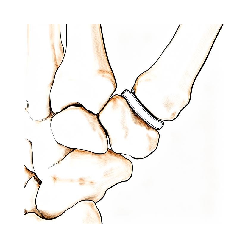
Esta operación reemplaza la superficie desgastada de una articulación pequeña por una pieza de carbono lisa y moldeada. Generalmente se propone para la artritis de larga evolución que no ha mejorado con modificaciones en las actividades, terapia de mano o el uso de férulas. Normalmente intentamos primero el tratamiento no quirúrgico y consideramos la cirugía cuando este no ha aportado suficiente mejoría. En algunos problemas estructurales, puede recomendarse la cirugía de entrada.
La operación tiene como objetivo aliviar el dolor manteniendo la movilidad de la articulación. También busca conservar la altura natural del pulgar y preservar la fuerza de pinza y de agarre. En la articulación de la base del pulgar, el 91% de los implantes siguen funcionando bien en el seguimiento a medio plazo. Hablaremos con usted sobre esta opción y decidiremos juntos si se adapta a su mano y a sus objetivos.
Antes de la operación
Durante las siete horas previas a la operación, no coma ni beba nada. Pedimos siete horas en lugar de seis para poder adelantar su cirugía si el programa quirúrgico avanza antes de lo previsto. Su cirujano le indicará cuáles de sus medicamentos habituales debe suspender y cuándo, y es útil traer una lista por escrito de todo lo que toma. Organice que alguien lo lleve a casa después y use ropa holgada y cómoda. Normalmente bastan las radiografías para planificar la operación, aunque a veces también se utiliza una resonancia magnética o una ecografía. Si padece otras enfermedades, es posible que necesite análisis de sangre o una evaluación con el anestesista, el médico que administra la anestesia. La mayoría de los pacientes no necesitan ninguna de las dos cosas.
El día de la intervención
Acudirá a la unidad de admisiones quirúrgicas del hospital, donde se le registrará y preparará para la cirugía. Conocerá al anestesista, el médico que se encarga de su anestesia. Esta intervención se realiza bajo anestesia general. Usted permanecerá completamente dormido durante la operación. En algunos pacientes también se aplica un bloqueo nervioso regional para aliviar el dolor postoperatorio; el anestesista decide al respecto el mismo día, según sus circunstancias individuales.
Posteriormente, se le llevará al quirófano, donde se lleva a cabo la operación. Después despertará en la sala de recuperación, donde las enfermeras le supervisarán mientras la anestesia va desapareciendo. Una vez que se halle estable, será trasladado a la planta de hospitalización o podrá volver a casa, según el tipo de intervención y su evolución.
¿En qué consiste la operación?
El cirujano realiza una única incisión sobre la zona de la mano que se va a operar. A través de esta abertura, retira las superficies desgastadas y dañadas de la articulación. En lugar de esas superficies desgastadas, coloca una pieza de carbono lisa y moldeada que queda situada entre los huesos. Este separador asume la función de la superficie articular desgastada, de modo que los huesos se deslizan suavemente en lugar de rozar entre sí.
En la articulación del pulgar situada en la base de la mano, el cirujano conserva la altura natural del pulgar colocando el separador entre los huesos. A continuación, la articulación se repara y se refuerza para que cicatrice en una buena posición. Es posible que el pulgar se mantenga inmovilizado en posición recta mientras cicatriza.
La incisión se cierra con puntos de sutura. Se coloca un vendaje sobre la herida, y nuestro equipo le explicará cómo cuidarla durante los primeros días.
Después de la operación
La mayoría de los pacientes permanecen una noche en el hospital tras esta operación, aunque algunos pueden volver a casa el mismo día. Despertará en la sala de recuperación y después será trasladado a la habitación. Las enfermeras se encargarán de que esté cómodo y le darán analgésicos si los necesita. Su mano llevará un vendaje y una férula, que protegen la zona operada mientras cicatriza. Puede levantarse de la cama y moverse en cuanto se sienta estable. Por favor, organice que alguien se quede con usted durante las primeras 24 horas en casa. Dejamos el vendaje puesto durante unos 10 días; por favor, no lo retire antes de ese plazo a menos que se lo indiquemos. Lo cambiaremos o lo retiraremos cuando venga a consulta.
Recuperación
Es de esperar cierto dolor e hinchazón en la mano durante los primeros días y semanas. Esto forma parte normal de la cicatrización y mejora poco a poco. Mantener la mano elevada cuando esté en reposo ayuda, y los analgésicos que le damos aliviarán las molestias. La mayoría de las personas notan que lo peor del dolor pasa pronto, y a partir de ahí el bienestar mejora de forma constante.
Su mano llevará un vendaje y una férula, que protegen la articulación mientras cicatriza. Dejamos el vendaje puesto durante unos 10 días y lo cambiaremos o lo retiraremos cuando venga a consulta. La rehabilitación tras una cirugía de mano es terapia de mano, no fisioterapia. Su terapia de mano después de la cirugía se realizará con Ruby Doolan en Extend Rehabilitation. Ruby es terapeuta de mano: le guiará en los ejercicios y le confeccionará cualquier férula que necesite a medida que su mano se recupera. Al principio, los ejercicios son suaves y se centran en controlar la hinchazón. A medida que vuelve el movimiento, se van intensificando para recuperar la capacidad de doblar, estirar y agarrar.
Podrá realizar tareas cotidianas ligeras en casa dentro de los límites de la férula. No podrá agarrar, hacer pinza ni levantar peso con la mano operada mientras esté inmovilizada. Una vez retirada la férula y con la autorización de su cirujano, podrá empezar a usar la mano con más normalidad, y después podrá volver a conducir. Consulte nuestra página sobre Conducción tras cirugía de miembro superior para conocer los detalles.
La recuperación varía de una persona a otra. Su cronograma podría ser diferente, y su cirujano y su terapeuta le guiarán a lo largo del proceso.
Qué puede salir mal
La mayoría de los pacientes evolucionan bien, pero en ocasiones pueden surgir problemas. Su cirujano y el equipo lo vigilarán de cerca para detectar cualquier anomalía a tiempo.
A veces el implante cambia de posición o se hunde en el hueso. Es posible que note un dolor nuevo en la articulación, la sensación de que el dedo o el pulgar no está recto, o un cambio en la forma en que se mueve. Si nota cualquiera de estas cosas, coméntelo en su próxima revisión.
El implante también puede aflojarse. Normalmente se nota como un dolor que reaparece después de un buen periodo de mejoría, a menudo peor al agarrar o hacer pinza. Si esto ocurre, comuníqueselo a su cirujano.
Puede aparecer rigidez en la articulación. Es posible que el dedo o el pulgar no se doble ni se estire tanto como antes, y el tendón del dorso de la articulación puede quedar adherido por tejido cicatricial. Mencione esto en su revisión para que su terapeuta pueda ajustar sus ejercicios.
La articulación puede volverse inestable o puede reaparecer la deformidad original. Es posible que vea que el dedo vuelve a desviarse. Coméntelo en su próxima cita.
La infección es poco frecuente, pero requiere atención rápida. Llame a la clínica ese mismo día si tiene fiebre, enrojecimiento que se extiende alrededor de la herida, o salida de líquido o pus por ella. Un dolor que sigue empeorando a pesar de los analgésicos también significa que debe llamar a la clínica ese mismo día.
Durante este tipo de cirugía pueden irritarse o lesionarse estructuras cercanas. Un nervio próximo al pulgar puede volverse sensible y provocar hormigueo o alteraciones de la sensibilidad en la piel. Pueden verse afectados la arteria del lado del pulgar de la muñeca o un tendón de la parte delantera de la muñeca. Si nota entumecimiento o no puede mover los dedos una vez que haya pasado el efecto del bloqueo nervioso, a las 24 horas aproximadamente, llame a la clínica. Es normal tener entumecimiento y debilidad durante las primeras 24 horas después de un bloqueo nervioso.
En ocasiones, un hueso de la mano puede romperse durante la cirugía o después de ella. Esto provoca un dolor y una hinchazón repentinos. Si esto ocurre, llame a la clínica.
Si un implante falla y no puede salvarse, otras operaciones pueden ayudar. Se puede retirar el implante y tratar la articulación de otra forma, lo que alivia el dolor de manera fiable. En casos poco frecuentes, se puede fusionar la articulación, es decir, dejarla rígida de forma permanente, o puede ser necesario amputar el dedo.
En la tabla de complicaciones de esta página se detallan las tasas típicas, por si desea conocer los datos exactos.
¿Cuándo deben llamarnos?
La mayoría de los problemas aparecen pronto, y actuar con rapidez facilita su solución. Llame a la clínica ese mismo día si tiene fiebre, enrojecimiento que se extiende alrededor de la herida, o salida de líquido o pus por ella. Llame también a la clínica ese mismo día si el dolor sigue empeorando a pesar de los analgésicos. Llame a la clínica si nota entumecimiento o no puede mover los dedos una vez que haya pasado el efecto del bloqueo nervioso, a las 24 horas aproximadamente. Es normal tener entumecimiento y debilidad durante las primeras 24 horas después de un bloqueo nervioso.
Acuda al servicio de urgencias si tiene hinchazón o dolor en la pantorrilla, o dificultad para respirar o dolor en el pecho. Pueden ser signos de un coágulo de sangre. Acuda al servicio de urgencias si los dedos, la mano o el pulgar se ponen pálidos, fríos, blancos, azulados u oscuros. Puede tratarse de un problema de circulación.
Si no puede contactar con la clínica, fuera del horario de atención o durante el fin de semana, acuda al servicio de urgencias más cercano.
¿Dónde leer más sobre esta afección?
Esta página trata sobre la intervención quirúrgica en sí. La afección que se trata, así como las evidencias sobre cuándo la cirugía resulta útil y cuándo no, se explican con mayor detalle en la página Artritis basal del pulgar.
Evidence & references
This is the clinical evidence summary written for health professionals. It is technical, and it lists the research this page was built from. You do not need to read it to understand your treatment or to make a decision about it.
Overview
- Pyrocarbon interpositional arthroplasty provided pain relief and high patient satisfaction with good implant longevity in a study with minimum 8-year follow-up [1].
- Partial trapeziectomy with pyrocarbon arthroplasty may prove to be a successful option for the treatment of trapeziometacarpal joint osteoarthritis, providing excellent pain relief and high patient satisfaction [2].
- PyroCarbon implant hemiarthroplasty is a promising surgical technique for stages II and III trapeziometacarpal arthritis and can provide a stable, mobile, and pain-free joint [3].
- The Pyrocardan implant is not superior to other arthroplasty methods and has a high complication rate [4].
- The Pyrocardan implant appears as a simple alternative to the Dubert procedure for ulnar carpo-metacarpal osteoarthritis, particularly in cases involving the 4th ray [5].
- The PyroDisk implant for treating advanced trapeziometacarpal arthritis did not demonstrate superiority over published outcome data of trapeziectomy with or without ligament reconstruction and tendon interposition [6].
- The good functional and radiographic outcomes, and the absence of surgical complications are evidence that the Pyrocardan resurfacing implant is a valid option for treating STT osteoarthritis [7].
- Arthroplasty is recommended for patients with post-trauma painful PIP joints [8].
- Active patients, under 65 years of age with Eaton-Littler stage III osteoarthritis of the thumb CMC joint who are treated with anatomic pyrolytic carbon resurfacing hemiarthroplasty and stabilisation may achieve sustained improvement in pain and hand function for up to 6.5 years [9].
- The insertion of pyrolytic carbon implants for PIPJ arthroplasty is a technically demanding procedure, but it has the potential to achieve pain relief, stability, satisfactory ROM, and correction of the deformity [10].
- The results of pyrolytic carbon PIPJ arthroplasty in review were unpredictable and may not be superior to those achieved with other methods of arthroplasty [10].
- A study with long-term follow-up after pyrocarbon disc interposition for the treatment of CMC1 osteoarthritis grade 2 to 3 in more than 150 thumbs was conducted [11].
- The PyroDisk interposition arthroplasty confirmed good 5-year clinical results [12, 13].
- PyroDisk interposition has merit in patients with early disease unresponsive to conservative measures, in whom trapeziectomy may be considered too destructive and hemi or total arthroplasty overzealous [14].
- The good results, the simple surgical procedure, the absence of complications, and the lack of a need for any fixation or ligamentoplasty confirm the advantages of this pyrocarbon implant in the treatment of STT arthrosis [15].
- All subjective and objective outcomes were similar following LRTI and pyrolytic interpositional arthroplasty in patients with TMC arthritis, except pinch strength, which was more improved following pyrolytic interpositional arthroplasty [16].
- The encouraging mid-term clinical results of the interposition implant (APSI) in the context of proximal pole scaphoid pseudarthrosis make this technique a proven alternative, without precluding a definitive therapeutic option in case of failure [18].
- The Pyrocardan implant may be contraindicated in patients with symptomatic STT OA who intend to continue high-load activities, particularly those involving wrist extension under load, such as tennis [20].
Anatomy & Pathophysiology
Bony Anatomy
- The skeleton of the hand and wrist consists of 27 bones, of which 19 are long bones [28].
- The radial ray or first ray is the shortest, made up of only three bones: a metacarpal and two phalanges [28].
- The trapezium is clearly angled out in front of the carpal plane so that the first metacarpal makes an angle of about 45 degrees with the second metacarpal in the sagittal plane [28].
- The thumb metacarpal is the shortest, while the index metacarpal is by far the longest [28].
- The epiphyseal plates are located at the proximal ends of the phalanges and the first metacarpal, whereas they are located at the distal ends of the other metacarpals [28].
- The thumb metacarpal is independent and articulates with the trapezium [35].
- The index metacarpal is the most firmly fixed [35].
- The ring metacarpal has about 10 degrees of mobility in flexion and extension [35].
- The fifth metacarpal has a range of flexion–extension of approximately 20 degrees [35].
Soft Tissue & Vascular Anatomy
- The extrinsic extensors run through six different fibroosseous retinacular compartments at the wrist level [27].
- The first (most radial) compartment contains the abductor pollicis longus and the extensor pollicis brevis [27].
- The second extensor compartment contains the extensor carpi radialis longus and the extensor carpi radialis brevis [27].
- The third compartment contains the extensor pollicis longus, which turns abruptly radialward about Lister tubercle [27].
- The fourth extensor compartment contains the extensor indicis proprius lying deep to the four tendons of the extensor digitorum communis [27].
- The fifth compartment contains the extensor digiti quinti [27].
- The sixth compartment contains the extensor carpi ulnaris tendon, which inserts at the base of the little finger metacarpal [27].
- The extrinsic finger flexors are the flexor digitorum profundus and the flexor digitorum superficialis [27].
- The flexor digitorum profundus inserts on the proximal volar aspect of the distal phalanx [27].
- The flexor digitorum superficialis acts as a flexor of the proximal interphalangeal and metacarpophalangeal joints [27].
- There are seven interosseous muscles, four dorsal and three volar [32].
- The dorsal interossei are abductors [32].
- The volar interossei are adductors [32].
- The middle finger has two dorsal interossei (abductors) and no volar interossei (adductors) because the central axis of the hand lies within it [32].
- The "princeps pollicis" artery is the terminal branch of the radial artery and crosses the first intermetacarpal space [36].
- The princeps pollicis runs along the ulnar side of the first metacarpal bone and along the volar surface of the adductor muscle [36].
- The princeps pollicis divides into two terminal rami, namely the collateral palmar arteries of the thumb, at the level of the cutaneous flexion crease of the metacarpophalangeal joint [36].
- An arcade located deep in the flexor tendon joins together the two arteries at the level of the distal metaphysis of the first phalanx [36].
- The dorsal arteries of the thumb originate from the palmar arteries (princeps, commissural or anastomoses of the superficial arcade) at the level of the first metacarpal [36].
Functional Mechanics
- The wrist has three axes of movement, permitting the hand to be positioned in any spatial configuration [28].
- The thumb ray is more mobile, shorter, and more proximal than the other rays, allowing it to project in front of the plane of the palm to oppose itself to the other four rays [28].
- The skeleton of the hand presents a longitudinal and transverse concavity, giving it the shape of a cup with a palmar concavity when the thumb is placed next to the index finger [28].
- The transverse axis of the palm forms an acute angle of approximately 75 degrees with the longitudinal axis [28].
- The metacarpal arch is endowed with a great deal of adaptability because of the mobility of the peripheral metacarpals [35].
- The heads of the second and third metacarpals are fixed [35].
- The mobility of the ulnar metacarpals allows "flexion" of 10 degrees for the fourth metacarpal, and "flexion" of 20 degrees for the fifth metacarpal [35].
- The keystones of the longitudinal arches are the metacarpophalangeal articulations, whose thick anterior glenoid capsules, the volar plates, prevent hyperextension [35].
- The dorsal skin slides distally to allow metacarpophalangeal joint flexion [40].
- Interphalangeal flexion is accomplished by means of a special arrangement of skin folds on the dorsum of each articulation [40].
- The gliding mechanism for tendons in narrow crowded areas is assured by the synovial sheath, which allows a considerable amplitude of movement [40].
- The fibrous sheath assumes the role of a pulley when the tendon changes direction [40].
- The position of each articulation depends on the equilibrium of forces acting at that level, and this equilibrium is subject to the position of the immediately proximal articulation [40].
- Single articular movements around a fixed perpendicular axis simply do not exist in the hand [40].
- Almost all movements in the hand are around oblique and variable axes, resulting in combined movements permitting optimal orientation of the phalanges at the time of prehension [40].
Indications and Disease Staging
- PyroCarbon implant hemiarthroplasty is a promising surgical technique for stages II and III trapeziometacarpal arthritis [3].
- The Pyrocardan resurfacing implant is a valid option for treating scaphotrapeziotrapezoid (STT) osteoarthritis [7].
- The pyrocarbon implant is indicated for the treatment of STT arthrosis [15].
- The pyrocarbon interposition implant (APSI) is an alternative for the treatment of proximal pole scaphoid pseudarthrosis [18].
- The Pyrocardan implant may be contraindicated in patients with symptomatic STT osteoarthritis who intend to continue high-load activities, particularly those involving wrist extension under load, such as tennis [20].
Patient Selection and Demographics
- Active patients under 65 years of age with Eaton-Littler stage III osteoarthritis of the thumb CMC joint are candidates for anatomic pyrolytic carbon resurfacing hemiarthroplasty and stabilisation [9].
- Pyrocarbon disc interposition is used for the treatment of CMC1 osteoarthritis grade 2 to 3 [11].
- Total trapezectomy associated with the placement of a NuGrip pyrocarbon implant is a procedure for the replacement of the TMC joint [22].
Comparative Classification and Efficacy
- The insertion of pyrolytic carbon implants for PIPJ arthroplasty has the potential to achieve pain relief, stability, satisfactory ROM, and correction of the deformity; however, the results in this review were unpredictable and may not be superior to those achieved with other methods of arthroplasty [10].
Clinical Presentation
- Pyrocarbon interpositional arthroplasty is indicated for trapeziometacarpal osteoarthritis [1].
- Pyrocarbon interpositional arthroplasty is indicated for trapeziometacarpal joint osteoarthritis [2].
- PyroCarbon implant hemiarthroplasty is a surgical technique for stages II and III trapeziometacarpal arthritis [3].
- The Pyrocardan implant is used for trapeziometacarpal osteoarthritis [4].
- The Pyrocardan implant is used for ulnar carpo-metacarpal osteoarthritis, particularly in cases involving the 4th ray [5].
- The PyroDisk implant is used for advanced trapeziometacarpal arthritis [6].
- The Pyrocardan resurfacing implant is used for scaphotrapeziotrapezoid (STT) osteoarthritis [7].
- Pyrocarbon PIP arthroplasty is used for post-trauma painful joints [8].
- Anatomic pyrolytic carbon resurfacing hemiarthroplasty is used for Eaton-Littler stage III osteoarthritis of the thumb CMC joint in patients under 65 years of age [9].
- Pyrolytic carbon implants are used for proximal interphalangeal joint (PIPJ) arthroplasty [10].
- Pyrocarbon disc interposition is used for CMC1 osteoarthritis grade 2 to 3 [11].
- PyroDisk interposition arthroplasty is used for trapeziometacarpal osteoarthritis [12].
- PyroDisk interposition is indicated for patients with early disease unresponsive to conservative measures, in whom trapeziectomy may be considered too destructive and hemi or total arthroplasty overzealous [14].
- The pyrocarbon implant is used for STT arthrosis [15].
- Pyrolytic interpositional arthroplasty is used for TMC arthritis [16].
- Pi2 pyrocarbon spacer arthroplasty is used for carpometacarpal joint osteoarthritis [17].
- The interposition implant (APSI) is used for pseudarthrosis of the proximal pole of the scaphoid [18].
- A pyrocarbon implant is used for Kienböck disease [19].
- Total trapeziectomy associated with the placement of a pyrocarbon type Nugrip implant is used after replacement of the TMC joint [22].
- The Pyrocardan implant is used for stage 2 trapeziometacarpal osteoarthritis [23].
- The Pi2 pyrocarbon implant is used in total trapeziectomy and suspension ligamentoplasty [24].
- The Pi2 pyrocarbon implant is used in destructions of the capitolunate joint space [25].
Investigations
- Clinical evaluation of the hand and wrist requires combining patient history with a careful physical examination to pinpoint or narrow the scope of possible pathologic processes [26].
- Diagnostic tests such as imaging and serum laboratory studies are useful in determining hand pathology but can be expensive, time-consuming, and often nonspecific [26].
- A systematic method to approaching the physical examination of the hand is essential due to the number of structures in a small space [26].
Treatment
Trapeziometacarpal Joint Osteoarthritis
- Pyrocarbon interpositional arthroplasty provided pain relief and high patient satisfaction with good implant longevity in a minimum 8-year follow-up study [1].
- Partial trapeziectomy with pyrocarbon arthroplasty provides excellent pain relief and high patient satisfaction for trapeziometacarpal joint osteoarthritis [2].
- PyroCarbon implant hemiarthroplasty is a promising surgical technique for stages II and III trapeziometacarpal arthritis that can provide a stable, mobile, and pain-free joint [3].
- The Pyrocardan implant is not superior to other arthroplasty methods and has a high complication rate for trapeziometacarpal osteoarthritis [4].
- Long-term follow-up data for pyrocarbon disc interposition for the treatment of CMC1 osteoarthritis grade 2 to 3 is available in a study involving more than 150 thumbs [11].
- Interposition arthroplasty with PyroDisk confirmed good 5-year clinical results [12, 13].
- The modified procedure of partial trapeziectomy and pyrocarbon interpositional arthroplasty improves functional results by increasing active and passive range of movement at the TMCJ, providing joint stability and pain relief comparable to standard techniques, while preserving thumb length and key pinch strength [29].
- Pyrocarbon interposition arthroplasty for trapeziometacarpal osteoarthritis does not preclude subsequent conventional arthroplasty techniques in the event of failure [23].
Scaphotrapeziotrapezoid Joint Osteoarthritis
- The Pyrocardan resurfacing implant is a valid option for treating STT osteoarthritis, evidenced by good functional and radiographic outcomes and the absence of surgical complications [7].
- The good results, simple surgical procedure, absence of complications, and lack of need for fixation or ligamentoplasty confirm the advantages of the pyrocarbon implant in the treatment of STT arthrosis [15].
Other Indications
- Pyrocarbon PIP arthroplasty is recommended for post-trauma painful joints [8].
- The insertion of pyrolytic carbon implants for PIPJ arthroplasty is a technically demanding procedure with the potential to achieve pain relief, stability, satisfactory ROM, and correction of deformity, though results were unpredictable and may not be superior to other methods of arthroplasty [10].
- Range of motion improved after Pi2 pyrocarbon spacer arthroplasty in carpometacarpal joint osteoarthritis, especially in opposition with a mean Kapandji score of 9.60, with no major aggravation of metacarpophalangeal extension [17].
- The interposition implant (APSI) for proximal scaphoid pseudarthrosis provides encouraging mid-term clinical results and serves as a proven alternative without precluding a definitive therapeutic option in case of failure [18].
- There were no changes in range of motion after surgery for Kienböck disease treated with a pyrocarbon implant, and the average grip strength was 76.5% of the contralateral side [19].
Complications
- The Pyrocardan implant has a high complication rate [4].
- The Pyrocardan resurfacing implant for STT osteoarthritis was associated with the absence of surgical complications [7].
- The pyrocarbon implant for STT arthrosis was associated with the absence of complications [15].
Recovery
- Range of motion improved after Pi2 pyrocarbon spacer arthroplasty, especially in opposition, with a mean Kapandji score of 9.60 [17].
- There was no major aggravation of metacarpophalangeal extension following Pi2 pyrocarbon spacer arthroplasty [17].
- The average grip strength was 76.5% of the contralateral side after pyrocarbon implant treatment for Kienböck disease [19].
- There were no changes in range of motion after surgery for Kienböck disease treated with a pyrocarbon implant [19].
Key Evidence
- [L4] Pyrocarbon interpositional arthroplasty provided pain relief and high patient satisfaction with good implant longevity. [1] (10.1177/1753193420906805)
- [L3] Partial trapeziectomy with pyrocarbon arthroplasty may prove to be a successful option for the treatment of trapeziometacarpal joint osteoarthritis, providing excellent pain relief and high patient satisfaction. [2] (10.1177/1753193413519384)
- [L4] PyroCarbon implant hemiarthroplasty is a promising surgical technique for stages II and III trapeziometacarpal arthritis and can provide a stable, mobile, and pain-free joint. [3] (10.1097/bth.0b013e318271aab7)
- [L4] The study concludes that the Pyrocardan implant is not superior to other arthroplasty methods and has a high complication rate. [4] (10.1016/j.hansur.2016.09.004)
- [L4] The Pyrocardan implant appears as a simple alternative to the Dubert procedure for ulnar carpo-metacarpal osteoarthritis, particularly in cases involving the 4th ray. [5] (10.1016/j.hansur.2018.10.044)
- [L4] The PyroDisk implant for treating advanced trapeziometacarpal arthritis did not demonstrate superiority over published outcome data of trapeziectomy with or without ligament reconstruction and tendon interposition. [6] (10.1016/j.jhsa.2014.07.011)
- [L3] The good functional and radiographic outcomes, and the absence of surgical complications are evidence that the Pyrocardan1 resurfacing implant is a valid option for treating STT osteoarthritis. [7] (10.1016/j.hansur.2017.01.003)
- [L4] We recommend arthroplasty for this group of patients. [8] (10.1016/s0363-5023(03)80384-2)
- [L3] Active patients, under 65 years of age with Eaton-Littler stage III osteoarthritis of the thumb CMC joint who are treated with anatomic pyrolytic carbon resurfacing hemiarthroplasty and stabilisation may achieve sustained improvement in pain and hand function for up to 6.5 years. [9] (10.1142/s2424835518500443)
- [L4] The insertion of pyrolytic carbon implants for PIPJ arthroplasty is a technically demanding procedure, but it has the potential to achieve pain relief, stability, satisfactory ROM, and correction of the deformity; however, the results in this review were unpredictable and may not be superior to those achieved with other methods of arthroplasty. [10] (10.1016/j.jhsa.2006.02.018)
- [L4] This is the first study with long-term follow-up after pyrocarbon disc interposition for the treatment of CMC1 osteoarthritis grade 2 to 3 in more than 150 thumbs. [11] (10.1016/j.jhsa.2018.06.086)
- [L4] Our study confirms the good 5-year clinical results of interposition arthroplasty with PyroDisk. [12] (10.1177/1558944716660555n)
- [L4] Our study confirms the good 5-year clinical results of interposition arthroplasty with PyroDisk. [13] (10.1177/1558944716660555m)
- [L4] PyroDisk interposition has merit in patients with early disease unresponsive to conservative measures, in whom trapeziectomy may be considered too destructive and hemi or total arthroplasty overzealous. [14] (10.1177/1753193420981552)
- [L4] The good results, the simple surgical procedure, the absence of complications, and the lack of a need for any fixation or ligamentoplasty confirm the advantages of this pyrocarbon implant in the treatment of STT arthrosis. [15] (10.1016/j.main.2005.04.010)
- [L3] All subjective and objective outcomes were similar following LRTI and pyrolytic interpositional arthroplasty in patients with TMC arthritis, except pinch strength, which was more improved following pyrolytic interpositional arthroplasty. [16] (10.1155/2019/7961507)
- [L3] Range of motion improved, especially in opposition (mean Kapandji score, 9.60), with no major aggravation of metacarpophalangeal extension. [17] (10.1016/j.hansur.2016.05.002)
- [L4] Les résultats cliniques encourageants à moyen terme de l’implant d’interposition (APSI), dans le cadre d’une pseudarthrose du pôle proximal du scaphoïde, font de cette technique une alternative probante, sans interdire une option thérapeutique définitive en cas d’échec. [18] (10.1016/j.main.2011.10.031)
- [L5] There were no changes in range of motion after surgery and the average grip strength was 76.5% of the contralateral side. [19] (10.1016/j.main.2014.09.002)
- [Case_report] The Pyrocardan implant may be contraindicated in patients with symptomatic STT OA who intend to continue high-load activities, particularly those involving wrist extension under load, such as tennis. [20] (10.1016/j.jhsg.2026.100964)
- [L4] La trapézectomie totale associée à la mise en place d'un implant en pyrocarbone type Nugrip après le remplacement de l'articulation TMC semble être une procédure attrayante. [22] (10.1016/j.hansur.2018.10.166)
- [L3] Il ne coupe pas les ponts en cas d'éventuel échec à des techniques d'arthroplastie plus conventionnelles. [23] (10.1016/j.main.2011.10.132)
- [L3] The implant Pi2 seems to contribute keeping trapezial height, without functional advantages and with a risk of dislocation. [24] (10.1016/j.main.2013.04.003)
- [L4] Cette technique a l'avantage de préserver les mobilités médio-carpiennes et les ligaments extrinsèques du poignet. [25] (10.1016/j.hansur.2016.10.125)
- [L4] The modified procedure improves functional results by increasing active and passive range of movement at the TMCJ, providing joint stability and pain relief comparable to standard techniques, while preserving thumb length and key pinch strength. [29] (10.1177/1753193414553368)
References
[1] Partial trapeziectomy and pyrocarbon interpositional arthroplasty for trapeziometacarpal osteoarthritis: minimum 8-year follow-up. Journal of Hand Surgery (European Volume). 2020. DOI: 10.1177/1753193420906805
[2] Partial trapeziectomy and pyrocarbon interpositional arthroplasty for trapeziometacarpal joint osteoarthritis: results after minimum 2 years of follow-up. Journal of Hand Surgery (European Volume). 2014. DOI: 10.1177/1753193413519384
[3] PyroCarbon Implant Hemiarthroplasty for Trapeziometacarpal Arthritis. Techniques in Hand & Upper Extremity Surgery. 2013. DOI: 10.1097/bth.0b013e318271aab7
[4] Early outcomes of Pyrocardan® implants for trapeziometacarpal osteoarthritis. Hand Surgery and Rehabilitation. 2016. DOI: 10.1016/j.hansur.2016.09.004
[5] L’implant en Pyrocarbone (Pyrocardan®) dans le traitement de l’arthrose carpo-métacarpienne ulnaire post-traumatique. Hand Surgery and Rehabilitation. 2018. DOI: 10.1016/j.hansur.2018.10.044
[6] Pyrocarbon Interposition (PyroDisk) Implant for Trapeziometacarpal Osteoarthritis: Minimum 5-Year Follow-Up. The Journal of Hand Surgery. 2014. DOI: 10.1016/j.jhsa.2014.07.011
[7] Treatment of scaphotrapeziotrapezoid osteoarthritis with the Pyrocardan® implant: Results with a minimum follow-up of 2 years. Hand Surgery and Rehabilitation. 2017. DOI: 10.1016/j.hansur.2017.01.003
[8] Pyrocarbon PIP arthroplasty for post trauma painful joints. The Journal of Hand Surgery. 2003. DOI: 10.1016/s0363-5023(03)80384-2
[9] Anatomic Pyrocarbon Hemiarthroplasty for Thumb Carpometacarpal Osteoarthritis in Patients under 65 Years: Mid Term Results. The Journal of Hand Surgery (Asian-Pacific Volume). 2018. DOI: 10.1142/s2424835518500443
[10] Pyrolytic Carbon Proximal Interphalangeal Joint Resurfacing Arthroplasty. The Journal of Hand Surgery. 2006. DOI: 10.1016/j.jhsa.2006.02.018
[11] Long-Term Follow-Up After Pyrocarbon Disc Interposition for Thumb CMC Osteoarthritis. The Journal of Hand Surgery. 2018. DOI: 10.1016/j.jhsa.2018.06.086
[12] Pyrocarbon Interposition Arthroplasty of the Trapeziometacarpal Joint With the PyroDisk. HAND. 2016. DOI: 10.1177/1558944716660555n
[13] PyroDisk Interposition Implant for Trapeziometacarpal Osteoarthritis. HAND. 2016. DOI: 10.1177/1558944716660555m
[14] Re: Smeraglia F, et al. Partial trapeziectomy and pyrocarbon interpositional arthroplasty for trapeziometacarpal osteoarthritis: minimum 8-year follow-up. J Hand Surg Eur. 2020, 45: 472–6. Journal of Hand Surgery (European Volume). 2021. DOI: 10.1177/1753193420981552
[15] Traitement de l'arthrose S.T.T. par un Implant en pyrocarbone. Premiers résultats. Chirurgie de la Main. 2005. DOI: 10.1016/j.main.2005.04.010
[16] Tendon versus Pyrocarbon Interpositional Arthroplasty in the Treatment of Trapeziometacarpal Osteoarthritis. BioMed Research International. 2019. DOI: 10.1155/2019/7961507
[17] A ten-year prospective outcome study of Pi2 pyrocarbon spacer arthroplasty in carpometacarpal joint osteoarthritis. Hand Surgery and Rehabilitation. 2016. DOI: 10.1016/j.hansur.2016.05.002
[18] Pseudarthrose du scaphoïde : résection proximale et interposition d’un implant en pyrocarbone, à propos de 20 cas. Chirurgie de la Main. 2011. DOI: 10.1016/j.main.2011.10.031
[19] Le traitement de la maladie de Kienböck par un implant en pyrocarbone : fait clinique. Chirurgie de la Main. 2014. DOI: 10.1016/j.main.2014.09.002
[20] Game, Set… Revision! A Case Report of a Tennis Player Who Smashed His Scaphotrapeziotrapezoid-Joint Pyrocardan Implant Twice. Journal of Hand Surgery Global Online. 2026. DOI: 10.1016/j.jhsg.2026.100964
[22] Reprise de prothèses totales trapézo-métacarpiennes par un implant en Pyrocarbone type NuGrip. À propos de 6 cas. Hand Surgery and Rehabilitation. 2018. DOI: 10.1016/j.hansur.2018.10.166
[23] L’implant pyrocardan : interposition libre en pyrocarbone trapezometacarpienne pour l’arthrose de stade 2. Chirurgie de la Main. 2011. DOI: 10.1016/j.main.2011.10.132
[24] Trapézectomie totale et ligamentoplastie de suspension : une interposition par un implant Pi2® en pyrocarbone a-t-elle un intérêt ?. Chirurgie de la Main. 2013. DOI: 10.1016/j.main.2013.04.003
[25] Utilisation de l’implant en pyrocarbone de type Pi2 dans les destructions de l’interligne articulaire capito-lunaire. Hand Surgery and Rehabilitation. 2016. DOI: 10.1016/j.hansur.2016.10.125
[26] Orthopaedic Knowledge Update 13 Ebook Without Multimedia. Anatomy, Evaluation, Clinical Examination, and Imaging > Evaluation and Clinical Examination: Current Concepts.
[27] A Lange Medical Book Current Diagnosis Treatment In Orthopedics Fifth Edition. 9Hand Surgery > Image DISORDERS OF THE MUSCULATURE OF THE HAND.
[28] Exam Of The Hand Wrist 2Ed. 1.1 SKELETON OF THE HAND > The osseous skeleton.
[29] Re: Mariconda et al. Partial trapeziectomy and pyrocarbon interpositional arthroplasty for trapeziometacarpal joint osteoarthritis: results after minimum 2 years of follow-up. J Hand Surg Eur. 2014, 39: 604–610. Journal of Hand Surgery (European Volume). 2014. DOI: 10.1177/1753193414553368
[32] Green S Operative Hand Surgery. Interosseous and Hypothenar Muscles.
[35] Exam Of The Hand Wrist 2Ed. The arches of the hand > The metacarpal arch.
[36] Exam Of The Hand Wrist 2Ed. Techniques of investigation of the arterial supply by J P Melki > Vascularization of the thumb > Palmar aspect.
[40] Exam Of The Hand Wrist 2Ed. 1.3 MOVEMENTS OF THE HAND AND WRIST > Gliding mechanisms.