Patients › Hand
Deformidad en ojal
Extensor central-slip injury at the middle finger joint; early splinting prevents the zigzag deformity, established cases need surgery.
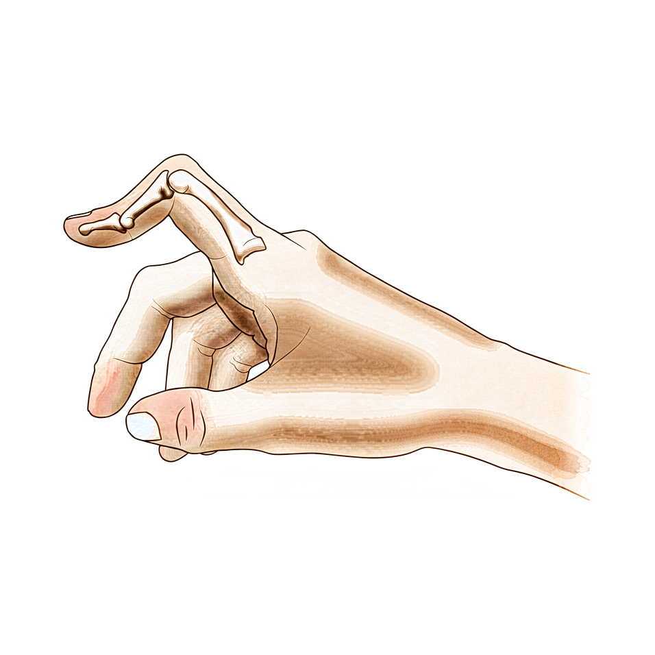
Qué está sintiendo
En una deformidad en ojal, la articulación media del dedo se dobla hacia abajo, hacia la palma, y la punta del dedo se dobla hacia atrás, en sentido contrario. El dedo puede verse hinchado sobre la articulación media, y también puede doler en esa zona. Enderezar la articulación media se vuelve difícil, y cuanto más lo intenta, más parece resistirse el dedo.
El problema se nota sobre todo en las tareas que requieren que el dedo se estire y luego vuelva a doblarse. Recoger una moneda, sujetar una taza por el asa, escribir en un teclado, abrocharse una camisa o girar una llave pueden volverse incómodos. El agarre en sí suele seguir funcionando, y por eso muchas personas conviven con el dedo durante semanas antes de buscar ayuda.
Si la deformidad se debe a una lesión, la articulación media puede doler después de usar la mano, y la rigidez puede ser peor por la mañana, antes de que el dedo se suelte. En la artritis reumatoide, la misma postura doblada se desarrolla lentamente porque el revestimiento de la articulación sigue inflamado e hinchado durante meses.
Algunos signos requieren atención rápida. Si el dedo o la mano se ponen calientes, rojos, hinchados y dolorosos, sobre todo si tiene fiebre, acuda al servicio de urgencias ese mismo día. Lo mismo se aplica si un dedo se pone pálido, frío, blanco o azulado, o si pierde de repente la sensibilidad o el movimiento en él después de una lesión. Si, en cambio, los síntomas no mejoran, empeoran a lo largo de las semanas, le despiertan por la noche o le impiden trabajar o usar la mano, consulte a su médico de cabecera o pida una valoración por un especialista.
¿Qué está ocurriendo realmente?
Por el dorso de cada dedo discurre un conjunto de tendones que enderezan las articulaciones. Imagínelos como cuerdas que pasan bajo una capucha en el dorso del dedo. La cuerda que endereza la articulación media se llama tendón central, y se inserta en la base del hueso medio. Otras dos cuerdas, llamadas bandas laterales, discurren a su lado y ayudan a enderezar tanto la articulación media como la punta del dedo.
En una deformidad en ojal, el tendón central se ha roto o se ha estirado y separado del hueso medio. La articulación media ya no puede ser traccionada hasta quedar recta, así que cae en una posición doblada. Las dos bandas laterales se deslizan entonces desde la parte superior de la articulación y bajan hacia la palma. Una vez que quedan por debajo del punto de giro de la articulación, esas mismas cuerdas empiezan a doblar la articulación media en lugar de enderezarla, y tiran de la punta del dedo hacia atrás, en sentido contrario. Por eso el dedo se queda con la articulación media doblada y la punta arqueada que se han descrito antes, y por eso cada intento de enderezarlo parece hacer que las cuerdas tiren con más fuerza en su contra.
Hace falta que se dañe más de una estructura para que se produzca esta postura. El tendón central, el ligamento que une las dos cuerdas sobre el dorso del dedo y las fibras de la capucha que mantienen todo en su sitio deben lesionarse antes de que se desarrolle la deformidad. Una rotura del tendón central por sí sola puede dejar la articulación media débil sin que aparezca la deformidad completa, y esa es una de las razones por las que el tratamiento temprano es importante: puede evitar por completo que la deformidad se forme.
El mismo resultado final puede tener distintos puntos de partida. Puede producirlo una lesión, como un golpe en el dedo o un corte sobre el dorso de la articulación, y también la inflamación continua de la artritis reumatoide, que va desgastando lentamente las mismas estructuras. Un dedo también puede tener este aspecto sin ninguna lesión del tendón, cuando el problema está en cambio en el lado de la palma de la articulación, y esa variante se trata de otra manera; por eso lo primero es determinar cuál de las dos tiene usted.
Qué podemos hacer al respecto
El Dr. Kieran Hirpara, cirujano de extremidades superiores en el Mater Private Hospital Rockhampton, comienza con las opciones menos invasivas que se adapten a su condición. Por lo general, los pacientes son derivados a nuestra clínica por su médico de cabecera; si un fisioterapeuta le ha sugerido que nos consulte, igualmente necesitará una derivación de su médico de cabecera para poder acceder al reembolso de Medicare. En la clínica, tomamos su historia clínica, examinamos su mano y solicitamos estudios de imagen si es necesario para determinar qué está ocurriendo. Como esta deformidad puede tener varios puntos de partida distintos, determinar cuál es el suyo es lo primero, antes de cualquier tratamiento.
El primer paso suele ser el uso de férulas y la terapia de mano. En una lesión reciente, una férula que mantenga recta la articulación media puede evitar por completo que la deformidad se forme, y cuanto antes se empiece, mejor: las personas tratadas dentro de las seis semanas posteriores a la lesión evolucionan mejor que las tratadas más tarde. La férula mantiene recta solo la articulación media. La punta del dedo, el nudillo y los demás dedos siguen moviéndose, porque inmovilizar todo el dedo hace que la punta se ponga rígida. En una deformidad que lleva tiempo presente, podemos usar yesos para enderezar la articulación media tanto como sea posible, seguidos de tres meses con una férula que le permite doblar el dedo mientras trabaja. Vale la pena probar este enfoque antes de plantearse la cirugía.
Cuando las férulas y la terapia no han logrado una mejoría suficiente, puede plantearse la cirugía. La operación reconstruye el tendón que endereza la articulación media, a veces usando como injerto un pequeño trozo de tendón de su propio antebrazo. En algunos casos, los tendones que se han deslizado hacia la palma se recolocan sobre la articulación, donde deben estar. El objetivo es el mismo en todos los casos: que pueda volver a enderezar la articulación media y usar el dedo sin que se enganche ni se doble hacia abajo. La operación tiene su propia página, y le explicaremos en qué consiste para su dedo en particular antes de tomar cualquier decisión.
Qué esperar
El pronóstico depende mucho del momento. Si se detecta a tiempo, a menudo puede frenarse una deformidad en ojal antes de que se instale. Una férula que mantenga recta la articulación media poco después de una lesión puede evitar por completo que la deformidad se forme, y las personas tratadas dentro de las seis semanas evolucionan mejor que las tratadas más tarde. Sin tratamiento, la deformidad tiende a empeorar en lugar de mantenerse igual, porque los tendones que se han salido de su sitio siguen tirando del dedo cada vez más hacia su postura doblada. Una lesión que parecía leve puede dar lugar semanas después a un dedo rígido y fijo.
En una deformidad que lleva tiempo presente, el tratamiento no quirúrgico sigue teniendo un papel real. Los yesos para enderezar la articulación media tanto como sea posible, seguidos de tres meses con una férula que le permite doblar el dedo mientras lo usa, pueden funcionar bien. El dedo responde a este enfoque cuando los yesos consiguen dejar la articulación media casi recta y usted usa activamente la mano con la férula durante esos tres meses, recuperando la flexión a lo largo del proceso. En las lesiones del pulgar, una férula que mantenga recto el nudillo puede ayudar si se empieza antes de que se desarrolle la deformidad.
La cirugía tiene como objetivo que pueda volver a enderezar la articulación media. En un grupo publicado de 18 personas a las que se les hizo un injerto de tendón para corregir una deformidad de larga evolución, 16 obtuvieron resultados buenos o excelentes. Se ha comunicado que una técnica más reciente, que permite mover el dedo pronto, funcionó en 11 casos. La cirugía también puede ayudar en la forma congénita de esta deformidad, en la que está presente desde el nacimiento.
Hay que ser honestos: no todas las correcciones se mantienen. En los dedos afectados por artritis reumatoide, la cirugía sobre los tejidos blandos por sí sola puede no mantenerse en las fases avanzadas, y los resultados a largo plazo son menos previsibles. Cuando una deformidad reaparece o persiste, otro procedimiento distinto puede ser la mejor respuesta. Sea cual sea el punto de partida, el objetivo realista es un dedo que pueda enderezar y usar, no la promesa de un dedo perfectamente normal.
Cuándo consultar a un especialista
La deformidad en ojal responde bien a la atención temprana, así que conviene saber cuándo actuar. Si se ha lesionado el dedo y la articulación media no se endereza, o si el dedo sigue doblado e hinchado varios días después de una lesión, acuda a su médico de cabecera y pida una valoración por un especialista. Cuanto antes se empiece con la férula, más posibilidades hay de frenar la deformidad antes de que se instale.
Algunos signos requieren atención más rápida. Acuda a un servicio de urgencias ese mismo día si el dedo o la mano se ponen calientes, rojos, hinchados y dolorosos, sobre todo si tiene fiebre. Lo mismo se aplica si un dedo se pone pálido, frío, blanco o azulado, o si pierde de repente la sensibilidad o el movimiento en él después de una lesión. Si hay una herida abierta en la piel sobre una articulación lesionada, o el dedo se ve claramente deformado, acuda también.
Si, en cambio, los síntomas no mejoran, empeoran a lo largo de las semanas, le despiertan por la noche o le impiden trabajar o usar la mano, consulte a su médico de cabecera o pida una valoración por un especialista. Si no puede comunicarse con la clínica, fuera del horario de atención o en fin de semana, acuda al servicio de urgencias más cercano.
En profundidad
Advanced reading: the deeper science (optional)
Esta sección va más allá de lo necesario para tomar decisiones terapéuticas por su cuenta. La deformidad en ojal merece una lectura adicional, ya que el tratamiento instintivo para un dedo doloroso, hinchado y doblado —dejarlo en reposo y mantenerlo inmóvil— es precisamente lo único que la evidencia específicamente no respalda.
Movilización temprana, no inmovilización prolongada
Una revisión sistemática de los tratamientos para lesiones del tendón central concluyó que la base de evidencia es limitada; además, el papel de las distintas estrategias tanto en lesiones abiertas como cerradas no está bien sustentado [1]. A pesar de dicha limitación, la evidencia proveniente de estudios individuales apoya de forma tentativa la movilización temprana y no respalda la inmovilización prolongada [1].
Esto merece ser expresado con claridad, pues va en contra de la reacción instintiva habitual. La diferencia radica en proteger el tendón central en proceso de curación, lo cual exige mantener la articulación media recta; en contraste, la inmovilización total del dedo provoca rigidez de la articulación distal y hace que todo el dedo se vuelva rígido. La férula tiene como finalidad mantener una sola articulación inmóvil mientras las demás siguen moviéndose.
¿Por qué la deformidad empeora en lugar de permanecer estable?
La deformidad en ojal es progresiva; esto resulta poco común y merece ser comprendido. El tendón central se inserta en la base del hueso medio y estira la articulación media. Cuando este tendón falla, dicha articulación se flexiona, y las dos bandas laterales, que normalmente discurren por encima del eje articular, desplazanse por debajo de él.
Una vez debajo del eje articular, los mismos tendones que antes ayudaban a estirar la articulación media ahora la flexionan, al mismo tiempo que tiran de la punta del dedo hacia una hiperextensión. De este modo, la deformidad se auto-reforzar: cada intento de enderezar el dedo tensa aún más las estructuras que lo mantienen flexionado. Por eso, una lesión que parecía leve puede derivar en una deformidad fija semanas después; también explica por qué el período adecuado para aplicar férulas simples es muy temprano.
La deformidad que no es boutonnière
Un dedo puede adoptar la misma postura sin que exista lesión alguna en el tendón central; en ese caso, los tratamientos son completamente distintos. Las deformidades boutonnière y pseudoboutonnière difieren en cuanto a patoanatomía, diagnóstico y tratamiento [2]. La variante pseudoboutonnière se origina por una lesión de la placa volar en la articulación media; en este caso, la articulación distal del dedo conserva una flexión pasiva normal, en lugar de quedar en hiperextensión.
Para diferenciarlas en la práctica clínica, hay que observar la punta del dedo. En una boutonnière verdadera, la punta permanece en hiperextensión y resiste la flexión, especialmente cuando la articulación media está estirada; en una pseudoboutonnière, en cambio, la punta se flexiona con facilidad. Colocar una férula en una pseudoboutonnière como si fuera una boutonnière real implica tratar la articulación equivocada.
Pertenece a una familia de lesiones definidas según el lugar donde falla el tendón
La lesión en “boutonnière” se encuentra junto con la lesión en “mallet finger” y la lesión de la banda sagital como una de las tres lesiones del mecanismo extensor cerrado, clasificadas según la zona en la que se produce el fallo del mecanismo [3]. Las tres pueden manifestarse como un dedo hinchado que permanece en una posición anormal pocos días después de la lesión; además, cada una requiere una posición específica de la férula. Este es el motivo práctico por el cual vale la pena llegar a un diagnóstico preciso antes de someter al paciente a semanas de uso de férula.
Referencias
[1] Geoghegan L, Wormald JCR, Adami RZ, Rodrigues JN. Lesiones del tendón extensor de la porción central: revisión sistemática de los tratamientos. J Hand Surg Eur Vol. 2019;44(8):825-32. https://doi.org/10.1177/1753193419845311
[2] Hanson ZC, Thompson RG, Andrews JR, Lourie GM. Deformidades en ojal versus pseudodeformidades en ojal: patoanatomía, diagnóstico y tratamiento. J Hand Surg Am. 2023;48(5):489-97. https://doi.org/10.1016/j.jhsa.2022.10.019
[3] Lin JD, Strauch RJ. Lesiones cerradas del mecanismo extensor de partes blandas (dedo en martillo, deformidad en ojal y lesión de la banda sagital). J Hand Surg Am. 2014;39(5):1005-11. https://doi.org/10.1016/j.jhsa.2013.11.018
Evidence & references
This is the clinical evidence summary written for health professionals. It is technical, and it lists the research this page was built from. You do not need to read it to understand your treatment or to make a decision about it.
Overview
- A staged technique for the repair of traumatic boutonniere deformity allows the surgeon to divide the deformity into various categories and use the operative procedure indicated for each particular problem, often avoiding unnecessary surgery [1].
- Differentiating a true boutonniere deformity from a pseudoboutonniere injury is critical in determining clinical management [2].
- A retrospective study of fifty-four longstanding post-burn boutonnière deformities was carried out to evaluate the problems associated with the surgical procedures [3].
- An understanding of the anatomy, clinical presentation, treatment options, and expected outcomes is crucial for optimal treatment of posttraumatic boutonnière and swan neck deformities [4].
- Early treatment of central slip injuries can prevent the deformity [5].
- Transection of the terminal extensor tendon is a useful procedure for salvage of the boutonniere deformity [6].
- No boutonniere deformities were produced in a series of 20 patients who had chronic mallet finger deformity treated with central slip tenotomy [7].
- Reconstruction of the central slip by the transverse retinacular ligament is not indicated for boutonnière deformities with severe contracture of the lateral band [8].
- The best indication for reconstruction of the central slip by the transverse retinacular ligament is in cases in which the central slip is defective and conservative treatment is not effective [8].
- Similar results occurred for chronic boutonniere deformity using serial casting for adequate extension followed by 3 months of relative motion flexion (RMF) orthotic use, which should be attempted prior to surgical intervention [9].
- A new surgical technique with early mobilization is an effective and simplified treatment of Boutonniere deformity [10].
- The natural history of the boutonnière deformity in rheumatoid arthritis is outlined, and a simple method of repair is described [11].
- Chronic boutonniere deformity will respond to relative motion flexion splinting if serial casting can place the proximal interphalangeal joint in less than -20° extension, and the patient actively uses the hand in a relative motion flexion orthosis for 3 months, recovering flexion [12].
- The Y-shaped tendon graft can be a useful procedure for the correction of chronic boutonniere deformity [13].
- In a patient series of 18 patients, the Y-shaped tendon graft provided good or excellent results in 16 patients [13].
- Extension splinting of the metacarpophalangeal (MP) joint may be effective if traumatic thumb injuries are treated before development of a boutonnière deformity [14].
- Congenital boutonniere deformity is understood as various embryonic developmental failures [15].
- A reliable surgical option for congenital boutonniere deformity involves central slip reconstruction [15].
- Extensor tenotomy to correct a boutonnière deformity of long standing markedly lessened disability with restoration of flexion of the distal joint [16].
- In chronic boutonniere deformity, when the lateral bands are deficient or damaged, the cross-lateral band reconstruction technique using palmaris longus autograft is a treatment option with satisfactory results [17].
- Extension splinting may be effective if traumatic thumb injuries are treated before deformity development [18].
- Surgical treatment with advancement of the extensor pollicis brevis tendon and imbrication of the dorsoradial capsule is necessary once a traumatic thumb boutonnière deformity is established [18].
- The prevalence of boutonnière deformity without rheumatoid arthritis or trauma is approximately 13% [19].
- Current concepts relating to the presentation and treatment of closed soft tissue injuries of the extensor mechanism in zones I, III, and V, commonly known as mallet finger, boutonniere deformities, and sagittal band injuries, are summarized [20].
- Long-term results following soft tissue reconstruction for boutonniere finger deformity in rheumatoid arthritis are unreliable [24].
- Recurrent or persistent deformity in rheumatoid arthritis is best treated with a salvage procedure [24].
Anatomy & Pathophysiology
Extensor Mechanism Anatomy
- The extensor mechanism of the finger at the level of the proximal interphalangeal (PIP) joint consists of both intrinsic and extrinsic contributions [28].
- The extensor tendon entering the digit is the continuation of the extensor digitorum communis (EDC), with contributions from the extensor indicis proprius in the index finger and the extensor digiti quinti in the small finger [28].
- In 30% of cases, the EDC has a separate insertion into the base of the proximal phalanx, but extension of the metacarpophalangeal (MP) joint typically occurs through the pull of the sagittal bands [28].
- The principal bony insertion of the extrinsic digital extensors is on the dorsal proximal aspect of the middle phalanx [32].
- Metacarpophalangeal joint extension is provided by extrinsic extensor force transmitted through the sagittal bands [32].
- Distal interphalangeal joint extension is achieved through conjoined lateral bands composed of tendinous slips from both extrinsic and intrinsic tendons [32].
- The deep head of each dorsal interosseous muscle forms a lateral tendon, or lateral band, at the level of the MP joint [41].
- At the level of the middle of the proximal phalanx, transverse fibers arch dorsally from each lateral band to join over the dorsum of the finger, flexing the proximal phalanx [41].
- Oblique fibers (spiral fibers) from the lateral bands sweep over the distal third of the proximal phalanx to insert onto the lateral tubercles at the base of the middle phalanx, extending the middle phalanx [41].
- The lateral bands are joined by the lateral slips of the extensor tendon to form the conjoined lateral band [41].
- The two conjoined lateral bands to each finger unite at the distal third of the middle phalanx to form the terminal tendon, which inserts at the base of the distal phalanx to extend it [41].
- The volar interossei form the ulnar lateral band of the index finger and the radial lateral band of the ring and little fingers [41].
- The flexor digiti quinti brevis is structurally and functionally similar to the deep head of the dorsal interossei, forming the ulnar lateral band of the little finger [41].
Pathophysiology & Biomechanics
- A boutonniere deformity requires subluxation of the lateral bands volar to the axis of rotation of the PIP joint [22].
- Damage to the central slip alone does not cause a boutonniere deformity [22].
- A boutonniere deformity occurs only when the central slip, triangular ligament, and transverse and oblique fibers of the interosseous hood are all damaged [22].
- Detachment of the central slip from the middle phalanx produces a decrease in extension of the PIP joint [22].
- When the transverse and oblique fibers of the interosseous hood are divided in addition to the central slip, extension at the PIP joint is further decreased [22].
- Early treatment of central slip injuries can prevent the development of the deformity [5].
- The extensor mechanism is a complex structure that demands a delicate balance from intrinsic and extrinsic tendons of the hand [28].
- Injury to the extensor mechanism can disrupt this delicate balance, leading to significant sequelae [28].
- Understanding the anatomy, clinical presentation, treatment options, and expected outcomes is crucial for optimal treatment of posttraumatic boutonnière and swan neck deformities [4].
Classification
- A staged management plan allows the surgeon to divide traumatic boutonniere deformity into various categories and use the operative procedure indicated for each particular problem [1].
- The choice of surgical procedure for rheumatoid boutonniere deformity is dependent on accurate staging of the deformity, which is based on the flexibility of the proximal interphalangeal joint and the state of the articular cartilage [23].
- In a cadaveric model, damage to the central slip alone does not cause a boutonniere deformity [22].
- The boutonniere deformity requires subluxation of the lateral bands volar to the axis of rotation of the PIP joint [22].
- In a cadaveric model, the central slip, triangular ligament, and oblique fibers need to all be damaged to cause subluxation of the lateral bands which leads to an acute boutonniere deformity [29].
- Anterior dislocation of the proximal interphalangeal joint is associated with ruptures of the central slip of the extensor mechanism and results in boutonnière deformity unless repaired [27].
- The incidence of uncorrectable swan-neck and boutonniere deformities is estimated to be between 8% and 16% during the first 2 years after the onset of systemic disease in rheumatoid arthritis [23].
- The prevalence of boutonniere deformities in patients with established rheumatoid arthritis is approximately 36% [23].
Clinical Presentation
Diagnostic Differentiation and Classification
- The surgeon can divide the boutonniere deformity into various categories to use the operative procedure indicated for each particular problem, often avoiding unnecessary surgery [1].
Etiology and Pathomechanics
- The cause of boutonniere deformity in rheumatoid arthritis is chronic synovitis of the proximal interphalangeal joint [23].
- Combined injury of the central slip, triangular ligament, and transverse and oblique fibers of the interosseous hood causes a boutonniere deformity [22].
Prevalence and Natural History
- A swan-neck or boutonniere deformity occurs in approximately half of patients with rheumatoid arthritis [23].
- The incidence of uncorrectable swan-neck and boutonniere deformities is estimated to be between 8% and 16% during the first 2 years after the onset of systemic disease [23].
- The prevalence of finger deformities in patients with established rheumatoid arthritis is approximately 36% for boutonniere deformities [23].
- The natural history of the boutonnière deformity in rheumatoid arthritis is outlined in the literature [11].
Clinical Assessment and Testing
- The central slip tenodesis test detects disruption of the central slip before the boutonniere deformity becomes established [34].
- The central slip tenodesis test is applicable in uncooperative children and unconscious patients [34].
- Treating swan neck and boutonniere deformities of the PIP joint requires understanding the cause, biomechanical changes, and articular status [26].
Investigations
Clinical Examination and Diagnostic Tests
- A careful physical examination is essential to direct care and future testing if indicated [31].
- Diagnostic tests such as imaging and serum laboratory studies are useful in the determination of pathology but can be expensive, time consuming, and often nonspecific [31].
Biomechanical and Pathomechanical Assessment
Imaging and Advanced Modalities
- A computational 3D model validation reassures the reliability of research in understanding the pathomechanics of boutonniere deformity [21].
Treatment
Non-Operative
- Early treatment of central slip injuries can prevent the development of boutonniere deformity [5].
- Extension splinting of the metacarpophalangeal joint may be effective for traumatic thumb boutonniere deformity if treated before the deformity develops [14].
- Extension splinting may be effective for traumatic thumb boutonniere deformity if treated before deformity development [18].
- Chronic boutonniere deformity responds to relative motion flexion splinting if serial casting places the proximal interphalangeal joint in less than -20° extension and the patient actively uses a relative motion flexion orthosis for 3 months [12].
- Serial casting for adequate extension followed by 3 months of relative motion flexion orthotic use should be attempted prior to surgical intervention for chronic boutonniere deformity [9].
- The use of relative motion flexion orthoses is effective in increasing active distal interphalangeal joint flexion and improving proximal interphalangeal joint extension in patients with Burton stage 1 chronic boutonniere deformity [30].
- Conservative treatment of closed central slip injuries involves immobilization of the proximal interphalangeal, distal interphalangeal, and metacarpophalangeal joints in full extension for four weeks followed by a Capener spring splint for two further weeks [92].
- In a review of 115 central slip injuries, 17 of 24 patients treated conservatively achieved an outcome of less than a 20° extension deficit at the proximal interphalangeal joint and more than 80% return of proximal interphalangeal joint flexion [92].
- Patients treated for central slip injuries within six weeks of injury had better outcomes than those treated later [92].
Operative
- A staged technique allows the surgeon to divide traumatic boutonniere deformity into various categories and use the operative procedure indicated for each particular problem, often avoiding unnecessary surgery [1].
- Reconstruction of the central slip by the transverse retinacular ligament is best indicated in cases in which the central slip is defective and conservative treatment is not effective [8].
- A simple method of repair for the rheumatoid boutonnière deformity is described in the literature [11].
- In a patient series, the Y-shaped tendon graft provided good or excellent results in 16 of 18 patients with chronic boutonniere deformity [13].
- Surgical treatment with advancement of the extensor pollicis brevis tendon and imbrication of the dorsoradial capsule is necessary once a traumatic boutonnière deformity of the thumb is established [18].
- The cross-lateral band reconstruction technique using palmaris longus autograft is a treatment option with satisfactory results for chronic boutonniere deformity when the lateral bands are deficient or damaged [17].
- A reliable surgical option involving central slip reconstruction is suggested for the congenital form of boutonniere deformity [15].
- A simple staged procedure using a central slip facilitation device is a valid alternative in the management of severe Dupuytren's proximal interphalangeal joint contracture, demonstrating reliable, reproducible correction of the deformity and acceptable patient outcomes [38].
Complications
- In a patient series of 18 patients, the Y-shaped tendon graft provided good or excellent results in 16 of 18 patients for the correction of chronic boutonniere deformity [13].
- Extension splinting of the MP joint may be effective if traumatic thumb injuries are treated before development of a boutonnière deformity [14].
- The case extends understanding of congenital boutonniere deformity as various embryonic developmental failures and suggests a reliable surgical option involving central slip reconstruction [15].
- Surgical treatment with advancement of the extensor pollicis brevis tendon and imbrication of the dorsoradial capsule is necessary once a boutonnière deformity is established in the thumb [18].
- This article summarizes current concepts relating to the presentation and treatment of closed soft tissue injuries of the extensor mechanism in zones I, III, and V, commonly known as mallet finger, boutonniere deformities, and sagittal band injuries [20].
- This validation reassures the reliability of the research in understanding the pathomechanics of boutonniere deformity [21].
- In a cadaver model, detachment of the central slip from the middle phalanx produced a decrease in extension of the PIP joint [22].
- When the transverse and oblique fibers of the interosseous hood were also divided after central slip detachment, extension at the PIP joint was further decreased [22].
- A boutonniere deformity occurred only when all 3 structures (central slip, transverse and oblique fibers of the interosseous hood, and triangular ligament) were damaged [22].
- Division of the central slip leads to loss of extension at the PIP joint [22].
- The prevalence of finger deformities in patients with established rheumatoid arthritis is approximately 14% for swan-neck deformities and 36% for boutonniere deformities [23].
- In the later stages of both swan-neck and boutonniere deformities, soft-tissue procedures alone may not result in lasting operative correction [23].
- Long-term results following soft tissue reconstruction are unreliable [24].
- Recurrent or persistent deformity is best treated with a salvage procedure [24].
- Treating swan neck and boutonniere deformities of the PIP joint is a difficult challenge that requires understanding the cause, biomechanical changes, and articular status [26].
- Anterior dislocation of the proximal interphalangeal joint is associated with ruptures of the central slip of the extensor mechanism [27].
- Anterior dislocation of the proximal interphalangeal joint results in boutonnière deformity unless repaired [27].
Recovery
Non-Operative Management
- Serial casting for adequate extension followed by 3 months of relative motion flexion (RMF) orthotic use should be attempted prior to surgical intervention for chronic boutonniere deformity [9].
- Chronic boutonniere deformity responds to relative motion flexion splinting if serial casting places the proximal interphalangeal joint in less than -20° extension and the patient actively uses the hand in a relative motion flexion orthosis for 3 months, recovering flexion [12].
- Extension splinting of the metacarpophalangeal joint may be effective for traumatic thumb boutonniere injuries if treated before the development of a boutonnière deformity [14].
Operative Management
- A staged technique allows the surgeon to divide the traumatic boutonniere deformity into various categories and use the operative procedure indicated for each particular problem, often avoiding unnecessary surgery [1].
- The Y-shaped tendon graft provided good or excellent results in 16 of 18 patients for the correction of chronic boutonniere deformity [13].
- Reconstruction of the central slip by the transverse retinacular ligament is indicated in cases in which the central slip is defective and conservative treatment is not effective, but is not indicated for boutonnière deformities with severe contracture of the lateral band [8].
- A simple method of repair for the correction of the rheumatoid boutonnière deformity has been described [11].
- A new surgical technique with early mobilization for chronic boutonniere deformity was reported to be an effective and simplified treatment in 11 cases [10].
- Central slip reconstruction is a reliable surgical option for the congenital form of boutonniere deformity [15].
Outcomes and Prognosis
- Early treatment of central slip injuries can prevent the boutonniere deformity [5].
- Long-term results following soft tissue reconstruction for boutonniere finger deformity in rheumatoid arthritis are unreliable, and recurrent or persistent deformity is best treated with a salvage procedure [24].
- In the later stages of rheumatoid boutonniere deformities, soft-tissue procedures alone may not result in lasting operative correction [23].
- A patient with boutonniere deformity of the second toe after planter dislocation of the proximal interphalangeal joint reported neither deformity nor difficulty when last seen 22 months after the operation [95].
Key Evidence
- [L4] With this plan of management, the surgeon is able to divide the boutonniere deformity into various categories and then use the operative procedure indicated for each particular problem, often avoiding unnecessary surgery. [1] (10.1016/s0363-5023(83)80009-4)
- [L5] Differentiating a true boutonniere deformity from a pseudoboutonniere injury is critical in determining clinical management. [2] (10.1016/j.jhsa.2022.10.019)
- [L4] A retrospective study of fifty-four longstanding post-burn boutonnière deformities was carried out to evaluate the problems associated with the surgical procedures. [3] (10.1016/0266-7681(86)90006-9)
- [L5] An understanding of the anatomy, clinical presentation, treatment options, and expected outcomes is crucial for optimal treatment of posttraumatic boutonnière and swan neck deformities. [4] (10.5435/jaaos-d-14-00272)
- [L5] The paper outlines the anatomy, pathoanatomy, and treatment concepts for boutonniere deformity, emphasizing that early treatment of central slip injuries can prevent the deformity. [5] (10.1016/s0749-0712(21)00060-3)
- [L4] Transection of the terminal extensor tendon is a useful procedure for salvage of the boutonniere deformity. [6] (10.1016/s0749-0712(21)00065-2)
- [L4] No boutonniere deformities were produced in this series of 20 patients who had chronic mallet finger deformity. [7] (10.1016/s0363-5023(87)80205-8)
- [L4] This procedure is not indicated for boutonnière deformities with severe contracture of the lateral band; its best indication is in cases in which the central slip is defective and conservative treatment is not effective. [8] (10.1016/0266-7681(90)90080-n)
- [L4] Similar results occurred for chronic boutonniere deformity using serial casting for adequate extension followed by 3 months of RMF orthotic use, which should be attempted prior to surgical intervention. [9] (10.1016/j.jht.2023.02.005)
- [L4] Our technique is an effective and simplified treatment of Boutonniere deformity. [10] (10.1097/bth.0000000000000152)
- [L4] The natural history of the boutonnière deformity in rheumatoid arthritis is outlined, and a simple method of repair is described. [11] (10.2106/00004623-196951070-00009)
- [L4] Chronic boutonniere deformity will respond to relative motion flexion splinting if serial casting can place the proximal interphalangeal joint in less than -20° extension, and the patient actively uses the hand in a relative motion flexion orthosis for 3 months, recovering flexion. [12] (10.1097/sap.0000000000002307)
- [L4] The Y-shaped tendon graft can be a useful procedure for the correction of chronic boutonniere deformity; in our patient series, this provided good or excellent results in 16 of 18 patients. [13] (10.1016/j.jhsa.2021.01.003)
- [L4] Extension splinting of the MP joint may be effective if these injuries are treated before development of a boutonnière deformity. [14] (10.1016/s0266-7681(00)80024-8)
- [Case_report] The case extends understanding of congenital boutonniere deformity as various embryonic developmental failures and suggests a reliable surgical option involving central slip reconstruction. [15] (10.1016/j.jhsa.2014.05.030)
- [L4] In chronic boutonniere deformity, when the lateral bands are deficient or damaged, the cross-lateral band reconstruction technique using palmaris longus autograft is a treatment option with satisfactory results. [17] (10.1016/j.jhsa.2017.04.010)
- [L4] Extension splinting may be effective if treated before deformity development, but surgical treatment with advancement of the extensor pollicis brevis tendon and imbrication of the dorsoradial capsule is necessary once a boutonnière deformity is established. [18] (10.1054/jhsb.1999.0303)
- [L3] The prevalence of boutonnière deformity without rheumatoid arthritis or trauma is approximately 13%. [19] (10.1177/1753193417704610)
- [L5] This article summarizes current concepts relating to the presentation and treatment of closed soft tissue injuries of the extensor mechanism in zones I, III, and V, commonly known as mallet finger, boutonniere deformities, and sagittal band injuries. [20] (10.1016/j.jhsa.2013.11.018)
- [L5] This validation reassures the reliability of the research in understanding the pathomechanics of boutonniere deformity. [21] (10.1186/s13018-025-06329-3)
- [L5] [22] (10.1016/j.jhsa.2017.07.011)
- [L5] [23] (10.5435/00124635-199903000-00002)
- [L5] Long-term results following soft tissue reconstruction are unreliable, and recurrent or persistent deformity is best treated with a salvage procedure. [24] (10.1016/j.jhsa.2011.05.029)
- [L4] Treating swan neck and boutonniere deformities of the PIP joint is a difficult challenge that requires understanding the cause, biomechanical changes, and articular status. [26] (10.1016/j.hcl.2017.12.006)
- [Paper] [28] (10.1016/j.hcl.2012.05.044)
- [Paper] In a cadaveric model, the central slip, triangular ligament, and oblique fibers need to all be damaged to cause subluxation of the lateral bands which leads to an acute boutonniere deformity. [29] (10.1016/s0363-5023(12)60014-8)
- [L4] The use of RMFO is effective in increasing active distal interphalangeal joint flexion and improving PIP extension in patients with Burton stage 1 chronic boutonniere deformity. [30] (10.1016/j.jhsa.2022.08.007)
- [Paper] The test detects disruption of the central slip before the boutonniere deformity becomes established and is applicable in uncooperative children and unconscious patients. [34] (10.1016/0266-7681(94)90057-4)
- [L4] The simple staged procedure is a valid alternative in the management of severe Dupuytren's PIPJ contracture, demonstrating reliable, reproducible correction of the deformity and acceptable patient outcomes. [38] (10.1177/1753193412439673)
- [L4] [92] (10.1177/1758998318822663)
- [Case_report] The patient reported neither deformity nor difficulty when last seen 22 months after the operation. [95] (10.1007/s00402-009-0816-x)
References
[1] A staged technique for the repair of the traumatic boutonniere deformity. The Journal of Hand Surgery. 1983. DOI: 10.1016/s0363-5023(83)80009-4
[2] Boutonniere Versus Pseudoboutonniere Deformities: Pathoanatomy, Diagnosis, and Treatment. The Journal of Hand Surgery. 2023. DOI: 10.1016/j.jhsa.2022.10.019
[3] Reconstructive surgery of the post-burn boutonnière deformity. The Journal of Hand Surgery: Journal of the British Society for Surgery of the Hand. 1986. DOI: 10.1016/0266-7681(86)90006-9
[4] Posttraumatic Boutonnière and Swan Neck Deformities. Journal of the American Academy of Orthopaedic Surgeons. 2015. DOI: 10.5435/jaaos-d-14-00272
[5] BOUTONNIERE DEFORMITY. Hand Clinics. 1995. DOI: 10.1016/s0749-0712(21)00060-3
[6] TREATMENT OF THE CHRONIC BOUTONNIERE DEFORMITY BY EXTENSOR TENOTOMY. Hand Clinics. 1995. DOI: 10.1016/s0749-0712(21)00065-2
[7] Central slip tenotomy for chronic mallet finger deformity. The Journal of Hand Surgery. 1987. DOI: 10.1016/s0363-5023(87)80205-8
[8] Reconstruction of the Central Slip by the Transverse Retinacular Ligament for Boutonnière Deformity. Journal of Hand Surgery. 1990. DOI: 10.1016/0266-7681(90)90080-n
[9] The relative motion concept in acute and chronic boutonniere deformity: Invited commentary. Journal of Hand Therapy. 2023. DOI: 10.1016/j.jht.2023.02.005
[10] Chronic Boutonniere Deformities, Supple, or Stiff: A New Surgical Technique With Early Mobilization in 11 Cases. Techniques in Hand & Upper Extremity Surgery. 2017. DOI: 10.1097/bth.0000000000000152
[11] Correction of the Rheumatoid Boutonnière Deformity. The Journal of Bone & Joint Surgery. 1969. DOI: 10.2106/00004623-196951070-00009
[12] A Paradigm Shift in Managing Acute and Chronic Boutonniere Deformity. Annals of Plastic Surgery. 2020. DOI: 10.1097/sap.0000000000002307
[13] Y-Shaped Tendon Graft—A Technique in the Reconstruction of Posttraumatic Chronic Boutonniere Deformity. The Journal of Hand Surgery. 2021. DOI: 10.1016/j.jhsa.2021.01.003
[14] Traumatic Boutonnière Deformity of the Thumb. Journal of Hand Surgery. 2000. DOI: 10.1016/s0266-7681(00)80024-8
[15] Restoration of the Central Slip in Congenital Form of Boutonniere Deformity: Case Report. The Journal of Hand Surgery. 2014. DOI: 10.1016/j.jhsa.2014.05.030
[16] Extensor Tenotomy for Chronic Boutonniere Deformity of the Finger: REPORT OF TWO CASES.. The Journal of Bone and Joint Surgery. American Volume. 1965.
[17] Chronic Boutonniere Deformity: Cross-Lateral Band Technique Using Palmaris Longus Autograft. The Journal of Hand Surgery. 2017. DOI: 10.1016/j.jhsa.2017.04.010
[18] TRAUMATIC BOUTONNIÈRE DEFORMITY OF THE THUMB. The Journal of Hand Surgery: Journal of the British Society for Surgery of the Hand. 2000. DOI: 10.1054/jhsb.1999.0303
[19] Thumb boutonnière deformity without rheumatoid arthritis or trauma. Journal of Hand Surgery (European Volume). 2017. DOI: 10.1177/1753193417704610
[20] Closed Soft Tissue Extensor Mechanism Injuries (Mallet, Boutonniere, and Sagittal Band). The Journal of Hand Surgery. 2014. DOI: 10.1016/j.jhsa.2013.11.018
[21] Assessment of the ligamentous stress distribution in the pathomechanics of the boutonniere deformity through a computational 3D model. Journal of Orthopaedic Surgery and Research. 2025. DOI: 10.1186/s13018-025-06329-3
[22] Biomechanics of the Acute Boutonniere Deformity. The Journal of Hand Surgery. 2018. DOI: 10.1016/j.jhsa.2017.07.011
[23] Operative Correction of Swan-Neck and Boutonniere Deformities in the Rheumatoid Hand. Journal of the American Academy of Orthopaedic Surgeons. 1999. DOI: 10.5435/00124635-199903000-00002
[24] Treatment of Boutonniere Finger Deformity in Rheumatoid Arthritis. The Journal of Hand Surgery. 2011. DOI: 10.1016/j.jhsa.2011.05.029
[26] Treating the Proximal Interphalangeal Joint in Swan Neck and Boutonniere Deformities. Hand Clinics. 2018. DOI: 10.1016/j.hcl.2017.12.006
[27] Anterior dislocation of the proximal interphalangeal joint. A cause of rupture of the central slip of the extensor mechanism.. The Journal of bone and joint surgery. American volume. 1970.
[28] Boutonnière and Pulley Rupture in Elite Athletes. Hand Clinics. 2012. DOI: 10.1016/j.hcl.2012.05.044
[29] Biomechanics of the Acute Boutonniere Deformity. The Journal of Hand Surgery. 2012. DOI: 10.1016/s0363-5023(12)60014-8
[30] The Use of Relative Motion Flexion Orthoses for Chronic Boutonniere Deformity. The Journal of Hand Surgery. 2024. DOI: 10.1016/j.jhsa.2022.08.007
[31] Orthopaedic Knowledge Update 13 Ebook Without Multimedia. Anatomy, Evaluation, Clinical Examination, and Imaging > Evaluation and Clinical Examination: Current Concepts.
[32] A Lange Medical Book Current Diagnosis Treatment In Orthopedics Fifth Edition. 9Hand Surgery > Image DISORDERS OF THE MUSCULATURE OF THE HAND.
[34] The Central Slip Tenodesis Test for Early Diagnosis of Potential Boutonnière Deformities. Journal of Hand Surgery. 1994. DOI: 10.1016/0266-7681(94)90057-4
[38] Management of severe Dupuytren’s contracture of the proximal interphalangeal joint with use of a central slip facilitation device. Journal of Hand Surgery (European Volume). 2012. DOI: 10.1177/1753193412439673
[41] Green S Operative Hand Surgery. Interosseous and Hypothenar Muscles.
[92] Therapeutic management of closed central slip injuries: Outcome of a service evaluation. Hand Therapy. 2019. DOI: 10.1177/1758998318822663
[95] Boutonniere deformity of the second toe after planter dislocation of proximal interphalangeal joint: a case report. Archives of Orthopaedic and Trauma Surgery. 2009. DOI: 10.1007/s00402-009-0816-x