Patients › Elbow
Liberación del túnel cubital
Cubital tunnel release relieves ulnar nerve compression—addressing numbness/weakness in the ring & little fingers.
¿Por qué se ha recomendado esta operación?
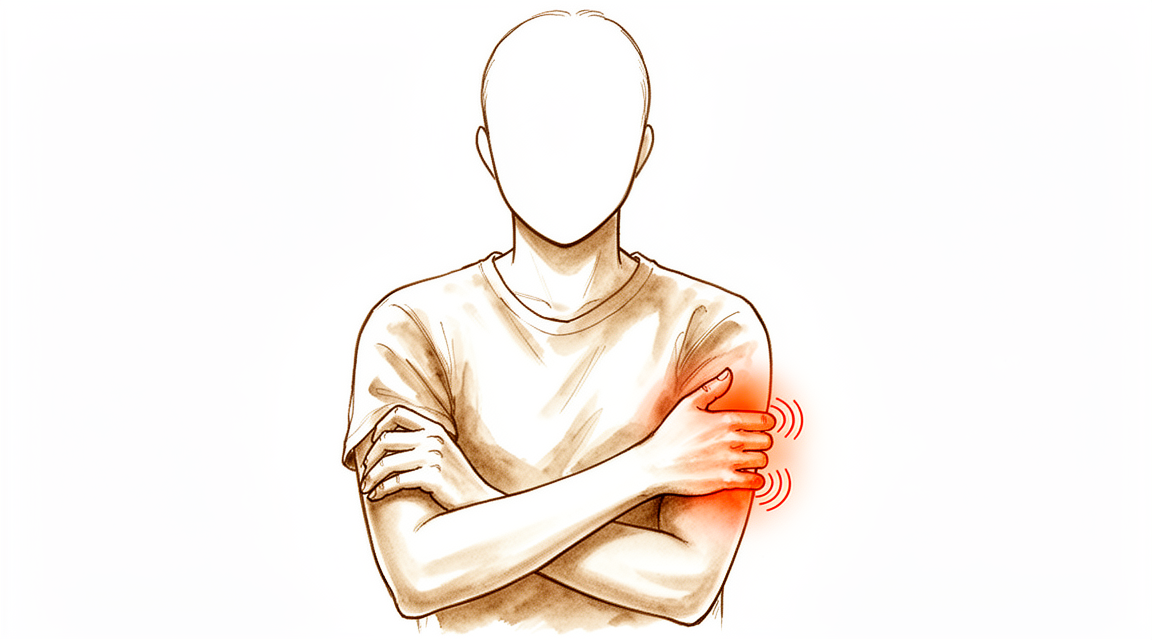
El Dr. Kieran Hirpara, cirujano de extremidades superiores en el Mater Private Hospital Rockhampton, comienza por proponer las opciones menos invasivas que se adapten a su condición. Por lo general, los pacientes son derivados a nuestra clínica por su médico de cabecera; si un fisioterapeuta le ha recomendado consultarnos, igualmente necesitará una derivación de su médico de cabecera para poder acceder al reembolso de Medicare. En su consulta, tomamos su historia clínica, examinamos su brazo y, si es necesario, solicitamos estudios de imagen o pruebas nerviosas. El síndrome del túnel cubital se refiere a la presión que se ejerce sobre un nervio en la parte interna del codo. Esta presión provoca entumecimiento u hormigueo en el dedo meñique y en el lado del dedo anular de la mano; este síntoma puede empeorar cuando el codo permanece doblado durante largos periodos. En casos crónicos como este, normalmente probamos primero tratamientos no quirúrgicos: modificación de actividades, fisioterapia o terapia de la mano, y uso de férulas. Consideramos la cirugía cuando dichas medidas no logran mejorar suficientemente sus síntomas.
La operación se denomina liberación del túnel cubital. Consiste en aliviar la presión sobre el nervio para que sus síntomas desaparezcan o disminuyan. Más del 90 % de las personas sometidas a esta cirugía quedan curadas o experimentan una mejora. Hablaremos de aquello que le preocupa y decidiremos juntos si este es el siguiente paso adecuado para usted.
Antes de la operación
Una vez que haya programado la cirugía, le proporcionaremos instrucciones claras a seguir. Deberá abstenerse de comer y beber durante siete horas antes de la operación. Pedimos siete horas en lugar de seis para que, si el programa quirúrgico avanza antes de lo previsto, sea posible adelantar la hora de su intervención. Es posible que sea necesario suspender algunos medicamentos antes de la cirugía; su cirujano le indicará cuáles y cuándo. Lleve consigo una lista de todos los fármacos que toma, incluyendo pastillas, inyecciones y cremas. Organice que alguien lo lleve a casa después de la operación, ya que no podrá conducir usted mismo. Use ropa holgada y cómoda, preferiblemente con una manga fácil de quitar. Es posible que se hayan solicitado estudios de imagen como radiografías, ecografías o resonancias magnéticas para planificar la intervención. Si padece otras enfermedades, podría ser necesario realizar análisis de sangre o una consulta con el anestesista; sin embargo, la mayoría de los pacientes no requieren esto.
El día de la intervención
Llega usted a la unidad de admisiones quirúrgicas del hospital, donde se le registrará y preparará para la cirugía. Conocerá al anestesista, el médico encargado de garantizar su comodidad y seguridad durante la operación. Esta intervención se realiza bajo anestesia general; usted permanecerá completamente dormido durante todo el procedimiento. En algunos pacientes también se aplica un bloqueo nervioso regional para aliviar el dolor postoperatorio; el anestesista decide al respecto ese mismo día, según sus circunstancias individuales. Posteriormente, se le lleva al quirófano, donde se lleva a cabo la operación.
Despertará en la sala de recuperación, donde las enfermeras lo vigilarán mientras la anestesia va desapareciendo. Una vez que se halle estable, será trasladado a la planta de hospitalización o podrá volver a casa, según el tipo de intervención y su recuperación. Muchas personas regresan a su hogar el mismo día. Antes de irse, le explicaremos cómo cuidar su brazo y qué esperar en los próximos días.
¿En qué consiste la operación?
La operación se denomina liberación del túnel cubital. El cirujano realiza una incisión en la cara interna del codo, justo detrás del bulto óseo que se puede palpar allí. A través de dicha incisión, localiza el nervio cubital, el nervio que ha estado comprimido y que provoca la sensación de hormigueo en el dedo meñique y el anular. Este nervio discurre por un túnel de tejido cerca del codo, similar a un cable que pasa por un conducto estrecho. El cirujano abre ese túnel para que el nervio deje de estar comprimido; esto se conoce como descompresión, es decir, liberar al nervio de la presión.
En ocasiones, también es necesario trasladar el nervio a una nueva posición en la parte delantera del codo, de modo que no se vea afectado al doblar el brazo. Durante la operación, el cirujano decidirá si con la simple liberación del túnel basta, o si el traslado del nervio resultará más beneficioso. Esa decisión dependerá del examen clínico y de lo que el cirujano observe durante la intervención.
Una vez liberado el nervio, el cirujano cierra la incisión. Primero se coloca sobre la herida una malla autoadhesiva fina que mantiene unidos los bordes de la piel; posteriormente se aplica un pegamento cutáneo líquido sobre dicha malla, el cual se solidifica y sella la herida por completo. Este material permanece en su sitio durante una o dos semanas, tras lo cual se desprende por sí solo, sin necesidad de retirarlo.
Después de la operación
Despertará en la sala de recuperación, donde las enfermeras lo vigilarán mientras el efecto de la anestesia desaparece. Este es un procedimiento ambulatorio, por lo que podrá volver a casa el mismo día. El dolor suele ser leve a moderado y se puede controlar con analgésicos sencillos; le explicaremos cuáles debe tomar antes de irse. Su brazo tendrá un vendaje suave sobre la herida, y podrá mover los dedos, la mano y el hombro según le resulte cómodo. No se coloca ningún yeso ni férula, y no necesitará un cabestrillo a menos que se lo indiquemos. Por favor, organice que alguien se quede con usted durante las primeras 24 horas después de llegar a casa. Dejamos el vendaje puesto durante unos 10 días; no lo retire antes de ese momento a menos que se lo indiquemos. Lo cambiaremos o lo retiraremos cuando venga a la consulta.
Recuperación
La mayoría de las personas notan que el hormigueo en el dedo meñique y el anular desaparece rápidamente después de la cirugía. Muchos relatan que duermen mejor a los pocos días, ya que la molestia nocturna disminuye pronto. Si sus síntomas eran graves, el entumecimiento o la debilidad pueden tardar más en desaparecer. La sensibilidad suele volver de forma gradual; esto puede llevar semanas, meses o incluso más tiempo, dependiendo de cuánto se haya comprimido el nervio. La mayoría de quienes presentaban síntomas severos también experimentan mejoría, aunque la recuperación sea lenta.
Su brazo llevará un vendaje suave, que dejamos puesto durante unos 10 días. Puede mover los dedos, la mano y el hombro según le resulte cómodo. Mantenga la herida seca y siga las instrucciones de cuidado que le demos antes de irse a casa. La terapia de mano posterior a la cirugía la realizará Ruby Doolan en Extend Rehabilitation. Su terapeuta de mano le guiará en los ejercicios y le confeccionará una férula si fuera necesario. Los movimientos suaves ayudan al deslizamiento del nervio y evitan la rigidez del codo.
Puede realizar la mayoría de las tareas diarias en casa en cuanto se sienta cómodo, utilizando el otro brazo cuando sea preciso. Una vez retirado el vendaje y estabilizada la herida, podrá volver a actividades ligeras. Por lo general, puede conducir cuando se haya quitado cualquier férula y sea capaz de sujetar el volante y reaccionar rápidamente sin dolor; consulte nuestra página sobre Conducción tras cirugía de miembro superior. El regreso al trabajo y a la práctica deportiva depende del tipo de actividad que realice; la tabla anterior indica los plazos habituales.
La recuperación varía de una persona a otra. Su cronograma personal puede ser distinto, y nosotros le guiaremos durante todo el proceso.
Qué puede salir mal
La mayoría de los pacientes evolucionan bien, pero en ocasiones pueden surgir problemas. Su cirujano y el equipo lo vigilarán de cerca para detectar cualquier incidencia a tiempo.
A veces los síntomas iniciales no desaparecen o reaparecen tras un período de alivio. Es posible que note que el hormigueo o entumecimiento en el dedo meñique y el anular vuelve, o que persista el dolor en la cara interna del codo. Si esto ocurre, hágalo saber en su próxima consulta. Se puede realizar otra cirugía para volver a liberar el nervio; muchas personas siguen experimentando mejoría, aunque los resultados son menos predecibles que tras la primera operación.
La herida quirúrgica puede infectarse. Esté atento a un aumento del dolor alrededor de la incisión, enrojecimiento que se extiende desde ella, hinchazón o secreción. Si observa cualquiera de estos signos, comuníquese con la clínica sin demora. Por lo general, las infecciones se tratan con antibióticos y cuidados de la herida; por eso es importante una evaluación temprana.
Un nervio pequeño situado cerca de la cara interna del codo puede irritarse durante la cirugía. Esto puede dejar una zona de entumecimiento o un punto sensible en la cara interna del antebrazo u, ocasionalmente, un pequeño bulto doloroso donde el nervio se ha fusionado con el tejido cicatricial. Si nota entumecimiento o sensibilidad nueva en esa zona, menciónelo en su cita de revisión.
El propio nervio cubital, en ocasiones, se vuelve inestable tras la liberación, desplazándose hacia adelante y atrás sobre el bulto óseo al doblar el codo. Es posible sentir o ver un movimiento brusco en la cara interna del codo. Informe a su cirujano si nota esto.
El hábito de fumar puede influir negativamente en la recuperación; por eso conviene comentarlo con nosotros antes de la cirugía. Una fractura o luxación previa en el mismo codo también aumenta la probabilidad de necesitar otra intervención; este punto lo analizaremos al planificar su operación.
En la tabla de complicaciones de esta página se detallan las tasas habituales, por si desea conocer los datos específicos.
¿Cuándo deben llamarnos?
La mayoría de los problemas aparecen en las primeras etapas; preferimos que nos informen lo antes posible. Llámenos si tiene fiebre, si el enrojecimiento alrededor de la herida se extiende o si hay secreción proveniente de ella. También llámenos si el dolor empeora en lugar de mejorar, o si aparece entumecimiento nuevo en la mano. Acuda a urgencias si presenta hinchazón o dolor en la pantorrilla, dificultad para respirar, dolor intenso y repentino en el brazo, o si no puede mover el brazo en absoluto.
¿Dónde leer más sobre esta afección?
Esta página trata sobre la intervención quirúrgica en sí. La afección que se trata, así como las evidencias sobre cuándo la cirugía resulta útil y cuándo no, se explican con mayor detalle en la página de Síndrome del túnel cubital.
Evidence & references
This is the clinical evidence summary written for health professionals. It is technical, and it lists the research this page was built from. You do not need to read it to understand your treatment or to make a decision about it.
Anatomy & Pathophysiology
Epidemiology & Clinical Presentation
- Cubital tunnel syndrome is the second most common entrapment neuropathy of the upper extremity behind carpal tunnel syndrome [1].
- The incidence of cubital tunnel syndrome is reported to be nearly 21 cases per 100,000 people per year [1].
- The prevalence of cubital tunnel syndrome in the United States population is between 1.8% and 5.9% [9].
- Pain is not a common symptom of cubital tunnel syndrome, but an aching pain localized to the elbow or proximal forearm may be reported [1].
- Patients typically present with worsening sensory numbness of the hand and digits in an ulnar distribution [1].
- Clinical progression may involve motor weakness of the hypothenar musculature and clawing of the hand due to loss of intrinsic musculature [1].
- Dysesthesias in the small finger and ulnar side of the ring finger are exacerbated by prolonged elbow flexion [8].
- Advanced clinical findings include intrinsic muscle weakness of the hand, hypothenar wasting, and resulting functional impairment [8].
- Patients with cubital tunnel release often have a history of trauma to the anatomic site of the cubital tunnel [9].
- Male gender is a risk factor for cubital tunnel syndrome [9].
Anatomical & Biomechanical Factors
- The ulnar nerve’s posterior location and superficial course make it susceptible to irritation, compression, and traction with elbow motion [8].
- Elbow flexion diminishes the volume of the cubital tunnel and elongates the nerve [8].
- Both compression and nerve tension can contribute to cubital tunnel syndrome [8].
- Ulnar nerve compression may stem from space-occupying lesions [8].
- Compression may occur proximally at the ligament of Struthers (medial intermuscular septum) [8].
- Compression may occur distally from fascial bands between the ulnar and humeral heads of the flexor carpi ulnaris [8].
- Compression may occur about the roof of the cubital tunnel in patients with an anconeus epitrochlearis [8].
- Subluxation or dislocation of the ulnar nerve during elbow flexion can be palpated and may be associated with increased symptoms [1].
- Ulnar nerve mobility may be associated with dislocation of the medial head of the triceps [1].
Diagnostic Localization
- The dorsal ulnar cutaneous nerve innervates the ulnar aspect of the dorsum of the hand and arises from the ulnar nerve approximately 6 cm proximal to the wrist [1].
- Diminished sensation in the dorsal ulnar cutaneous nerve territory localizes the lesion proximal to this branch, likely within the cubital tunnel [1].
- Preserved dorsal sensation may localize the lesion to Guyon canal in the wrist [1].
- Ultrasonography sensitivity for diagnosing ulnar neuropathy at the elbow ranges from 46% to 100% [1].
- Ultrasonography specificity for diagnosing ulnar neuropathy at the elbow ranges from 43% to 97% [1].
- MRI sensitivity for diagnosing ulnar neuropathy at the elbow is as high as 95% [1].
- MRI specificity for diagnosing ulnar neuropathy at the elbow is 80% [1].
- MRI can demonstrate enlarged, T2-hyperintense nerve lesions proximal to a point of compression with distal muscle atrophy [1].
- Ultrasonography can demonstrate hypoechoic, enlarged nerve fascicles [1].
- Electrodiagnostic studies are critical in the differential diagnosis and localization of the level of compression [7].
- If sensory loss far exceeds motor complaints, a sensory neuropathy should be considered [7].
- If motor complaints exceed sensory complaints, compression in the Guyon canal should be considered, especially if proximal extrinsic hand muscles are normal and there is no thenar atrophy [7].
- If there is additional thenar atrophy, radiculopathy or compression in the thoracic outlet should be considered [7].
- If extrinsic motors are equally involved, brachial plexus neuritis or upper motor neuron disease may be considered if deep tendon reflexes are increased [7].
Clinical Presentation
- Cubital tunnel syndrome is the second most common peripheral nerve entrapment disorder of the upper extremity after carpal tunnel syndrome [8].
- Pain is not a common symptom of cubital tunnel syndrome, but an aching pain localized to the elbow or proximal forearm may be reported by patients [1].
- Clinical manifestations include dysesthesias in the small finger and ulnar side of the ring finger exacerbated by prolonged elbow flexion [8].
- Patients may report numbness and tingling in the ulnar one and one-half digits of the affected upper extremity [8].
- Patients may report pain and numbness along the ulnar forearm and elbow [8].
- The disease progresses to involve motor weakness of the hypothenar musculature [1].
- Advanced findings include intrinsic muscle weakness of the hand, hypothenar wasting, and resulting functional impairment [8].
- Later in the disease process, patients may complain of grip weakness and hand atrophy [8].
- Clawing of the hand due to loss of intrinsic musculature is a typical presentation feature [1].
- In mild-to-moderate cubital tunnel syndrome, patients are instructed to avoid prolonged elbow flexion for sleeping [9].
- The severity of cubital tunnel syndrome is divided into three categories: mild dysfunction (intermittent paresthesias and subjective weakness), moderate dysfunction (intermittent paresthesias and measurable weakness), and severe dysfunction (persistent paresthesias and measurable weakness) [9].
- A detailed ulnar nerve examination should assess the presence or absence of sensation in the distribution of the dorsal ulnar cutaneous nerve [1].
- If sensation is diminished in the dorsal ulnar cutaneous nerve territory, the localization is proximal to this nerve and likely within the cubital tunnel [1].
- With preserved dorsal sensation, the lesion may be localized to Guyon canal in the wrist [1].
- Identification of dislocating structures is important as it may affect surgical decision making [1].
- Many conditions can mimic cubital tunnel syndrome, including amyotrophic lateral sclerosis and Pancoast tumors [7].
- If there is additional thenar atrophy, radiculopathy or rarely compression in the thoracic outlet with the very rare Gilliatt “true” neurogenic thoracic outlet syndrome should be considered [7].
- If extrinsic motors are equally involved, brachial plexus neuritis can be considered or upper motor neuron disease if the deep tendon reflexes are increased [7].
- Electrodiagnostic studies by an experienced examiner are critical in the differential diagnosis and localization of the level of compression [7].
- EMG/NCS may be ordered to confirm the extent of compression as well as sites, including identifying additional sites of compression such as above the elbow in the cervical spine [8].
- Electrophysiology is helpful to ensure proper diagnosis and targeted treatment [1].
Investigations
- Diagnosis of cubital tunnel syndrome is made by clinical history and physical examination, with adjunct electrophysiology and imaging as needed [1].
- Diminished sensation in the dorsal ulnar cutaneous nerve territory localizes the lesion proximal to this branch and likely within the cubital tunnel [1].
- Ultrasonography demonstrates hypoechoic, enlarged nerve fascicles in ulnar neuropathy at the elbow [1].
- Ultrasonography sensitivity for diagnosing ulnar neuropathy at the elbow is reported between 46% and 100% [1].
- Ultrasonography specificity for diagnosing ulnar neuropathy at the elbow is reported between 43% and 97% [1].
- MRI demonstrates enlarged, T2-hyperintense nerve lesions proximal to a point of compression with distal muscle atrophy in ulnar neuropathy at the elbow [1].
- MRI and ultrasonography can be used to evaluate recurrent or persistent symptoms after surgery [1].
Treatment
Non-Operative Management
- Conservative management for cubital tunnel syndrome includes activity modification, anti-inflammatory medications, and therapy [1].
- Many cases of cubital tunnel syndrome require surgical intervention despite attempts at conservative management [1].
Surgical Techniques: In Situ Decompression
- In situ decompression is a commonly utilized technique for ulnar nerve surgery within the cubital tunnel [1].
- Techniques for in situ decompression vary, with some surgeons unroofing the cubital tunnel and others performing circumferential neurolysis from the distal third of the arm (arcade of Struthers) through the cubital tunnel to distal structures (Osborne fascia) [1].
- The reported success rate for in situ decompression is 65.3% to 94.1% [1].
- The complication rate for in situ decompression is reported to be 3% [1].
- The rate of secondary surgery for in situ decompression is 2.5% [1].
- In patients with ulnar neuropathy and no evidence of active subluxation, in situ decompression is feasible and cost-effective [1].
- A recent trend favors performing in situ decompression rather than ulnar nerve transposition due to similar outcomes but higher complication and revision surgery rates associated with transposition [1].
Surgical Techniques: Anterior Transposition
- Anterior transposition is a commonly utilized technique for ulnar nerve surgery within the cubital tunnel [1].
- Anterior transposition can be performed subcutaneously, intramuscularly, or in a submuscular fashion [1].
- The reported success rate for subcutaneous transposition is 77.7% to 94% [1].
- Complication rates for anterior transposition are reported to be up to 14%, likely due to the more extensive dissection required [1].
- The rate of secondary surgery for anterior transposition is 11.1% [1].
- Anterior transposition involves more mobilization of the nerve and more extensive degrees of dissection of the soft tissues around the elbow compared to in situ decompression [1].
- Anterior transposition should be considered in patients where ulnar nerve subluxation or dislocation is apparent and reproducible during elbow flexion on physical examination [1].
- Structures that may tether the nerve distally after transposition include branches of the medial antebrachial cutaneous nerve, vascular branches from the ulnar artery, Osborne fascia, ulnar motor branches to the flexor carpi ulnaris, the distal intermuscular septum, the flexor-pronator muscle origin, and the investing fascia of the flexor digitorum superficialis overlying the ulnar nerve [1].
Surgical Techniques: Endoscopic Release
- Endoscopic techniques are being increasingly utilized for cubital tunnel release [1].
- Outcomes for endoscopic cubital tunnel release have been similar to in situ decompression for symptom relief and return to work [1].
- Patients undergoing endoscopic cubital tunnel release report higher scar satisfaction compared to other techniques [1].
- For endoscopic cubital tunnel release, the patient is placed supine with the shoulder abducted and externally rotated, and the arm on an arm table [4].
- A tourniquet is placed high on the brachium to avoid interfering with the surgical release during endoscopic cubital tunnel release [4].
- The incision for endoscopic cubital tunnel release is 2 cm long, made through the skin over the cubital tunnel just posterior to the medial epicondyle [4].
- During endoscopic cubital tunnel release, the dissection is carried down to the medial epicondyle while protecting superficial nerves and avoiding violation of the deep fascia during initial exposure [4].
- The ulnar nerve is identified by palpation directly posterior to the medial epicondyle, and an incision is made through the roof of the canal [4].
- A spatula is used to open the space between the ulnar nerve and the roof of the canal (fascia) during endoscopic release [4].
- A trocar/cannula is placed into the canal, with an attached retractor sliding on the external surface of the fascia to elevate superficial nerves [4].
- The roof of the canal (fascia) is divided with a blade along the superior slot of the cannula during endoscopic release [4].
Surgical Techniques: Open Decompression with Epicondylectomy
- An 8-cm skin incision is made along the course of the ulnar nerve centered over the posterior aspect of the medial epicondyle for open decompression with epicondylectomy [4].
- The medial epicondyle is exposed subperiosteally, incising the common flexor-pronator origin while protecting the ulnar collateral ligament [4].
- The entire medial epicondyle and a portion of the supracondylar ridge are removed with an osteotome or rongeur to release the insertion of the medial intermuscular septum [4].
- The medial intermuscular septum is exposed and excised proximally to the insertion of the coracobrachialis muscle to release the arcade of Struthers [4].
- A bone rasp is used to ensure that no bony ridges remain in the area of the osteotomy [4].
- The periosteum is reattached to the common flexor-pronator tendon to separate the raw cancellous surface from the ulnar nerve [4].
- The ulnar nerve is allowed to seek its own position adjacent to the medial humeral condyle after decompression [4].
- Postoperative care involves protecting the wound in a soft bulky dressing and allowing early range of motion as tolerated [4].
Comparative Outcomes and Complications
- A Cochrane review demonstrated no difference in symptom severity scores at 6 and 12 months follow-up between in situ decompression and anterior transposition techniques [1].
- There was insufficient evidence in the Cochrane review to recommend a best treatment between in situ decompression and anterior transposition [1].
- Complications from both in situ decompression and anterior transposition include ulnar instability, infection, and medial antebrachial cutaneous nerve injury [1].
- Cohorts reporting success rates for in situ decompression and subcutaneous transposition are small and subject to significant publication bias [1].
Revision Surgery
- Recurrent or persistent symptoms after cubital tunnel surgery can be difficult to treat [1].
- In patients with recurrent symptoms, 77% can experience either motor and/or sensory improvement after revision cubital tunnel decompression [1].
- 23% of patients with recurrent symptoms achieve complete recovery from symptoms at final follow-up after revision cubital tunnel decompression [1].
- Improvement in recurrent symptoms after revision surgery may be due to pain relief even when neurologic findings do not improve [1].
Complications
- Complications from in situ decompression and anterior transposition include ulnar instability, infection, and medial antebrachial cutaneous nerve injury [1].
- The reported complication rate for in situ decompression is 3% [1].
- The reported complication rate for anterior transposition is up to 14% [1].
- Higher complication rates for anterior transposition are likely due to the more extensive dissection required to complete the nerve transposition [1].
- Rates of secondary surgery were higher in patients undergoing anterior transposition compared to in situ release (11.1% vs 2.5%) [1].
- In patients with recurrent symptoms after surgery, 77% can experience either motor and/or sensory improvement after revision cubital tunnel decompression [1].
- In patients with recurrent symptoms after surgery, 23% achieve complete recovery from symptoms at final follow-up after revision cubital tunnel decompression [1].
References
[1] Orthopaedic Knowledge Update 13 Ebook Without Multimedia. Neuropathies, Vascular Conditions: Buerger’s, Raynaud’s; Degenerative Conditions > Upper Extremity Neuropathies > Cubital Tunnel Syndrome.
[4] Campbell S Operative Orthopaedics 4 Volume Set. COMPRESSIVE NEUROPATHIES OF THE HAND, FOREARM, AND ELBOW > ENDOSCOPIC CUBITAL TUNNEL RELEASE.
[7] Green S Operative Hand Surgery. Median Nerve Compression at the Elbow and Forearm > Recurrent Cubital Tunnel Syndrome.
[8] Orthopaedic Knowledge Update 13 Ebook Without Multimedia. Elbow Degenerative Conditions and Nerve Disorders > Nerve Disorders > Cubital Tunnel Syndrome.
[9] Campbell S Operative Orthopaedics 4 Volume Set. COMPRESSIVE NEUROPATHIES OF THE HAND, FOREARM, AND ELBOW > CUBITAL TUNNEL SYNDROME AND TARDY ULNAR NERVE PALSY.