Patients › Hand
Artritis de la articulación interfalángica distal
Qué está sintiendo
La artritis de la articulación de la punta del dedo, la más cercana a la uña, provoca un dolor que se localiza justo en el extremo del dedo. La articulación suele hincharse y volverse sensible, y a su alrededor pueden formarse unos bultos óseos firmes llamados nódulos de Heberden. Estos bultos son un signo frecuente de artritis en esta articulación.
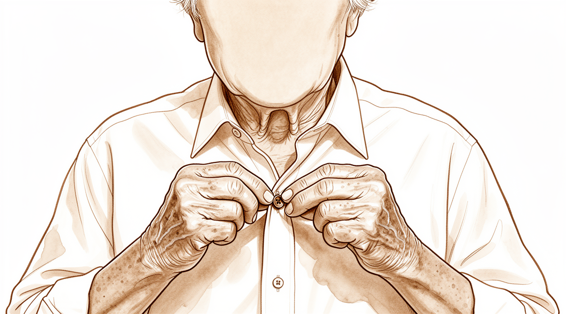
El dolor suele empeorar después de usar el dedo, y puede notarlo dolorido al despertar por la mañana. Las tareas finas que requieren la misma punta del dedo se vuelven difíciles: abrocharse una camisa, coger monedas pequeñas, enhebrar una aguja o sujetar una llave para abrir una cerradura. Escribir a mano también puede doler. Con el tiempo, la articulación puede volverse rígida y perder parte de su flexión, y el dedo puede ir desviándose poco a poco de su alineación.
Algunas personas notan un pequeño bulto lleno de líquido cerca de la uña, llamado quiste mucoso. Crece a partir de la articulación artrítica que está debajo, por lo que a menudo reaparece incluso después de drenarlo.
Si la artritis apareció tras una lesión del dedo, como un dedo en martillo en el que el tendón que endereza el dedo se desgarró o arrancó un fragmento de hueso, la articulación puede desgastarse más rápido de lo que lo habría hecho de otro modo. Ese desgaste provoca el mismo dolor y la misma rigidez, y el dedo puede perder movilidad aunque la lesión en sí haya curado.
Esté atento a algunos signos de alarma. Si el dedo se pone caliente, rojo, hinchado y doloroso, sobre todo si tiene fiebre, acuda al servicio de urgencias ese mismo día. Puede tratarse de una infección que necesita atención el mismo día, y no necesita antes una derivación de su médico de cabecera. Si el dolor no mejora con el paso de las semanas, empeora o le despierta por la noche, consulte a su médico de cabecera o solicite una evaluación especializada.
¿Qué está ocurriendo realmente?
Cada dedo es una cadena de pequeños huesos. La articulación situada en la misma punta, justo detrás de la uña, es donde el último hueso se une con el hueso contiguo. Una capa lisa y resbaladiza recubre los extremos de ambos huesos y les permite deslizarse uno sobre otro. Piense en ella como un amortiguador incorporado que además mantiene la superficie deslizante.
En la artritis, esa capa resbaladiza se adelgaza o se deteriora. Entonces los huesos rozan entre sí, y la articulación reacciona generando hueso adicional alrededor de sus bordes. Esas protuberancias óseas son los bultos firmes que puede notar cerca de la uña, los descritos en la sección anterior. El roce causa el dolor, y las superficies desgastadas junto con las protuberancias explican por qué la articulación se hincha, se vuelve rígida y se va desviando poco a poco de su alineación.
La articulación también se mantiene unida gracias a pequeñas bandas de tejido a cada lado y a una banda plana de fibras tendinosas que pasa por encima y se inserta justo más allá de la articulación. Cuando la artritis altera la articulación, estas bandas y tendones pueden perder su equilibrio. Esa es una de las razones por las que el dedo puede doblarse demasiado en la articulación media mientras la punta se curva hacia abajo, un cambio de forma que tiende a aparecer a medida que la artritis avanza.
A veces el desgaste se debe a una lesión y no simplemente al desgaste por el uso. Un golpe fuerte, un aplastamiento o una lesión de tendón como el dedo en martillo pueden dañar las superficies de la articulación. Entonces la articulación se desgasta antes de lo que lo habría hecho de otro modo, y por eso el dolor y la rigidez pueden aparecer años después de que una lesión pareciera haber curado.
La artritis en esta articulación también puede formar un pequeño bulto lleno de líquido junto a la uña, el quiste mucoso mencionado antes. Crece a partir de la propia articulación irritada, y por eso tiende a reaparecer después de drenarlo.
La buena noticia es que esta articulación hace poco del trabajo de agarre con fuerza. Su función es el control fino en la punta del dedo, y por eso el tratamiento se centra en calmar el dolor y en mantener ese control fino.
Qué podemos hacer al respecto
El Dr. Kieran Hirpara, cirujano de extremidades superiores en el Mater Private Hospital Rockhampton, comienza con las opciones menos invasivas que se adapten a su condición. Por lo general, los pacientes son derivados a nuestra clínica por su médico de cabecera; si un fisioterapeuta le ha sugerido que nos consulte, igualmente necesitará una derivación de su médico de cabecera para poder acceder al reembolso de Medicare. En su primera consulta, tomamos su historia clínica, examinamos su dedo y solicitamos estudios de imagen si son necesarios. Como se trata de un problema de desgaste de larga duración, normalmente probamos primero el tratamiento no quirúrgico y solo hablamos de cirugía si este no ha logrado una mejoría suficiente.
El primer paso suele ser una pequeña férula que mantiene recta la articulación de la punta del dedo. La férula alivia el dolor en la articulación y ayuda a que el dedo se enderece. Es sencilla y segura, y puede llevarla durante su día normal. Normalmente la combinamos con medicación antiinflamatoria, comprimidos que calman la hinchazón y el dolor. La terapia de la mano también puede ayudarle a mantener la punta del dedo funcional para las tareas finas mientras se calma la irritación. Dé a estas medidas una oportunidad razonable antes de decidir nada más.
Si después de eso el dolor y la rigidez siguen limitándole, la cirugía pasa a ser una opción. La intervención estándar es una fusión, también llamada artrodesis, en la que se retiran las superficies articulares desgastadas y se unen los dos huesos para que consoliden en uno solo. Después, la articulación ya no se dobla, pero no duele y es estable, y el dedo queda fijado en una posición que funciona bien para las tareas cotidianas. La fusión también endereza un dedo que se ha desviado de su alineación, y es el tratamiento que impide que un quiste mucoso vuelva a aparecer. Si para usted es más importante conservar algo de movimiento en la punta del dedo que eliminar el dolor por completo, existe otra intervención en la que se recortan los bultos óseos y se limpia la articulación, dejándola con movilidad. Hablaremos de qué opción se adapta a su dedo y a lo que usted quiere que haga su mano, y lo decidiremos juntos.
Qué esperar
La artritis de la articulación de la punta del dedo suele desarrollarse lentamente a lo largo de los años. El dolor suele aparecer y desaparecer: empeora después de usar el dedo y luego vuelve a calmarse con el reposo. Sin tratamiento, la articulación a menudo se vuelve más rígida y el dedo puede desviarse más con el tiempo. Algunas personas notan que el dolor mejora por sí solo a medida que la articulación se vuelve rígida, pero en muchas sigue dando molestias, sobre todo con tareas finas como abrocharse una camisa o girar una llave.
La mayoría de las personas obtienen un buen alivio con las medidas sencillas descritas antes: una férula, medicación antiinflamatoria y terapia de la mano. Estas medidas calman el dolor mientras sigue usando el dedo para las tareas cotidianas. Si no han ayudado tras una prueba razonable, la cirugía suele aliviar bien el dolor. Una articulación de la punta del dedo fusionada no duele y es estable, y en general las personas vuelven a usar la mano con normalidad, aunque la punta ya no se dobla. Si para usted es más importante conservar algo de movimiento, la intervención de limpieza articular deja el dedo con capacidad de doblarse, con un alivio del dolor que es bueno, pero no siempre completo.
La recuperación de la cirugía lleva semanas o meses, no días. Al principio el dedo se mantiene inmovilizado mientras los huesos se unen, y su terapeuta de mano le guiará mientras recupera la fuerza de agarre y el control fino. La mayoría de las personas comprueban que el resultado final es un dedo que cumple su función sin dolor, aunque la punta quede más recta que antes.
Algunas advertencias sinceras. La fusión no siempre consolida en el primer intento, y algunas personas necesitan otra operación para retirar el material metálico o repetir la fusión. Fumar retrasa la consolidación ósea, por lo que dejar de fumar antes de la cirugía da a su dedo la mejor oportunidad de que los huesos se unan bien. Si el dolor no mejora con el paso de las semanas, empeora o le despierta por la noche, consulte a su médico de cabecera o solicite una evaluación especializada.
Cuándo consultar a un profesional
La mayoría de las artritis de la articulación de la punta del dedo se desarrollan lentamente, por lo que para empezar basta con una consulta habitual con su médico de cabecera. Acuda a su médico de cabecera si el dolor no mejora con el paso de las semanas, empeora o le despierta por la noche. Lo mismo se aplica si el dedo le impide trabajar o realizar tareas finas como abrocharse una camisa.
Algunos signos requieren atención más rápida. Acuda a un servicio de urgencias ese mismo día si el dedo se pone caliente, rojo, hinchado y doloroso, sobre todo si tiene fiebre. Ese cuadro sugiere una infección, y no necesita antes una derivación de su médico de cabecera.
Acuda a un servicio de urgencias después de una lesión si la piel sobre la articulación está abierta, se ve el hueso o el dedo tiene una forma claramente anormal. Lo mismo se aplica si el dedo se pone pálido, frío, blanco o azul, o si pierde de repente la sensibilidad o el movimiento del dedo después de un golpe.
Un quiste mucoso que reaparece una y otra vez, o un dedo que se va desviando de su alineación, merece una evaluación especializada.
En mayor profundidad
Advanced reading: the deeper science (optional)
Esta sección profundiza más de lo necesario para que usted tome decisiones sobre su propio tratamiento. La artritis de la articulación distal del dedo merece una lectura adicional, ya que la artrodesis, la intervención quirúrgica estándar y generalmente descrita como sencilla, presenta una tasa de complicaciones superior a la que su reputación sugiere; además, existe una alternativa que preserva el movimiento y rara vez se menciona.
La fusión no es tan sencilla como parece
La fusión de la articulación de la punta del dedo se presenta como una intervención fiable; en cuanto al alivio del dolor, efectivamente lo es. Sin embargo, el perfil de complicaciones no es tan benigno. Un estudio que analizó los factores de riesgo en 173 pacientes lo afirma claramente: la artrodesis de la articulación interfalángica distal suele generar complicaciones, siendo la artrosis, la artrodesis de revisión y el tabaquismo los factores de riesgo identificados [1].
El hecho de que el tabaquismo figure en esa lista merece atención, pues es el único factor que el paciente puede controlar. La consolidación ósea depende del riego sanguíneo; se trata de una articulación pequeña, rodeada de una capa delgada de tejidos blandos, situada al final del trayecto vascular del dedo.
El implante que logra una mejor unión también genera problemas que los implantes más baratos no causan
En el debate sobre los métodos de fijación, la respuesta es sorprendentemente clara: se trata de un intercambio, no de una solución definitiva. En un total de 1,125 pacientes, los tornillos de compresión sin cabeza parecen aumentar las tasas de unión ósea, pero se asocian a complicaciones que no se observan con otras técnicas más económicas y ya consolidadas; además, aparte de la unión ósea, no existen pruebas suficientes que demuestren la superioridad de dichos tornillos [2].
Las complicaciones específicas relacionadas con estos tornillos se deben a la anatomía. El tornillo discurre a lo largo del eje de la punta del dedo, por lo que pasa cerca del lecho ungueal y puede provocar deformidad de la uña; además, en falanges distales muy pequeñas puede no haber suficiente hueso para fijarlo adecuadamente. Los alambres son más baratos y evitan esos problemas específicos, aunque logran una unión ósea algo menos fiable.
Cuando el objetivo principal es lograr la unión ósea, realizar una fusión de revisión o tratarse de pacientes fumadores, la ventaja de los tornillos resulta más evidente. En cambio, cuando el hueso es pequeño y la integridad de la uña es importante, no queda tan claro que sean la opción adecuada.
La alternativa que preserva el movimiento
La fusión articular no es la única opción, aunque esta alternativa no se discute con frecuencia. En pacientes cuya principal queja son los bultos óseos y el dolor que estos provocan, más que la artritis en toda la articulación, se pueden extirpar los osteofitos prominentes manteniendo la integridad de la articulación.
La quilectomía abierta de la articulación de la punta del dedo se describe como una alternativa segura y eficaz a la artrodesis en pacientes con artrosis sintomática que desean conservar el movimiento articular, en un total de 78 pacientes [3].
Esto es importante porque la articulación de la punta del dedo contribuye poco a la fuerza de agarre, pero sí en gran medida a la manipulación fina y al aspecto estético de la mano. Para quienes sus síntomas se deben precisamente a los nódulos de Heberden, aceptar una articulación de la punta del dedo permanentemente rígida supone un sacrificio mayor de lo que parece; sin embargo, existe una opción intermedia.
El quiste que suele acompañarlo
La artritis en esta articulación suele generar un quiste mucoso, una pequeña protuberancia llena de líquido que se origina en la articulación afectada, generalmente al lado de la uña. Dado que depende de la articulación subyacente, en ese sentido se comporta como el ganglio de la muñeca: drenarlo reduce la hinchazón, pero no elimina la causa raíz. Este tema se aborda por separado, pero es importante conocer esta relación, ya que la aparición recurrente de este quiste es señal de la artritis subyacente y no un problema aislado.
Referencias
[1] Runkel A, Bonaventura B, Sundermann B, Zajonc H, Eisenhardt S, Leibig N. Factores de riesgo en la artrodesis de la articulación interfalángica distal de la mano: un estudio retrospectivo. J Hand Surg Eur Vol. 2022;47(9):907-14. https://doi.org/10.1177/17531934221111641
[2] Dickson D, Mehta S, Nuttall D, Ng C. Revisión sistemática sobre la artrodesis de la articulación interfalángica distal. J Hand Microsurg. 2014;6(2):74-84. https://doi.org/10.1007/s12593-014-0163-1
[3] Lin EA, Papatheodorou LK, Sotereanos DG. Quirurgia de cheilectomía para el tratamiento de la osteoartritis sintomática de la articulación interfalángica distal: revisión de 78 casos. J Hand Surg Am. 2017;42(11):889-93. https://doi.org/10.1016/j.jhsa.2017.07.006
Evidence & references
This is the clinical evidence summary written for health professionals. It is technical, and it lists the research this page was built from. You do not need to read it to understand your treatment or to make a decision about it.
Overview
- Osteoarthritis is the most common indication for distal interphalangeal joint arthrodesis [19].
- In a cohort of 149 cases, postoperative complications for distal interphalangeal and thumb interphalangeal joint arthrodesis occurred at a rate similar to that reported in existing literature [19].
- Finger distal interphalangeal and thumb interphalangeal joint arthrodesis generally results in favorable outcomes in terms of bony union regardless of underlying medical condition or technical details of the surgical operation [6].
- The X-fuse implant provides excellent stability with minimal hardware problems and a high rate of union in patients with finger distal interphalangeal and thumb interphalangeal joint arthritis [1].
- In select patients, percutaneous distal interphalangeal joint arthrodesis is advantageous in comparison with open fusion techniques [2].
- Reamed percutaneous distal interphalangeal joint arthrodesis is associated with higher union rates achieved in a shorter time and reduced postoperative pain compared with the non-reamed percutaneous technique [8].
- Successful fusions of the distal interphalangeal joint at an angle up to 35 degrees can be achieved using small diameter headless compression screws [14].
- The Mini-Acutrak screw is suitable for distal interphalangeal joint fusion in all fingers with the exception of the small finger [22].
- The nonaxial multiple small screws technique is a feasible option for distal interphalangeal and thumb interphalangeal joint arthrodesis, especially when a small finger is indicated and a significant flexion angle is required [24].
- Given the lack of difference in biomechanical performance between K-wires and compression screws, consideration should be given to other factors such as cost and complication profiles when choosing an implant for distal interphalangeal joint fusion [34].
- The smile incision and reverse shotgun approach may be a good surgical option for distal interphalangeal joint arthrodesis when more volar part joint preparation and more volar implant insertion sites are necessary [17].
- Results from a lateral approach and plate fixation for distal interphalangeal joint arthrodesis are equivalent to traditional methods but with fewer major complications [11].
- For persistently symptomatic, unstable distal interphalangeal joints, arthrodesis should be considered [4].
- In the distal interphalangeal joint, there is probably good reason to proceed to arthrodesis immediately for synovial chondromatosis [3].
- The swan neck deformity can progress significantly with time because of increasing distal interphalangeal joint flexion contracture [5].
- Silicone interpositional arthroplasty of the distal interphalangeal joint is an acceptable alternative to arthrodesis, achieving excellent pain relief and a range of movement of 30–40 degrees with a low overall complication rate of 5% [13].
- Swanson hinge implant arthroplasty can be the surgical treatment of choice for the patient who desires continued mobility, albeit limited, of the involved osteoarthritic distal interphalangeal joint [9].
- Open distal interphalangeal joint cheilectomy is a safe and effective alternative to distal interphalangeal joint arthrodesis in patients with symptomatic osteoarthritis who wish to preserve joint motion [28].
- Denervation with cheilectomy of the distal interphalangeal joint presents a compelling motion-preserving alternative to arthrodesis for symptomatic distal interphalangeal joint osteoarthritis [10].
- A surgical technique for treating symptomatic distal interphalangeal joint arthritis reduces pain while preserving distal interphalangeal joint motion [18].
- The authors recommend simultaneous surgical intervention in case of severe painful osteoarthritis of the proximal interphalangeal and distal interphalangeal joints of the same digit [7].
- The combination of distal interphalangeal arthrodesis and proximal interphalangeal Swanson arthroplasty resulted in a favorable outcome in terms of simultaneous bony union and flexibility [12].
- Combined distal interphalangeal arthrodesis and proximal interphalangeal procedures present unique challenges regarding hardware conflict, where K-wires offer the easiest compatibility and headless screws must ideally not reach proximal to the midpoint of the middle phalanx [90].
- A customized structural bone graft addresses issues of bone stock loss and medullary absence in failed distal interphalangeal joint silicone arthroplasty, achieving reliable union rates and high patient satisfaction [21].
Anatomy & Pathophysiology
Bony Anatomy & Morphology
- The skeleton of the hand consists of 27 bones, of which 19 are long bones [47].
- The skeleton is divided into five rays, each ray making up a polyarticulated chain comprising the metacarpals and phalanges [47].
- The thumb ray is made up of only three bones—a metacarpal and two phalanges [47].
- The other four digital rays are formed by four skeletal segments—a metacarpal and three phalanges [47].
- The thumb metacarpal is the shortest, and the index metacarpal is by far the longest [47].
- The proximal and middle phalanges of the long and ring fingers are longer than those of the index finger [47].
- The epiphyseal plates are located at the proximal ends of the phalanges and the first metacarpal [47].
- An examination of 100 specimens of the terminal joints of human fingers was described in a 1982 study [75].
- A substantial number of distal phalanges are too small to accommodate commonly available headless compression screws, particularly in females and the small finger [39].
- Understanding of DIP joint morphology may lend insight into the biomechanics and disease progression within the DIP joints [23].
Soft Tissue Anatomy & Extensor Mechanism
- Distal interphalangeal joint extension is achieved through the conjoined lateral bands that are composed of tendinous slips from the extrinsic and intrinsic tendons [46].
- The principal bony insertion of the extrinsic digital extensors is on the dorsal proximal aspect of the middle phalanx [46].
- The deep head of each dorsal interosseous muscle forms a lateral tendon, or lateral band, at the level of the MP joint [59].
- The lateral bands are joined by the lateral slips of the extensor tendon to form the conjoined lateral band [59].
- The two conjoined lateral bands to each finger unite at the distal third of the middle phalanx to form the terminal tendon [59].
- The terminal tendon inserts at the base of the distal phalanx to extend it [59].
- The flexor digitorum profundus inserts on the proximal volar aspect of the distal phalanx, flexing the distal interphalangeal joint as well as the proximal interphalangeal and metacarpophalangeal joints [46].
- The flexor digitorum profundus provides digital flexion at both the proximal and distal interphalangeal joints [66].
- The A5 annular pulley is located over the distal interphalangeal joint [66].
- The vinculum breve of the flexor digitorum profundus and check-rein ligaments may account for the greater amount of passive hyperextension that can be achieved at the distal than the proximal interphalangeal joint [80].
Biomechanics & Kinematics
- The wrist influences the position of the metacarpophalangeal joint; the metacarpophalangeal joint also affects the position of the proximal interphalangeal joint, which in turn affects the distal interphalangeal joint [67].
- Almost all movements in the hand are around oblique and variable axes, resulting in combined movements permitting optimal orientation of the phalanges at the time of prehension [67].
- The axes of flexion are so arranged that flexion of all the metacarpophalangeal and proximal interphalangeal joints causes the fingers to converge toward the scaphoid [70].
- In a combinatorial relationship, intrinsic muscles produce steep inclination of extensor forces at the DIP joint [73].
- There were no significant differences in comparisons among loads (200, 400, 600, and 800 g) regarding the inclination slope of intrinsic extensor forces at the DIP joint [73].
- Immobilization of the distal interphalangeal joint of any finger reduces the overall grip strength of the hand, with the effect becoming progressively more pronounced from the index to the little fingers [37].
- Positioning the middle finger DIP joint in either extension or 20° of flexion did not significantly affect grip strength or dexterity [49].
- Biomechanically, dynamic tenodesis for the DIP joint using the remaining FDP tendon results in a flexion angle greater than 30 degrees [76].
Pathophysiology & Etiology
- Osteoarthritis occurs with the highest prevalence in the distal interphalangeal joint of the hand and has been divided into an erosive and a nonerosive form [26].
- The pathogenesis of the early stages of osteoarthritis is poorly understood, but considerable emphasis has been placed on the role of cartilage and subchondral bone as well as soft tissue structures such as collateral ligaments and tendons [26].
- Osteoarthritis is a complex disease resulting in the failure of articular cartilage due to a combination of genetic, metabolic, biochemical, and biomechanical factors [26].
- Cartilage loss and reactive new bone formation at joint margins is accompanied by the proliferation of osteoarticular tissue in the capsule [26].
- Heberden's nodes are more common in women who engage in cooking in school and the incidence increases with the number of tasks involved [25].
- Cooking as an occupation is an aetiological factor in the pathogenesis of Heberden's nodes [25].
- Work-load, sports, and repeated minimal trauma contribute to the development of Heberden's nodes [25].
- Repetitive injuries to a joint lead to an increased risk of developing posttraumatic arthritis [48].
- Athletes have a higher incidence of degenerative joint changes compared to the general population [48].
- Mallet finger is caused by forced flexion of an extended fingertip [51].
- Less common hyperextension or hyperextension/axial loading tends to cause the larger fractures and subluxation in mallet injuries [51].
- One-third of all mallet fingers are associated with a fracture [51].
- Mallet fractures associated with large fragments may result in volar subluxation of the distal phalanx as the collateral ligaments remain attached to the fracture fragment [51].
- Fracture fragments that are less than 43% of the distal phalanx articular surface usually do not allow volar subluxation [51].
- Fracture fragments involving larger than 52% of the joint surface consistently allow subluxation [51].
- Palmar subluxation of a DIP joint without preexisting arthritic deformity is expected when more than one half of the dorsal articular surface is injured [20].
- The mechanism of displaced intra-articular osteochondral fracture causing irreducible dislocation of the DIP joint probably involved hyperextension of the DIP joint [15].
- The palmar plate was torn at the attachment to the distal phalanx in cases of displaced intra-articular osteochondral fracture [15].
- Attempts to perform a closed reduction of displaced intra-articular osteochondral fractures can lead to further displacement of the fracture, with folding of the fragment on the articular cartilage hinge [15].
- The cause of irreducible palmar dislocation of the DIP joint can be an entrapment of the extensor tendon in front of the head of the middle phalanx [41].
- Irreducibility was more commonly seen in dorsal than in volar dislocations of the DIP joint [85].
- Volar dislocations of the DIP joint carried a higher risk of instability immediately after reduction compared to dorsal dislocations [85].
- Some loss of motion in small joints of the fingers after hyperflexion injuries would be expected [79].
Classification
- Osteoarthritis of the distal interphalangeal joint has been divided into an erosive and a nonerosive form [26].
- Radiographic evaluation represents the most standardized method to quantify disease progression in distal interphalangeal joint osteoarthritis, with different systems developed for defining and grading radiographic features [26].
- The interrater reliability of the Kellgren & Lawrence and OARSI classification systems for post-traumatic osteoarthritis in the distal interphalangeal joint after mallet finger fractures is considerably lower than initially assumed [86].
Clinical Presentation
- Osteoarthritis occurs with the highest prevalence in the distal interphalangeal joint of the hand [26].
- Heberden's nodes are more common in women who engage in cooking in school, with incidence increasing with the number of tasks involved [25].
- There is no confirmed difference in the incidence of disease between right and left hands in elementary school cooks [25].
- Radiographic evaluation represents the most standardized method to quantify disease progression in distal interphalangeal joint osteoarthritis [26].
- The pathogenesis of the early stages of distal interphalangeal joint osteoarthritis is poorly understood [26].
- Considerable emphasis has been placed on the role of cartilage and subchondral bone in the pathogenesis of distal interphalangeal joint osteoarthritis [26].
- Soft tissue structures such as collateral ligaments and tendons play a role in the pathogenesis of distal interphalangeal joint osteoarthritis [26].
- Cartilage loss and reactive new bone formation at joint margins are accompanied by the proliferation of osteoarticular tissue in the capsule [26].
- Radiological osteoarthritis after a mallet finger fracture is similar to the natural degenerative process in the distal interphalangeal joint [27].
- Radiological osteoarthritis after a mallet finger fracture is accompanied by a decrease in range of motion of the distal interphalangeal joint [27].
- The decrease in range of motion following mallet finger fracture does not clinically affect patient-reported outcomes [27].
- Palmar subluxation of a distal interphalangeal joint without preexisting arthritic deformity is expected when more than one half of the dorsal articular surface is injured [20].
- Primary synovial chondromatosis of the distal interphalangeal joint is an extremely rare entity that requires accurate diagnosis to distinguish from other arthropathies [50].
- Adnexal tumours may masquerade as a distal interphalangeal joint ganglion and should enter the differential diagnosis when examining tumours of the digits [89].
- Floating distal interphalangeal joint injuries can be misdiagnosed initially due to minimal deformity [33].
- Osteoarthritis may develop following floating distal interphalangeal joint injuries [33].
- The mechanism of displaced intra-articular osteochondral fracture causing irreducible dislocation of the distal interphalangeal joint probably involved hyperextension of the joint [15].
- The palmar plate is torn at the attachment to the distal phalanx in displaced intra-articular osteochondral fractures of the distal interphalangeal joint [15].
- Attempts to perform a closed reduction of a displaced intra-articular osteochondral fracture can lead to further displacement of the fracture with folding of the fragment on the articular cartilage hinge [15].
- The cause of irreducibility in palmar dislocation of the distal interphalangeal joint can be entrapment of the extensor tendon in front of the head of the middle phalanx [41].
- Interrater reliability for measurements of the distal interphalangeal joint motions of the index and long digits is high [16].
- Interrater reliability for distal interphalangeal joint motion measurements is slightly higher for the dorsal method of placement than for the lateral method [16].
Investigations
- Radiographic evaluation is the most standardized method to quantify disease progression in distal interphalangeal joint osteoarthritis [26].
- Different systems have been developed for defining and grading radiographic features of distal interphalangeal joint osteoarthritis [26].
- Interrater reliability was high for measurements of the proximal and distal interphalangeal joint motions of the index and long digits [16].
- Interrater reliability was slightly higher for the dorsal method of goniometer placement than for the lateral method [16].
- Primary synovial chondromatosis of the distal interphalangeal joint requires accurate diagnosis to distinguish from other arthropathies [50].
- A distinct collagen septum exists between the extensor tendon and skin at the distal interphalangeal joint [53].
- The existence of a distinct collagen septum between the extensor tendon and skin at the distal interphalangeal joint was confirmed using MRI and histology [53].
Treatment
Non-Operative
- Splinting of the distal interphalangeal joint reduces pain and improves extension at the joint [29].
- Splinting for DIP joint arthritis does not give rise to non-compliance, increased stiffness, or restriction of range of motion [29].
- Splinting and anti-inflammatory medications are the mainstay of nonoperative treatment for DIP joint arthritis [48].
- Injection with Collagenase Clostridium histolyticum is an option for the treatment of DIP joint contractures in Dupuytren disease, though the potential risk for recurrence should be carefully weighed prior to its use [93].
Operative: Arthrodesis
- Arthrodesis is generally the most accepted surgical option for treatment of degenerative and traumatic conditions involving the distal interphalangeal joint of the fingers or the interphalangeal joint of the thumb [38].
- The ideal position of DIP arthrodesis is thought to be slightly flexed to improve power, fine pinch, and grip [38].
- In select patients, percutaneous DIP joint arthrodesis is advantageous in comparison with open fusion techniques [2].
- Reamed percutaneous DIPJ arthrodesis is associated with higher union rates achieved in a shorter time and reduced postoperative pain compared with the non-reamed percutaneous technique [8].
- The X-fuse implant provides excellent stability with minimal hardware problems and a high rate of union in patients with finger DIPJ and thumb IPJ arthritis [1].
- Finger DIP and thumb IP joint arthrodesis generally resulted in favourable outcome in terms of bony union regardless of the underlying medical condition or technical details of the surgical operation [6].
- Osteoarthritis was the most common indication for DIP and thumb IP arthrodesis, and postoperative complications occurred at a rate similar to that reported in existing literature [19].
- Successful fusions of the DIP joints at an angle up to 35 degrees were achieved using small diameter headless compression screws, providing benefits including early mobilization and favourable functional outcome scores [14].
- The Mini-Acutrak screw is suitable for DIPJ fusion in all fingers with the exception of the small finger [22].
- The nonaxial multiple small screws technique could be used as a feasible option in DIPJ and thumb IPJ arthrodesis, especially when a small finger is indicated and a significant flexion angle is required [24].
- Given the lack of difference in biomechanical performance between K-wires and compression screws, consideration should be given to other factors such as cost and complication profiles when choosing an implant for DIPJ fusion [34].
- DIP joint preparation is not necessary before performing arthrodesis in fingers with stage IV chondropathy [44].
- The use of a buried break-away compression screw avoids complications related to bulky hardware in DIP arthrodesis [44].
- Dorsal plate fixation allows for the performance of a distal finger joint arthrodesis in slight flexion [38].
- The smile incision and reverse shotgun approach may be a good surgical option for DIPJ arthrodesis when more volar part joint preparation and more volar implant insertion sites are necessary [17].
- Lateral approach and plate fixation for DIP joint arthrodesis yields results equivalent to traditional methods but with fewer major complications [11].
- For persistently symptomatic, unstable DIP joints, arthrodesis should be considered [4].
- In the DIP joint, there is probably good reason to proceed to arthrodesis immediately in cases of synovial chondromatosis [3].
- A customized structural bone graft addresses issues of bone stock loss and medullary absence in failed DIPJ silicone arthroplasty, achieving reliable union rates and high patient satisfaction [21].
- All joints had fused radiologically in a series of proximodistal interphalangeal arthrodesis of the little finger [32].
- Arthrodesis of the DIP joint provides stability and pain relief and can correct deformity [48].
- A painless, stable, distal interphalangeal joint that is in a functional position is more important than the ability to move the joint [48].
- The authors recommend simultaneous surgical intervention in case of severe painful OA of the PIP and DIP joints of the same digit [7].
- The combination of DIP arthrodesis and PIP Swanson arthroplasty resulted in a favourable outcome in terms of simultaneous bony union and flexibility [12].
- For patients prioritizing hand aesthetics or with unstable joints, distal interphalangeal joint arthrodesis is preferable [31].
Operative: Arthroplasty and Motion-Preserving
- Silicone interpositional arthroplasty of the DIP joint is an acceptable alternative to arthrodesis, achieving excellent pain relief and a range of movement of 30–40 degrees with a low overall complication rate of 5% [13].
- Swanson hinge implant arthroplasty can be the surgical treatment of choice for the patient who desires continued mobility, albeit limited, of the involved osteoarthritic DIP joint [9].
- Open DIP joint cheilectomy is a safe and effective alternative to DIP joint arthrodesis in patients with symptomatic osteoarthritis who wish to preserve joint motion [28].
- Denervation with cheilectomy of the distal interphalangeal joint presents a compelling motion-preserving alternative to arthrodesis for symptomatic DIP joint osteoarthritis [10].
- A surgical technique for DIP joint arthritis reduces pain while preserving DIP joint motion [18].
- Volar plate arthroplasty of the distal interphalangeal joint allows for a painless functional arc of motion without residual joint subluxation [40].
Complications
- The X-fuse implant provides excellent stability with minimal hardware problems [1].
- The X-fuse implant has a high rate of union in patients with finger DIPJ and thumb IPJ arthritis [1].
- Reamed percutaneous DIPJ arthrodesis is associated with reduced postoperative pain compared with the non-reamed percutaneous technique [8].
- The swan neck deformity progressed significantly with time because of increasing DIPJ flexion contracture [5].
- Postoperative complications occurred at a rate similar to that reported in the existing literature in a retrospective cohort of 149 cases of DIP and thumb IP joint arthrodesis [19].
- Arthrodesis of the distal interphalangeal joint often leads to complications [30].
- Silicone interpositional arthroplasty of the DIP joint has a low overall complication rate of 5% [13].
- Bony union of the joint was delayed in a series using the Herbert screw for DIPJ arthrodesis [56].
- Follow-up to 5 years of DIP joint flexible implant arthroplasty showed only one complication [92].
Recovery
Arthrodesis Outcomes and Complications
- Postoperative complications in DIP and thumb IP joint arthrodesis occurred at a rate similar to that reported in existing literature [19].
- Finger DIP and thumb IP joint arthrodesis generally resulted in favourable outcomes in terms of bony union regardless of underlying medical condition or technical details of the surgical operation [6].
- Reamed percutaneous DIPJ arthrodesis is associated with higher union rates achieved in a shorter time compared with the non-reamed percutaneous technique [8].
- Successful fusions of the DIP joints at an angle up to 35 degrees were achieved using small diameter headless compression screws [14].
- Small diameter headless compression screws for DIP arthrodesis provide benefits including early mobilization and favourable functional outcome scores [14].
- Although bony union of the joint was delayed in a minimally invasive technique using the Herbert Screw, firm bone union was ultimately obtained in all joints [56].
Arthroplasty Outcomes
- Silicone interpositional arthroplasty of the DIP joint achieves a range of movement of 30–40 degrees [13].
Non-Operative Management
- Splinting of the DIP joint does not give rise to non-compliance, increased stiffness, or restriction of range of motion [29].
Key Evidence
- [L4] The X-fuse implant seems to provide excellent stability with minimal hardware problems with a high rate of union in a wide array of patients with finger DIPJ and thumb IPJ arthritis. [1] (10.1097/bth.0b013e31829ba688)
- [L4] In select patients, this percutaneous DIP joint arthrodesis is advantageous in comparison with open fusion techniques. [2] (10.1007/s11552-010-9265-9)
- [L5] In the DIP joint there is probably good reason to proceed to arthrodesis immediately. [3] (10.1016/s0363-5023(09)91115-7)
- [L4] For persistently symptomatic, unstable DIP joints, arthrodesis should be considered. [4] (10.1016/j.jhsb.2005.09.003)
- [L5] The swan neck deformity in this individual progressed significantly with time because of increasing DIPJ flexion contracture. [5] (10.1016/j.jht.2009.11.005)
- [L3] In our cohort finger DIP and thumb IP joint arthrodesis generally resulted in favourable outcome in terms of bony union regardless of the underlying medical condition or technical details of the surgical operation. [6] (10.1142/s2424835520500216)
- [L3] The authors recommend simultaneous surgical intervention in case of severe painful OA of the PIP and DIP joints of the same digit. [7] (10.1177/17531934231191255)
- [L3] Reamed percutaneous DIPJ arthrodesis is associated with higher union rates achieved in a shorter time and reduced postoperative pain compared with the non-reamed percutaneous technique. [8] (10.1177/15589447261487482)
- [L5] Swanson hinge implant arthroplasty can be the surgical treatment of choice for the patient who desires continued mobility, albeit limited, of the involved osteoarthritic DIP joint. [9] (10.1016/s0894-1130(98)80061-6)
- [L4] It presents a compelling motion-preserving alternative to arthrodesis for symptomatic DIP joint osteoarthritis. [10] (10.1016/j.jhsa.2026.01.027)
- [L4] The results obtained in this small series are equivalent to the traditional methods of DIP joint arthrodesis but with fewer major complications. [11] (10.1016/j.jhsa.2007.09.004)
- [L4] The combination of DIP arthrodesis and PIP Swanson arthroplasty resulted in a favourable outcome in terms of simultaneous bony union and flexibility. [12] (10.1177/17531934231215790)
- [L4] The study confirms that silicone interpositional arthroplasty of the DIP joint is an acceptable alternative to arthrodesis, achieving excellent pain relief and a range of movement of 30–40 degrees with a low overall complication rate of 5%. [13] (10.1177/1753193411422679)
- [L4] In this series, successful fusions of the DIP joints, at an angle up to 35 degrees were achieved using small diameter headless compression screws, which provided benefits including early mobilization and favourable functional outcome scores. [14] (10.1142/s2424835518500406)
- [L5] [15] (10.1016/s0363-5023(82)80018-x)
- [L5] Interrater reliability was high for measurements of the PIP and DIP joint motions of the index and long digits and slightly higher for the dorsal method of placement than for the lateral method. [16] (10.1016/s0894-1130(01)80021-1)
- [L4] This technique may be a good surgical option for DIPJ arthrodesis when more volar part joint preparation and more volar implant insertion sites are necessary. [17] (10.1186/s12891-024-08016-6)
- [L4] This surgical technique reduces pain while preserving DIP joint motion. [18] (10.1177/1558944716660555i)
- [L3] Osteoarthritis was the most common indication for arthrodesis and postoperative complications occurred at a rate similar to that reported in the existing literature. [19] (10.1186/s12891-024-07361-w)
- [L5] Palmar subluxation of a DIP joint without preexisting arthritic deformity is expected when more than one half of the dorsal articular surface is injured. [20] (10.1016/j.jhsa.2007.09.006)
- [L4] A customized structural bone graft using the described technique addresses issues of bone stock loss and medullary absence in failed DIPJ silicone arthroplasty, achieving reliable union rates and high patient satisfaction. [21] (10.1177/17531934231151217)
- [L4] The authors conclude that the Mini-Acutrak screw is suitable for DIPJ fusion in all fingers with the exception of the small finger. [22] (10.1016/j.jhsa.2005.09.009)
- [L5] Our understanding of morphology may lend insight into the biomechanics and disease progression within the DIP joints. [23] (10.1007/s11552-014-9605-2)
- [L4] Thus, the NMSS technique could be used as a feasible option in DIPJ and thumb IPJ arthrodesis, especially when a small finger is indicated and a significant flexion angle is required. [24] (10.1186/s12891-022-05473-9)
- [L4] [25] (10.1016/0266-7681(93)90167-e)
- [L5] [26] (10.1016/j.jhsa.2010.09.003)
- [L4] Radiological OA after an MFF is similar to the natural degenerative process in the DIP joint and is accompanied by a decrease in range of motion of the DIP joint, which does not clinically affect PROMs. [27] (10.1016/j.jhsa.2023.03.027)
- [L4] Open DIP joint cheilectomy is a safe and effective alternative to DIP joint arthrodesis in patients with symptomatic osteoarthritis who wish to preserve joint motion. [28] (10.1016/j.jhsa.2017.07.006)
- [L2] It does not give rise to non-compliance, increased stiffness or restriction of range of motion. [29] (10.1016/j.jht.2013.08.004)
- [L3] Arthrodesis of the distal interphalangeal joint often leads to complications. [30] (10.1177/17531934221111641)
- [L3] For patients prioritizing hand aesthetics or with unstable joints, distal interphalangeal joint arthrodesis is preferable. [31] (10.1177/1753193420917818)
- [L4] All joints had fused radiologically. [32] (10.1016/j.hansur.2016.06.003)
- [Case_report] Floating DIP joint injuries can be misdiagnosed initially due to minimal deformity; open reduction and internal fixation is a viable treatment option for chronic cases, though osteoarthritis may develop. [33] (10.1016/j.jhsa.2010.05.025)
- [L5] Given the lack of difference in biomechanical performance between K-wires and compression screws, consideration should be given to other factors such as cost and complication profiles when choosing an implant for DIPJ fusion. [34] (10.1177/1558944715627211)
- [L4] Immobilization of the distal interphalangeal joint of any finger reduces the overall grip strength of the hand, with the effect becoming progressively more pronounced from the index to the little fingers. [37] (10.1177/1753193418765068)
- [L4] [38] (10.1016/j.jhsa.2018.03.049)
- [L4] A substantial number of distal phalanges are too small to accommodate commonly available headless compression screws, particularly in females and the small finger. [39] (10.1007/s11552-014-9679-x)
- [L4] The technique allows for a painless functional arc of motion without residual joint subluxation. [40] (10.1053/jhsu.2001.26325)
- [L5] The cause of irreducibility was an entrapment of the extensor tendon in front of the head of the middle phalanx. [41] (10.1016/s0363-5023(87)80116-8)
- [L4] Our results show that DIP joint preparation is not necessary before performing arthrodesis in fingers with stage IV chondropathy and that the use of a buried break-away compression screw avoids complications related to bulky hardware. [44] (10.1016/j.main.2015.03.002)
- [L4] [48] (10.1142/s0218810417500149)
- [L2] Positioning the middle finger DIP joint in either extension or 20° of flexion did not significantly affect grip strength or dexterity, allowing other considerations such as appearance to be prioritized. [49] (10.1016/j.jhsa.2014.06.021)
- [Case_report] Primary synovial chondromatosis of the distal interphalangeal joint is an extremely rare entity that requires accurate diagnosis to distinguish from other arthropathies. [50] (10.1177/15589447211049520)
- [L5] [51] (10.1177/1753193414554772)
- [L5] We confirmed the existence of a distinct collagen septum between the extensor tendon and skin at the DIP joint using MRI and histology. [53] (10.1016/j.jhsa.2008.11.030)
- [L4] Although bony union of the joint was delayed, firm bone union was ultimately obtained in all joints. [56] (10.1097/00130911-200212000-00008)
- [L5] [73] (10.1002/jor.22021)
- [L5] An examination of 100 specimens of the terminal joints of human fingers is described. [75] (10.1016/s0363-5023(82)80084-1)
- [L5] Biomechanically, dynamic tenodesis for the DIP joint using the remaining FDP tendon is a valuable procedure because it results in a flexion angle greater than 30 degrees. [76] (10.1016/j.jhsg.2020.08.007)
- [L5] Therefore, some loss of motion in small joints of the fingers after hyperflexion injuries would be expected. [79] (10.1142/s2424835516720206)
- [L5] This may account for the greater amount of passive hyperextension that can be achieved at the distal than the proximal interphalangeal joint. [80] (10.1016/0266-7681(91)90058-v)
- [L4] Irreducibility was more commonly seen in dorsal than in volar dislocations, while volar dislocations carried a higher risk of instability immediately after reduction. [85] (10.1177/1753193415616957)
- [L4] The interrater reliability of the Kellgren & Lawrence and OARSI classification systems for post-traumatic osteoarthritis in the distal interphalangeal joint after mallet finger fractures is considerably lower than initially assumed. [86] (10.1016/j.jhsa.2024.03.012)
- [L5] Adnexal tumours may masquerade as a DIPJ ganglion, and should enter the differential diagnosis of the hand surgeon when examining tumours of the digits. [89] (10.1177/1753193415620179)
- [L5] Combined DIP arthrodesis and PIP procedures present unique challenges regarding hardware conflict; K-wires offer the easiest compatibility, while headless screws must ideally not reach proximal to the midpoint of the middle phalanx. [90] (10.1016/j.jhsa.2024.08.006)
- [L4] Follow-up to 5 years showed results that were good to excellent, with only one complication, and a high degree of patient satisfaction. [92] (10.1016/0363-5023(89)90184-6)
- [L4] Injection with CCH is an option for the treatment of DIP joint contractures in Dupuytren disease, though the potential risk for recurrence should be carefully weighed prior to its use. [93] (10.1016/j.jhsa.2018.07.004)
References
[1] Distal Interphalangeal Joint Arthrodesis Using Nitinol Intramedullary Fixation Implants. Techniques in Hand & Upper Extremity Surgery. 2013. DOI: 10.1097/bth.0b013e31829ba688
[2] Treatment of Symptomatic Distal Interphalangeal Joint Arthritis with Percutaneous Arthrodesis: A Novel Technique in Select Patients. HAND. 2010. DOI: 10.1007/s11552-010-9265-9
[3] Synovial chondromatosis in the distal interphalangeal joint. The Journal of Hand Surgery. 1990. DOI: 10.1016/s0363-5023(09)91115-7
[4] Management of Tophaceous Gout of the Distal Interphalangeal Joint. Journal of Hand Surgery. 2006. DOI: 10.1016/j.jhsb.2005.09.003
[5] Swan Neck Deformity after Distal Interphalangeal Joint Flexion Contractures: A Biomechanical Analysis. Journal of Hand Therapy. 2010. DOI: 10.1016/j.jht.2009.11.005
[6] Factors Influencing Bone Union in Finger Distal Interphalangeal and Thumb Interphalangeal Joint Arthrodesis. The Journal of Hand Surgery (Asian-Pacific Volume). 2020. DOI: 10.1142/s2424835520500216
[7] Does distal interphalangeal joint arthrodesis affect proximal interphalangeal joint arthroplasty outcomes in the same finger?. Journal of Hand Surgery (European Volume). 2023. DOI: 10.1177/17531934231191255
[8] Reamed Percutaneous Arthrodesis for Distal Interphalangeal Joint Arthritis Improves Bone Fusion and Pain Compared With a Non-Reamed Technique. HAND. 2026. DOI: 10.1177/15589447261487482
[9] Distal interphalangeal joint implant arthroplasty in a musician. Journal of Hand Therapy. 1998. DOI: 10.1016/s0894-1130(98)80061-6
[10] Denervation with Cheilectomy of the Distal Interphalangeal Joint: Technique and Medium-Term Results. The Journal of Hand Surgery. 2026. DOI: 10.1016/j.jhsa.2026.01.027
[11] Alternative to the Distal Interphalangeal Joint Arthrodesis: Lateral Approach and Plate Fixation. The Journal of Hand Surgery. 2008. DOI: 10.1016/j.jhsa.2007.09.004
[12] Simultaneous anterograde screw arthrodesis of distal interphalangeal joint and silastic proximal interphalangeal joint replacement for osteoarthritis. Journal of Hand Surgery (European Volume). 2023. DOI: 10.1177/17531934231215790
[13] Joint replacement in 131 painful osteoarthritic and post-traumatic distal interphalangeal joints. Journal of Hand Surgery (European Volume). 2011. DOI: 10.1177/1753193411422679
[14] A Functional Angle of Up to 35° at the Distal Interphalangeal Joint Can Be Achieved with Headless Compression Screw Fusion. The Journal of Hand Surgery (Asian-Pacific Volume). 2018. DOI: 10.1142/s2424835518500406
[15] Displaced intra-articular osteochondral fracture—Cause for irreducible dislocation of the distal interphalangeal joint. The Journal of Hand Surgery. 1982. DOI: 10.1016/s0363-5023(82)80018-x
[16] Goniometry of the proximal and distal interphalangeal joints, part II: Placement preferences, interrater reliability, and concurrent validity. Journal of Hand Therapy. 2001. DOI: 10.1016/s0894-1130(01)80021-1
[17] Smile incision and reverse shotgun approach in distal interphalangeal joint arthrodesis. BMC Musculoskeletal Disorders. 2024. DOI: 10.1186/s12891-024-08016-6
[18] Treatment of Symptomatic Distal Interphalangeal Joint Arthritis. HAND. 2016. DOI: 10.1177/1558944716660555i
[19] Arthrodesis of distal interphalangeal and thumb interphalangeal joint: a retrospective cohort study of 149 cases. BMC Musculoskeletal Disorders. 2024. DOI: 10.1186/s12891-024-07361-w
[20] A Biomechanical Study of Distal Interphalangeal Joint Subluxation After Mallet Fracture Injury. The Journal of Hand Surgery. 2008. DOI: 10.1016/j.jhsa.2007.09.006
[21] Salvage of failed Swanson’s arthroplasty of the distal interphalangeal joint. Journal of Hand Surgery (European Volume). 2023. DOI: 10.1177/17531934231151217
[22] Use of a Headless Compressive Screw for Distal Interphalangeal Joint Arthrodesis in Digits: Clinical Outcome and Review of Complications. The Journal of Hand Surgery. 2006. DOI: 10.1016/j.jhsa.2005.09.009
[23] Curvatures of the DIP Joints of the Hand. HAND. 2014. DOI: 10.1007/s11552-014-9605-2
[24] Distal interphalangeal joint arthrodesis with nonaxial multiple small screws: a biomechanical analysis with axial headless compression screw and clinical result of 15 consecutive cases. BMC Musculoskeletal Disorders. 2022. DOI: 10.1186/s12891-022-05473-9
[25] The Aetiological Significance of Work-Load in the Development of Osteoarthritis of the Distal Interphalangeal Joint. Journal of Hand Surgery. 1993. DOI: 10.1016/0266-7681(93)90167-e
[26] Osteoarthritis of the Distal Interphalangeal Joint. The Journal of Hand Surgery. 2010. DOI: 10.1016/j.jhsa.2010.09.003
[27] Posttraumatic Osteoarthritis of the Distal Interphalangeal Joint: A Follow-Up Study of 12 Years After Nonsurgical Treatment of Mallet Finger Fractures. The Journal of Hand Surgery. 2023. DOI: 10.1016/j.jhsa.2023.03.027
[28] Cheilectomy for Treatment of Symptomatic Distal Interphalangeal Joint Osteoarthritis: A Review of 78 Patients. The Journal of Hand Surgery. 2017. DOI: 10.1016/j.jhsa.2017.07.006
[29] Splinting of the Distal Interphalangeal Joint Reduces Pain and Improves Extension at the Joint; Results Front the Splint-OA Study. Journal of Hand Therapy. 2014. DOI: 10.1016/j.jht.2013.08.004
[30] Risk factors in distal interphalangeal joint arthrodesis in the hand: a retrospective study of 173 cases. Journal of Hand Surgery (European Volume). 2022. DOI: 10.1177/17531934221111641
[31] Silicone arthroplasty versus screw arthrodesis in distal interphalangeal joint osteoarthritis. Journal of Hand Surgery (European Volume). 2020. DOI: 10.1177/1753193420917818
[32] Proximodistal interphalangeal arthrodesis of the little finger: A series of 7 cases. Hand Surgery and Rehabilitation. 2016. DOI: 10.1016/j.hansur.2016.06.003
[33] Floating Distal Interphalangeal Joint Injury: Case Report. The Journal of Hand Surgery. 2010. DOI: 10.1016/j.jhsa.2010.05.025
[34] Biomechanical Analysis of Internal Fixation Methods for Distal Interphalangeal Joint Arthrodesis. HAND. 2016. DOI: 10.1177/1558944715627211
[37] Effect of immobilization of the distal interphalangeal joint of fingers on grip strength. Journal of Hand Surgery (European Volume). 2018. DOI: 10.1177/1753193418765068
[38] Dorsal Plate Fixation for Distal Interphalangeal Joint Arthrodesis of the Fingers and Thumb. The Journal of Hand Surgery. 2018. DOI: 10.1016/j.jhsa.2018.03.049
[39] Dimensional Analysis of the Distal Phalanx with Consideration of Distal Interphalangeal Joint Arthrodesis Using a Headless Compression Screw. HAND. 2014. DOI: 10.1007/s11552-014-9679-x
[40] Volar plate arthroplasty of the distal interphalangeal joint. The Journal of Hand Surgery. 2001. DOI: 10.1053/jhsu.2001.26325
[41] Irreducible palmar dislocation of the distal interphalangeal joint of the finger. The Journal of Hand Surgery. 1987. DOI: 10.1016/s0363-5023(87)80116-8
[44] Comparison of distal interphalangeal fusion with and without joint preparation in cases of stage IV chondropathy. Chirurgie de la Main. 2015. DOI: 10.1016/j.main.2015.03.002
[46] A Lange Medical Book Current Diagnosis Treatment In Orthopedics Fifth Edition. 9Hand Surgery > Image DISORDERS OF THE MUSCULATURE OF THE HAND.
[47] Exam Of The Hand Wrist 2Ed. 1.1 SKELETON OF THE HAND > The osseous skeleton.
[48] Arthrodesis of Little Finger Distal Interphalangeal Joint in Flexion to Regain Sporting Ability. The Journal of Hand Surgery (Asian-Pacific Volume). 2017. DOI: 10.1142/s0218810417500149
[49] Simulated Distal Interphalangeal Joint Fusion of the Index and Middle Fingers in 0° and 20° of Flexion: A Comparison of Grip Strength and Dexterity. The Journal of Hand Surgery. 2014. DOI: 10.1016/j.jhsa.2014.06.021
[50] Primary Distal Interphalangeal Joint Tenosynovial Chondromatosis of the Small Finger: A Case Report With Literature Review. HAND. 2022. DOI: 10.1177/15589447211049520
[51] Mallet fingers with bone avulsion and DIP joint subluxation. Journal of Hand Surgery (European Volume). 2014. DOI: 10.1177/1753193414554772
[53] Dorsal Digital Septum of the Distal Interphalangeal Joint. The Journal of Hand Surgery. 2009. DOI: 10.1016/j.jhsa.2008.11.030
[56] Distal Interphalangeal Joint Arthrodesis Using a Minimally Invasive Technique with the Herbert Screw. Techniques in Hand and Upper Extremity Surgery. 2002. DOI: 10.1097/00130911-200212000-00008
[59] Green S Operative Hand Surgery. Interosseous and Hypothenar Muscles.
[66] A Lange Medical Book Current Diagnosis Treatment In Orthopedics Fifth Edition. 9Hand Surgery > FLEXOR TENDON INJURY.
[67] Exam Of The Hand Wrist 2Ed. 1.3 MOVEMENTS OF THE HAND AND WRIST > Gliding mechanisms.
[70] Exam Of The Hand Wrist 2Ed. The digital rays.
[73] Combinatorial roles of extrinsic and intrinsic muscles in extension strength of the distal interphalangeal joint. Journal of Orthopaedic Research. 2011. DOI: 10.1002/jor.22021
[75] The distal interphalangeal joints of human fingers. The Journal of Hand Surgery. 1982. DOI: 10.1016/s0363-5023(82)80084-1
[76] The Effect of Flexor Digitorum Profundus Dynamic Tenodesis on the Distal Interphalangeal Joint: A Cadaver Study. Journal of Hand Surgery Global Online. 2020. DOI: 10.1016/j.jhsg.2020.08.007
[79] Simultaneous Volar Dislocation of Distal Interphalangeal Joint and Volar Fracture-Subluxation of Proximal Interphalangeal Joint of Little Finger: A New Mechanism of Injury. The Journal of Hand Surgery (Asian-Pacific Volume). 2016. DOI: 10.1142/s2424835516720206
[80] Accessory Roles of the Vinculum Breve of the Flexor Digitorum Profundus and Check-Rein Ligaments at the Distal Interphalangeal Joint. Journal of Hand Surgery. 1991. DOI: 10.1016/0266-7681(91)90058-v
[85] Differences between dorsal and volar dislocations of the distal interphalangeal joint of fingers: a report of 30 cases. Journal of Hand Surgery (European Volume). 2016. DOI: 10.1177/1753193415616957
[86] Rater Agreement of Post-Traumatic Osteoarthritis of the Distal Interphalangeal Joint 12 Years After a Mallet Finger Fracture. The Journal of Hand Surgery. 2024. DOI: 10.1016/j.jhsa.2024.03.012
[89] An adnexal tumour masquerading as a distal interphalangeal joint ganglion. Journal of Hand Surgery (European Volume). 2016. DOI: 10.1177/1753193415620179
[90] Combined Distal Interphalangeal Joint Arthrodesis With Proximal Interphalangeal Joint Arthroplasty or Arthrodesis: Technical Considerations. The Journal of Hand Surgery. 2025. DOI: 10.1016/j.jhsa.2024.08.006
[92] Distal interphalangeal joint flexible implant arthroplasty. The Journal of Hand Surgery. 1989. DOI: 10.1016/0363-5023(89)90184-6
[93] Collagenase Clostridium histolyticum for the Treatment of Distal Interphalangeal Joint Contractures in Dupuytren Disease. The Journal of Hand Surgery. 2019. DOI: 10.1016/j.jhsa.2018.07.004