Patients › Elbow
Osteoartritis del codo
Osteoarthritis of the elbow — primary and post-traumatic, conservative and surgical options.
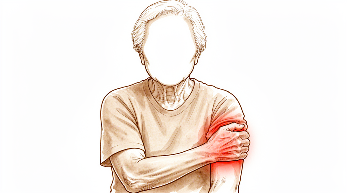
Qué está sintiendo
La artritis de codo suele manifestarse como rigidez y dolor en los extremos mismos del movimiento. Es posible que note que no puede estirar el brazo por completo o que no puede doblarlo del todo. El dolor tiende a localizarse en esos límites, no en la parte media del movimiento. Muchas personas también notan sensación de atrapamiento, chasquidos o bloqueo, a veces dolorosos, cuando los espolones óseos y pequeños fragmentos sueltos de cartílago o de hueso se atascan dentro de la articulación.
Las tareas cotidianas que requieren tener el brazo estirado o doblado por completo se vuelven difíciles. Alcanzar un estante alto, empujarse para levantarse de una silla o llevarse un tenedor a la boca pueden resultar incómodos. Girar el antebrazo, como al girar una llave o un destornillador, a menudo sigue siendo fácil al principio y solo se vuelve rígido más adelante. Cuánto le limite esto depende en gran medida de su trabajo y de sus exigencias diarias. El trabajo manual pesado, levantar peso o lanzar objetos someten la articulación a mucha más tensión que las tareas de oficina.
El dolor nocturno no es típico de la artritis de codo. Si el codo le despierta por la noche, eso apunta a una causa diferente y conviene que lo revisen. Un dolor que no se alivia, que empeora a lo largo de semanas o que le impide trabajar o usar el brazo debe ser evaluado por su médico de cabecera, o puede pedirnos una evaluación especializada.
Algunas personas con artritis de codo también desarrollan compresión del nervio cubital, el nervio que pasa por detrás de la parte interna del codo. Esto puede causar hormigueo o entumecimiento en los dedos anular y meñique, o debilidad en la mano. Si nota estos síntomas, menciónelos en su próxima revisión para que podamos evaluar el nervio.
El problema de fondo es el desgaste de la superficie articular, junto con la formación de hueso adicional alrededor de los bordes del codo. Ese hueso adicional, junto con una envoltura articular que se vuelve más tensa y los fragmentos sueltos, bloquea físicamente el codo en los extremos de su rango de movimiento. Curiosamente, el espacio dentro de la articulación a menudo se conserva, por lo que en una prueba de imagen la articulación puede verse mejor de lo que sugieren sus síntomas.
¿Qué está ocurriendo realmente?
Dentro de su codo, tres pequeñas articulaciones trabajan juntas. Una permite que el brazo se doble y se estire. Dos permiten que el antebrazo gire, para que pueda poner la palma de la mano hacia arriba o hacia abajo. La superficie de cada articulación está cubierta por una capa lisa y resbaladiza de cartílago, parecida al revestimiento antiadherente de una buena sartén. Cuando ese revestimiento está intacto, las superficies se deslizan unas sobre otras casi sin fricción.
En la artritis de codo, ese revestimiento se adelgaza en algunas zonas, y el cuerpo responde formando hueso adicional alrededor de los bordes de la articulación. Estas protuberancias óseas, llamadas osteofitos o espolones óseos, se acumulan en la parte delantera y trasera del codo. La envoltura articular, una funda de tejido que rodea la articulación, también se vuelve más tensa y se acorta. Pequeños trozos de cartílago o de hueso pueden desprenderse y quedar flotando dentro de la articulación como cuerpos sueltos.
En conjunto, estos cambios explican lo que usted está sintiendo. Los espolones óseos y la envoltura tensa actúan como un tope físico en los extremos del movimiento, por lo que el codo no se estira ni se dobla por completo. Los cuerpos sueltos son los que provocan el atrapamiento, los chasquidos o el bloqueo cuando se atascan entre las superficies en movimiento. Al principio, el dolor se localiza en esos límites del rango de movimiento porque es ahí donde se produce el bloqueo. A medida que se desgasta más el revestimiento de cartílago, el dolor también puede extenderse a la parte media del movimiento.
Hay dos aspectos que hacen que la artritis de codo sea algo distinta de la artritis de cadera o de rodilla. En primer lugar, el espacio dentro de la articulación a menudo se mantiene amplio, por lo que una radiografía puede verse mejor de lo que usted siente. En segundo lugar, el desgaste suele afectar más a una parte de la articulación que al resto: la zona donde el extremo superior de uno de los huesos del antebrazo se encuentra con el extremo del hueso del brazo que está por encima. Ese punto soporta mucha carga cuando usted empuja o levanta peso, y por eso el trabajo manual pesado, el levantamiento de pesas y los lanzamientos se relacionan con esta afección.
También puede aparecer tras una lesión antigua. Una fractura u otro daño en la articulación puede dejar las superficies ligeramente irregulares, de modo que después se desgastan de forma desigual.
Qué podemos hacer al respecto
El Dr. Kieran Hirpara, cirujano de extremidades superiores en el Mater Private Hospital Rockhampton, comienza con las opciones menos invasivas que se adapten a su condición. Por lo general, los pacientes son derivados a nuestra clínica por su médico de cabecera; si un fisioterapeuta le ha sugerido que nos consulte, igualmente necesitará una derivación de su médico de cabecera para poder acceder al reembolso de Medicare. En su primera visita, tomamos su historia clínica, examinamos su codo y solicitamos radiografías si son necesarias. Las radiografías muestran los espolones óseos en la parte delantera y trasera de la articulación. Una tomografía computarizada, que ofrece una imagen más detallada, puede ayudarnos a planificar la cirugía si se llega a ese punto.
Para un problema de larga evolución como este, normalmente probamos primero el tratamiento no quirúrgico. Cambiar la forma de trabajar y de levantar peso reduce la tensión sobre la articulación, y el reposo puede calmar los brotes. La terapia de la mano tiene como objetivo conservar el movimiento que usted tiene y fortalecer la zona alrededor del codo. Los comprimidos antiinflamatorios, un grupo común de medicamentos que alivian el dolor y la hinchazón, pueden ayudar con los síntomas. Damos a estas opciones un tiempo de prueba razonable antes de hablar de cirugía.
Si esas medidas no han producido una mejoría suficiente, puede valer la pena considerar la cirugía. Por lo general se ofrece cuando el dolor no se alivia, cuando la rigidez le impide realizar las tareas cotidianas, o cuando el codo se bloquea o se atrapa de forma dolorosa. La operación principal es una limpieza de la articulación, llamada desbridamiento. Consiste en eliminar los espolones óseos y los fragmentos sueltos que bloquean el movimiento y en liberar la envoltura articular tensa, conservando sus propias superficies articulares. Puede realizarse mediante pequeños cortes (cirugía artroscópica) o mediante una incisión abierta, según lo que sea necesario alcanzar. Hablaremos con usted sobre cuál es la más adecuada para su codo y decidiremos juntos el plan.
Qué esperar
La artritis de codo es una afección a largo plazo. Por lo general no desaparece por sí sola, pero a menudo sigue un patrón: periodos más tranquilos y, luego, brotes cuando se exige mucho al codo. Al principio, el dolor se localiza en los extremos del movimiento y el espacio dentro de la articulación a menudo se conserva. A medida que avanza el desgaste, el dolor puede extenderse a la parte media del movimiento y la rigidez puede ir aumentando.
Sin tratamiento, los espolones óseos y la envoltura articular tensa siguen bloqueando los extremos del movimiento, por lo que la rigidez tiende a ir aumentando con el tiempo. El tratamiento no quirúrgico sigue siendo el primer paso en el manejo inicial y puede aportar alivio en las primeras etapas. Cambiar la forma de trabajar y de levantar peso, la terapia de la mano y los comprimidos antiinflamatorios pueden calmar los síntomas lo suficiente como para que usted siga adelante. Si estas medidas dejan de funcionar, es señal de que la cirugía podría ayudar.
La cirugía para esta afección tiene como objetivo eliminar los bloqueos mecánicos, no reemplazar la articulación. Una limpieza de los espolones óseos y de los fragmentos sueltos, con liberación de la envoltura tensa, mejora el rango de movimiento y la función en la mayoría de las personas, con tasas bajas de complicaciones y de reintervención. La mejoría es real, pero no es una cura: no hace que el cartílago vuelva a crecer ni detiene el avance de la artritis. Algunas personas notan que su rango de movimiento vuelve a disminuir con los años tras una limpieza, y los cambios en la radiografía tienden a avanzar a lo largo de una década. El reemplazo completo del codo rara vez se ofrece para la artritis por desgaste y, por lo general, no se ofrece a personas menores de 65 años ni a personas físicamente activas, porque los implantes se desgastan con cargas elevadas.
Si su codo se pone caliente, rojo, hinchado y doloroso, sobre todo si tiene fiebre, acuda a urgencias ese mismo día. No necesita una derivación de su médico de cabecera. Si sus síntomas no se alivian, empeoran a lo largo de semanas, le despiertan por la noche o le impiden trabajar o usar el brazo, consulte a su médico de cabecera o pídanos una evaluación especializada.
¿Cuándo consultar a un especialista?
La mayoría de las artritis de codo avanzan lentamente, por lo que tiene tiempo para actuar. Consulte a su médico de cabecera, o pídanos una evaluación especializada, si la rigidez y el dolor no se alivian, empeoran a lo largo de semanas o le impiden trabajar o usar el brazo. También conviene que le evalúen si el codo se bloquea o se atrapa una y otra vez, porque los fragmentos sueltos dentro de la articulación pueden bloquear el movimiento. También debe mencionar el hormigueo o el entumecimiento en los dedos anular y meñique, ya que indica que el nervio cubital está siendo comprimido.
Hay un conjunto de signos que requiere atención el mismo día. Si su codo se pone caliente, rojo, hinchado y doloroso, sobre todo si tiene fiebre, acuda a urgencias. No necesita una derivación de su médico de cabecera. Una infección dentro de una articulación puede dañarla rápidamente, por lo que esto no puede esperar a una cita de rutina.
En profundidad
Advanced reading: the deeper science (optional)
Esta sección profundiza más de lo necesario para que usted tome sus propias decisiones terapéuticas. La osteoartritis del codo merece una lectura adicional, pues la intervención quirúrgica que parecería la solución definitiva —el reemplazo articular, tal como se realiza habitualmente en cadera y rodilla— se comporta de manera muy distinta en el codo; este único hecho determina todo el abanico de opciones de tratamiento.
¿Por qué el reemplazo de codo no es la opción predeterminada?
La artroplastia total de codo funciona, pero no tolera la carga de la misma manera que el reemplazo de cadera o rodilla. Al analizar los datos de 2,118 pacientes con artritis reumatoide, se observó que el reemplazo de codo seguía arrojando resultados satisfactorios; no obstante, se asoció con tasas de fallo del implante y complicaciones considerablemente más altas que en la artroplastia de cadera y rodilla [1].
Esto conlleva una restricción permanente para levantar pesos, en lugar de ser una medida preventiva únicamente durante la fase de recuperación. Dado que el codo se encuentra al final de una palanca larga, incluso pesos moderados en la mano generan fuerzas considerables sobre el implante, las cuales, con el tiempo, provocan su aflojamiento.
Por ello, la jerarquía de tratamientos para el codo es inversa a la de las extremidades inferiores: en la cadera, el reemplazo articular es el procedimiento estándar para casos avanzados de artritis; en el codo, en cambio, se reserva principalmente para pacientes mayores con menor demanda física, así como para casos de artritis inflamatoria en lugar de artritis por desgaste.
La causa es tan importante como la gravedad
Dos personas con codos igualmente desgastados pueden tener pronósticos distintos según la razón por la cual la articulación se deterioró. En un estudio con 679 pacientes, la etiología de la artritis influyó en los resultados, en particular en los modos específicos de fallo del implante; los pacientes con artritis reumatoide obtuvieron mejores resultados funcionales que aquellos sometidos a prótesis por causas postraumáticas [2].
Esto merece ser destacado, pues va en contra de lo que la mayoría esperaría: que una articulación dañada por una única lesión tenga un mejor pronóstico que una afectada por una enfermedad sistémica. La explicación radica en la demanda mecánica: la artritis postraumática suele aparecer en personas más jóvenes con estilos de vida físicamente exigentes, por lo que el implante debe soportar fuerzas para las cuales no fue diseñado.
El desbridamiento es el procedimiento que realiza la mayor parte del trabajo
En el caso de la artritis degenerativa primaria, el tratamiento principal no es el reemplazo articular, sino el desbridamiento: se eliminan los espolones óseos y los cuerpos sueltos que obstruyen el movimiento articular al final de su rango, y se libera la cápsula articular tensa, manteniendo intactas las superficies articulares.
La evidencia clínica es consistente. En 1,097 pacientes, el desbridamiento arrojó buenos resultados funcionales a mediano plazo, sin aumento alguno de complicaciones al emplear técnica artroscópica [3]. En 871 pacientes, tanto el desbridamiento osteocapsular abierto como el artroscópico mejoraron de manera fiable la flexión, la extensión y los índices funcionales, con tasas de complicaciones bajas [4]. Además, un metaanálisis realizado en 586 pacientes demostró que el desbridamiento resulta eficaz para aliviar los síntomas incapacitantes de la artrosis primaria del codo, con una tasa de complicaciones aceptable [5].
Es importante comprender qué se pretende lograr con el desbridamiento: no se reacondicionan las superficies articulares ni se detiene el proceso artrósico. Su función es eliminar los obstáculos mecánicos que aparecen al final del rango de movimiento. Por ello, resulta muy útil para aquellos pacientes cuya principal queja es la imposibilidad de estirar o doblar completamente el codo, así como el dolor intenso al alcanzar los límites de movimiento; en cambio, su efecto es menor en pacientes que experimentan dolor a lo largo de todo el rango de movilidad.
La elección entre cirugía abierta o artroscópica no es el factor decisivo
Al igual que ocurre con varias intervenciones quirúrgicas en el codo, la técnica genera más debate del que justifican las evidencias. Las revisiones mencionadas anteriormente consideraron que ambos métodos son seguros y eficaces; además, una revisión narrativa de 639 pacientes concluyó que no era posible determinar cuál procedimiento resulta superior [6].
El factor determinante en la práctica clínica es lo que se necesita abordar. La artroscopia resulta adecuada para intervenciones en los compartimentos anterior y posterior; sin embargo, en casos de codo rígido que requiera una liberación capsular extensa, o cuando la anatomía esté alterada y sea necesario visualizar y proteger algún nervio, la cirugía abierta podría ser más segura.
Referencias
[1] Chou TA, Ma H, Wang J, Tsai S, Chen C, Wu P, et al. Artroplastia total de codo en pacientes con artritis reumatoide: una revisión sistemática y metaanálisis. Bone Joint J. 2020;102-B(8):967-80. https://doi.org/10.1302/0301-620X.102B8.BJJ-2019-1465.R1
[2] Wang J, Ma H, Chou TA, Tsai S, Chen C, Wu P, et al. Resultados tras la artroplastia total de codo en casos de artritis reumatoide frente a condiciones postraumáticas: una revisión sistemática y metaanálisis. Bone Joint J. 2019;101-B(12):1489-97. https://doi.org/10.1302/0301-620X.101B12.BJJ-2019-0799.R1
[3] White CHR, Ravi V, Watson J, Badhrinarayanan S, Phadnis J. Revisión sistemática sobre el desbridamiento artroscópico frente al abierto en el codo artrítico. Arthroscopy. 2020;37(2):747-58. https://doi.org/10.1016/j.arthro.2020.09.005
[4] Guerrero EM, Bullock GS, Helmkamp JK, Madrid A, Ledbetter L, Richard MJ, et al. Impacto clínico del desbridamiento osteocapsular artroscópico frente al abierto en la artrosis primaria del codo: una revisión sistemática. J Shoulder Elbow Surg. 2020;29(4):689-98. https://doi.org/10.1016/j.jse.2019.12.003
[5] de Klerk HH, Welsink CL, Spaans AJ, Verweij LPE, van den Bekerom MPJ. Desbridamiento artroscópico y abierto en la artrosis primaria del codo: una revisión sistemática y metaanálisis. EFORT Open Rev. 2020;5(12):874-82. https://doi.org/10.1302/2058-5241.5.190095
[6] Poonit K, Zhou X, Zhao B, Sun C, Yao C, Zhang F, et al. Tratamiento de la artrosis del codo mediante desbridamiento abierto o artroscópico: una revisión narrativa. BMC Musculoskelet Disord. 2018;19(1). https://doi.org/10.1186/s12891-018-2318-x
Evidence & references
This is the clinical evidence summary written for health professionals. It is technical, and it lists the research this page was built from. You do not need to read it to understand your treatment or to make a decision about it.
Overview
Epidemiology and Demographics
- Primary osteoarthritis of the elbow is a relatively rare occurrence, seen in only 1% to 2% of patients with elbow arthrosis [3].
- Symptomatic primary osteoarthritis of the elbow affects 2% of the population [6].
- Primary osteoarthritis of the elbow is confined almost exclusively to men [3].
- Men are affected more often than women with a 4:1 ratio [6].
- The average age of presentation for primary elbow osteoarthritis is 50 years, with a range of 20 to 70 years [6].
- Hand dominance and strenuous manual labor are associated with primary osteoarthritis of the elbow [6].
- Secondary causes of elbow osteoarthritis include trauma, osteochondritis dissecans, and synovial osteochondromatosis [6].
Pathoanatomy
- Osteoarthritis of the elbow is characterized by osteophyte formation, capsular contracture, and loose bodies, often with relative preservation of the joint space [6].
- Periarticular hypertrophic osteophytes act as a mechanical block at the end ranges of flexion and extension [6].
- Advanced elbow osteoarthritis rarely presents with joint space narrowing [6].
- Osteoarthritis typically involves the radiocapitellar joint articular cartilage preferentially, with relative preservation of the ulnohumeral articular surfaces [6].
Clinical Presentation and Evaluation
- Patients typically present with loss of terminal extension and flexion and painful catching, clicking, or locking of the elbow [6].
- Pain in elbow osteoarthritis is typically noted at the end ranges of motion and not through the midrange [6].
- Night pain is not typical of elbow osteoarthritis; if present, an inflammatory cause of the arthritis should be considered [6].
- Ulnar neuropathy is present in up to 50% of patients with elbow osteoarthritis [6].
- Standard AP and lateral radiographs typically show osteophyte formation at the coronoid process (anterior and medial), coronoid fossa, radial fossa, radial head, olecranon tip, and olecranon fossa [6].
- Radiographs typically show preserved joint spaces at the ulnohumeral joint and mildly narrowed joint spaces at the radiocapitellar joint [6].
- Radiographs typically underestimate the number of loose bodies present in the elbow [6].
- CT may be useful for surgical planning, allowing a detailed assessment of osteophytes and the presence of loose bodies [6].
Treatment Principles
- Nonoperative treatment remains the first step in the early management of elbow osteoarthritis [4].
- The goal of treatment is to obtain a low level of pain with sufficient motion range to ensure good function, while preserving future surgical options and delaying elbow arthroplasty to the extent possible [1].
- The appropriate treatment for elbow arthritis depends on the etiology, severity, patient age, and functional demands [8].
- Treatment of elbow arthritis must be individualized based on etiology, severity, patient age, and functional demands [14].
- Nonsurgical management may provide relief in early stages of elbow arthritis [14].
Nonoperative Management
- Rest, NSAIDs, corticosteroid injections, and activity modification are the mainstays of nonsurgical treatment for elbow osteoarthritis [6].
Surgical Indications and Options
- Surgical indications for elbow osteoarthritis include failure to respond to nonsurgical interventions, loss of motion that interferes with activities of daily living, and painful locking or catching of the elbow [6].
- Joint-sparing procedures such as débridement, excision of osteophytes, capsular release, and removal of loose bodies are preferred surgical options [6].
- Total elbow arthroplasty is rarely indicated for elbow osteoarthritis and is not indicated for patients younger than 65 years or physically active patients because of concerns about implant longevity [6].
- Surgical options must be tailored to cartilage integrity and bone structure, with total elbow arthroplasty generally avoided in young, active patients due to poor durability [35].
- Total elbow arthroplasty is a treatment option for elbows severely affected by rheumatoid arthritis, with recent reports showing decreased prevalence of complications, maintenance of excellent pain relief, and functional restoration [32].
- Total elbow arthroplasty remains associated with substantial complication and reoperation rates [43].
Arthroscopic Procedures
- Elbow arthroscopic osteocapsular arthroplasty is a safe, efficacious treatment for patients with mild to moderate osteoarthritis [2].
- Arthroscopic treatment of elbow osteoarthritis provides good short-term outcomes in primary elbow osteoarthritis and is associated with a low complication rate [7].
- Arthroscopic debridement for primary degenerative osteoarthritis results in statistically significant and clinically relevant improvement in elbow range of motion and clinical outcomes with low complication and reoperation rates [15].
- Arthroscopic treatment of elbow osteoarthritis significantly improved 6-month clinical results for functional scores, pain, strength, and range of motion [30].
- Arthroscopic osteocapsular arthroplasty can be recommended for its favorable overall treatment outcomes for elbow osteoarthritis [29].
- Arthroscopic osteocapsular arthroplasty is comparable to the open procedure in managing primary osteoarthritis of the elbow [73].
- The open procedure shows better outcomes than arthroscopic osteocapsular arthroplasty in the improvement of flexion limitation [73].
- Contraindications for arthroscopic elbow procedures include severe contracture and periarticular heterotopic ossification [6].
- Relative contraindications for arthroscopic elbow procedures include prior ulnar nerve transposition and prior extensive open procedures [6].
- Osteocapsular arthroplasty refers to the arthroscopic technique for elbow joint débridement, involving capsular release, loose body removal, and excision of osteophytes [6].
- Miniopen ulnar nerve decompression is a safe and effective procedure that can be indicated for ulnar neuropathy in the setting of elbow osteoarthritis [117].
Open Procedures
- The Outerbridge-Kashiwagi arthroplasty is the classic open procedure, in which the olecranon fossa is trephinated and osteophytes are removed [6].
- Limitations of the Outerbridge-Kashiwagi procedure include incomplete anterior release and incomplete osteophyte removal anteriorly [6].
- The Outerbridge-Kashiwagi procedure is an effective and safe way of treating both posttraumatic arthritis and osteoarthritis of the elbow [13].
- Both open elbow debridement and the Outerbridge-Kashiwagi procedure had excellent survivorship until conversion to total elbow arthroplasty and are viable options in the treatment of primary elbow osteoarthritis and post-traumatic cases [22].
- A medial or lateral column approach can be used for open débridement, loose body removal, osteophyte resection, and capsulectomy depending upon the location of the pathology and concomitant procedures to be performed [6].
- Capsulectomy and debridement for primary osteoarthritis of the elbow through a medial trans-flexor approach is associated with a low rate of complications and is safe and effective [5].
Outcomes and Survivorship
- Patients with either posttraumatic or primary degenerative osteoarthritis can expect satisfactory elbow function and an improvement in pain with little chance of reoperation at the midterm of the follow-up duration [11].
- Open and arthroscopic debridement procedures seem to be safe and effective in the treatment of elbow osteoarthritis [61].
Complications
- Deep infections in the elbow are more common than other joints treated arthroscopically, with a rate of 0.8% to 2.2% [6].
- Transient nerve palsies complicate 1% to 3% of cases, with radial and ulnar nerves being the most common [6].
- Other complications of elbow osteoarthritis surgery include stiffness (heterotopic ossification), hematoma formation, and synovial ganglion formation [6].
Anatomy & Pathophysiology
Epidemiology & Demographics
- Primary osteoarthritis most commonly affects middle-aged males (approximately 50 years old), manual laborers, throwing athletes, and heavy weight lifters [70].
Etiology & Secondary Causes
- Posttraumatic arthritis is commonly seen after elbow injuries such as distal humerus fractures, radial head fractures, proximal ulna fractures, or elbow fracture-dislocations [12].
- Arthritis can develop in response to the initial cartilage insult or secondary to residual articular incongruities from injuries [12].
- Postinjury malunions, nonunions, or residual instability can alter kinematics and load across the elbow, predisposing the elbow to degenerative changes [12].
- Posttraumatic arthritis of the elbow is particularly challenging to treat due to associated bone loss and its frequent occurrence in younger populations with higher functional demands [12].
- Unlike primary osteoarthritis, rheumatoid arthritis frequently involves the elbow joint, can be bilateral, and often involves other joints [70].
- Rheumatoid arthritis causes an intense inflammatory synovitis that leads to progressive joint destruction, involving the ulnohumeral joint early in the disease process [70].
Pathoanatomy & Wear Patterns
- Rarely, advanced disease presents with joint space narrowing [6].
- Cadaveric studies have demonstrated that radiocapitellar degeneration is more common with increasing age [18].
- The posteromedial aspect of the radial head appears to be consistently involved with reciprocal changes seen on the capitellum in primary elbow osteoarthritis [18].
- Osteophytic change occurs predominantly in the ulnohumeral compartment of the elbow, whereas joint space narrowing more frequently affects the radiocapitellar articulation [53].
- Ulnohumeral osteophytes were found in 95% of patients with symptomatic osteoarthritis, while radiohumeral osteophytes were found in 59% [78].
- The primary pathology of elbow osteoarthritis is loss of articular cartilage with resulting osteophyte formation on the olecranon process, coronoid process, and their respective fossae [70].
- Secondary resultant changes in elbow osteoarthritis involve osteophyte formation along the margin of the radial head and formation of loose bodies that may result in mechanical block or crepitance [70].
- Three-dimensional computational models identified unique regions of bony impingement in elbow osteoarthritis, such as between the radial head and a posterior capitellar osteophyte in extension [54].
Clinical Presentation
- Pain is typically noted at the end ranges of motion and not through the midrange [6].
- Night pain is not typical; if present, an inflammatory cause of the arthritis should be considered [6].
- Forearm rotation is relatively preserved until later in the disease process [6].
- Most patients initially complain of pain at terminal limits of motion as a result of capsular stretch and osteophyte impingement at the extremes of motion [70].
- Later in the disease process, pain through the mid arc of motion develops as the extent of cartilage loss progresses [70].
Imaging Findings
- Radiographs typically show osteophyte formation at the coronoid process (anterior and medial), coronoid fossa, radial fossa, radial head, olecranon tip, and olecranon fossa [6].
- Joint spaces at the ulnohumeral joint are usually preserved, and those at the radiocapitellar joint are mildly narrowed [6].
- Loose bodies may be evident on radiographs, which typically underestimate the number present [6].
- The ulnohumeral articular joint space is preferentially preserved with osteoarthritis [71].
- Osteophytes are typically seen on the coronoid and olecranon tips, with loss of the concavity of the radial head, coronoid, and olecranon fossa [71].
- Ulnohumeral joint space loss suggests inflammatory or posttraumatic arthritis rather than primary osteoarthritis [71].
Classification
Radiographic Classification Systems
- The Broberg and Morrey classification system is based on osteophyte formation and joint space narrowing [18].
- The Hasting and Retting classification system focuses on radiocapitellar wear and subluxation [18].
- The Broberg and Morrey and Hasting and Retting classification systems are both X-ray based [18].
- Kwak et al. described a CT-based method of quantifying elbow arthritis [18].
- The CT-based classification demonstrated high correlation with the visual analog scale and the Mayo Elbow Performance Score [18].
- The CT-based classification demonstrated moderate correlation with range of motion [18].
- A new radiographic classification system was developed based on the lack of radiographic evidence for morphologic predisposition in primary elbow osteoarthritis [27].
- The extent of degenerative joint disease of the radiocapitellar joint had a negative effect on postoperative outcome [27].
- The developed radiographic classification system appears valid in predicting postoperative outcome [27].
- A bony landmarks classification system effectively delineated osteophyte distribution in elbow patients [58].
Reliability and Validity
- The Broberg and Morrey and Hasting and Retting classification systems demonstrated substantial intraobserver and interobserver reliability for evaluating radiographic severity of post-traumatic arthritis and primary osteoarthritis of the elbow [39].
- The CT-based staging system was highly reproducible and clinically feasible compared with previous plain radiograph-based staging systems for elbow osteoarthritis [28].
- The 4 grades of the Broberg and Morrey classification system have only fair interobserver reliability [118].
- Interobserver reliability for the Broberg and Morrey classification system is influenced by subspecialty and experience [118].
- Surgeons with more than 10 years of experience had greater agreement in arthrosis ratings than surgeons with less experience [118].
- Surgeons who treated more than 10 elbow fractures per year had better agreement in arthrosis ratings than those treating fewer fractures [118].
- Simplified binary rating systems for elbow arthrosis resulted in moderate agreement among observers [118].
- Interobserver agreement for radiographic assessment and classification in a cohort study of arthroscopic management yielded k values ranging from 0.81 to 0.85 [44].
Clinical Presentation
Epidemiology and Demographics
- The prevalence of primary elbow osteoarthritis in Japanese subjects aged 50-89 years was 25.2%, with most cases being asymptomatic [17].
- The prevalence of elbow osteoarthritis was 55.0% in respondents aged 40 years or older, with a symptomatic prevalence of 22.6% [25].
- Men are affected more often than women with primary elbow osteoarthritis at a 4:1 ratio [6].
- Older age, male sex, and a history of elbow trauma are identified as significant risk factors for elbow osteoarthritis [25].
Pathoanatomy
- Advanced disease rarely presents with joint space narrowing [6].
- Primary osteoarthritis of the elbow is unique due to relative preservation of articular cartilage and maintenance of joint space with hypertrophic osteophyte formation [21].
- Radiocapitellar degeneration is more common with increasing age [18].
- The posteromedial aspect of the radial head appears to be consistently involved in primary elbow osteoarthritis, with reciprocal changes seen on the capitellum [18].
History and Symptoms
- Night pain is not typical for elbow osteoarthritis; if present, an inflammatory cause of the arthritis should be considered [6].
- Most patients initially complain of pain at terminal limits of motion as a result of capsular stretch and osteophyte impingement at the extremes of motion, with or without mechanical symptoms [70].
- The degree of disability caused by osteoarthritis depends on the patient’s vocation and physical disability [6].
Physical Examination
- Inspection should check for prior surgical incisions and joint effusion at the lateral soft spot [6].
- Pain is usually felt at the end ranges of flexion and extension rather than throughout the arc during range of motion assessment [6].
Imaging
- Standard AP and lateral radiographs should be obtained for evaluation of elbow osteoarthritis [6].
- Joint spaces at the ulnohumeral joint are usually preserved on radiographs [6].
- Joint spaces at the radiocapitellar joint are mildly narrowed on radiographs [6].
- CT may be useful for surgical planning and allows a detailed assessment of osteophytes and the presence of loose bodies [6].
- CT scans with 3D reconstructions may be useful for evaluating the extent and location of disease and for surgical planning [101].
Investigations
History and Physical Examination
- Patients with elbow osteoarthritis typically present with loss of terminal extension and flexion, along with painful catching, clicking, or locking of the elbow [6].
- Pain in elbow osteoarthritis is typically noted at the end ranges of motion rather than through the midrange [6].
- The presence of night pain in a patient with elbow arthritis suggests an inflammatory cause rather than typical osteoarthritis [6].
- Physical examination for elbow osteoarthritis includes inspection for prior surgical incisions and joint effusion at the lateral soft spot [6].
- Forearm rotation is relatively preserved until later in the disease process of elbow osteoarthritis [6].
- The degree of disability caused by elbow osteoarthritis depends on the patient’s vocation and physical disability [6].
Imaging
- Standard AP and lateral radiographs are the initial imaging modality for evaluating elbow osteoarthritis [6].
- Radiographs of the elbow in osteoarthritis typically show osteophyte formation at the coronoid process, coronoid fossa, radial fossa, radial head, olecranon tip, and olecranon fossa [6].
- In elbow osteoarthritis, joint spaces at the ulnohumeral joint are usually preserved, while those at the radiocapitellar joint are mildly narrowed [6].
- CT imaging may be useful for surgical planning by allowing a detailed assessment of osteophytes and the presence of loose bodies [6].
- CT has greater sensitivity than radiographs for the detection of osteophytes and loose bodies in primary elbow osteoarthritis [139].
- A CT-based staging system for elbow osteoarthritis is highly reproducible and clinically feasible compared with plain radiograph-based staging systems [28].
- On imaging assessment for elbow arthritis, signs of impingement such as osteophytes and filling of fossae are more common than cartilage lesions like joint narrowing [145].
- Three-dimensional computational models can identify the locations and volumes of bony impingement in patients with elbow osteoarthritis [54].
- Pre-operative simulation results can be used as an index to determine which osteophytes to remove during arthroscopic surgery for elbow osteoarthritis [50].
- A bony landmarks classification system effectively delineates osteophyte distribution in patients with elbow osteoarthritis [58].
- The Broberg and Morrey classification for elbow arthritis is based on osteophyte formation and joint space narrowing [18].
- The Hasting and Retting classification system for elbow arthritis focuses on radiocapitellar wear and subluxation [18].
- Both the Broberg and Morrey and Hasting and Retting classification systems demonstrated substantial intraobserver and interobserver reliability for evaluating radiographic severity of elbow arthritis [39].
- A CT-based method of quantifying elbow arthritis described by Kwak et al. demonstrates high correlation with visual analog scale and the Mayo Elbow Performance Score, and moderate correlation with range of motion [18].
Treatment
Nonoperative Management
- Nonsurgical treatments can be effective for reducing symptoms but have limited effectiveness for improving range of motion limitations and pain related to impinging osteophytes [66].
Surgical Indications
- Surgical treatment for elbow arthritis is based on disease etiology, severity of degeneration, and patient age [10].
- Failure to respond to nonsurgical interventions is an indication for surgical treatment of elbow osteoarthritis [6].
- Loss of motion that interferes with activities of daily living is an indication for surgical treatment of elbow osteoarthritis [6].
- Painful locking or catching of the elbow is an indication for surgical treatment of elbow osteoarthritis [6].
- The goal of treatment for post-traumatic osteoarthritis of the elbow is to obtain a low level of pain with sufficient motion range to ensure good function, while preserving future surgical options and delaying elbow arthroplasty to the extent possible [1].
Joint-Sparing Procedures
- Joint-sparing procedures such as débridement, excision of osteophytes, capsular release, and removal of loose bodies are preferred for elbow osteoarthritis [6].
- Osteocapsular debridement is an effective surgical treatment option for patients with symptomatic primary elbow osteoarthritis who have failed conservative management [46].
- Surgical debridement is an effective treatment for the disabling symptoms of primary elbow OA with an acceptable complication rate [63].
- Elbow arthroscopic debridement for primary degenerative osteoarthritis results in statistically significant and clinically relevant improvement in elbow range of motion and clinical outcomes with low complication and reoperation rates [15].
- Arthroscopic debridement for elbow osteoarthritis provides satisfactory pain relief, improvement of elbow motion, and good functional outcome [64].
- Arthroscopic debridement for the elbow osteoarthritis provided satisfactory pain relief, improvement of elbow motion, and good functional outcome [68].
- Elbow AOA is a safe, efficacious treatment for patients with mild to moderate osteoarthritis [2].
- This minimally invasive technique provides good short-term outcomes in primary elbow osteoarthritis and is associated with a low complication rate [7].
- The study recommends arthroscopic debridement in the surgical management of patients with osteoarthritis of the elbow [9].
- This procedure is a valuable adjunct in the management of the arthritic elbow, serving as an intermediate step between nonoperative management and elbow replacement surgery [36].
- Both open elbow debridement and the OK procedure had excellent survivorship until conversion to total elbow arthroplasty and are viable options in the treatment of primary elbow osteoarthritis and post-traumatic cases [22].
- The OK procedure is an effective and safe way of treating both posttraumatic arthritis and osteoarthritis of the elbow [13].
- The modified Outerbridge-Kashiwagi procedure combining mini-open and arthroscopic techniques achieved short-term clinical success in pain relief and function restoration in patients with elbow osteoarthritis [105].
- Most patients had an uneventful postoperative course and have shown a painless elbow joint, with satisfactory functional recovery at short-term follow-up after open debridement and radiocapitellar replacement [34].
- The medial approach is effective for the treatment of advanced primary osteoarthritis of the elbow, especially in patients with ulnar nerve symptoms as well as medial osteophytes [19].
- Arthroscopic osteocapsular arthroplasty is comparable to the OPEN procedure in managing primary osteoarthritis of the elbow; however, the OPEN procedure shows the better outcome in improvement of flexion limitation [73].
- Open and arthroscopic debridement procedures seem to be safe and effective in the treatment of elbow OA [61].
- In subjects with slight or moderate OA and preserved elbow joint congruence, arthroscopy provides good-to-satisfactory results [44].
- The extent of degenerative joint disease of the radiocapitellar joint had a negative effect on postoperative outcome, and the developed classification system appears valid in predicting postoperative outcome [27].
- A reproducible triceps tendon-guided arthroscopic olecranon debridement technique can restore adequate joint space, mobility, and function to the patient with degenerative elbow osteoarthritis [23].
Arthroplasty and Salvage Procedures
- Total elbow arthroplasty is rarely indicated for elbow osteoarthritis, and it is not indicated for patients younger than 65 years or physically active patients because of concerns about implant longevity [6].
- Total elbow arthroplasty is best reserved for low demand, elderly patients who will be able to comply with the 5-lb weightlifting restriction imposed postoperatively to protect the implants from bearing wear, hardware loosening, or failure [100].
- Open or arthroscopic débridement may be effective in the treatment of early arthritis, whereas interposition arthroplasty or total elbow arthroplasty is best reserved for more advanced cases [100].
- Total elbow arthroplasty has become a reliable treatment option for elbows severely affected by rheumatoid arthritis, with recent reports showing decreased prevalence of complications, maintenance of excellent pain relief, and functional restoration [32].
- The study observed favorable midterm results with the capitellocondylar total elbow replacement, with good pain relief and improved function of the elbow [52].
- Although total elbow arthroplasty has become an accepted treatment for a variety of degenerative and traumatic elbow conditions, concerns regarding its durability in young and/or high-demand patients make elbow arthrodesis an acceptable alternative in this population [98].
- Elbow arthrodesis is reserved for patients with painful arthritis who are not candidates for total elbow arthroplasty, especially individuals who place high demands on the upper extremities, such as manual laborers [98].
- For unilateral arthrodesis of the elbow, a position of 90 to 100 degrees of flexion is desirable to provide the most powerful grip strength [98].
- Bilateral elbow arthrodesis rarely is indicated because of resultant functional limitations [98].
- If bilateral elbow arthrodesis is indicated, one elbow should be placed in 110 to 120 degrees of flexion to permit the patient to reach the mouth, and the other should be placed in 45 to 65 degrees to aid in personal hygiene [98].
- Interposition arthroplasty is considered for intrinsic contractures in young patients (20 to 50 years) with articular cartilage destruction in whom the anatomic architecture of the distal humerus and proximal ulna are relatively preserved [69].
Procedural Considerations and Contraindications
- Contraindications for arthroscopic procedures for elbow osteoarthritis include severe contracture and periarticular heterotopic ossification [6].
- Relative contraindications for arthroscopic procedures for elbow osteoarthritis include prior ulnar nerve transposition and prior extensive open procedures [6].
- Regardless of the type of procedure used, ulnar nerve transposition and release of the posterior bundle of the medial collateral ligament (MCL) should be considered for patients who have less than 90° to 100° of elbow flexion [6].
- Ulnar nerve decompression/transposition and release of the posterior bundle of the MCL should be considered for patients who have less than 90° to 100° of elbow flexion [66].
- During arthroscopic surgery, joint distention moves the capsule away from bone, but the distance between the neurovascular structures and the capsule remain unchanged; therefore, the nerves remain at risk with capsular work [6].
- Neurovascular structures at risk during portal placement, débridement, and capsular release include median nerve (anteromedial), the ulnar nerve (posteromedial), and the radial nerve (lies adjacent to the anterolateral capsule) [6].
- The brachialis muscle protects the median nerve and brachial artery during capsular procedures [6].
- The olecranon osteophytosis extends medially and laterally and not just at the tip, requiring resection to be extended along the medial and lateral aspects of the olecranon to allow maximal extension and prevent impingement [6].
- Coronoid osteophytosis extends medially and not just at the tip, requiring resection to be extended medially if necessary to maximize the restoration of flexion range of motion [6].
- Elbow arthroscopy is technically demanding, and several neurovascular structures that are at risk during the procedure include the radial, ulnar, and median nerves [66].
- Unlike other joints, arthroscopic débridement in the elbow can be very effective at reducing symptoms and improving range of motion [66].
Complications
- Deep infections in the elbow are more common than in other joints treated arthroscopically, with a rate of 0.8% to 2.2% [6].
- Infection can manifest as superficial minor wound complications or deep infection [6].
- Infection is associated with intraoperative corticosteroid injections [6].
- Transient nerve palsies complicate 1% to 3% of cases [6].
- Radial and ulnar nerves are the most commonly affected in transient nerve palsies [6].
- Heterotopic ossification is a complication associated with stiffness [6].
- Hematoma formation is a recognized complication [6].
- Synovial ganglion formation is a recognized complication [6].
- During arthroscopic surgery, joint distention moves the capsule away from bone, but the distance between neurovascular structures and the capsule remains unchanged, keeping nerves at risk during capsular work [6].
- The median nerve is at risk during portal placement, débridement, and capsular release due to its anteromedial location [6].
- The ulnar nerve is at risk during portal placement, débridement, and capsular release due to its posteromedial location [6].
- The radial nerve is at risk during portal placement, débridement, and capsular release as it lies adjacent to the anterolateral capsule [6].
- Total elbow arthroplasty is associated with substantial complication and reoperation rates [43].
- Total elbow replacement is associated with a high complication rate and may be warranted only for seriously disabled patients [113].
- Total elbow replacement for patients with rheumatoid arthritis has no more perioperative complications than for those who do not have rheumatoid arthritis [123].
Recovery
Nonoperative Management
- Rest, NSAIDs, corticosteroid injections, and activity modification are the mainstays of nonsurgical treatment [6].
Surgical Indications and Goals
- Surgical indications include failure to respond to nonsurgical interventions, loss of motion that interferes with activities of daily living, and painful locking or catching of the elbow [6].
Arthroscopic Outcomes
- Patients with primary or posttraumatic elbow osteoarthritis who underwent arthroscopic debridement can expect satisfactory elbow function and an improvement in pain with little chance of reoperation at the midterm of the follow-up duration [11].
- Clinical outcomes for patients with primary elbow osteoarthritis who underwent arthroscopic osteocapsular arthroplasty improved from preoperative assessment to short- and medium-term follow-up [31].
- Range of motion decreased between short- and medium-term follow-up after arthroscopic osteocapsular arthroplasty for primary elbow osteoarthritis [31].
Open Surgical Outcomes
- Most patients undergoing open debridement and radiocapitellar replacement for primary or post-traumatic arthritis of the elbow had an uneventful postoperative course and showed a painless elbow joint with satisfactory functional recovery at short-term follow-up [34].
- A tendency of advancing roentgenological osteoarthritic changes was recognized in most patients 10 years after the Outerbridge-Kashiwagi method [80].
Total Elbow Arthroplasty Outcomes
- Total elbow arthroplasty is rarely indicated and is not indicated for patients younger than 65 years or physically active patients because of concerns about implant longevity [6].
- The main objective of elbow joint replacement in arthritic diseases is to decrease pain, increase joint stability, and improve overall range of motion [51].
- In most cases, elbow function was maintained in the long-term without loosening of the implant following Kudo type-5 total elbow arthroplasty for rheumatoid arthritis [20].
- The capitellocondylar total elbow replacement provided significant long-term relief of pain and improvement in functional status and range of motion in patients with rheumatoid arthritis, with improvements not deteriorating over time [120].
- The mean Mayo elbow performance score significantly improved from 48 points preoperatively to 83 points at final follow-up for GSB III total elbow arthroplasty in patients with rheumatoid arthritis [75].
- In the course of total elbow arthroplasty, satisfactory supportive range of motion was restored to patients' elbow joints [76].
- Long-term follow-up showed acceptable results of the type-3 Kudo total elbow arthroplasty in patients with rheumatoid arthritis [72].
Complications
Key Evidence
- [L4] The goal of treatment is to obtain a low level of pain with sufficient motion range to ensure good function, while preserving future surgical options and delaying elbow arthroplasty to the extent possible. [1] (10.1016/j.otsr.2013.11.004)
- [L4] Elbow AOA is a safe, efficacious treatment for patients with mild to moderate osteoarthritis. [2] (10.1016/j.jhsa.2015.11.018)
- [L5] Primary osteoarthritis of the elbow is a relatively rare occurrence, seen in only 1% to 2% of patients with elbow arthrosis, and is confined almost exclusively to men. [3] (10.1016/s0894-1130(00)80037-x)
- [L5] Nonoperative treatment remains the first step in the early management of elbow osteoarthritis. [4] (10.2106/jbjs.e.00568)
- [L4] This approach is associated with a low rate of complications and is safe and effective for the treatment of primary osteoarthritis of the elbow. [5] (10.1016/j.jhsa.2011.07.018)
- [L4] This minimally invasive technique provides good short-term outcomes in primary elbow osteoarthritis and is associated with a low complication rate. [7] (10.1016/j.otsr.2019.09.003)
- [L5] The appropriate treatment for elbow arthritis depends on the etiology, severity, patient age, and functional demands. [8] (10.1016/j.jhsa.2009.02.019)
- [L4] The study recommends this technique in the surgical management of patients with osteoarthritis of the elbow. [9] (10.1302/0301-620x.96b2.30714)
- [L5] Surgical treatment for elbow arthritis is based on disease etiology, severity of degeneration, and patient age. [10] (10.1016/j.jhsa.2007.12.022)
- [L3] Patients with either pathology can expect satisfactory elbow function and an improvement in pain with little chance of reoperation at the midterm of the follow-up duration. [11] (10.1016/j.jseint.2021.07.018)
- [L5] [12] (10.1016/j.jhsa.2022.12.014)
- [L4] The OK procedure is an effective and safe way of treating both posttraumatic arthritis and osteoarthritis of the elbow. [13] (10.1016/j.jse.2015.11.052)
- [L5] Treatment of elbow arthritis must be individualized based on etiology, severity, patient age, and functional demands; nonsurgical management may provide relief in early stages, while surgical options range from arthroscopic debridement for pain at motion extremes to total elbow arthroplasty for pain throughout the arc of motion. [14] (10.1016/j.jhsa.2012.12.037)
- [L1] Elbow arthroscopic debridement for primary degenerative osteoarthritis results in statistically significant and clinically relevant improvement in elbow range of motion and clinical outcomes with low complication and reoperation rates. [15] (10.1016/j.arthro.2017.08.247)
- [L3] The prevalence of primary elbow osteoarthritis in Japanese subjects aged 50-89 years was 25.2%, with most cases being asymptomatic. [17] (10.1016/j.jse.2021.07.015)
- [L3] [18] (10.1177/17585732251327183)
- [L4] The medial approach is effective for the treatment of advanced primary osteoarthritis of the elbow, especially in patients with ulnar nerve symptoms as well as medial osteophytes. [19] (10.2106/jbjs.d.02684)
- [L3] In most cases, elbow function was maintained in the long-term without loosening of the implant. [20] (10.1302/0301-620x.99b6.bjj-2016-1033.r2)
- [L4] Primary osteoarthritis of the elbow is unique due to relative preservation of articular cartilage and maintenance of joint space with hypertrophic osteophyte formation. [21] (10.5435/00124635-200802000-00005)
- [L4] Both open elbow debridement and the OK procedure had excellent survivorship until conversion to total elbow arthroplasty and are viable options in the treatment of primary elbow osteoarthritis and post-traumatic cases. [22] (10.1016/j.jse.2022.01.138)
- [L5] In doing so, we present a reproducible technique that can restore adequate joint space, mobility, and function to the patient with degenerative elbow osteoarthritis. [23] (10.1016/j.eats.2024.103332)
- [L3] The prevalence of elbow OA was 55.0% in respondents aged 40 years or older, with a symptomatic prevalence of 22.6%; older age, male sex, and a history of elbow trauma were identified as significant risk factors. [25] (10.1016/j.jse.2018.02.049)
- [L4] The extent of degenerative joint disease of the radiocapitellar joint had a negative effect on postoperative outcome, and the developed classification system appears valid in predicting postoperative outcome. [27] (10.1016/j.jse.2007.03.014)
- [L4] CT-based staging system was highly reproducible and clinically feasible, compared with previous plain radiograph-based staging systems, for elbow osteoarthritis. [28] (10.1016/j.joca.2019.03.004)
- [L4] Arthroscopic osteocapsular arthroplasty can be recommended for its favorable overall treatment outcomes for elbow osteoarthritis. [29] (10.1016/j.jse.2019.09.036)
- [L3] Arthroscopic treatment of elbow osteoarthritis significantly improved 6-month clinical results for functional scores, pain, strength and range of motion. [30] (10.1016/j.otsr.2019.09.002)
- [L4] Serial assessment of patients with primary elbow OA who underwent arthroscopic OCA showed that the clinical outcomes improved from preoperative assessment to short- and medium-term follow-up, although ROM decreased between short- and medium-term follow-up. [31] (10.1177/23259671231162398)
- [L4] Total elbow arthroplasty has become a reliable treatment option for elbows severely affected by rheumatoid arthritis, with recent reports showing decreased prevalence of complications, maintenance of excellent pain relief, and functional restoration. [32] (10.2106/00004623-199805000-00008)
- [L4] Most patients had an uneventful postoperative course and have shown a painless elbow joint, with satisfactory functional recovery at short-term follow-up. [34] (10.1016/j.jse.2011.08.071)
- [L4] Surgical options must be tailored to cartilage integrity and bone structure, with total elbow arthroplasty generally avoided in young, active patients due to poor durability. [35] (10.1016/j.jhsg.2025.100736)
- [L4] This procedure is a valuable adjunct in the management of the arthritic elbow, serving as an intermediate step between nonoperative management and elbow replacement surgery. [36] (10.1016/s1058-2746(99)90131-3)
- [L3] Both the BM and HR classification systems demonstrated substantial intraobserver and interobserver reliability for evaluating radiographic severity of post-traumatic arthritis and primary osteoarthritis of the elbow. [39] (10.1016/j.jse.2014.10.015)
- [L4] Total elbow arthroplasty remains associated with substantial complication and reoperation rates. [43] (10.1016/j.jhsg.2026.100981)
- [L4] [44] (10.1007/s12306-015-0365-0)
- [L2] Osteocapsular debridement is an effective surgical treatment option for patients with symptomatic primary elbow osteoarthritis who have failed conservative management. [46] (10.1016/j.jse.2020.01.060)
- [L4] These results can be used as an index to determine the osteophytes to be removed during arthroscopic surgery for elbow osteoarthritis. [50] (10.1016/j.jseint.2026.101667)
- [L4] The main objective of elbow joint replacement in arthritic diseases is to decrease pain, increase joint stability and improve overall range of motion. [51] (10.1007/s12306-010-0070-y)
- [L4] The study observed favorable midterm results with the capitellocondylar total elbow replacement, with good pain relief and improved function of the elbow. [52] (10.1016/j.jse.2004.10.010)
- [L3] Osteophytic change occurs predominantly in the ulnohumeral compartment of the elbow, whereas joint space narrowing more frequently affects the radiocapitellar articulation. [53] (10.1016/j.jse.2006.08.005)
- [L4] Three-dimensional computational models identified the locations and volumes of bony impingement in patients with osteoarthritis of the elbow and highlighted unique regions of impingement, such as between the radial head and a posterior capitellar osteophyte in extension. [54] (10.1016/j.jhsa.2013.03.035)
- [L3] The bony landmarks classification system effectively delineated osteophyte distribution in elbow patients. [58] (10.1186/s13018-025-06145-9)
- [L5] However, from the data we obtained the open and arthroscopic debridement procedures seem to be safe and effective in the treatment of elbow OA. [61] (10.1186/s12891-018-2318-x)
- [L1] Surgical debridement is an effective treatment for the disabling symptoms of primary elbow OA with an acceptable complication rate. [63] (10.1302/2058-5241.5.190095)
- [L4] Arthroscopic debridement for elbow osteoarthritis provides satisfactory pain relief, improvement of elbow motion, and good functional outcome. [64] (10.1016/j.jse.2014.01.009)
- [L4] Arthroscopic debridement for the elbow osteoarthritis provided satisfactory pain relief, improvement of elbow motion, and good functional outcome. [68] (10.1016/s0363-5023(11)60056-7)
- [L3] This long-term follow-up study showed acceptable results of the type-3 Kudo total elbow arthroplasty in patients with rheumatoid arthritis. [72] (10.2106/00004623-200110000-00008)
- [L3] Arthroscopic osteocapsular arthroplasty is comparable to the OPEN procedure in managing primary osteoarthritis of the elbow; however, the OPEN procedure shows the better outcome in improvement of flexion limitation. [73] (10.1016/j.arthro.2018.11.057)
- [L4] The mean Mayo elbow performance score significantly improved from 48 points preoperatively to 83 points at final follow-up. [75] (10.1007/s10165-011-0509-5)
- [L4] In the course of total elbow arthroplasty, satisfactory supportive range of motion was restored to patients' elbow joints. [76] (10.1136/annrheumdis-2014-eular.2916)
- [L4] [78] (10.1016/j.jse.2008.03.012)
- [L4] However, a tendency of advancing roentgenological osteoarthritic changes was recognized in most patients 10 years after OKM. [80] (10.1007/bf01234111)
- [L4] The modified Outerbridge-Kashiwagi procedure combining mini-open and arthroscopic techniques achieved short-term clinical success in pain relief and function restoration in patients with elbow osteoarthritis. [105] (10.1016/j.jse.2020.05.007)
- [L3] Total elbow replacement is associated with a high complication rate and therefore may be warranted only for seriously disabled patients. [113] (10.2106/jbjs.d.02734)
- [L4] Miniopen ulnar nerve decompression was safe and effective procedure and can be indicated for ulnar neuropathy in the setting of elbow osteoarthritis. [117] (10.1016/j.arthro.2020.12.055)
- [L3] [118] (10.1016/j.jhsa.2011.12.043)
- [L3] The capitellocondylar total elbow replacement provided significant long-term relief of pain and improvement in functional status and range of motion in patients with rheumatoid arthritis, with improvements not deteriorating over time. [120] (10.2106/00004623-199304000-00004)
- [L3] Total elbow replacement for patients with RA has no more complications perioperatively than for those who do not have RA. [123] (10.1016/j.jse.2008.06.012)
- [L1] CT has greater sensitivity than radiographs for the detection of osteophytes and loose bodies in primary elbow osteoarthritis. [139] (10.1016/j.jse.2021.04.001)
- [L3] On the imaging assessment, signs of impingement (osteophytes and filling of fossae) are more common than cartilage lesions (joint narrowing). [145] (10.1016/j.otsr.2019.09.012)
References
[1] Post-traumatic osteoarthritis of the elbow. Orthopaedics & Traumatology: Surgery & Research. 2014. DOI: 10.1016/j.otsr.2013.11.004
[2] Outcomes of Elbow Arthroscopic Osteocapsular Arthroplasty. The Journal of Hand Surgery. 2016. DOI: 10.1016/j.jhsa.2015.11.018
[3] Osteoarthritis and traumatic arthritis of the elbow. Journal of Hand Therapy. 2000. DOI: 10.1016/s0894-1130(00)80037-x
[4] Management of Elbow Osteoarthritis. The Journal of Bone & Joint Surgery. 2006. DOI: 10.2106/jbjs.e.00568
[5] Capsulectomy and Debridement for Primary Osteoarthritis of the Elbow Through a Medial Trans-Flexor Approach. The Journal of Hand Surgery. 2011. DOI: 10.1016/j.jhsa.2011.07.018
[6] Aaos Comprehensive Orthopaedic Review 3. Arthritis and Arthroplasty of the Elbow > I. Osteoarthritis.
[7] Arthroscopic treatment of elbow osteoarthritis. Orthopaedics & Traumatology: Surgery & Research. 2019. DOI: 10.1016/j.otsr.2019.09.003
[8] Elbow Arthritis: Current Concepts. The Journal of Hand Surgery. 2009. DOI: 10.1016/j.jhsa.2009.02.019
[9] Arthroscopic debridement in the treatment of patients with osteoarthritis of the elbow, based on computer simulation. The Bone & Joint Journal. 2014. DOI: 10.1302/0301-620x.96b2.30714
[10] Surgical Options for the Arthritic Elbow. The Journal of Hand Surgery. 2008. DOI: 10.1016/j.jhsa.2007.12.022
[11] Midterm outcomes and survivorship of arthroscopic elbow debridement: a comparison of posttraumatic versus primary degenerative osteoarthritis. JSES International. 2022. DOI: 10.1016/j.jseint.2021.07.018
[12] Elbow Arthritis. The Journal of Hand Surgery. 2023. DOI: 10.1016/j.jhsa.2022.12.014
[13] The outerbridge-kashiwagi procedure for primary degenerative arthritis of the elbow vs. post traumatic arthritis. Journal of Shoulder and Elbow Surgery. 2016. DOI: 10.1016/j.jse.2015.11.052
[14] Elbow Arthritis: Current Concepts. The Journal of Hand Surgery. 2013. DOI: 10.1016/j.jhsa.2012.12.037
[15] Arthroscopic Debridement for Primary Degenerative Osteoarthritis of the Elbow Leads to Significant Improvement in Range of Motion and Clinical Outcomes: A Systematic Review. Arthroscopy. 2017. DOI: 10.1016/j.arthro.2017.08.247
[17] Prevalence and associated factors of primary elbow osteoarthritis in the Japanese general elderly population: a Japanese cohort survey randomly sampled from a basic resident registry. Journal of Shoulder and Elbow Surgery. 2022. DOI: 10.1016/j.jse.2021.07.015
[18] Sex-related differences in wear patterns in primary elbow osteoarthritis. Shoulder & Elbow. 2025. DOI: 10.1177/17585732251327183
[19] Débridement Arthroplasty for Primary Osteoarthritis of the Elbow. Journal of Bone and Joint Surgery. 2005. DOI: 10.2106/jbjs.d.02684
[20] Kudo type-5 total elbow arthroplasty for patients with rheumatoid arthritis. The Bone & Joint Journal. 2017. DOI: 10.1302/0301-620x.99b6.bjj-2016-1033.r2
[21] Primary Osteoarthritis of the Elbow: Current Treatment Options. Journal of the American Academy of Orthopaedic Surgeons. 2008. DOI: 10.5435/00124635-200802000-00005
[22] Long-term survivorship of open débridement and débridement arthroplasty for elbow arthritis: a retrospective chart review. Journal of Shoulder and Elbow Surgery. 2022. DOI: 10.1016/j.jse.2022.01.138
[23] Triceps Tendon‐Guided Arthroscopic Olecranon Debridement for Posterior Impingement Caused by Degenerative Elbow Osteoarthritis: A Technical Note. Arthroscopy Techniques. 2025. DOI: 10.1016/j.eats.2024.103332
[25] The prevalence of elbow osteoarthritis in Japanese middle-aged and elderly populations: the relationship between risk factors and function. Journal of Shoulder and Elbow Surgery. 2018. DOI: 10.1016/j.jse.2018.02.049
[27] Primary osteoarthritis of the elbow: Lack of radiographic evidence for morphologic predisposition, results of operative debridement at intermediate follow-up, and basis for a new radiographic classification system. Journal of Shoulder and Elbow Surgery. 2008. DOI: 10.1016/j.jse.2007.03.014
[28] Intraobserver and interobserver reliability of the computed tomography-based radiographic classification of primary elbow osteoarthritis: comparison with plain radiograph-based classification and clinical assessment. Osteoarthritis and Cartilage. 2019. DOI: 10.1016/j.joca.2019.03.004
[29] Arthroscopic osteocapsular arthroplasty for advanced-stage primary osteoarthritis of the elbow using a computed tomography–based classification. Journal of Shoulder and Elbow Surgery. 2020. DOI: 10.1016/j.jse.2019.09.036
[30] Arthroscopic debridement for osteoarthritis of the elbow: Results and analysis of predictive factors. Orthopaedics & Traumatology: Surgery & Research. 2019. DOI: 10.1016/j.otsr.2019.09.002
[31] Serial Changes in Clinical Outcomes After Arthroscopic Osteocapsular Arthroplasty for Primary Elbow Osteoarthritis: A Medium-term Follow-up Study. Orthopaedic Journal of Sports Medicine. 2023. DOI: 10.1177/23259671231162398
[32] Total Elbow Arthroplasty in Patients Who Have Juvenile Rheumatoid Arthritis. The Journal of Bone & Joint Surgery*. 1998. DOI: 10.2106/00004623-199805000-00008
[34] Open debridement and radiocapitellar replacement in primary and post-traumatic arthritis of the elbow: a multicenter study. Journal of Shoulder and Elbow Surgery. 2012. DOI: 10.1016/j.jse.2011.08.071
[35] Management of Severe Elbow Arthritis in a Young Patient. Journal of Hand Surgery Global Online. 2025. DOI: 10.1016/j.jhsg.2025.100736
[36] Arthroscopic management of the arthritic elbow: Indications, technique, and results. Journal of Shoulder and Elbow Surgery. 1999. DOI: 10.1016/s1058-2746(99)90131-3
[39] Reliability testing of two classification systems for osteoarthritis and post-traumatic arthritis of the elbow. Journal of Shoulder and Elbow Surgery. 2015. DOI: 10.1016/j.jse.2014.10.015
[43] Total Elbow Arthroplasty Complication, Reoperation, and Revision Rates: A Comparison Between Arthroplasty for Arthritis Versus Fracture. Journal of Hand Surgery Global Online. 2026. DOI: 10.1016/j.jhsg.2026.100981
[44] Arthroscopic joint debridement and capsular release in primary and post-traumatic elbow osteoarthritis: a retrospective blinded cohort study with minimum 24-month follow-up. MUSCULOSKELETAL SURGERY. 2015. DOI: 10.1007/s12306-015-0365-0
[46] The Clinical Impact of Different Approaches to Osteocapsular Debridement for Primary Osteoarthritis of the Elbow: A Systematic Review. Journal of Shoulder and Elbow Surgery. 2020. DOI: 10.1016/j.jse.2020.01.060
[50] Predicting impinging osteophytes based on pre-operative simulation results to guide arthroscopic débridement for elbow osteoarthritis. JSES International. 2026. DOI: 10.1016/j.jseint.2026.101667
[51] Linked semi-constrained total elbow prosthesis in chronic arthritis: results of 18 cases. MUSCULOSKELETAL SURGERY. 2010. DOI: 10.1007/s12306-010-0070-y
[52] Capitellocondylar total elbow replacement in late-stage rheumatoid arthritis. Journal of Shoulder and Elbow Surgery. 2005. DOI: 10.1016/j.jse.2004.10.010
[53] Radiographic changes at the elbow in primary osteoarthritis: A comparison with normal aging of the elbow joint. Journal of Shoulder and Elbow Surgery. 2007. DOI: 10.1016/j.jse.2006.08.005
[54] Identifying the Location and Volume of Bony Impingement in Elbow Osteoarthritis by 3-Dimensional Computational Modeling. The Journal of Hand Surgery. 2013. DOI: 10.1016/j.jhsa.2013.03.035
[58] Bony landmarks guided mapping of the osteophytes of the elbow osteoarthritis patients: a three dimensional computed tomograph based study. Journal of Orthopaedic Surgery and Research. 2025. DOI: 10.1186/s13018-025-06145-9
[61] Treatment of osteoarthritis of the elbow with open or arthroscopic debridement: a narrative review. BMC Musculoskeletal Disorders. 2018. DOI: 10.1186/s12891-018-2318-x
[63] Arthroscopic and open debridement in primary elbow osteoarthritis: a systematic review and meta-analysis. EFORT Open Reviews. 2020. DOI: 10.1302/2058-5241.5.190095
[64] Arthroscopic débridement for primary osteoarthritis of the elbow: analysis of preoperative factors affecting outcome. Journal of Shoulder and Elbow Surgery. 2014. DOI: 10.1016/j.jse.2014.01.009
[66] Aaos Comprehensive Orthopaedic Review 3. Arthritis and Arthroplasty of the Elbow > Osteoarthritis.
[68] Factors Affecting the Results of Arthroscopic Debridement for the Elbow Osteoarthritis. The Journal of Hand Surgery. 2011. DOI: 10.1016/s0363-5023(11)60056-7
[69] Aaos Comprehensive Orthopaedic Review 3. Elbow Stiffness* > VI. Treatment.
[70] Orthopaedic Knowledge Update 13 Ebook Without Multimedia. Elbow Degenerative Conditions and Nerve Disorders > Etiologies.
[71] Aaos Comprehensive Orthopaedic Review 3. Imaging of the Shoulder and Elbow > II. Elbow.
[72] Kudo Total Elbow Arthroplasty in Patients with Rheumatoid Arthritis. The Journal of Bone and Joint Surgery-American Volume. 2001. DOI: 10.2106/00004623-200110000-00008
[73] Clinical Outcome of Osteocapsular Arthroplasty for Primary Osteoarthritis of the Elbow: Comparison of Arthroscopic and Open Procedure. Arthroscopy: The Journal of Arthroscopic & Related Surgery. 2019. DOI: 10.1016/j.arthro.2018.11.057
[75] Clinical and radiological results of GSB III total elbow arthroplasty in patients with rheumatoid arthritis. Modern Rheumatology. 2011. DOI: 10.1007/s10165-011-0509-5
[76] AB0306 Does Total Elbow Arthroplasty Improve Disease Activity Scores and HAQ in Patients with Rheumatoid Arthritis Treated with Biologics?. Annals of the Rheumatic Diseases. 2014. DOI: 10.1136/annrheumdis-2014-eular.2916
[78] Pattern of osteophyte distribution in primary osteoarthritis of the elbow. Journal of Shoulder and Elbow Surgery. 2008. DOI: 10.1016/j.jse.2008.03.012
[80] Outerbridge-Kashiwagi's method for arthroplasty of osteoarthritis of the elbow — 44 elbows followed for 8–16 years. Journal of Orthopaedic Science. 1996. DOI: 10.1007/bf01234111
[98] Campbell S Operative Orthopaedics 4 Volume Set. ARTHROSCOPIC SHOULDER ARTHRODESIS FOR BRACHIAL PLEXUS INJURY > ELBOW ARTHRODESIS.
[100] Orthopaedic Knowledge Update 13 Ebook Without Multimedia. Elbow Degenerative Conditions and Nerve Disorders > Summary.
[101] Orthopaedic Knowledge Update 13 Ebook Without Multimedia. Elbow Degenerative Conditions and Nerve Disorders > Evaluation.
[105] Clinical outcomes of a combined arthroscopic and mini-open Outerbridge-Kashiwagi procedure for elbow osteoarthritis. Journal of Shoulder and Elbow Surgery. 2020. DOI: 10.1016/j.jse.2020.05.007
[113] Primary Souter-Strathclyde Total Elbow Prosthesis in Rheumatoid Arthritis. Journal of Bone and Joint Surgery. 2005. DOI: 10.2106/jbjs.d.02734
[117] Arthroscopic Osteocapsular Arthroplasty with Mini‐Open Ulnar Nerve Decompression for Elbow Arthritis. Arthroscopy. 2021. DOI: 10.1016/j.arthro.2020.12.055
[118] Radiographic Arthrosis After Elbow Trauma: Interobserver Reliability. The Journal of Hand Surgery. 2012. DOI: 10.1016/j.jhsa.2011.12.043
[120] Capitellocondylar total elbow replacement in rheumatoid arthritis. Long-term results.. The Journal of Bone & Joint Surgery. 1993. DOI: 10.2106/00004623-199304000-00004
[123] Comparison of perioperative complications in patients with and without rheumatoid arthritis who receive total elbow replacement. Journal of Shoulder and Elbow Surgery. 2009. DOI: 10.1016/j.jse.2008.06.012
[139] Effectiveness of radiographs and computed tomography in evaluating primary elbow osteoarthritis. Journal of Shoulder and Elbow Surgery. 2021. DOI: 10.1016/j.jse.2021.04.001
[145] Can preoperative imaging predict the outcomes after arthroscopic release for elbow arthritis?. Orthopaedics & Traumatology: Surgery & Research. 2019. DOI: 10.1016/j.otsr.2019.09.012