Patients › Wrist
Excisión de ganglio de la muñeca
Ganglion excision — when to consider surgery, arthroscopic vs open approaches, and expected recovery.
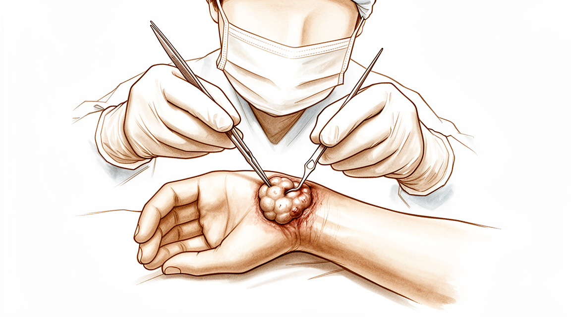
¿Por qué se ha recomendado esta operación?
El Dr. Kieran Hirpara, cirujano de extremidades superiores en el Mater Private Hospital Rockhampton, comienza por proponer las opciones menos invasivas que se adapten a su condición. Un quiste ganglionar de muñeca es un bulto lleno de líquido situado cerca de la articulación de la muñeca; su extirpación se denomina excisión. Por lo general, los pacientes son derivados a nuestra clínica por su médico de cabecera; si un fisioterapeuta le ha sugerido que nos visite, igualmente necesitará una derivación de su médico de cabecera para poder acceder al reembolso de Medicare. En su consulta, tomamos su historia clínica, examinamos su muñeca y, si es necesario, solicitamos estudios de imagen.
La mayoría de los quistes ganglionares de muñeca se tratan inicialmente sin cirugía. Normalmente intentamos modificar las actividades, aplicar terapia de mano, usar férulas o drenar el bulto mediante una aguja. La cirugía se considera únicamente cuando estas medidas no logran una mejora suficiente. Alrededor del 40 % de los quistes ganglionares de muñeca disminuyen de tamaño por sí solos durante los primeros 6 años; por eso, en un primer momento suele ser razonable esperar. El drenaje con aguja generalmente no evita que el quiste reaparezca, mientras que la extirpación quirúrgica presenta una tasa de recurrencia de aproximadamente el 10 %. El objetivo de la operación es eliminar el bulto y aliviar su dolor, de modo que su muñeca vuelva a funcionar correctamente.
Antes de la operación
La mayoría de las personas no necesitan mucha preparación. Se le indicará que no coma ni beba durante 7 horas antes de la cirugía. Pedimos 7 horas en lugar de las 6 habituales para poder adelantar su turno si la lista de cirugías avanza antes de lo previsto. Su cirujano le indicará qué medicamentos debe suspender y cuándo, y usted debe llevar una lista de todos los fármacos que toma. Organice que alguien lo lleve a casa después de la intervención, y use ropa holgada y cómoda. Es posible que ya se hayan realizado estudios de imagen como radiografías, ecografías o resonancias magnéticas para planificar la operación. Si padece otras afecciones médicas, es posible que necesite análisis de sangre o una consulta con el anestesista.
El día de la intervención
Llega usted a la unidad de admisiones quirúrgicas del hospital, donde se le registra y prepara para la cirugía. Allí conoce al anestesista. Esta operación se realiza bajo anestesia general; usted permanecerá completamente dormido durante todo el procedimiento. En algunos pacientes también se puede aplicar un bloqueo nervioso regional para aliviar el dolor postoperatorio; el anestesista decide al respecto ese mismo día según sus circunstancias individuales. Posteriormente, se le lleva al quirófano, donde se realiza la operación.
Despierta usted en la sala de recuperación, donde las enfermeras le vigilan mientras la anestesia va desapareciendo. Una vez que su estado se estabiliza, será trasladado a la planta de hospitalización o podrá regresar a casa, según el tipo de intervención y su recuperación. La mayoría de los pacientes vuelven a casa el mismo día. Si decide irse a casa, alguien debe llevarle, tal como se acordó previamente a la cirugía. Antes de marcharse, recibirá instrucciones sobre el cuidado de su muñeca y sobre la visita de seguimiento.
¿En qué consiste la operación?
La operación consiste en extirpar el quiste ganglionar, que es el bulto lleno de líquido, así como el pequeño tallo que lo conecta con la articulación de la muñeca. Es importante eliminar ese tallo, pues si se deja, el quiste tiene más probabilidades de volver a formarse.
El lugar donde el cirujano realiza la intervención depende de la ubicación del quiste. Si se encuentra en el dorso de la muñeca, la operación se efectúa mediante dos o tres pequeñas incisiones mínimamente invasivas, cada una de aproximadamente 1 cm de longitud. Se introduce una cámara delgada en la articulación de la muñeca para que el cirujano pueda visualizar el interior y extirpar el quiste y su tallo. Si el quiste está en la cara palmar de la muñeca, la operación se realiza mediante una sola incisión abierta sobre el quiste; el cirujano separa cuidadosamente el quiste de las estructuras circundantes. En la cara palmar, el quiste suele situarse cerca de una arteria que irriga la mano, y a veces lo rodea; por ello, esta parte de la intervención se lleva a cabo de forma lenta y delicada. Previamente, el cirujano verificará que el flujo sanguíneo hacia la mano sea adecuado a través de ambas arterias principales.
Una vez extraído el quiste, se cierran las incisiones con puntos de sutura. Se coloca un vendaje sobre ellas, el cual debe mantenerse durante unos 10 días. El cirujano le recomendará comenzar a mover la muñeca desde el principio, dentro de las primeras 2 semanas tras la cirugía, ya que el movimiento suave ayuda a prevenir la rigidez.
Después de la operación
Por lo general, se trata de una intervención ambulatoria, por lo que podrá volver a casa el mismo día; aunque en ocasiones los pacientes permanecen una noche en el hospital. Alguien debe acompañarle durante las primeras 24 horas. Es posible que sienta algo de dolor alrededor de las incisiones; el uso de analgésicos sencillos suele ser suficiente para aliviarlo. Al descansar, mantenga la mano elevada sobre un cojín para reducir la hinchazón. Puede moverse por casa en cuanto se sienta estable, y se recomienda realizar movimientos suaves de muñeca desde el principio. Dejamos el vendaje puesto durante unos 10 días; por favor, no lo retire antes de ese plazo a menos que se lo indiquemos. Lo cambiamos o lo retiramos cuando venga a la consulta. La mayoría de las personas vuelven a conducir en un plazo de dos a tres semanas, una vez que pueden sujetar el volante cómodamente.
Recuperación
Durante los primeros días, la muñeca le dolerá y estará un poco hinchada alrededor de las incisiones. Un analgésico sencillo suele aliviar esto, y mantener la mano elevada sobre una almohada ayuda a reducir la hinchazón. La molestia disminuye progresivamente durante las primeras dos semanas.
Deberá mantener el vendaje puesto durante unos 10 días; nosotros lo cambiamos o lo retiramos en su siguiente consulta. Los movimientos suaves de la muñeca comienzan pronto, dentro de las primeras 2 semanas, ya que moverla temprano ayuda a prevenir la rigidez. La terapia de la mano postoperatoria la realizará Ruby Doolan en Extend Rehabilitation. Su terapeuta de la mano le guiará en los ejercicios y supervisará la recuperación de su movilidad y fuerza de agarre. Puede moverse por la casa en cuanto se sienta estable, y utilizará la mano para tareas ligeras según le resulte cómodo.
A medida que la hinchazón disminuye, las actividades cotidianas se vuelven más fáciles. Una vez que pueda agarrar objetos sin dolor, escribir, comer y vestirse volverán a ser actividades normales. Por lo general, se puede volver a conducir en un plazo de dos a tres semanas, una vez que la herida esté cómoda y pueda sujetar el volante con comodidad; consulte nuestra página sobre conducir tras una cirugía de miembro superior. La mayoría de las personas retoman sus labores y actividades habituales a medida que recuperan movilidad y fuerza.
La recuperación varía según cada persona, por lo que su cronograma podría ser distinto. Su cirujano y su terapeuta de la mano le guiarán en cada consulta; puede esperar una mejora constante a medida que su muñeca se recupera.
Qué puede salir mal
La mayoría de los pacientes evolucionan bien, pero en ocasiones pueden surgir problemas. Su cirujano y el equipo lo vigilarán de cerca para detectar cualquier incidencia a tiempo.
El bulto puede reaparecer después de la cirugía. Es posible que note que vuelve a formarse ese mismo bulto blando cerca de la muñeca, a veces semanas o meses después. Si observa o siente que vuelve a aparecer, mencione este hecho en su próxima consulta. Su cirujano le explicará las opciones disponibles si fuera necesario un tratamiento adicional.
Algunas personas experimentan dolor en la muñeca tras la operación. Esto es más probable si ya sentía dolor en la muñeca antes de la cirugía, o si su trabajo o deporte implica flexiones bruscas y fuertes hacia atrás de la muñeca. El dolor suele manifestarse como una molestia profunda que empeora con la actividad y no mejora por completo con analgésicos comunes. Asimismo, algunas personas notan que su mano no funciona tan fluidamente como esperaban; realizar agarres o tareas cotidianas les resulta más difícil que antes. Si el dolor o la debilidad persisten más allá de la fase inicial de recuperación, informe a su cirujano en la siguiente visita para que se considere la posibilidad de terapia de mano u otras medidas.
Durante la cirugía artroscópica en la parte dorsal de la muñeca, es poco frecuente que se produzca lesión en los tendones encargados de estirar los dedos. Esto se manifestaría como dificultad para estirar uno o varios dedos, o como un dedo que queda más bajo que los demás. Si no consigue estirar un dedo correctamente, comuníquese de inmediato con la clínica.
En el caso de bultos situados en la cara palmar de la muñeca, existen complicaciones poco frecuentes relacionadas con la proximidad del bulto a una arteria que irriga la mano. Las señales de alerta incluyen una mano de color pálido, azulado o más fría de lo habitual, así como entumecimiento y hormigueo persistentes. Si observa alguno de estos síntomas, acuda a urgencias en lugar de esperar.
En la tabla de complicaciones de esta página se detallan las tasas típicas; si desea conocer los datos exactos, puede consultarla.
¿Cuándo deben llamarnos?
Llámenos si tiene fiebre, o si la piel alrededor de las heridas se vuelve más roja, hinchada o comienza a exudar líquido. Llámenos si el dolor empeora en lugar de mejorar, o si no puede enderezar un dedo. Acuda a urgencias si su mano se vuelve pálida, azulada o fría; si pierde la sensibilidad en la mano; o si no puede moverla. También acuda a urgencias si presenta hinchazón en la pantorrilla o dificultad para respirar.
¿Dónde leer más sobre esta afección?
Esta página trata sobre la operación en sí. La afección que se trata, así como las evidencias sobre cuándo la cirugía resulta útil y cuándo no, se explican con mayor detalle en la página Quistes sinoviales de la muñeca.
Evidence & references
This is the clinical evidence summary written for health professionals. It is technical, and it lists the research this page was built from. You do not need to read it to understand your treatment or to make a decision about it.
Anatomy & Pathophysiology
Bony Anatomy
- The wrist includes the distal radioulnar, radiocarpal, and ulnocarpal joints and the eight carpal bones and their proximal and distal articulations [4].
- The proximal row of carpal bones consists of the scaphoid, lunate, triquetrum, and pisiform [4].
- The distal row of carpal bones consists of the trapezium, trapezoid, capitate, and hamate [4].
- The pisiform and trapezoid are the smallest carpal bones, while the capitate is the largest [4].
- The radiocarpal joints are formed by the articulation of the distal radius with the scaphoid and lunate through their respective concave facets on the distal radius [4].
- The triquetrum articulates with the triangular fibrocartilage at the radiocarpal joint [4].
- The distal concave articular surfaces of the proximal carpal row form the midcarpal articulations with the distal row [4].
- The distal row articulates with the metacarpals, allowing mobility in the thumb, stability in the index and long finger metacarpals, and increased mobility in the ring and little finger metacarpals [4].
- The distal ulnar convexity articulates at the lesser sigmoid notch of the distal radius [4].
- The sigmoid notch articular surface accommodates the ulnar head through two thirds of its arc [4].
- There is about a 20-degree inclination of the distal ulna at its articulation with the radius [4].
- The ulnar styloid lies dorsal to the ulnar head and extends distally [4].
- The distal radius has three articular components: the scaphoid fossa, the lunate fossa, and the sigmoid notch [8].
- A ridge corresponding with the scapholunate interval is located between the scaphoid and lunate fossae on the distal radius [8].
- The radial styloid allows attachment of the brachioradialis tendon [8].
- The radial styloid is the origin of the radial scapholunate and radial lunocapitate ligaments [8].
- The concave elliptical distal radius is oriented in the sagittal plane with an average of 11 degrees of volar tilt [8].
- In the frontal plane, the average radial inclination of the distal radius is 23 degrees [8].
- Radial length is measured from the tip of the radial styloid to the ulnar articular surface and averages 13 mm [8].
- The ulnar shaft remains fixed in its rotation at the ulnohumeral joint, while the radius rotates around the ulna in pronation and supination [8].
- The radius has a lateral bow that is crucial to the maintenance of full pronation and supination [8].
Ligamentous Anatomy
- The triangular fibrocartilage complex (TFCC) attaches to the base of the ulnar styloid and separates the hyaline cartilage–covered ulnar head from the styloid [4].
- The TFCC includes the ulnar collateral ligament, dorsal and volar radioulnar ligaments, articular disc, meniscal homologue, extensor carpi ulnaris sheath, and ulnolunate and ulnotriquetral ligaments [4].
- Interosseous intrinsic ligaments connect the carpal bones in the proximal and distal carpal rows [4].
- The scapholunate and lunotriquetral interosseous ligaments connect the proximal carpal row [4].
- Ligaments connecting the trapezium to the trapezoid, trapezoid to the capitate, and capitate to the hamate connect the distal carpal row [4].
- Extrinsic ligaments extend from the radius and ulna distally across the carpal rows [4].
- The radial collateral ligament extends from the radial styloid to the scaphoid waist [4].
- The ulnar collateral ligament extends from the base of the ulnar styloid to the pisiform [4].
- The transverse carpal ligament is an extrinsic ligament [4].
- Volar extrinsic ligaments include the radioscapocapitate, radiolunotriquetral, and radioscapolunate ligaments on the radial side [4].
- The ulnolunate and ulnotriquetral components of the TFCC are volar extrinsic ligaments on the ulnar side [4].
- The space of Poirier is a relatively thin area on the palmar side of the carpus between the radiolunotriquetral and radioscapocapitate ligaments [4].
- The space of Poirier overlies the palmar surface of the lunate [4].
- Dorsal extrinsic ligaments include the dorsal radiocarpal and dorsal intercarpal ligaments [4].
- The trapezoidal dorsal radiocarpal ligament attaches along the dorsal radial articular margin of the lunate fossa, from the Lister tubercle to the lesser sigmoid notch [4].
- The trapezoidal dorsal radiocarpal ligament spans the lunotriquetral joint and inserts on the dorsal surface of the triquetrum [4].
- The dorsal intercarpal ligament is attached to the distal, dorsal surface of the triquetrum and passes across the midcarpal joint to attach to the dorsal surfaces of the scaphoid waist and the trapezoid [4].
- Extrinsic carpal ligaments connect the radius or the ulna to the carpus [9].
- Volar ligaments are generally stronger than dorsal ligaments [9].
- The radioscaphocapitate ligament connects to the waist of the scaphoid and limits ulnar translation of the carpus [9].
- The long radiolunate ligament helps limit ulnar translocation of the carpus [9].
- The short radiolunate ligament helps control lunate position [9].
- The radioscapholunate ligament is a vascular conduit and not a true ligament, also known as the ligament of Testut [9].
- The ulnolunate ligament attaches to the palmar radioulnar ligament and the lunate [9].
- The ulnocapitate ligament attaches to the ulnar head and is the most superficial or palmar palmar ulnocarpal ligament [9].
- The ulnotriquetral ligament attaches to the palmar radioulnar ligament and the triquetrum [9].
- The dorsal radiocarpal ligament has a trapezoidal shape and passes from the dorsal rim of the distal radius to the lunate and triquetrum [9].
- Fibers of the dorsal radiocarpal ligament insert onto the dorsal lunotriquetral interosseous ligament [9].
- The scapholunate interosseous ligament is a major stabilizer of the wrist and the most commonly injured wrist ligament [9].
- The scapholunate interosseous ligament is C-shaped, consisting of dorsal, palmar, and interosseous portions, with the dorsal portion being the strongest and thickest [9].
- The scapholunate interosseous ligament provides a flexion force on the lunate given its attachment to the scaphoid [9].
- The lunotriquetral interosseous ligament is C-shaped, with the volar portion being the thickest and strongest [9].
- The lunotriquetral interosseous ligament provides an extension moment on the lunate given its attachment to the triquetrum [9].
- The capitohamate ligament is a thick ligament with a 5 × 5 mm cross section and extensions to the third or fourth metacarpals [9].
- The dorsal intercarpal ligament passes from the dorsal tubercle of the triquetrum to the distal pole of the scaphoid [9].
- The dorsal intercarpal ligament reinforces the elastic dorsal wrist capsule and helps stabilize the scapholunate articulation with a contribution to the dorsal scapholunate interosseous ligament from its deep fibers [9].
- The space of Poirier is an area adjacent to the proximal capitate without ligamentous attachment, situated ulnar to the radioscaphocapitate ligament and radial to the long radiolunate in the floor of the carpal tunnel [9].
- The space of Poirier is a weak area vulnerable to instability, through which the distal carpal row separates from the lunate during a perilunate dislocation [9].
Soft Tissue Anatomy
- Six dorsal compartments contain wrist and digital extensor tendons on the dorsal surface of the wrist [8].
- The contents of the carpal tunnel reside on the volar surface, including nine flexor tendons and the median nerve [8].
- The flexor carpi ulnaris tendon can be palpated near its insertion on the pisiform on the ulnar surface [8].
- The boundaries of Guyon’s canal are the volar carpal ligament and transverse carpal ligament, the hook of the hamate radially, and the pisiform ulnarly [8].
- Guyon’s canal contains the ulnar artery and nerve [8].
- The interosseous membrane connects the shafts of the radius and ulna in the interosseous space [8].
- The central portion of the interosseous membrane is thickened and important in force transmission between the radius and ulna [8].
Ganglion Pathophysiology
- The dorsal wrist ganglion accounts for 60% to 70% of all hand and wrist ganglions [3].
- The main cyst of a dorsal wrist ganglion is usually located directly over the scapholunate ligament [3].
- Dorsal wrist ganglions may occur anywhere else between the extensor tendons [3].
- Failure to identify and excise the pedicle attachment to the scapholunate ligament increases the likelihood of recurrence for dorsal wrist ganglions [3].
- A small, mucin-filled duct is invariably seen piercing the transverse fibers of the scapholunate ligament, connecting the underlying scapholunate joint with the main cyst [3].
- The volar wrist ganglion is the second most common ganglion of the hand and wrist, accounting for 18% to 20% [1].
- The majority of volar ganglions occur either directly over the distal edge of the radius or slightly more distally over the scaphoid tubercle [1].
- Volar ganglions arising over the distal edge of the radius arise from the capsular and ligamentous fibers of the radiocarpal joint [1].
- Volar ganglions arising over the distal edge of the radius occur under the volar wrist crease between the flexor carpi radialis and abductor pollicis longus tendons [1].
- The main cyst of a volar wrist ganglion arising from the radiocarpal joint may be intertwined with bifurcating branches of the radial artery [1].
- Another type of volar ganglion arises from the capsule of the scaphotrapezial joint [1].
- Volar ganglions may appear small clinically but can be surprisingly extensive at surgery [1].
- Multiloculated volar ganglion cysts may extend under the thenar muscles, along the flexor carpi radialis tendon, into the carpal canal, and under the first extensor compartment adjacent to the dorsal branch of the radial artery [1].
- Extensions of volar ganglions may reach as far dorsally as the first web space [1].
- The radial artery is frequently intimately attached to the wall of a volar ganglion and may be completely encircled by it [1].
Biomechanics and Kinematics
- The eight carpal bones represent the most complex articular system in the human body [5].
- The human wrist allows precise positioning of the hand and optimization of power and prehensile tasks throughout a nearly hemispherical arc of wrist motion [5].
- The wrist joint’s motion planes include flexion, extension, radial deviation, ulnar deviation, and circumduction [9].
- There is minimal carpal motion with pronosupination [9].
- Approximately 62° of wrist extension occurs through the radiocarpal joint [9].
- 62% of wrist flexion occurs through the midcarpal joint [9].
- The midcarpal joint is mostly responsible for 20° of radial deviation and 40° of ulnar deviation [9].
- The midcarpal joint is responsible for the “dart thrower’s motion,” which involves moving from radial extension into ulnar flexion positioning of the wrist [9].
- The radius bears 80% of the axial load transmitted through the radiocarpal joint in neutral ulnar variance [9].
- The ulna bears 20% of the axial load transmitted through the radiocarpal joint in neutral ulnar variance [9].
- The proximal row of carpal bones forms an intercalated segment between the distal carpal row and the distal radius, bound into a functional unit by the scapholunate and lunotriquetral interosseous ligaments [9].
- The distal row is rigid with little motion between its bones due to stout intercarpal ligaments, acting as a functional unit with the scaphoid bridging both rows [9].
- During wrist flexion from neutral, the distal row flexes and ulnarly deviates slightly while the scaphoid pronates [9].
- During wrist flexion from neutral, the proximal row flexes differentially, with more rotation through the scaphoid, followed by the triquetrum and the lunate [9].
- The proximal row translates dorsally during wrist flexion [9].
- During wrist extension from neutral, the distal row extends and radially deviates slightly while the scaphoid supinates [9].
- During wrist extension from neutral, the proximal row extends differentially, with more motion in the scaphoid, followed by the triquetrum and then the lunate [9].
- The proximal row translates palmarly during wrist extension [9].
Clinical Presentation
- Volar wrist ganglions are the second most common ganglion of the hand and wrist, accounting for 18% to 20% of cases [1].
- The majority of volar ganglions occur directly over the distal edge of the radius or slightly more distally over the scaphoid tubercle [1].
- Ganglions arising from the distal edge of the radius originate from capsular and ligamentous fibers of the radiocarpal joint [1].
- Radiocarpal volar ganglions occur under the volar wrist crease between the flexor carpi radialis and abductor pollicis longus tendons [1].
- The main cyst of a radiocarpal volar ganglion may be intertwined with bifurcating branches of the radial artery [1].
- A second type of volar ganglion arises from the capsule of the scaphotrapezial joint [1].
- Multiloculated cysts may extend under the thenar muscles, along the flexor carpi radialis tendon, into the carpal canal, and under the first extensor compartment adjacent to the dorsal branch of the radial artery [1].
- Extensions of volar ganglions can reach as far dorsally as the first web space [1].
- Extensions of volar ganglions can often be appreciated preoperatively by careful palpation and digital compression [1].
- The patency of the radial and ulnar arteries must be assessed prior to surgery [1].
- The Allen test should be performed routinely to exclude ulnar artery occlusion [1].
- Preservation of the radial artery is particularly important in patients with a radial-dominant circulation [1].
Investigations
Magnetic Resonance Imaging
- MRI for hand and wrist imaging was first reported in 1986 [10].
- The primary advantages of MRI compared with CT and radiography are improved tissue characterization, especially of soft tissues such as ligamentous structures in the wrist and synovium in the hand, and the lack of ionizing radiation [10].
- MRI is the modality of choice for imaging radiographically occult fractures of the hand and wrist [10].
- Early MRI was limited by low magnetic field strength (0.15 T) and limited image options and image processing [10].
- Modern MRI is generally at 1.5T or 3T with a wide variety of imaging options and powerful image processing and postprocessing available [10].
- 3T is much preferred for hand and wrist imaging, especially for imaging small fields of view [10].
- Dedicated extremity magnets have been marketed, but image quality is poor compared with conventional MR imaging [10].
- 7T MRI has recently become approved for clinical use [10].
- 7T MRI has the potential to become a powerful tool for hand and wrist imaging as applications are developed [10].
- MRI with contrast enhancement is most commonly used to determine whether soft-tissue lesions are solid or cystic [10].
- In the case of rheumatologic imaging, MRI with contrast enhancement is used to better visualize erosions and synovial burden [10].
- Dynamic contrast enhancement has been used with inconsistent results to assess for the presence of avascular necrosis in the lunate or scaphoid after injury [10].
- MR angiography of the hand and wrist can be helpful in situations such as diagnosis of the hypothenar hammer syndrome [10].
- MR arthrography can be performed for evaluation of the triangular fibrocartilage and intercarpal ligament tears [10].
- MR arthrography is generally unnecessary with the increasing availability of high field MRI [10].
Computed Tomography and Other Imaging Modalities
- Hybrid SPECT/CT has been used for the diagnosis of radiographic occult fractures of the wrist [6].
- Cone-beam CT has been used in the diagnosis of scaphoid fractures [6].
- CT and MRI have a diagnostic impact on wrist injuries in young adults [6].
- Cine MRI is a new approach to the diagnosis of scapholunate dissociation [6].
- Cone-beam computed tomography arthrography is an innovative modality for the evaluation of wrist ligament and cartilage injuries [6].
- 3D analysis of the wrist has been described [6].
- Normal and variant anatomy of the wrist and hand can be evaluated on MR imaging [6].
- A practical guide to diagnostic imaging of the upper extremity has been published [7].
- 3-Tesla imaging of the wrist and hand has specific techniques and applications [7].
- MR imaging of the human hand and wrist at 7 T has been described [7].
- Magnetic resonance imaging can be used for the anterior radiocarpal ligaments [7].
- The carpal contents/canal ratio can be established by means of magnetic resonance imaging [7].
- MR imaging can be used for trauma of the elbow and wrist [7].
- Magnetic resonance imaging of the wrist has been described [7].
- MR imaging can be used for ulnar impaction [7].
- Rupture of the distal biceps tendon can be evaluated with MR imaging [7].
- MR imaging can be used for the wrist and elbow in clinical sports medicine [7].
- Magnetic resonance imaging of the musculoskeletal system, including the elbow, has been described [7].
- The efficacy of magnetic resonance imaging of the elbow has been evaluated [7].
- Sports and occupational injuries of the elbow can be evaluated with MR imaging findings [7].
- Magnetic resonance imaging of the elbow has been described [7].
- Magnetic resonance imaging can be used in Kienbock disease [7].
- Magnetic resonance imaging of the elbow, including normal anatomy, imaging technique, and osseous abnormalities, has been described [7].
- Magnetic resonance imaging of the elbow, including abnormalities of the ligaments, tendons, and nerves, has been described [7].
- Low-field MRI can be used for scaphoid fracture [7].
- MR imaging can be used for traumatic and overuse injuries of the wrist and hand in athletes [7].
- MR imaging diagnosis of TFC pathology can be correlated with arthroscopy [7].
- MR imaging of the elbow in baseball pitchers has been described [7].
- Overuse syndromes and injuries involving the elbow can be evaluated with MR imaging findings [7].
- Occult wrist fractures can be detected by magnetic resonance imaging [7].
- Ulnar collateral ligament injury in the throwing athlete can be evaluated with saline-enhanced MR arthrography [7].
- Magnetic resonance imaging of the triangular fibrocartilage complex has been described [7].
- Preoperative evaluation of the ulnar collateral ligament by magnetic resonance imaging and computed tomography arthrography has been evaluated in baseball players with surgical confirmation [7].
- Histologic and magnetic resonance imaging correlations in Kienbock’s disease have been described [7].
- MRI can be used for the diagnosis of occult dorsal wrist ganglion [7].
Radiography and Stress Views
- Distal radioulnar joint stress radiography can be used for detecting radioulnar ligament injury [6].
- Radiographic stress views can be compared for scapholunate dynamic instability in a cadaver model [6].
- Imaging of radial wrist pain involves various imaging modalities and anatomy [6].
- Intrinsic ligament and triangular fibrocartilage complex tears of the wrist can be compared using MDCT arthrography, conventional 3-T MRI, and MR arthrography [6].
- Simple plain radiographic signs and measures can be used to diagnose acute scapholunate ligament injuries of the wrist [11].
- Radiographic clues can be used for determining carpal instability and treatment protocol for scaphoid fractures [11].
- The role of imaging in diagnosing diseases of the distal radioulnar joint, triangular fibrocartilage complex, and distal ulna has been described [11].
Diagnostic Accuracy and Systematic Reviews
- The efficacy of magnetic resonance imaging and clinical tests in diagnostics of wrist ligament injuries has been evaluated in a systematic review [11].
- The role of magnetic resonance imaging in scaphoid fractures has been described [11].
Treatment
Dorsal Wrist Ganglion
- Failure to identify and excise the pedicle attachment to the scapholunate ligament increases the likelihood of recurrence [3].
- Careful preoperative palpation of the cyst with digital compression often reveals its extent and the direction of the pedicle [3].
- Transillumination or aspiration confirms the diagnosis preoperatively [3].
- Review of preoperative radiographs to rule out an interosseous component is recommended before dissection [3].
- Most dorsal ganglions can be approached through a transverse incision over the proximal carpal row [3].
- A modified incision or second transverse incision may be necessary for ganglions not directly over the scapholunate ligament [3].
- The diagnosis of ganglion cyst should be made before commitment to a transverse incision because this incision is not readily incorporated into a limb-sparing incision if a subsequent diagnosis of a malignant soft tissue tumor is made [3].
- The extensor pollicis longus and extensor digitorum communis tendons are retracted radially and ulnarly, respectively, to expose the dorsal ganglion [3].
- The joint capsule is opened along the border of the radius and scaphoid's proximal pole with the wrist in volar flexion [3].
- The capsule is elevated and retracted distally to expose the capsular attachments to the scapholunate ligament [3].
- Smaller intraarticular cysts are often seen attached to the scapholunate ligament [3].
- The capsular incision is continued around the ganglion, leaving all capsular attachments to the ligament intact [3].
- The capsular incision is extended laterally if capsular ducts are encountered, which can be identified by small amounts of mucin drainage [3].
- The ganglion and its capsular attachments are tangentially excised off the scapholunate ligament [3].
- A small, mucin-filled duct is invariably seen piercing the transverse fibers of the scapholunate ligament [3].
- This duct appears to connect the underlying scapholunate joint with the main cyst [3].
- Synovial and capsular attachments along the distal margin of the scapholunate ligament are excised to give an unobstructed view of the head and neck of the capitate [3].
Volar Wrist Ganglion
- Volar ganglions arising from the distal edge of the radius occur under the volar wrist crease between the flexor carpi radialis and abductor pollicis longus tendons [1].
- The main cyst of a volar ganglion may be intertwined with bifurcating branches of the radial artery [1].
- Extensions of volar ganglions can be appreciated preoperatively by careful palpation and digital compression [1].
- The Allen test should be performed routinely to assess the patency of the radial and ulnar arteries [1].
- Ulnar artery occlusion should be excluded preoperatively [1].
- The radial artery must be preserved, particularly in patients with a radial-dominant circulation [1].
- Exposure and precise identification of the capsular attachments of a volar ganglion are more difficult than for a dorsal ganglion [1].
- The incision for volar ganglion excision must be planned to allow for extension into the carpal tunnel or base of the thenar muscles [1].
- Longitudinal incisions allow for optimal visualization during volar ganglion excision [1].
- The forearm fascia is incised longitudinally to identify and mobilize the dome of the cyst [1].
- The radial artery is frequently intimately attached to the wall of the ganglion and may be completely encircled by it [1].
- Loupe magnification aids in the dissection to protect the radial artery [1].
- The pedicle is traced to the volar joint capsule, usually the scaphotrapezial or radiocarpal ligament [1].
- The joint is opened and explored, and the ganglion attachments are excised approximately 3 ± 4 mm [1].
- Surrounding tissues are compressed digitally after excision to rule out further mucin-filled pockets [1].
- Unidentified extensions of the ganglion must be excised if present [1].
- Capsular closure is unnecessary and only delays early mobilization [1].
- A bulky bandage and elevation of the hand ensure early postoperative comfort [1].
- Motion of the wrist should begin within the first 2 weeks after surgery [1].
- Unexpected branches of the radial sensory or lateral antebrachial cutaneous nerves may be injured, leading to troublesome neuromas [1].
- Extensions of the routine incision into the carpal canal must avoid injury to the palmar cutaneous branch of the median nerve [1].
- Injuries to the radial artery can be repaired microscopically [1].
- Some authors recommend leaving a portion of the cyst wall attached to the artery to avoid arterial injury [1].
- Stiffness of the wrist is less common than with dorsal ganglions but can occur if early motion is not encouraged [1].
- Curved incisions appear to consistently provide more attractive scars, especially near the volar wrist creases [1].
References
[1] Green S Operative Hand Surgery. BOX 59.1 Ganglions of the Hand and Wrist > Operative Treatment > Volar Wrist Ganglion.
[3] Green S Operative Hand Surgery. BOX 59.1 Ganglions of the Hand and Wrist > Operative Treatment > Dorsal Wrist Ganglion.
[4] Campbell S Operative Orthopaedics 4 Volume Set. NERVE INJURIES AT THE LEVEL OF THE HAND AND WRIST > ANATOMY.
[5] Green S Operative Hand Surgery. INTRODUCTION.
[6] Campbell S Operative Orthopaedics 4 Volume Set. ANATOMIC RECONSTRUCTION OF THE DISTAL RADIOULNAR LIGAMENTS > RADIOGRAPHIC TECHNIQUES.
[7] Campbell S Operative Orthopaedics 4 Volume Set. ELBOW, WRIST, AND HAND.
[8] A Lange Medical Book Current Diagnosis Treatment In Orthopedics Fifth Edition. 2Musculoskeletal Trauma Surgery > FRACTURES AND DISLOCATIONS OF THE DISTAL AND MID-FOREARM.
[9] Aaos Comprehensive Orthopaedic Review 3. Carpal Instability* > II. Anatomy and Biomechanics (See Chapter 92).
[10] Orthopaedic Knowledge Update 13 Ebook Without Multimedia. Anatomy, Evaluation, Clinical Examination, and Imaging > Imaging: Advances in Imaging of the Hand and Upper Extremity > Magnetic Resonance Imaging.
[11] Campbell S Operative Orthopaedics 4 Volume Set. ANATOMIC RECONSTRUCTION OF THE DISTAL RADIOULNAR LIGAMENTS > DIAGNOSIS AND EVALUATION.