Patients › Wrist
Quistes ganglionares de la muñeca
Wrist ganglia are common, fluid-filled lumps – often painless – and this page covers observation, aspiration, and excision.
Qué está sintiendo
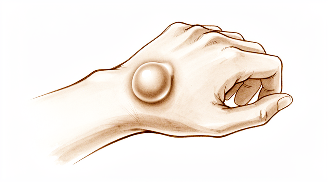
Un quiste ganglionar de muñeca es un bulto lleno de líquido que se forma a partir de una articulación o de la vaina de un tendón de la muñeca. No es un crecimiento que se convierta en algo dañino. Muchas personas lo notan primero como un bulto liso en la parte dorsal de la muñeca, aunque también puede aparecer en la cara palmar. Puede notarse firme, y su tamaño puede cambiar con el tiempo.
El bulto en sí suele ser indoloro. Cuando hay dolor, tiende a localizarse en la profundidad de la muñeca, cerca del bulto, y a menudo empeora con la actividad. Las tareas que doblan o cargan la muñeca, como levantarse de una silla empujando con las manos, cargar bolsas de la compra o agarrar una sartén pesada, pueden provocar molestias. Algunas personas notan la muñeca más débil o más rígida de lo habitual, lo que puede dificultar escribir en el teclado, escribir a mano o abrir frascos.
Como el bulto se sitúa cerca de los nervios de la muñeca, un quiste ganglionar a veces puede presionarlos. Esto puede causar hormigueo, entumecimiento o debilidad en la mano. Si nota cualquiera de estos síntomas nerviosos, conviene que le revisen la muñeca cuanto antes, ya que hay que liberar al nervio de esa presión.
Si el bulto o el dolor no se calman, empeoran a lo largo de semanas o le impiden usar la mano o trabajar, consulte a su médico de cabecera o solicite una evaluación con un especialista.
¿Qué está ocurriendo realmente?
Un quiste ganglionar se forma a partir de una pequeña válvula unidireccional en el revestimiento de la articulación de la muñeca. El líquido que normalmente lubrica la articulación sale empujado a través de esta válvula, pero no puede volver. Se acumula en un saco bajo la piel, y ese saco es el bulto que usted puede ver y palpar. El bulto está conectado a la articulación mediante un tallo fino; por eso su tamaño puede cambiar según la articulación produzca más o menos líquido.
La mayoría de los quistes ganglionares se forman a partir del ligamento escafolunar, una banda corta de tejido que mantiene unidos dos de los pequeños huesos de la muñeca. Funciona un poco como una junta entre ellos. El revestimiento que rodea esta banda se irrita, a menudo tras una distensión leve o un esfuerzo repetido, y es de ahí de donde se escapa el líquido. Esto también explica por qué las molestias se localizan en la profundidad de la muñeca y no en la piel: el problema está en la propia articulación, no en el bulto.
La posición del bulto explica el resto de sus síntomas. En la parte dorsal de la muñeca, suele situarse justo encima de ese ligamento, cerca de los tendones que estiran los dedos. En la cara palmar, a menudo se forma cerca de la arteria principal y de los nervios que llegan a la mano; por eso puede aparecer hormigueo o entumecimiento cuando el quiste los presiona. Algunos bultos son pequeños y están ocultos, y solo aparecen cuando la muñeca se dobla hacia delante, pero aun así pueden causar un dolor profundo.
Conviene saber que los quistes ganglionares son frecuentes. Muchas personas los tienen sin ningún síntoma, y aparecen en las pruebas de imagen de muñecas sin dolor con más frecuencia de lo que cabría esperar. Tener uno no significa que haya algún problema en la estructura de la propia articulación de la muñeca.
Qué podemos hacer al respecto
El Dr. Kieran Hirpara, cirujano de extremidades superiores en el Mater Private Hospital Rockhampton, comienza con las opciones menos invasivas que se adapten a su condición. Por lo general, los pacientes son derivados a nuestra clínica por su médico de cabecera; si un fisioterapeuta le ha sugerido consultarnos, igualmente necesitará una derivación de su médico de cabecera para poder acceder al reembolso de Medicare. Una evaluación en la consulta, con su historia clínica, una exploración y estudios de imagen cuando son necesarios, establece el diagnóstico.
Como un quiste ganglionar es benigno, no hacer nada es un primer paso razonable. Muchos quistes ganglionares desaparecen por sí solos. Entre el 28 % y el 58 % de los quistes ganglionares de muñeca desaparecen sin ningún tratamiento. En los niños menores de 10 años, los quistes ganglionares suelen situarse en la cara palmar de la muñeca, y entre el 69 % y el 79 % se reducen hasta desaparecer por sí solos en un plazo de 12 a 18 meses. Observar y esperar, a veces con una férula para mayor comodidad, es adecuado para la mayoría de los niños con un quiste ganglionar en la mano o la muñeca.
Si el bulto le molesta, podemos drenarlo con una aguja. Esto se llama aspiración. Extraer el líquido elimina hasta el 85 % de los quistes ganglionares cuando se realiza una, dos o tres veces. A menudo vuelve algo de líquido después, porque el drenaje vacía el bulto pero deja la válvula que lo formó. Aun así, el 74 % de las personas quedan satisfechas tras la aspiración y unos consejos sencillos sobre el bulto. Drenarlo primero también deja la operación como una opción posterior en lugar de como el primer paso. Un quiste ganglionar oculto en la parte dorsal de la muñeca, que causa dolor profundo pero no se puede ver, suele tratarse primero sin cirugía, inmovilizando la muñeca y con inyecciones de esteroides en el revestimiento de la articulación.
Cuando estas medidas no han dado suficiente alivio, la cirugía puede extirpar el quiste ganglionar. La operación extirpa el bulto y su tallo, la fina conexión con la articulación de la muñeca, porque dejar el tallo hace más probable que el bulto reaparezca. Puede realizarse como una operación abierta o mediante cirugía artroscópica a través de pequeños portales. Le explicaremos qué opción se adapta mejor a su muñeca, y usted decidirá junto con nosotros si la cirugía le merece la pena.
Qué esperar
Muchos quistes ganglionares de muñeca cambian con el tiempo en lugar de permanecer igual. Pueden reducirse, hincharse o aparecer y desaparecer. Algunos desaparecen por sí solos: aproximadamente el 40 % de los quistes ganglionares de muñeca disminuyen durante los primeros 6 años desde su primera evaluación, y entre el 39 % y el 42 % de los quistes ganglionares de la parte dorsal de la muñeca desaparecen sin ningún tratamiento. En los niños pequeños, la mayoría de los quistes de la mano y la muñeca desaparecen por sí solos, normalmente en un plazo de un año. Si el suyo no le causa muchas molestias, esperar es una opción razonable, y el bulto no se convertirá en nada dañino si lo deja como está.
El tratamiento cambia las probabilidades. Drenar el bulto con una aguja lo vacía, pero la mayoría de los quistes ganglionares reaparecen después de la aspiración, porque la válvula que formó el bulto sigue ahí. La cirugía que extirpa el bulto y su tallo tiene una probabilidad de recurrencia mucho menor: aproximadamente el 10 % de los quistes ganglionares de muñeca reaparecen después de la cirugía, y entre el 3 % y el 12 % reaparecen tras la extirpación abierta de un quiste ganglionar de la parte dorsal de la muñeca. La mayoría de las personas que se operan notan que sus síntomas desaparecen. Sin embargo, puede persistir algo de dolor después, sobre todo si su trabajo o su deporte exigen doblar la muñeca hacia atrás con fuerza.
Si opta por la cirugía, la recuperación suele ser sencilla. Al principio, la mano se envuelve en un vendaje voluminoso y se mantiene en alto para que esté cómoda, y el movimiento de la muñeca comienza pronto, dentro de las primeras 2 semanas tras la cirugía de un quiste ganglionar de la cara palmar. Mover la muñeca pronto es importante: la rigidez es poco frecuente, pero puede aparecer si no se fomenta el movimiento temprano. La terapia de la mano después de la cirugía es con Ruby Doolan en Extend Rehabilitation, que le guiará en los ejercicios y le confeccionará cualquier férula que necesite. Una zona firme o sensible donde estaba el bulto puede persistir hasta 3 semanas después de la cirugía artroscópica en la cara palmar, y masajear esa zona según se le indique ayuda a que se calme. Las cicatrices forman parte de cualquier operación, y las incisiones curvas cerca de los pliegues de la muñeca tienden a cicatrizar con un aspecto más estético.
Si un bulto tratado o sus síntomas no se calman, o empeoran a lo largo de semanas, consulte a su médico de cabecera o solicite una evaluación con un especialista.
¿Cuándo consultar a un especialista?
La mayoría de los quistes ganglionares de muñeca son benignos, y muchos desaparecen por sí solos, por lo que un bulto por sí solo no es una urgencia. Consulte a su médico de cabecera o solicite una evaluación con un especialista si el bulto o el dolor no se calman a lo largo de semanas, empeoran o le impiden trabajar o usar la mano. Consulte antes si tiene hormigueo, entumecimiento o debilidad en la mano, ya que eso indica que el quiste está presionando un nervio. Acuda al servicio de urgencias si la mano o los dedos se ponen pálidos, fríos, blancos o azules, o si pierde de repente la sensibilidad o el movimiento de la mano después de una lesión. Estos signos son raros con un quiste ganglionar, pero requieren atención ese mismo día.
En mayor profundidad
Advanced reading: the deeper science (optional)
Esta sección profundiza más de lo necesario para que usted tome sus propias decisiones terapéuticas. Los quistes ganglionares de la muñeca merecen una lectura más detallada, pues en realidad la elección radica entre aceptar el riesgo de recurrencia o someterse a una intervención quirúrgica; además, las cifras que sustentan esta decisión son lo suficientemente claras como para tomar una determinación.
La aspiración vuelve a producirse; la excisión, en cambio, no, con frecuencia
Los dos tratamientos activos son extraer el quiste mediante una aguja y extirparlo quirúrgicamente junto con su tallo. Tras analizar 2,239 pacientes, se observa que la excisión quirúrgica abierta presenta una probabilidad significativamente menor de recurrencia que la aspiración. La excisión artroscópica ha arrojado resultados prometedores, pero los datos de ensayos comparativos son limitados y no han demostrado su superioridad [1].
Este mecanismo explica dicha diferencia. Un quiste ganglionar no es un saco de líquido flotante libremente; está conectado mediante un tallo a la cápsula articular subyacente, y la articulación sigue produciendo líquido. La aspiración vacía el reservorio pero deja intacta su fuente de origen; por ello, el rellenado del quiste es un resultado frecuente en lugar de un fracaso terapéutico. La excisión, en cambio, busca localizar y extirpar el tallo en su punto de origen.
La extirpación mediante técnica mínimamente invasiva y la abierta arrojan resultados similares
Cuando se opta por la extirpación, se han comparado directamente ambos métodos. En un total de 910 pacientes, los resultados en cuanto a recurrencia y complicaciones fueron comparables entre la técnica artroscópica y la abierta. Los autores abogan por la realización de estudios estandarizados y con suficiente potencia estadística [2].
Por ello, la decisión entre ambos métodos debe basarse en criterios secundarios: la presencia de cicatrices, la familiaridad del cirujano con cada técnica, y si es necesario examinar otras patologías intraarticulares simultáneamente, más que en el riesgo de recurrencia.
Nadie se pone de acuerdo sobre qué hacer después, y quizás eso no importe
Un hallazgo sencillo y honesto: una revisión sistemática y una encuesta realizadas entre cirujanos de mano revelaron que existe división de opiniones respecto a si se debe inmovilizar la muñeca tras la extirpación de un ganglio dorsal; además, en cuanto a los resultados funcionales, no existen datos concluyentes que indiquen que alguna estrategia sea superior [3].
Vale la pena saber que las diferencias en las indicaciones dadas por los cirujanos reflejan realmente una incertidumbre clínica, y no que alguno de ellos esté equivocado.
El argumento más contundente suele ser no hacer nada
Ninguno de los puntos anteriores demuestra que un ganglio deba ser tratado. Se trata de quistes benignos que con frecuencia varían en tamaño; además, una parte de ellos desaparece sin necesidad de intervención alguna. No evolucionan hacia ninguna otra patología.
Esto cambia por completo la perspectiva de la decisión terapéutica. Las razones para tratarlo son el dolor, la interferencia con el movimiento de la muñeca o la capacidad de agarre, la presión sobre algún nervio cercano, o un tamaño que realmente moleste al paciente; no la mera presencia del bulto. Teniendo en cuenta que la aspiración conlleva una tasa significativa de recurrencia y que la extirpación implica los riesgos propios de una intervención quirúrgica en la cápsula articular, la observación cuidadosa constituye una opción válida como primera medida, y merece ser mencionada explícitamente en lugar de considerarse una falta de acción.
La excepción se da cuando el ganglio provoca síntomas neurológicos: entumecimiento, debilidad o dolor que se irradia a la mano. En esos casos, el quiste comprime una estructura que no puede tolerar dicha presión indefinidamente; por lo tanto, la observación cuidadosa deja de ser la opción de menor riesgo.
Referencias
[1] Head L, Gencarelli JR, Allen M, Boyd KU. Tratamiento de los quistes ganglionares de la muñeca: revisión sistemática y metaanálisis. J Hand Surg Am. 2015;40(3):546-553.e8. https://doi.org/10.1016/j.jhsa.2014.12.014
[2] Crawford C, Keswani A, Lovy AJ, Levy I, Lutz K, Kim J, et al. Excisión artroscópica versus abierta de los quistes ganglionares dorsales: revisión sistemática y metaanálisis. J Hand Surg Eur Vol. 2017;43(6):659-64. https://doi.org/10.1177/1753193417734428
[3] Wong CR, Karpinski M, Hatchell AC, McRae MH, Murphy J, McRae MC. Inmovilización de la muñeca tras la excisión de quistes ganglionares dorsales: revisión sistemática y encuesta. Hand (N Y). 2021;18(2):254-63. https://doi.org/10.1177/15589447211014631
Evidence & references
This is the clinical evidence summary written for health professionals. It is technical, and it lists the research this page was built from. You do not need to read it to understand your treatment or to make a decision about it.
Overview
Epidemiology and Natural History
- The dorsal wrist ganglion is the prototype of all hand and wrist ganglions, accounting for 60% to 70% of all hand and wrist ganglions [1].
- Volar wrist ganglia are the second most common ganglion of the hand and wrist, accounting for 18% to 20% of cases [11].
- In an asymptomatic population, 51% of 103 wrists scanned demonstrated ganglia on MRI [7].
- In an asymptomatic population evaluated by MRI, volar wrist ganglia are more common than dorsal wrist ganglia [2].
- Neither surgical excision nor aspiration provides a clear long-term benefit over the natural history of the dorsal wrist ganglion [6].
- Pediatric ganglions of the hand have a greater rate of resolution than ganglions of the wrist [4].
Clinical Presentation and Diagnosis
- The main cyst of a dorsal wrist ganglion is usually located directly over the scapholunate ligament [1].
- Dorsal wrist ganglions may occur anywhere between the extensor tendons and can be connected to the scapholunate ligament through an elongated pedicle [1].
- Careful preoperative palpation of a dorsal wrist ganglion with digital compression often reveals its extent and the direction of the pedicle [1].
- Transillumination or aspiration confirms the diagnosis of a wrist ganglion preoperatively [1].
- In patients with recalcitrant dorsal wrist pain in the area of the scapholunate interval, occult dorsal wrist ganglions had a prevalence of 11 out of 14 [3].
- Routine wrist radiography is not cost-effective in the evaluation and treatment decision-making process for patients with a wrist ganglion due to a low prevalence of therapeutically significant findings [5].
- A systematic X-ray should be performed for painful dorsal wrist ganglia [13].
- Preoperative x-rays should be obtained as part of the routine workup for a dorsal wrist ganglion [22].
- Volar wrist ganglions may appear small clinically but can be surprisingly extensive at surgery, with multiloculated cysts extending under the thenar muscles, along the flexor carpi radialis tendon, into the carpal canal, and under the first extensor compartment [11].
- The Allen test should be performed routinely to assess the patency of the radial and ulnar arteries in patients with volar wrist ganglions [11].
Operative Considerations
- Failure to identify and excise the pedicle attachment to the scapholunate ligament increases the likelihood of recurrence for dorsal wrist ganglions [1].
- A small, mucin-filled duct is invariably seen piercing the transverse fibers of the scapholunate ligament, connecting the underlying scapholunate joint with the main cyst [1].
- The radial artery is frequently intimately attached to the wall of a volar wrist ganglion and may be completely encircled by the ganglion [11].
- Some authors recommend leaving a portion of the volar ganglion cyst wall attached to the radial artery to avoid arterial injury [11].
- The best indication for arthroscopic treatment of a volar wrist ganglion is a sessile type arising from the radiocarpal joint and centered on the proximal wrist crease [12].
- Mobile volar ganglia with a long pedicle are poor candidates for arthroscopic surgery [12].
- Open excision of dorsal wrist ganglia leads to a lower recurrence rate than arthroscopic excision [25].
- Routine midcarpal joint exploration during arthroscopic excision of dorsal wrist ganglions appeared to reduce recurrence at 1 year without negatively impacting patient outcomes [35].
- Arthroscopic resection of dorsal wrist ganglia has low morbidity, with unnoticeable scars and wrist mobility and strength close to normal by three months [10].
- Arthroscopic resection of radiocarpal volar ganglia is a reasonable alternative to open excision with decreased postoperative morbidity, easy technique, and a high rate of good results [56].
- Arthroscopic ganglionectomy through an intrafocal cystic portal is a safe and efficacious option for the treatment of painful wrist ganglia [39].
- Surgical excision of primary wrist ganglia significantly reduces patient symptoms with low recurrence rates and high patient satisfaction [18].
- Open surgical excision offers a significantly lower chance of recurrence compared with aspiration in the treatment of wrist ganglions [59].
- Female patients who have preoperative pain around dorsal wrist ganglia are the most likely to have residual pain after surgery [8].
- Patients whose occupation or activities require forceful wrist extension should be counseled on the considerable risk of residual pain and functional limitations that may occur after open dorsal wrist ganglion excision [26].
- Hand surgeons are divided regarding the need to immobilize the wrist after dorsal wrist ganglion excision [63].
Pediatric Management
- There is no consensus within the literature regarding the best management of pediatric wrist ganglia, and no single treatment modality confers a particular advantage or disadvantage over another [14].
- Surgical excision of pediatric hand and wrist ganglions is indicated for those that are symptomatic, do not resolve after approximately 2 months of observation and/or splinting, or recur [31].
Anatomy & Pathophysiology
Dorsal Wrist Ganglion
- Dorsal wrist ganglions may occur anywhere else between the extensor tendons, connected to the scapholunate ligament through an elongated pedicle [1].
- Failure to identify and excise the pedicle attachment to the scapholunate ligament increases the likelihood of recurrence [1].
- Although ganglions have been reported in other carpal joints, they are rare, and attachments to the scapholunate joint must be ruled out before a dissection is considered complete [1].
- Dorsal wrist ganglions can be confused with carpal bosses, which are osteoarthritic spurs or prominences that develop at the base of the second or third carpometacarpal joints [23].
- A small ganglion is associated with a carpal boss in 30% of cases [23].
- Unlike protruding dorsal ganglions, smaller, occult dorsal ganglions are easily overlooked and can often only be palpated with the involved wrist in marked volar flexion [64].
- An occult ganglion may be the cause of unexplained wrist pain and is disproportionately tender [64].
- Dorsal ganglions occasionally occur in association with an underlying scapholunate diastasis [64].
- Dorsal prominence of the proximal pole of the scaphoid secondary to intercarpal instability may be confused with a painful occult ganglion [64].
- Chronic tenosynovitis of the extensor tendons can be confused with a dorsal ganglion but is distinguished by the diffuse nature of the swelling and the puckering seen with digital extension, known as the tuck sign [64].
- Ganglions arising on or within extensor tendons typically occur over the metacarpals and are distinguished by their proximal motion with the fingers in extension [125].
- Two anomalous muscles, the anomalous extensor indicis proprius and the extensor digitorum brevis manus, may be found in the area of the dorsal wrist ganglion and can complicate management [151].
Volar Wrist Ganglion
- The volar wrist ganglion is the second most common ganglion of the hand and wrist, accounting for 18% to 20% of cases [11].
- The majority of volar ganglions occur either directly over the distal edge of the radius or slightly more distally over the scaphoid tubercle [11].
- Volar ganglions arising from the distal edge of the radius originate from the capsular and ligamentous fibers of the radiocarpal joint and occur under the volar wrist crease between the flexor carpi radialis and abductor pollicis longus tendons [11].
- The main cyst of a volar ganglion may be intertwined with bifurcating branches of the radial artery [11].
- Another type of volar ganglion arises from the capsule of the scaphotrapezial joint [11].
- Volar ganglions may appear small clinically but can be surprisingly extensive at surgery, with multiloculated cysts extending under the thenar muscles, along the flexor carpi radialis tendon, into the carpal canal, and under the first extensor compartment [11].
- The radial artery is frequently intimately attached to the wall of a volar ganglion and may even be completely encircled by the ganglion [11].
- The radioscaphocapitate and long radiolunate ligament interval may be the site of origin for the volar wrist ganglion [27].
- Ganglia arising from the interval between the radioscapocapitate (RSC) and long radiolunate (LRL) ligament appear at the more lateral aspect of the distal radius on arthrogram [12].
- Ganglia arising from the interval between the long radiolunate (LRL) and short radiolunate (SRL) ligaments appear at a more central position of the distal radius on arthrogram [12].
- Ganglia arising from the midcarpal joint or scaphotrapeziotrapezoid (STT) joint will not be revealed by an arthrogram of the radiocarpal joint unless an interosseous ligament at the proximal carpal row is ruptured [12].
Intraneural Ganglion
- Involvement of the peripheral nerves of the upper extremity by intraneural ganglion is rare [42].
- The ulnar nerve is the most commonly involved nerve in the upper extremity for intraneural ganglions [42].
- Most patients with intraneural ganglions are aged 30 to 50 years, though occurrence in children has also been reported [42].
- Most patients with intraneural ganglions present with a painless mass, while some may present with symptoms of nerve irritation or entrapment neuropathy [42].
- The articular (synovial) theory has gained worldwide acceptance as a scientifically proven explanation for the formation of intraneural ganglions [42].
Wrist Anatomy & Biomechanics
- The wrist is the anatomic region between the forearm and the hand, including the distal radioulnar, radiocarpal, and ulnocarpal joints and the eight carpal bones [69].
- The eight carpal bones include the scaphoid, lunate, triquetrum, and pisiform in the proximal row and the trapezium, trapezoid, capitate, and hamate in the distal row [69].
- The radiocarpal joints are formed by the articulation of the distal radius with the scaphoid and lunate through their respective concave facets on the distal radius and the triquetrum on the triangular fibrocartilage [69].
- The distal concave articular surfaces of the proximal carpal row form the midcarpal articulations with the distal row [69].
- The triangular fibrocartilage complex (TFCC) attaches to the base of the ulnar styloid and separates the hyaline cartilage–covered ulnar head from the styloid [69].
- The scapholunate interosseous ligament is C-shaped in the sagittal plane, with the dorsal third being the thickest and strongest portion [70].
- The volar portion of the lunotriquetral ligament is the thickest [70].
- The proximal carpal row has no muscular or tendinous attachments and is an intercalary segment [70].
- With axial loading through the neutral wrist, approximately 80% of forces are transmitted through the distal radius (60% scaphoid facet, 40% lunate facet) and 20% through the distal ulna [70].
- With wrist flexion, 60% of the motion is midcarpal and 40% is radiocarpal [70].
- With wrist extension, 33% of the motion is midcarpal and 66% is radiocarpal [70].
- The wrist can essentially be considered to be a two-joint system linking the hand to the forearm around the highly mobile bones of the proximal carpal row [71].
- The joints within the proximal carpal row govern 35% of the wrist extension-flexion arc and 33% of the wrist radial-ulnar deviation arc [94].
- Wrist ligaments co-ordinate the positioning of the bones in the mid-range of carpal motions and restrict further motion in extreme positions of the wrist joint [97].
- The terminal branches of the radial, ulnar, and anterior interosseous arteries provide extraosseous blood supply to the carpus through three dorsal and three palmar transverse arterial arches with longitudinal connections [72].
- The dorsal intercarpal arch is the largest of the dorsal arches and supplies the distal carpal row and, through anastomoses with the radiocarpal arch, the lunate and triquetrum [72].
- The deep palmar arch at the level of the metacarpal bases is consistent and communicates with the dorsal basal metacarpal arch and the palmar metacarpal arteries [72].
- The distal radial articular surface has a double obliquity of 12–15 degrees in the lateral view and 15–20 degrees in the anteroposterior view [78].
- The carpus is more stable in flexion than extension due to its anterior concavity [78].
- The stability of the carpal bones relies largely on interosseous ligaments and bony configuration rather than tendon insertions, with the exception of the pisiform [78].
- The scaphoid presents a long axis inclined by 45 degrees to the long axis of the radius [78].
- The lunate sits on the capitate and has anterior and posterior horns, with a line drawn between them lying perpendicular to the long axis of the wrist in neutral position [78].
- The ulnar head sits proximal to the distal radius and has only an indirect effect on stability of the wrist [78].
- Extension of the wrist is dependent on the extensor carpi radialis longus (ECRL), extensor carpi radialis brevis (ECRB), and extensor carpi ulnaris (ECU) [79].
- The ECRB is the primary wrist extensor because it has the greatest tension and the most favorable moment arm [79].
- The ECU is an extensor of the wrist in supination and primarily causes ulnar deviation of the wrist in pronation [79].
- The moment arms for extension of the wrist are 16.30 mm for the ECRB and 12.50 mm for the ECRL [79].
- The moment arm for extension of the wrist by the ECU is 6.3 mm in supination and becomes zero when the wrist is in complete pronation [79].
- The most frequently used wrist movements are not in the axis of flexion–extension but in semipronation, with an oblique axis between the ECRL/ECRB and the flexor carpi ulnaris [79].
- The superficial branch of the radial nerve averages 16 mm (range 5 to 22 mm) from the 3-4 arthroscopic portal [27].
- The dorsal sensory branch of the ulnar nerve averages 8 mm (range 0 to 14 mm) from the 6R arthroscopic portal [27].
- The 1-2 arthroscopic portal carries a high risk of injury to the superficial branch of the radial nerve [27].
- The 6U arthroscopic portal carries a high risk of injury to the dorsal sensory branch of the ulnar nerve [27].
- The ideal working portal for arthroscopic excision of a dorsal wrist ganglion is the 3-4 portal with visualization from the 4-5 or 6R portals [27].
Pathophysiology & Diagnostic Associations
- Preoperative x-rays should be obtained as part of the routine workup for a dorsal wrist ganglion to rule out interosseous components or instability [22].
- MRI is useful in confirming the diagnosis of an occult dorsal ganglion and differentiating pain related to scapholunate ligament injury [64].
- Excising a dorsal ganglion alone might not alleviate all the patient’s preoperative pain if other causes of wrist pain and tenderness, especially directly over the scapholunate ligament, are present [64].
- An occult dorsal ganglion is best initially treated conservatively by immobilization and steroid injections directly into the dorsal capsule if other causes of wrist pain are excluded [64].
- Excision of the posterior interosseous nerve at the level of the radiocarpal joint may help alleviate pain and add to the patient’s postoperative comfort in cases of dorsal ganglion [64].
- Arthroscopic resection was an effective treatment method for well-selected volar wrist ganglion arising from the radiocarpal joint in the long run [20].
- Abnormal synovitis at the interligamentous sulcus is a hallmark of a volar ganglion from the radiocarpal joint [47].
- Pressing the ganglion externally should demonstrate bulging of the origin of the ganglion as viewed intraarticularly during arthroscopy [47].
- The complex nature of the wrist has plagued clinicians and hampered the ability to formulate concise, yet thorough, algorithmic approaches to evaluation and treatment [45].
- A wrist joint should be considered biomechanically unstable when it is not able to bear loads and does not exhibit normal kinematics throughout its arc of motion [67].
- The longitudinal 'columnar' concept of wrist kinematics does not fit in with many of the recent findings related to carpal instabilities [68].
- Traction (distraction) changes normal carpal kinematics and motion of the wrist combined with traction induces abnormal patterns of motion [73].
- Distal radial fractures that result in a loss of normal palmar tilt lead to progressive load on the ulnocarpal and radioscaphoid articulations [103].
- The most significant effect on radiocarpal joint contact characteristics occurs with a depression of the scaphoid side of the joint [110].
- Findings of abnormalities in the scapholunate joint were observed in 10 of 16 wrists with painful dorsal ganglia based on arthroscopic assessment [149].
- In most cases of dorsal wrist ganglion, there is a mild chronic 'sprain' of the scapholunate ligament which does not give rise to the well-recognised radiological and clinical features associated with scapholunate instability [149].
- Only one of 16 wrist radiographs showed radiological scapholunate widening in a study of painful dorsal ganglia [149].
- The anatomical configuration of ulnar nerve compression by a synovial cyst should be investigated if clinical signs do not improve following surgical decompression of the ulnar nerve at the wrist [32].
Classification
- In a selected patient group with recalcitrant dorsal wrist pain in the area of the scapholunate interval, occult dorsal wrist ganglions had a high prevalence of 11 of 14 [3].
- There is a significant prevalence of asymptomatic wrist ganglia, with 51% of 103 wrists scanned demonstrating ganglia [7].
- In a series of 543 ganglions of the hand and wrist, 57% occurred over the dorsal aspect of the wrist [65].
- In a series of 543 ganglions of the hand and wrist, 17% occurred over the volar aspect of the wrist [65].
- In a series of 543 ganglions of the hand and wrist, 26% occurred on the fingers [65].
- In a series of 543 ganglions of the hand and wrist, 7% were mucous cysts [65].
- Approximately 65% of anterior wrist ganglia arise from the radioscaphoid/scapholunate interval [48].
- Approximately 34% of anterior wrist ganglia arise from the scaphotrapezial joint [48].
- Ganglions in pediatric populations most commonly affect the dorsal wrist [28].
- In a longitudinal follow-up of 14 children less than 10 years old, 79% of all hand and wrist cysts spontaneously resolved, with the majority resolving within a year [37].
- The incidence of dorsal wrist ganglia was higher in the military population compared with the civilian population [40].
- In a series of 543 ganglions, 80% of patients were between twenty and fifty years of age [65].
- In a series of 543 ganglions, there were 363 females and 180 males, an almost 2 to 1 ratio [65].
- In a series of 543 ganglions, only one-fourth of patients complained of pain when first seen [65].
- In a series of 543 ganglions, the left and right hands were equally involved (242/257) [65].
- In a series of 543 ganglions, 90% were less than two centimeters in diameter [65].
- In a series of 543 ganglions, 50% were between 0.5 and 2.0 centimeters in diameter [65].
- In a series of 543 ganglions, the majority had been present for less than two years when patients sought attention [65].
- In a series of 543 ganglions, specific injury related to the onset of the ganglion occurred in only fifty patients [65].
- Ganglions may arise from a variety of additional sites over the dorsal wrist capsule, particularly in the region of the capitate, although a scapholunate origin is usual [15].
- Ganglions arising from wrist joints can present as painless masses in the center of the palm without signs or symptoms of median or ulnar nerve compression [17].
- The ulnar nerve is the most commonly involved nerve in the upper extremity by intraneural ganglion [42].
- Most patients with intraneural ganglions are aged 30 to 50 years, but occurrence in children has also been reported [42].
- Most patients with intraneural ganglions present with a painless mass, though some may present with symptoms of nerve irritation or entrapment neuropathy [42].
Clinical Presentation
Dorsal Wrist Ganglion
- The dorsal wrist ganglion accounts for 60% to 70% of all hand and wrist ganglions [1].
- Dorsal wrist ganglions may occur anywhere else between the extensor tendons, connected to the scapholunate ligament by an elongated pedicle [1].
- Transillumination or aspiration confirms the diagnosis of a dorsal wrist ganglion preoperatively [1].
- Review of preoperative radiographs is wise to rule out an interosseous component in dorsal wrist ganglions [1].
- MRI scans provide relatively good reliability in establishing the diagnosis of an occult dorsal wrist ganglion, with a sensitivity of 83% when using intra-operative findings as the standard [38].
- Female patients who have preoperative pain around dorsal wrist ganglia were the most likely to have residual pain after surgery [8].
- Although a scapholunate origin is usual, ganglia may also arise from a variety of additional sites over the dorsal wrist capsule, particularly in the region of the capitate [15, 16].
- A dorsal wrist ganglion can present preoperatively as an anterior wrist ganglion [19].
- The incidence of dorsal wrist ganglia was higher in the military compared with the civilian population [40].
Volar Wrist Ganglion
- The volar wrist ganglion is the second most common ganglion of the hand and wrist, accounting for 18% to 20% [11].
- Volar ganglions arising from the distal edge of the radius occur under the volar wrist crease between the flexor carpi radialis and abductor pollicis longus tendons [11].
- Volar ganglions arising from the scaphotrapezial joint capsule are a distinct type from those arising from the radiocarpal joint [11].
- Although volar ganglions may appear small clinically, they can be surprisingly extensive at surgery, with multiloculated cysts extending under the thenar muscles, along the flexor carpi radialis tendon, into the carpal canal, and under the first extensor compartment [11].
- Extensions of volar ganglions can often be appreciated preoperatively by careful palpation and digital compression [11].
- Unlike previous surgical and pathological series, an MRI study showed volar wrist ganglia are more common than dorsal wrist ganglia in an asymptomatic population [2].
- Wrist joint ganglions can present as painless masses in the center of the palm without signs or symptoms of median or ulnar nerve compression [17].
General Characteristics and Epidemiology
- There is a significant prevalence of asymptomatic wrist ganglia, with 51% of the 103 wrists scanned demonstrating ganglia [7].
- Ganglions in pediatric populations, which most commonly affect the dorsal wrist, demonstrate a female predilection [28].
- In children aged <10 years, ganglions mainly occur on the volar wrist [33].
- In children aged <10 years, 69% to 79% of ganglions display spontaneous regression within a span of 12-18 months [33].
- In a cohort of 14 children less than 10 years old, 79% of all cysts spontaneously resolved, the majority within a year [37].
- Ganglia are the most common benign soft tissue tumours of the hand [44].
- The majority of ganglia resolve spontaneously over 1 to 2 years [44].
- In a study of 50 patients with ganglia, 38% were concerned about the cosmetic appearance and 28% were concerned that their ganglion was a malignant growth [44].
- A minority of patients sought advice and treatment for ganglia because of pain [44].
Differential Diagnosis and Imaging
- The study concludes that routinely performing wrist radiography is not cost-effective in the evaluation and treatment decision-making process for patients with a wrist ganglion due to a low prevalence of therapeutically significant findings [5].
- It is suggested that preoperative x-rays should be obtained as part of the routine workup for a dorsal wrist ganglion [22].
- Sonography can localize occult ganglia [66].
- Preoperative ultrasound scanning helps to confirm the articular origin and nature of the cystic mass for volar wrist ganglions [12].
Investigations
Clinical Examination and Physical Diagnosis
- Careful preoperative palpation of the cyst with digital compression often reveals its extent and the direction of the pedicle [1].
- Transillumination or aspiration confirms the diagnosis preoperatively [1].
- A firm, bony, nonmobile, tender mass visible and palpable at the base of the carpometacarpal joints, especially when the wrist is flexed, is characteristic of a carpal boss [23].
- Dorsal wrist ganglions can be confused with carpal bosses [23].
Radiography
- Review of the patient's preoperative radiographs to rule out an interosseous component is wise [1].
- The mass of a carpal boss is best visualized radiologically with the hand in 30 to 40 degrees of supination and 20 to 30 degrees of ulnar deviation ("carpal boss view") [23].
Magnetic Resonance Imaging (MRI)
- In a selected patient group with recalcitrant dorsal wrist pain in the area of the scapholunate interval, occult dorsal wrist ganglions had a high prevalence (11 of 14) [3].
- MRI is an excellent diagnostic modality to evaluate rapidly growing upper extremity masses and distinguish ganglions from malignant processes [109].
- Convolutional neural networks (CNNs) can detect ganglion cysts in wrist MRI [136].
- Unlike previous surgical and pathological series, an MRI study of an asymptomatic population showed volar wrist ganglia are more common than dorsal wrist ganglia [2].
Arthrography and Arthroscopy
- A wrist arthrogram can be performed immediately prior to arthroscopic intervention or as a separate investigative procedure for volar wrist ganglions [12].
- Intraoperative arthrogram of a ganglion arising from the radiocarpal joint involves injecting 3 to 5 cc of nonionic contrast solution admixed with 2% lidocaine into the radiocarpal joint at the 3-4 portal site under fluoroscopic guidance [12].
- The relative position of the contrast-filling stalk on arthrogram can aid the identification of the true stalk during the actual arthroscopic procedure [12].
- Ganglia arising from the midcarpal joint or scaphotrapeziotrapezoid (STT) joint will not be revealed by arthrogram of the radiocarpal joint unless an interosseous ligament at the proximal carpal row is ruptured and creates a free communication between the radiocarpal and midcarpal joints [12].
- Arthroscopy is not the gold standard for the diagnosis of carpal ganglia because the majority of the cyst is found either subcutaneously or at the point of the capsular attachment to the proximal carpal row, external to the wrist joint [62].
- Sonography-assisted arthroscopic resection is a safer and more reliable method for treating volar wrist ganglia [21].
Treatment
Non-Operative Management
- The spontaneous resolution rate for wrist ganglia is reported as 28% to 58% [106].
- Aspiration treatment of ganglion cysts of the wrist and hand can safely remove 85% of these tumors if one, two, or three separate treatments are administered [95].
- Aspiration with or without cortisone injection has an average success rate of 35% to 50% [106].
- In a study of 50 patients, 74% were satisfied with aspiration of the ganglion and general advice [44].
- Although aspiration with triamcinolone acetonide injection plus wrist immobilization is an alternative method, surgery was the most successful form of treatment when considering the cure rate of dorsal wrist ganglion [84].
- In children aged <10 years, ganglions mainly occur on the volar wrist and can be treated expectantly, with 69% to 79% displaying spontaneous regression within a span of 12-18 months [33].
- While observation and/or splinting will likely be helpful in resolution of a majority of pediatric hand and wrist ganglions, surgical excision is indicated for those that are symptomatic, do not resolve after approximately 2 months of observation and/or splinting, or recur [31].
- An occult dorsal ganglion is best initially treated conservatively by immobilization and steroid injections directly into the dorsal capsule, which can also aid in diagnosis [64].
Operative Management: Dorsal Wrist Ganglion
- Failure to identify the pedicle and excise its attachment to the scapholunate ligament increases the likelihood of recurrence [1].
- Most dorsal ganglions can be approached through a transverse incision over the proximal carpal row [1].
- The diagnosis of ganglion cyst should be made before commitment to a transverse incision because this type of incision is not readily incorporated into a limb-sparing incision in the event of a subsequent diagnosis of a malignant soft tissue tumor [1].
- Typically, a dorsal ganglion appears between the extensor pollicis longus and extensor digitorum communis tendons [1].
- The main cyst and its pedicle are mobilized down to the underlying joint capsule [1].
- With the wrist in volar flexion, the joint capsule is opened along the border of the radius and scaphoid's proximal pole [1].
- The capsular incision is extended more laterally if any capsular ducts, which can be identified by small amounts of mucin drainage, are encountered during the dissection [1].
- The ganglion and its capsular attachments are tangentially excised off the scapholunate ligament [1].
- A small, mucin-filled duct is invariably seen piercing the transverse fibers of the scapholunate ligament [1].
- Synovial and capsular attachments along the distal margin of the scapholunate ligament are excised to give an unobstructed view of the head and neck of the capitate [1].
- The key to successful open treatment is excision of the ganglion stalk based at the scapholunate ligament and its associated dorsal-capsular attachments [106].
- Open ganglionectomy creates a defect in the capsule of 1 to 1.5 cm, which is left open [106].
- The rate of recurrence for open ganglionectomy can be as high as 40% [106].
- Stiffness has been reported from prolonged immobilization following open ganglionectomy [106].
- Excision of the posterior interosseous nerve at the level of the radiocarpal joint may help alleviate the pain and add to the patient’s postoperative comfort in cases of occult dorsal ganglion [64].
- Excising the ganglion alone might not alleviate all the patient’s preoperative pain if an occult dorsal ganglion is associated with underlying scapholunate diastasis [64].
- Dorsal ganglions occasionally occur in association with an underlying scapholunate diastasis, and they may be blamed for the carpal instability after their excision [64].
- In the treatment of dorsal wrist ganglion, surgery was the most successful form of treatment when considering the cure rate compared to aspiration with triamcinolone acetonide injection plus wrist immobilization [84].
- The quality of care would not be compromised by abandoning the practice of routine submission of surgical specimens for pathological examination after excision of the ganglion cyst in patients with the clinical diagnosis of wrist ganglion cyst [137].
- Following a dorsal incision distal to Lister’s tubercle, the extensor retinaculum between the tendons of the second and fourth extensor compartments is incised, the ganglion identified and its root followed to the dorsal capsule, and a small arthrotomy is performed over the scapholunate ligament [131].
- Complete excision of the ganglion along with its capsule is performed with local synovectomy [131].
- The capsule is either fenestrated or sutured, depending on the surgeon [131].
- After skin closure, a compressive dressing is applied and left in place for 3 days [131].
- Active mobilization of the wrist starts after the dressing is changed by the hand therapist [131].
- Sutures are removed 10 to 14 days postoperatively [131].
- Splints are not used on a regular basis after dorsal wrist ganglion excision [131].
- After 3 weeks, patients start with a stabilizing and strength programme under the guidance of the hand therapist [131].
Operative Management: Volar Wrist Ganglion
- The main cyst of a volar ganglion arising from the radiocarpal joint may be intertwined with bifurcating branches of the radial artery [11].
- The Allen test should be performed routinely and ulnar artery occlusion excluded before volar ganglion excision [11].
- The surgeon must be aware of the importance of preserving the radial artery, particularly in patients with a radial-dominant circulation [11].
- Longitudinal incisions allow for optimal visualization during volar ganglion excision [11].
- The radial artery is frequently intimately attached to the wall of the volar ganglion and may even be completely encircled by the ganglion [11].
- Loupe magnification aids in the dissection of the radial artery from the volar ganglion [11].
- The pedicle is traced to the volar joint capsule, usually the scaphotrapezial or radiocarpal ligament [11].
- The joint is opened and explored and the ganglion attachments are excised approximately 3 ± 4 mm [11].
- Once the ganglion has been excised, the surrounding tissues can be compressed digitally to rule out further mucin-filled pockets [11].
- Capsular closure is unnecessary and only delays early mobilization after volar ganglion excision [11].
- Motion of the wrist should begin within the first 2 weeks after volar ganglion surgery [11].
- Unexpected branches of the radial sensory or lateral antebrachial cutaneous nerves may be injured during volar ganglion excision and lead to troublesome neuromas [11].
- Extensions of the routine incision into the carpal canal must avoid injury to the palmar cutaneous branch of the median nerve [11].
- Injuries to the radial artery can be repaired microscopically [11].
- Some authors recommend leaving a portion of the cyst wall attached to the radial artery to avoid arterial injury [11].
- Stiffness of the wrist is less common than with dorsal ganglions but can occur if early motion is not encouraged [11].
- Curved incisions appear to consistently provide more attractive scars, especially near the volar wrist creases [11].
- Arthroscopic resection of radiocarpal volar ganglia is a reasonable alternative to open excision with decreased postoperative morbidity, easy technique, and high rate of good results [56].
- The best indication for arthroscopic volar wrist ganglionotomy is a sessile type of volar wrist ganglion arising from the radiocarpal joint and centered on the proximal wrist crease [12].
- Mobile ganglia with long pedicle are poor candidates for arthroscopic surgery [12].
- Preoperative ultrasound scanning helps to confirm the articular origin and nature of the cystic mass for volar ganglions [12].
- Arthroscopic drainage of a ganglion from the STT joint is feasible with the addition of new portals [12].
- A wrist arthrogram can be performed immediately prior to the arthroscopic intervention or as a separate investigative procedure to confirm the connection of the ganglion with the wrist joint [12].
- Ganglia arising from the interval between the RSC and the LRL ligament will show up at the more lateral aspect of the distal radius during arthrogram [12].
- Ganglia arising from the interval between the LRL and SRL ligaments will show up at a more central position of the distal radius during arthrogram [12].
- Ganglia arising from the midcarpal joint or STT joint will not be revealed by arthrogram of the radiocarpal joint unless one of the interosseous ligaments at the proximal carpal row is ruptured [12].
- For a right-handed surgeon operating on the right wrist, the scope entry site is typically 1-2 and the working portal is 3-4 [12].
- The author prefers to perform arthroscopic volar wrist ganglionotomy under PSLA without the use of a tourniquet to monitor any possible iatrogenic damage to the radial artery [12].
- Pressing the ganglion externally should demonstrate bulging of the origin of the ganglion as viewed intraarticularly [47].
- The ligament interval with abnormal synovitis should be shaved, but not the ligament itself, to abolish the abnormal check-valve of the ganglion [47].
- The cyst wall should not be shaved during arthroscopic volar ganglionotomy as the radial artery is at risk [47].
- The tip of the shaver should be palpated through the volar surface of the wrist to avoid accidental damage to the flexor tendons and radial artery [47].
- If the surgery is performed under PSLA without tourniquet, the radial pulse is checked intermittently to ensure its integrity [47].
- Overaggressive shaving may cause injury to the volar radiocarpal ligaments [47].
- Attempted shaving of the cyst wall may cause radial artery damage and bleeding [47].
- Inadequate instruction to the patient on massaging the ganglionotomy site at the postoperative period may lead to persistence of pseudorecurrence of the ganglion [47].
- No stitch is required for wound closure to optimize the scar appearance after arthroscopic volar ganglionotomy [47].
- Compressive bandaging is applied for 2 days to reduce saline swelling [47].
- The patient should be informed about the possibility of pseudorecurrence phenomenon of up to 3 weeks postoperative and be instructed to do daily regular massage at the ganglionotomy site [47].
- Further shaving of the stalk until a capsular defect of 5 to 10 mm is made, with an aim to abolish the pathologic valve phenomenon [47].
- One should avoid passing the shaver too anteriorly to endanger the superficial branch of the radial artery during STT ganglion decompression [47].
- The decompression of a ganglion arising from the STT interval is generally more difficult compared with the standard volar ganglion [47].
- A small arthroscope of 1.9 mm and small shaver of 2 mm are mandatory for STT interval ganglion decompression [47].
Operative Management: Arthroscopic Dorsal Wrist Ganglion
- Arthroscopic excision of dorsal wrist ganglia leads to a higher recurrence rate than open excision [25].
- The outcomes, recurrence, and complications rates after 4 years of follow-up support the use of arthroscopy as a treatment for dorsal wrist ganglion [29].
- The proposed classification of ganglia helps minimize the area of resection required in arthroscopic treatment [36].
- Arthroscopic treatment of a dorsal wrist ganglion is a good alternative to open surgery, though it is a difficult procedure requiring adequate experience [99].
- Arthroscopic resection of wrist ganglia is a safe technique allowing a satisfactory resection of the cyst and the adjoining joint capsule [101].
- Advancements in wrist arthroscopy have lowered the recurrence rates of dorsal wrist ganglions [124].
- The superficial branch of the radial nerve averages 16 mm (5 to 22 mm) from the 3-4 portal [27].
- The dorsal sensory branch of the ulnar nerve averages 8 mm (0 to 14 mm) from the 6R portal [27].
- The 1-2 portal carries a high risk of injury to the superficial branch of the radial nerve [27].
- The 6U portal carries a high risk of injury to the dorsal sensory branch of the ulnar nerve [27].
- Arthroscopy of the wrist typically is safe, with minor and transient complications [27].
- Nerve injury is a complication related to portal placement or suture of the TFCC, typically affecting the dorsal sensory branch of the radial or ulnar nerve [27].
- Infection is an uncommon complication of wrist arthroscopy [27].
- ECU tendinitis may be related to portal placement or the suture knot after TFCC repair [27].
- Tendon injury may result from improper portal placement [27].
- Metacarpophalangeal joint pain is typically caused by overdistraction and is transient [27].
- Wrist stiffness is an uncommon complication of uncertain etiology [27].
- The author prefers arthroscopic dorsal wrist ganglionotomy to be performed under PSLA as it minimizes the anesthetic risk to the patient [127].
- Tourniquet is not essential for arthroscopic dorsal wrist ganglionotomy as bleeding is rarely a problem [127].
- A 1.9 mm arthroscope is preferred to improve scar appearance in arthroscopic dorsal wrist ganglionotomy [127].
- Two portals are made at 1-2 and 6R for arthroscopic dorsal wrist ganglionotomy [127].
- Lidocaine is infiltrated around the stalk of the ganglion near 3-
Complications
Dorsal Wrist Ganglion Excision
- Patients whose occupation or activities require forceful wrist extension should be counseled on the considerable risk of residual pain and functional limitations after open dorsal wrist ganglion excision [26].
- Female patients with preoperative pain around dorsal wrist ganglia are the most likely to have residual pain after surgery [8].
- Surgical intervention for wrist ganglions has about a 10% recurrence rate, leaves scars, and carries some risk for adverse events [114].
Volar Wrist Ganglion Excision
- Unexpected branches of the radial sensory or lateral antebrachial cutaneous nerves may be injured during volar wrist ganglion excision, leading to troublesome neuromas [11].
- Injuries to the radial artery during volar wrist ganglion excision can be repaired microscopically [11].
- Wrist stiffness is less common with volar ganglion excision than with dorsal ganglion excision but can occur if early motion is not encouraged [11].
- Unpleasant scars are not an uncommon problem following volar wrist ganglion excision, particularly on the curved volar aspect of the wrist [11].
- Operation-related complications after arthroscopic volar wrist ganglionectomy are associated with the anatomical location of the ganglion being distal to the bifurcation of the radial artery and concurrently penetrated up to the superficial fascia layer [120].
Arthroscopic Complications
- Nerve injury during wrist arthroscopy is related to portal placement or suture of the TFCC and typically affects the dorsal sensory branch of the radial or ulnar nerve [27].
- Improper portal placement during wrist arthroscopy may result in tendon injury [27].
- Wrist stiffness is an uncommon complication of wrist arthroscopy of uncertain etiology [27].
- Overaggressive shaving during arthroscopic volar wrist ganglionotomy may cause injury to the volar radiocarpal ligaments [47].
- Attempted shaving of the cyst wall during arthroscopic volar wrist ganglionotomy may cause radial artery damage and bleeding [47].
- Inadequate instruction to the patient on massaging the ganglionotomy site postoperatively may lead to persistence of pseudorecurrence of the ganglion [47].
General and Pediatric Considerations
- In children aged <10 years, 69% to 79% of volar wrist ganglions display spontaneous regression within a span of 12-18 months [33].
Recovery
Postoperative Care and Mobilization
- A bulky bandage and elevation of the hand are used to ensure early postoperative comfort following volar wrist ganglion excision [11].
- Wrist motion should begin within the first 2 weeks after volar wrist ganglion surgery [11].
- Capsular closure is unnecessary after volar wrist ganglion excision and only delays early mobilization [11].
- Wrist mobility and strength are close to normal by three months following arthroscopic resection of dorsal wrist ganglia [10].
Outcomes and Functional Recovery
- At 8-month follow-up, a patient with a dorsal wrist ganglion associated with extensor digitorum brevis manus remained pain free with no ganglion recurrence or functional limitations [118].
Complications and Adverse Events
- Stiffness of the wrist is less common than with dorsal ganglions but can occur if early motion is not encouraged after volar wrist ganglion excision [11].
- Unexpected branches of the radial sensory or lateral antebrachial cutaneous nerves may be injured during volar wrist ganglion excision and lead to troublesome neuromas [11].
- Unpleasant scars are not an uncommon problem after volar wrist ganglion excision, particularly on the curved volar aspect of the wrist [11].
- Arthroscopic resection of dorsal wrist ganglia has low morbidity, with unnoticeable scars [10].
Key Evidence
- [L4] Unlike previous surgical and pathological series, our study showed volar wrist ganglia are more common than dorsal wrist ganglia. [2] (10.1197/j.jht.2003.10.037)
- [L4] In our selected patient group (recalcitrant dorsal wrist pain in the area of the scapholunate interval), occult dorsal wrist ganglions had a high prevalence (11 of 14). [3] (10.1016/s0363-5023(05)80288-6)
- [L4] Pediatric ganglions of the hand have a greater rate of resolution than ganglions of the wrist. [4] (10.1016/j.jhsa.2023.07.002)
- [L4] The study concludes that routinely performing wrist radiography is not cost-effective in the evaluation and treatment decision-making process for patients with a wrist ganglion due to a low prevalence of therapeutically significant findings. [5] (10.1007/s11552-007-9032-8)
- [L2] Neither surgical excision nor aspiration provides a clear long-term benefit over the natural history of the dorsal wrist ganglion. [6] (10.1016/j.jhse.2007.05.007)
- [L4] There is a significant prevalence of asymptomatic wrist ganglia, with 51% of the 103 wrists scanned demonstrating ganglia. [7] (10.1016/j.jhsb.2005.02.012)
- [L4] Female patients who have preoperative pain around dorsal wrist ganglia were the most likely to have residual pain after surgery. [8] (10.1016/j.arthro.2013.04.002)
- [L4] Arthroscopic resection of dorsal wrist ganglia has low morbidity, with unnoticeable scars and wrist mobility and strength close to normal by three months. [10] (10.1016/j.main.2006.07.006)
- [L4] A systematic X-ray should be performed for painful dorsal wrist ganglia. [13] (10.1016/j.main.2003.12.005)
- [L4] There is no consensus within the literature regarding the best management of pediatric wrist ganglia, and no single treatment modality confers a particular advantage or disadvantage over another. [14] (10.1177/1558944720966716)
- [L4] Although a scapholunate origin is usual, ganglia may also arise from a variety of additional sites over the dorsal wrist capsule, particularly in the region of the capitate. [15] (10.1016/0266-7681_88_90135-0)
- [L4] Although a scapholunate origin is usual, ganglia may also arise from a variety of additional sites over the dorsal wrist capsule, particularly in the region of the capitate. [16] (10.1016/0266-7681(88)90135-0)
- [L4] We report 2 cases of ganglions arising from wrist joints that presented as painless masses in the center of the palm without signs or symptoms of median or ulnar nerve compression. [17] (10.1016/j.jhsa.2003.11.006)
- [L4] Surgical excision of primary wrist ganglia significantly reduces patient symptoms with low recurrence rates and high patient satisfaction. [18] (10.1177/1753193411434376)
- [L5] The case demonstrates that a dorsal wrist ganglion can present preoperatively as an anterior wrist ganglion. [19] (10.1016/0363-5023(94)90096-5)
- [L4] Arthroscopic resection was an effective treatment method for well-selected volar wrist ganglion arising from the radiocarpal joint in long run. [20] (10.1016/j.main.2006.07.028)
- [Paper] This method is safer and more reliable for treating volar wrist ganglia. [21] (10.1016/j.eats.2011.12.007)
- [L5] It is suggested that preoperative x-rays should be obtained as part of the routine workup for a dorsal wrist ganglion. [22] (10.1016/s0363-5023(83)80096-3)
- [L3] This study suggests that open excision of dorsal wrist ganglia leads to a lower recurrence rate than does arthroscopic excision. [25] (10.1177/15589447211003184)
- [L4] Patients whose occupation or activities require forceful wrist extension should be counseled on the considerable risk of residual pain and functional limitations that may occur after open dorsal wrist ganglion excision. [26] (10.1016/j.jhsa.2015.05.030)
- [L2] Ganglions in pediatric populations, which most commonly affect the dorsal wrist, demonstrate a female predilection. [28] (10.1016/j.jhsa.2021.02.026)
- [L4] The outcomes, recurrence, and complications rates after 4 years of follow-up presented in this study support the use of arthroscopy as a treatment for dorsal wrist ganglion. [29] (10.1177/1558944717743601)
- [L4] While observation and/or splinting will likely be helpful in resolution of a majority of pediatric hand and wrist ganglions, surgical excision is indicated for those that are symptomatic, do not resolve after approximately 2 months of observation and/or splinting, or recur. [31] (10.1007/s11552-008-9122-2)
- [L5] This anatomical configuration should be investigated if clinical signs do not improve following surgical decompression of the ulnar nerve at the wrist. [32] (10.1016/s1297-3203(02)00123-3)
- [L4] In children aged <10 years, ganglions mainly occur on the volar wrist and can be treated expectantly, with 69% to 79% displaying spontaneous regression within a span of 12-18 months. [33] (10.1016/j.jhsa.2021.12.015)
- [L3] Routine midcarpal joint exploration during arthroscopic excision of dorsal wrist ganglions appeared to reduce recurrence at 1 year without negatively impacting patient outcomes. [35] (10.1177/17531934251405730)
- [L4] The proposed classification of ganglia helps minimize the area of resection required. [36] (10.1054/jhsb.2001.0620)
- [L4] [37] (10.1053/jhsu.2001.26141)
- [L3] MRI scans provide relatively good reliability in establishing the diagnosis of an occult dorsal wrist ganglion, with a sensitivity of 83% when using intra-operative findings as the standard. [38] (10.1177/1753193408092041)
- [L4] Arthroscopic ganglionectomy through an intrafocal cystic portal is a safe and efficacious option for the treatment of painful wrist ganglia. [39] (10.1016/j.arthro.2009.08.021)
- [L3] The incidence of dorsal wrist ganglia was higher in the military compared with the civilian population. [40] (10.1016/j.jhsg.2020.08.001)
- [L4] [42] (10.1016/j.jhsa.2015.05.025)
- [L4] [44] (10.1054/jhsb.2000.0504)
- [Paper] The complex nature of the wrist has plagued us clinically and hampered our ability to formulate concise, yet thorough, algorithmic approaches to evaluation and treatment. [45] (10.1016/s0894-1130(96)80065-2)
- [L4] This study demonstrates that approximately 65% of anterior wrist ganglia arise from the radioscaphoid/scapholunate interval, with 34% arising from the scaphotrapezial joint. [48] (10.1016/0363-5023(92)90358-v)
- [L1] The arthroscopic resection of radiocarpal volar ganglia is a reasonable alternative to open excision with decreased postoperative morbidity, easy technique, and high rate of good results. [56] (10.1080/02844310802210897)
- [L1] Open surgical excision offers significantly lower chance of recurrence compared with aspiration in the treatment of wrist ganglions. [59] (10.1016/j.jhsa.2014.12.014)
- [L5] They argue that arthroscopy is not the gold standard for the diagnosis of carpal ganglia because the majority of the cyst is found either subcutaneously or at the point of the capsular attachment to the proximal carpal row, external to the wrist joint. [62] (10.1016/j.jhsa.2004.06.002)
- [L2] The systematic review and survey of Canadian hand surgeons reveal that hand surgeons are divided regarding the need to immobilize the wrist after dorsal wrist ganglion excision. [63] (10.1177/15589447211014631)
- [L4] [65] (10.2106/00004623-197254070-00009)
- [L5] A wrist joint should be considered biomechanically unstable when it is not able to bear loads and does not exhibit normal kinematics throughout its arc of motion. [67] (10.1053/jhsu.1999.0866)
- [L5] The longitudinal 'columnar' concept of wrist kinematics does not fit in with many of the recent findings related to carpal instabilities. [68] (10.1016/s0363-5023(83)80025-2)
- [L5] Traction (distraction) changes normal carpal kinematics and motion of the wrist combined with traction induces abnormal patterns of motion. [73] (10.1053/jhsu.1999.jhsu24a0113)
- [Paper] Although aspiration with triamcinolone acetonide injection plus wrist immobilization is an alternative method, surgery was the most successful form of treatment when considering the cure rate of dorsal wrist ganglion. [84] (10.1007/s12593-011-0039-6)
- [L5] The joints within the proximal carpal row govern 35% of the wrist extension-flexion arc and 33% of the wrist radial-ulnar deviation arc. [94] (10.1016/0363-5023(90)90101-v)
- [L2] Aspiration treatment of ganglion cysts of the wrist and hand can safely remove 85% of these tumors if one, two or three separate treatments are administered. [95] (10.1016/s0363-5023(87)80221-6)
- [L5] Wrist ligaments co-ordinate the positioning of the bones in the mid-range of carpal motions, and restrict further motion in extreme positions of the wrist joint. [97] (10.1016/0266-7681(93)90015-8)
- [L4] Arthroscopic treatment of a dorsal wrist ganglion is a good alternative to open surgery, though it is a difficult procedure requiring adequate experience. [99] (10.1054/jhsb.1999.0290)
- [L4] Arthroscopic resection of wrist ganglia is a safe technique allowing a satisfactory resection of the cyst and the adjoining joint capsule. [101] (10.1016/s0363-5023(03)80308-8)
- [L5] Distal radial fractures that result in a loss of normal palmar tilt lead to progressive load on the ulnocarpal and radioscaphoid articulations. [103] (10.1016/s0363-5023(87)80202-2)
- [L4] [106] (10.1016/s0749-0712(21)00020-2)
- [L4] MRI is an excellent diagnostic modality to evaluate rapidly growing upper extremity masses and distinguish ganglions from malignant processes. [109] (10.1007/s11552-007-9083-x)
- [L5] The most significant effect on radiocarpal joint contact characteristics occurs with a depression of the scaphoid side of the joint. [110] (10.1016/s0363-5023(96)80003-7)
- [L5] Current best evidence suggests that about 40% of lesions decrease over the first 6 years after evaluation by a hand surgeon, that most ganglions recur after aspiration, and that surgical intervention has about a 10% recurrence rate, leaves scars, and has some risk for adverse events. [114] (10.1016/j.jhsa.2010.11.048)
- [L5] At 8-month follow-up, the patient remained pain free with no ganglion recurrence or functional limitations. [118] (10.1142/s0218810417200222)
- [L3] The operation-related complications after arthroscopic volar wrist ganglionectomy are associated with its anatomical location: distal to the bifurcation of the radial artery and concurrently penetrated up to the superficial fascia layer. [120] (10.1186/s12891-025-08766-x)
- [L2] [131] (10.1177/17531934231153029)
- [L4] CNNs can detect ganglion cysts in wrist MRI. [136] (10.1186/s12891-025-09011-1)
- [L4] In patients with the clinical diagnosis of wrist ganglion cyst, the quality of care would not be compromised by abandoning the practice of routine submission of surgical specimens for pathological examination after excision of the ganglion cyst. [137] (10.1016/s0363-5023(10)60107-4)
- [L4] [149] (10.1080/028443101750523267)
- [L5] [151] (10.1016/s0363-5023(79)80111-2)
References
[1] Green S Operative Hand Surgery. BOX 59.1 Ganglions of the Hand and Wrist > Operative Treatment > Dorsal Wrist Ganglion.
[2] The prevalence of wrist ganglia in an asymptomatic population: magnetic resonance imaging evaluation. Journal of Hand Therapy. 2004. DOI: 10.1197/j.jht.2003.10.037
[3] Evaluating dorsal wrist pain: MRI diagnosis of occult dorsal wrist ganglion. The Journal of Hand Surgery. 1995. DOI: 10.1016/s0363-5023(05)80288-6
[4] Natural History of Pediatric Hand and Wrist Ganglion Cysts: Longitudinal Follow-Up of a Prospective, Dual-Center Cohort. The Journal of Hand Surgery. 2023. DOI: 10.1016/j.jhsa.2023.07.002
[5] The Use of Routine Wrist Radiography is Not Useful in the Evaluation of Patients with a Ganglion Cyst of the Wrist. HAND. 2007. DOI: 10.1007/s11552-007-9032-8
[6] The Natural History of Untreated Dorsal Wrist Ganglia and Patient Reported Outcome 6 Years after Intervention. Journal of Hand Surgery (European Volume). 2007. DOI: 10.1016/j.jhse.2007.05.007
[7] The Prevalence of Wrist Ganglia in an Asymptomatic Population: Magnetic Resonance Evaluation. Journal of Hand Surgery. 2005. DOI: 10.1016/j.jhsb.2005.02.012
[8] Arthroscopic Excision of Dorsal Wrist Ganglion: Factors Related to Recurrence and Postoperative Residual Pain. Arthroscopy. 2013. DOI: 10.1016/j.arthro.2013.04.002
[10] Traitement arthroscopique des kystes synoviaux dorsaux du poignet. Chirurgie de la Main. 2006. DOI: 10.1016/j.main.2006.07.006
[11] Green S Operative Hand Surgery. BOX 59.1 Ganglions of the Hand and Wrist > Operative Treatment > Volar Wrist Ganglion.
[12] Green S Operative Hand Surgery. AUTHOR'S PREFERRED METHOD OF TREATMENT: ARTHROSCOPIC PARTIAL WRIST FUSION > Volar Wrist Ganglion.
[13] Les kystes synoviaux intra-osseux du semi-lunaire : problèmes diagnostiques. Chirurgie de la Main. 2004. DOI: 10.1016/j.main.2003.12.005
[14] Wrist Ganglion Cysts in Children: An Update and Review of the Literature. HAND. 2022. DOI: 10.1177/1558944720966716
[15] The Treatment of Dorsal Wrist Ganglia by Radical Excision. Journal of Hand Surgery. 1988. DOI: 10.1016/0266-7681_88_90135-0
[16] The treatment of dorsal wrist ganglia by radical excision. The Journal of Hand Surgery: Journal of the British Society for Surgery of the Hand. 1988. DOI: 10.1016/0266-7681(88)90135-0
[17] Wrist joint ganglion presenting as a painless mass in the palm: report of 2 cases. The Journal of Hand Surgery. 2004. DOI: 10.1016/j.jhsa.2003.11.006
[18] Patient outcomes following wrist ganglion excision surgery. Journal of Hand Surgery (European Volume). 2012. DOI: 10.1177/1753193411434376
[19] Dorsal wrist ganglion presenting as anterior wrist ganglion. The Journal of Hand Surgery. 1994. DOI: 10.1016/0363-5023(94)90096-5
[20] Résection des kystes synoviaux palmaires par arthroscopie. Chirurgie de la Main. 2006. DOI: 10.1016/j.main.2006.07.028
[21] Sonography‐Assisted Arthroscopic Resection of Volar Wrist Ganglia: A New Technique. Arthroscopy Techniques. 2012. DOI: 10.1016/j.eats.2011.12.007
[22] Rotatory subluxation of the scaphoid after excision of dorsal carpal ganglion and wrist manipulation—A case report. The Journal of Hand Surgery. 1983. DOI: 10.1016/s0363-5023(83)80096-3
[23] Green S Operative Hand Surgery. BOX 59.1 Ganglions of the Hand and Wrist > Carpmetacarpal Boss.
[25] Recurrence Rates of Dorsal Wrist Ganglion Cysts After Arthroscopic Versus Open Surgical Excision: A Retrospective Comparison. HAND. 2021. DOI: 10.1177/15589447211003184
[26] Outcomes of Open Dorsal Wrist Ganglion Excision in Active-Duty Military Personnel. The Journal of Hand Surgery. 2015. DOI: 10.1016/j.jhsa.2015.05.030
[27] Aaos Comprehensive Orthopaedic Review 3. Wrist Arthroscopy > V. Complications.
[28] Clinical Presentation and Characteristics of Hand and Wrist Ganglion Cysts in Children. The Journal of Hand Surgery. 2021. DOI: 10.1016/j.jhsa.2021.02.026
[29] Arthroscopic Resection of Dorsal Wrist Ganglion: Results and Rate of Recurrence Over a Minimum Follow-up of 4 Years. HAND. 2017. DOI: 10.1177/1558944717743601
[31] Pediatric Ganglion Cysts of the Hand and Wrist: An Epidemiologic Analysis. HAND. 2008. DOI: 10.1007/s11552-008-9122-2
[32] Compression de la branche profonde du nerf ulnaire par un kyste synovial. À propos d'un cas. Chirurgie de la Main. 2002. DOI: 10.1016/s1297-3203(02)00123-3
[33] Pediatric Ganglions of the Hand and Wrist: A Review of Current Literature. The Journal of Hand Surgery. 2022. DOI: 10.1016/j.jhsa.2021.12.015
[35] Arthroscopic resection of dorsal wrist ganglions with or without midcarpal exploration. Journal of Hand Surgery (European Volume). 2025. DOI: 10.1177/17531934251405730
[36] Arthroscopic Diagnosis and Treatment of Dorsal Wrist Ganglion. Journal of Hand Surgery. 2001. DOI: 10.1054/jhsb.2001.0620
[37] Longitudinal observation of pediatric hand and wrist ganglia. The Journal of Hand Surgery. 2001. DOI: 10.1053/jhsu.2001.26141
[38] Magnetic Resonance Imaging in the Diagnosis of Occult Dorsal Wrist Ganglions. Journal of Hand Surgery (European Volume). 2008. DOI: 10.1177/1753193408092041
[39] Arthroscopic Ganglionectomy Through an Intrafocal Cystic Portal for Wrist Ganglia. Arthroscopy. 2010. DOI: 10.1016/j.arthro.2009.08.021
[40] Epidemiology of Symptomatic Dorsal Wrist Ganglia in Active Duty Military and Civilian Populations. Journal of Hand Surgery Global Online. 2020. DOI: 10.1016/j.jhsg.2020.08.001
[42] Intraneural Ganglions of the Hand and Wrist. The Journal of Hand Surgery. 2015. DOI: 10.1016/j.jhsa.2015.05.025
[44] Ganglia: The Patient’s Perception. Journal of Hand Surgery. 2000. DOI: 10.1054/jhsb.2000.0504
[45] The Mysterious Wrist. Journal of Hand Therapy. 1996. DOI: 10.1016/s0894-1130(96)80065-2
[47] Green S Operative Hand Surgery. AUTHOR'S PREFERRED METHOD OF TREATMENT: ARTHROSCOPIC PARTIAL WRIST FUSION > Arthroscopic Volar Wrist Ganglionotomy.
[48] Anterior wrist ganglia from the scaphotrapezial joint. The Journal of Hand Surgery. 1992. DOI: 10.1016/0363-5023(92)90358-v
[56] Articular ganglia of the volar aspect of the wrist: Arthroscopic resection compared with open excision. A prospective randomised study. Scandinavian Journal of Plastic and Reconstructive Surgery and Hand Surgery. 2008. DOI: 10.1080/02844310802210897
[59] Wrist Ganglion Treatment: Systematic Review and Meta-Analysis. The Journal of Hand Surgery. 2015. DOI: 10.1016/j.jhsa.2014.12.014
[62] Arthroscopic ganglionectomy?. The Journal of Hand Surgery. 2004. DOI: 10.1016/j.jhsa.2004.06.002
[63] Immobilization of the Wrist After Dorsal Wrist Ganglion Excision: A Systematic Review and Survey of Current Practice. HAND. 2021. DOI: 10.1177/15589447211014631
[64] Green S Operative Hand Surgery. BOX 59.1 Ganglions of the Hand and Wrist > Operative Treatment > Occult Dorsal Carpal Ganglion.
[65] Ganglions of the Wrist and Hand. The Journal of Bone & Joint Surgery. 1972. DOI: 10.2106/00004623-197254070-00009
[66] Exam Of The Hand Wrist 2Ed. Special techniques for imaging the hand and wrist.
[67] Position Statement: Definition of Carpal Instability. The Journal of Hand Surgery. 1999. DOI: 10.1053/jhsu.1999.0866
[68] Management of chronic rotary subluxation of the scaphoid by scapho-trapezio-trapezoid arthrodesis. The Journal of Hand Surgery. 1983. DOI: 10.1016/s0363-5023(83)80025-2
[69] Campbell S Operative Orthopaedics 4 Volume Set. NERVE INJURIES AT THE LEVEL OF THE HAND AND WRIST > ANATOMY.
[70] Aaos Comprehensive Orthopaedic Review 3. Anatomy of the Hand and Wrist > VII. The Wrist.
[71] Green S Operative Hand Surgery. WRIST BIOMECHANICS > Carpal Kinematics.
[72] Campbell S Operative Orthopaedics 4 Volume Set. NERVE INJURIES AT THE LEVEL OF THE HAND AND WRIST > CIRCULATION.
[73] The effects of wrist distraction on carpal kinematics. The Journal of Hand Surgery. 1999. DOI: 10.1053/jhsu.1999.jhsu24a0113
[78] Exam Of The Hand Wrist 2Ed. 1.2 SKELETON OF THE WRIST.
[79] Exam Of The Hand Wrist 2Ed. Extension of the wrist > Muscles of wrist extension.
[84] Surgical Excision Versus Aspiration Combined with Intralesional Triamcinolone Acetonide Injection Plus Wrist Immobilization Therapy in the Treatment of Dorsal Wrist Ganglion; A Randomized Controlled Trial. Journal of Hand and Microsurgery. 2011. DOI: 10.1007/s12593-011-0039-6
[94] Segmental motion of the proximal carpal row: Their global effect on the wrist motion. The Journal of Hand Surgery. 1990. DOI: 10.1016/0363-5023(90)90101-v
[95] Management of ganglion cysts of the hand by simple aspiration. The Journal of Hand Surgery. 1987. DOI: 10.1016/s0363-5023(87)80221-6
[97] Kinematic Analysis of Relative Motion within the Proximal Carpal Row. Journal of Hand Surgery. 1993. DOI: 10.1016/0266-7681(93)90015-8
[99] Arthroscopic Resection of Dorsal Wrist Ganglia and Treatment of Recurrences. Journal of Hand Surgery. 2000. DOI: 10.1054/jhsb.1999.0290
[101] Arthroscopic resection of wrist ganglia. The Journal of Hand Surgery. 2003. DOI: 10.1016/s0363-5023(03)80308-8
[103] A biomechanical study of distal radial fractures. The Journal of Hand Surgery. 1987. DOI: 10.1016/s0363-5023(87)80202-2
[106] ARTHROSCOPIC RESECTION OF DORSAL GANGLION OF THE WRIST. Hand Clinics. 1995. DOI: 10.1016/s0749-0712(21)00020-2
[109] Compression Neuropathy of the Radial Nerve Due to Ganglion Cysts. HAND. 2008. DOI: 10.1007/s11552-007-9083-x
[110] Effects of intra-articular distal radius depression on wrist joint contact characteristics. The Journal of Hand Surgery. 1996. DOI: 10.1016/s0363-5023(96)80003-7
[114] Wrist Ganglions. The Journal of Hand Surgery. 2011. DOI: 10.1016/j.jhsa.2010.11.048
[118] Dorsal Wrist Ganglion Associated with Extensor Digitorum Brevis Manus: Case Report and Review of the Literature. The Journal of Hand Surgery (Asian-Pacific Volume). 2017. DOI: 10.1142/s0218810417200222
[120] Anatomical location of volar wrist ganglion in preoperative MRI is a risk factor for operation-related complications after arthroscopic ganglionectomy. BMC Musculoskeletal Disorders. 2025. DOI: 10.1186/s12891-025-08766-x
[124] Orthopaedic Knowledge Update Sports Medicine 6. Hand and Wrist Injuries > Summary.
[125] Green S Operative Hand Surgery. BOX 59.1 Ganglions of the Hand and Wrist > Carpmetacarpal Boss > Ganglions of Extensor Tendons.
[127] Green S Operative Hand Surgery. AUTHOR'S PREFERRED METHOD OF TREATMENT: ARTHROSCOPIC PARTIAL WRIST FUSION > Arthroscopic Wrist Synovectomy > Dorsal Wrist Ganglion.
[131] Factors associated with self-reported pain and hand function following dorsal wrist ganglion excision. Journal of Hand Surgery (European Volume). 2023. DOI: 10.1177/17531934231153029
[136] Automated detection of wrist ganglia in MRI using convolutional neural networks. BMC Musculoskeletal Disorders. 2025. DOI: 10.1186/s12891-025-09011-1
[137] Necessity of Routine Pathological Examination following Surgical Excision of Wrist Ganglions. The Journal of Hand Surgery. 2010. DOI: 10.1016/s0363-5023(10)60107-4
[149] Arthroscopic findings in patients with painful wrist ganglia. Scandinavian Journal of Plastic and Reconstructive Surgery and Hand Surgery. 2001. DOI: 10.1080/028443101750523267
[151] The dorsal ganglion with anomalous muscles. The Journal of Hand Surgery. 1979. DOI: 10.1016/s0363-5023(79)80111-2